Báo cáo y học: " Different regulation of cigarette smoke induced inflammation in upper versus lower airways" pot
Bạn đang xem bản rút gọn của tài liệu. Xem và tải ngay bản đầy đủ của tài liệu tại đây (522.94 KB, 9 trang )
RESEARC H Open Access
Different regulation of cigarette smoke induced
inflammation in upper versus lower airways
Wouter Huvenne
1*
, Claudina A Pérez-Novo
1
, Lara Derycke
1
, Natalie De Ruyck
1
, Olga Krysko
1
, Tania Maes
2
,
Nele Pauwels
2
, Lander Robays
2
, Ken R Bracke
2
, Guy Joos
2
, Guy Brusselle
2
, Claus Bachert
1
Abstract
Background: Cigarette smoke (CS) is known to initiate a cascad e of mediator release and accumulation of
immune and inflammatory cells in the lower airways. We investigated and compared the effects of CS on upper
and lower airways, in a mouse model of subacute and chronic CS exposure.
Methods: C57BL/6 mice were whole-body exposed to mainstream CS or air, for 2, 4 and 24 weeks.
Bronchoalveolar lavage fluid (BAL) was obtained and tissue cryosections from nasal turbinates were stained for
neutrophils and T cells. Furthermore, we evaluated GCP-2, KC, MCP-1, MIP-3a, RORc, IL-17, FoxP3, and TGF-b1in
nasal turbinates and lungs by RT-PCR.
Results: In both upper and lower airways, subacute CS-exposure induced the expression of GCP-2, MCP-1, MIP-3a
and resulted in a neutrophilic influx. However, after chronic CS-exposure, there was a significant downregulation of
inflammation in the upper airways, while on the contrary, lower airway inflammation remained present. Whereas
nasal FoxP3 mRNA levels already increased after 2 weeks, lung FoxP3 mRNA increased only after 4 weeks,
suggesting that mechanisms to suppress inflammation occur earlier and are more efficient in nose than in lungs.
Conclusions: Altogether, these data demonstrate that CS induced inflammation may be differently regulated in
the upper versus lower airways in mice. Furthermore, these data may help to identify new therapeutic targets in
this disease model.
Background
Tobacco smoking can induce bronchial inflammation
and structural changes, and is one of the major causes
of Chronic Obstructive Pulmonary Disease (COPD),
which is characterized by a slowly progressive develop-
ment of airflow limitation that is not fully reversible [1].
There is growing eviden ce that the disea se process is
not confined to the lower airway s, which is perhaps not
surprising given the fact that the entire airway is
exposed to tobacco smoke. Epidemiologi cal data suggest
that 75% of the COPD patients have concomitant nasal
symptoms and more than 1/3 of patients with sinusitis
also have lower airway s ymptoms of asthma or COPD
[2]. These arguments stress the significant sinonasal
inflammation in patients with lower airway complaints,
beyond the scope of allergic inflammation [3-5].
We know from human and murine research that both
inflammatory and structural cells actively participate in
the inflammatory response that characterizes COPD. An
accumulation of inflamma tory cells such as neutrophils,
macro phages, dendritic cells and CD8+ T lymphocytes is
seen, although the cellular and molecular pathways
behind this increased cellular influx are still incompletely
unraveled. However, CC-chemok ines (MIP-1al pha, MIP-
3alpha, RANTES and MCP-1) [6] and CXC-chemokines
(IL-8, GCP-2) [7], binding to their respective receptors
play an important role. Moreover, the role of lympho-
cytes in t he development of COPD is demonstrated by
the fact that chronic cigarette smoke (CS) exposure leads
to an increase in peribronchial lymphoid follicles in both
mice and humans [8,9], although the importance of these
lymphoid follicles remains unclear [10].
COPD is frequently considered a Th1/Tc1 disease
[11], although recent developments in cytokine biology
imply that COPD might be better explained by the
pro-inflammatory T helper 17 (Th17) phenotype [12],
* Correspondence:
1
Upper Airways Research Laboratory (URL), ENT Department, Ghent
University Hospital, Ghent University, Belgium
Huvenne et al. Respiratory Research 2010, 11:100
/>© 2010 Huvenne et al; licensee BioMed Central Ltd. This is an Open Access article distributed under the terms of the Creative
Commons Attribution License ( which permits unrestricted use, distribution, and
reproduction in any medium, provided the original work is properly cited.
therefore suggesting a role of the interleukin (IL)-17
family members in COPD [13]. Alternatively, T regula-
tory cells which are widely investigated in the pathogen-
esis of asthma, might be involved in a possible
autoimmune base of COPD [14]. These cells, expressing
the transcription factor FoxP3, are involved in the inter-
play between lymphocyte subpopulations in order to
control the cigarette smoke induced inflammation,
including the activity of autoreactive lymphocytes [15].
Compared to lungs, the direct effect of CS on upper
airways is less extensively studied, although the link
between upper and lower airway smoke induced inflam-
mation is illustrated by increased nasal IL-8 concentra-
tions correlating with IL-8 in sputum of COPD patients
[2]. Moreover, these patients report a high prevalence of
nasal symptoms and sinusitis, and nasal and bronchial
inflammation coexist in smokers and is characterized by
infiltration of CD8+ T lymphocytes [16]. In upper air-
ways, CS may act as a local ir ritant, influencing the local
inflammatory process. It has been described that nicotine
has an effect on the nasal epithelium, regulating physiolo-
gical processes and influencing cell transport systems
[17], although an individual variability in response has
bee n reported. CS can increase nasal resistance [18], an d
the direct use of tobacco could also be linked to an
increased prevalence of sinusitis [19]. In addition,
a correlation between duration of secondhand smoke
exposure and sinusitis has recently been described [20].
Also in mice, obligatory nose breathers, little knowl-
edge has been gathered on the effects of CS o n upper
airways, especially in comparison to the lower airways.
We therefore aimed to investigate the inflammatory
response of the upper airways in a murine model of
COPD in comparison to the lower airway response after
exposure to mainstream cigarette smoke.
Methods
Mouse model of Cigarette Smoke exposure
Groups of 8 Male C57BL/6 mice, 6-8-week old were
exposed t o the tobacco smoke of five cigarettes (Refer-
ence Cigarette 2R4F without filter; University of Ken-
tucky, Lexington, KY, USA) four times per day with 30
min smoke-free intervals as described previously [6].
The animals were exposed to mai nstream ci garette
smoke by whole body exposure, 5 days per week for
2 weeks, 4 weeks and 24 we eks. The control groups
(8 age-matched male C57BL/6 mice) were exposed to
air. All experimental procedures were approved by the
local ethical committee for animal experiments (Faculty
of Medicine and Health Sciences, Ghent University).
Bronchoalveolar lavage
Twenty-four hours after the last exposure, mice were
weighed and sacrificed with an overdose of pentobarbital
(Sanofi-Synthelabo), and a tracheal cannula was inserted.
Atotalof3×300μl, followed by 3 × 1 ml of HBSS,
free of ionized calcium and magnesium, but supplemen-
ted with 0.05 mM sodium EDTA, was instilled via the
tracheal cannula and recovered by gentle manual aspira-
tion. The six lav age fractions were pooled and centri-
fuged, and the cell pellet was washed twice and finally
resuspended in 1 ml of HBSS. A total cell count was
performed in a Bürcker chamber, and the differential
cell counts (on at least 400 cells) were performed on
cytocentrifuged preparations using standard morpholo-
gic criteria after May-Grünwald-Giemsa staining.
Quantitative real time PCR
RNA and cDNA synthesis
Total RNA was isolated from mouse inferior turbinate
orlungtissuebyusingtheAurumTotalRNAMiniKit
(BioRad Laboratories, CA, USA). Single stranded cDNA
was then synthesized from 2 μgoftotalRNAwiththe
iScript cDNA Synthesis Kit (BioRad Laboratories, CA,
USA). Primer sequences are listed in table 1.
PCR amplifications using SYBR Green
PCR reactions contained 30 ng cDNA (total RNA
equivalent) of ea ch sample in duplicate, 1× SYBR Green
I Master mix (BioRad laboratories, CA, USA) and 250
nM of specific primer pairs (table 1) in a final volume of
20 μl. Real time amplifications were performed on the
iQ5 Real-Time PCR Detection System (BioRad labora-
tories, CA, USA) with a protocol consisting of 1 cycle at
95°C for 10 minutes followed b y 40 cyc les at 95°C for
30 seconds and at 62°C for 1 minute. At the end of
each PCR run, a melting curve analysis to control for
unspecific amplification was performed by increasing
the temperature by 0.4°C for 10 seconds starting from
62°C until 95°C.
PCR amplifications using TaqMan probes
PCR reactions contained 30 ng cDNA (total RNA
equivalent) of each sample in duplicate, 1× TaqMan
Master mix (BioRad laboratories, CA, USA), 100 nM of
TaqMan probe and 250 nM of specific primer pairs
(table 1) in a final volume of 20 μl. Real time amplifica-
tions were performed on the iQ5 Real-Time PCR Detec-
tion System (BioRad laboratories, CA, USA) with a
protocol consisting of 1 cycle at 95°C for 90 seconds fol-
lowedby50cyclesat95°Cfor15seconds,62°Cfor
1 minute and 72°C for 1 minute.
PCR amplifications using Assay on demand kits
PCR reactions contained 30 ng cDNA (total RNA
equivalent) of each sample in duplicate and 1× TaqMan
Master mix (BioRad laboratories, CA, USA). Primers
were obtained from Applied Biosystems inventoried
TaqMan Gene Expression Assay (table 1). Real time
amplifications were performed on the iQ5 Real-Time
PCR Detection System (BioRad laboratories, CA, USA)
Huvenne et al. Respiratory Research 2010, 11:100
/>Page 2 of 9
with a protocol consisting of 1 cycle at 95°C for 90
seconds followed by 5 0 cycles at 95°C for 15 seconds
and 60°C for 1 minute.
Normalization and data analysis
Quantification cycles (Cq) values were sele cted and
analyzed using the iQ5 Real-Time PCR software (BioRad
laboratories, CA, USA). Then, the relative expression
of each gene was calculated with the qBase software
(version 1.3.5, University of Ghent, Belgium) [21].
Results (expressed as relative expression units/30 ng
cDNA) were then normalized to the quantities of gene
beta-actin (ACTB) to correct for transcription and
amplification variations among samples.
Immunohistochemistry
Presence of lymphoid follicles
To evaluate the presence of lymphoid infilt rates in lung
tissues, sections obtained from formalin-fixed, paraffin-
embedded lung lobes were subjected to an immuno-
histological CD3/B220 double-staining, as described
previously [6].
Inferior turbinate stainings
After removal of the palate, nasal turbinates were
obta ined, snap frozen and stored at -80°C until analysis.
Cryosections were prepared (3-5 μm) and mounted on
Sup erFrost Plus glass slides (Menz el Glaeser, Braunsch-
weig, Germany), packed in aluminum paper and stored
at -20°C until staining.
Sections were fixed in acetone and incubated with
peroxidase blocking reagent. Then, primary biotinylated
antibodies (anti-CD3 (DakoCytomation, CA, USA) and
neutrophil 7/4 clone (Serotec, Düsseldorf, Germany)) or
isotype control were added, followed by anti-rabbit poly-
mer HRP (DakoCytomation). Finally, ready-to-use AEC+
substrate-chromogen-solution was added, sections were
counterstained with hematoxylin and coverslips were
mounted with aquatex. Slides were evaluated by light
microscopy (Olympus CX40) at magnification of x400
for the number of positive cells per field, and this was
done for the entire surface of the tissue cryosection by
two independent observers (on average, 12.43 ± 1.00
number of fields were counted per mouse).
Nasal epithelial cell isolation
Nasal epithelial cells were isolated in order to determine
their contribution to the overall nasal FoxP3 expression.
Therefore, pooled inferior turbinates were incubated in
collagenase/DNAse solution for 30 min at 37°C. Then,
mechanical digestion was performed, and supernatant
was discarded. The pellet was washed and incubated
for 30 min at 4°C with Fc blocking solution. Next,
Dynabeads (sheep anti-mouse IgG, Dynal, Invitrogen,
Belgium) coated with anti-pan cyto keratin (catalog nr C
1801, Sigma, Belgium) were for 30 min at 4°C during
gentle rotation and tubes were placed in the magnet for
2 min. The two fractions containing epithelial and sube-
pithelial cells respectively, were resuspended in 75 μl
RNA lysis buffer (Qiagen, Venlo, The Netherlands) in
separate tubes. Finally, tubes containing sube pithelial
cells were centrifuged, and tubes containing epithelial
cells were put again in the magnet. Supernatant was
taken to store at -80°C.
In order to isolate total RNA from nasal epithelial
cells and subepithelial cells, we used the RNeasy Micro
kit (Qiagen) according to the manufacturer’s specifica-
tions. Single stranded cDNA was then synthesized from
2 μg of total RNA with the iScript cDNA Synthesis Kit
(BioRad Laboratories).
Statistical analysis
Statistical analysis was performed wit h the Medcalc
software 9.2.0.1 (F. Schoonjans, B elgium, http://www.
medcalc.be). Data are expressed as mean with error bars
expressing standard error of the mean. All outcome vari-
ables were compared using non-parametrical tests (Krus-
kal-Wallis; Mann Whitney U test for unpaired data). The
significance level was set at a = 0.05. A Bonferoni correc-
tion was used in case of multiple statistical comparisons.
Table 1 Primer sequences used for real time PCR amplification
Forward primer (5’! 3’) Reverse primer (5’! 3’) TaqMan probe (5’-6-FAM ! TAMRA-3’) Amplicon
size
Genbank
Accession
number
ACTB AGAGGGAAATCGTGCGTGAC CAATAGTGATGACCTGGCCGT CACTGCCGCATCCTCTTCCTCCC 139 NM_007393
GCP-2 GCTGCCCCTTCCTCAGTCAT CACCGTAGGGCACTGTGGA 129 NM_009141
MCP-1 CTTCTGGGCCTGCTGTTCA CCAGCCTACTCATTGGGATCA CTCAGCCAGATGCAGTTAACGCCCC 126 NM_011 333
MIP-3a CCAGGCAGAAGCAAGCAACT TCGGCCATCTGTCTTGTGAA TGTTGCCTCTCGTACATACAGACGCCA 71 AJ222694 1
TGF-b1 TGACGTCACTGGAGTTGTACGG GGTTCATGTCATGGATGGTGC TTCAGCGCTCACTGCTCTTGTGACAG 170 M13177
RORc: Applied Biosystems - TaqMan Gene Expres sion Assays - Mm00441139_m1
KC (CXCL1): Applied Biosystems - TaqMan Gene Expression Assays - Mm00433859_m1
FoxP3: Applied Biosystems - TaqMan Gene Expression Assays - Mm00475156_m1
IL-17: Applied Biosystems - TaqMan Gene Expression Assays - Mm00439619_m1
Huvenne et al. Respiratory Research 2010, 11:100
/>Page 3 of 9
Results
BAL fluid analysis
2-wk, 4-wk and 24-wk CS exposure caused a significant
increase in the absolute numbers of total cell s, lympho-
cytes and neutrophils in t he BAL fluid (table 2). Signifi-
cant increase in alveolar macrophages was seen at 4-wk
and 24-wk CS exposure.
Immunohistochemistry
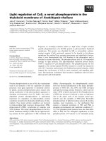
CS induced neutrophilic inflammation in upper airways
We analyzed the presence of neu trophils in the nasal
turbinate tissue of subacute (2-wk and 4-wk) a nd
chronic (24-wk) CS exposed mice by immunohisto-
chemistry, evaluating the average number of neutrophils
per high power field, for the entire section. The increase
in neutrophils w as seen only after 4-wk CS exposure,
compared to air exposed littermates (Fig. 1 B). Interest-
ingly, the number of neutrophils in the nasal turbinate
decreased when the mice were chronically (24-wk)
exposed, resulting in a significant lower amount of neu-
trophils per field in the CS exposed group compared to
the air exposed group (Fig. 1C).
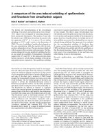
Scattered CD3+ T cells in nasal turbinates versus
(CS-induced) lymphoid follicles in lungs
The presence of peribronchial lymphoid follicles has been
shown both i n mice after chronic CS exposure and
patients with severe COPD. We c ould demonstrate the
presence of these lymphoid follicles in lungs after chronic
CS exposure, using a CD3/B220 double staining
(Fig. 2A). Lymphoid aggregates, absent i n the broncho-
vascular lung regio ns of air -exposed mice, were strongly
induced upon chronic CS exposure. In nasal turbinate
tissue on the other hand, the number of CD3+ cells did
not differ at any time point when air and smoke exposed
mice were compared (Fig . 3). Moreover, CD3+ cells were
not organized in lymphoid follicles - in contrast to find-
ings in lower airways upon chronic exposure - but w ere
scattered throughout the tissue section (Fig. 2B).
Real time Quantitative PCR analysis
Gene expression analysis in nasal turbinate
Neutrophilic chemoattraction related genes In the
nasal turbinates, no significant difference could be
found in Granulocyte Chemotacti c Protein (GCP)- 2 and
keratinocyte chemoattractant (KC - mouse IL-8 homolo-
gue) levels after 2-wk CS exposure (Fig. 4A). Continued
exposure (4-wk) however resulted in significant up-regu-
lation of GCP-2 representing the neutrophilic chemoat-
tractant signal in the CS group co mpared to the air
group, since levels of KC did not differ between groups
(Fig. 4B). This increase in GCP-2 expression disappeared
at chronic (24-wk) CS exposure; moreover KC leve ls
were significant lower in the CS group at that time
point (Fig. 4C).
Monocyte/Macrophag e chemoattraction related genes
We also found an interesting kinetics in the levels of
MCP-1 and MIP-3a. At 2-wk, a significant up-regula-
tion of MCP-1 mRNA in the CS-exp osed group and
a similar tendency for MIP-3a was seen (p = 0.08,
Fig. 4A). This increa se disappeared on continued expo-
sure at 4-wk, both for MCP-1 and MIP-3a (Fig. 4B).
Moreover, a significant lower expression of MCP-1 and
asimilartendencyforMIP-3a werenoticedatchronic
(24-wk) CS exposure (Fig. 4C).
TcellrelatedgenesInterestingly, FoxP3 was already
significantly increased after 2-wk and 4-wk CS exposure
- although this was not the case for TGF-b1-butnot
after 24-wk.
Levels of RORc and subsequent IL-17 were signifi-
cantly down-regulated after 2-wk CS exposure (Fig. 4A),
but this finding disappeared when CS exposure was
prolonged.
Gene expression analysis in lung
Neutrophilic chemoattraction related genes Significant
up-regulation of both GCP-2 and KC in the CS group
remained consistent throughout the entire study, repre-
senting the neutrophilic chemoattractant signal triggered
by CS exposure (Fig. 5A-C).
Monocyte/Macrophag e chemoattraction related genes
Both MCP-1 and MIP-3a were significantly increased in
the CS group at every time point (except for MIP-3a at
24 wk, p = 0.05) (Fig. 5A-C).
T cell related genes In contrast to the nose, 2-wk CS
exposure did not result in increased FoxP3 expression
in the lungs (Fig. 5A). At 4-wk and 24-wk h owever,
significantly higher FoxP3 levels were found in the CS
Table 2 Bronchoalveolar analysis
2-wk Air 2-wk Smoke 4-wk Air 4-wk Smoke 24-wk Air 24-wk Smoke
Total cell number, (× 10
3
) 602.5 ± 41.20 797.53 ± 74.96* 410.00 ± 144.12 1046.00 ± 154.98
†
432.50 ± 37.97 845.00 ± 114.25
†
Neutrophils, (× 10
3
) 0.00 ± 0.00 62.59 ± 10.47
‡
0.00 ± 0.00 200.23 ± 50.97
‡
0.16 ± 0.16 99.75 ± 30.04
†
Macrophages, (× 10
3
) 598.55 ± 38.90 723.66 ± 61.59 408.71 ± 142.94 797.55 ± 103.16* 429.46 ± 37.56 719.10 ± 80.27
†
Lymphocytes, (× 10
3
) 2.49 ± 066 8.32 ± 1.42
†
1.29 ± 1.21 47.82 ± 8.19
‡
2.46 ± 0.32 26.15 ± 8.42
†
Eosinophils, (× 10
3
) 1.46 ± 1.46 2.96 ± 1.49 0.00 ± 0.00 0.39 ± 0.26 0.27 ± 0.18 0.00 ± 0.00
Subacute (4-wk) and chronic (24-wk) CS exposure caused a significant increase in the absolute numbers of total cells, alveolar macrophages, lymphocytes and
neutrophils in the BAL fluid, compared to air exposed littermates. (Values are reported as mean ± SEM; n = 8 mice/group, *p < 0.05 versus Air,
†
p < 0.01 versus
Air,
‡
p < 0.001 versus Air)
Huvenne et al. Respiratory Research 2010, 11:100
/>Page 4 of 9
exposed groups although we could only find higher
TGF-b1 levels at 4-wk (Fig. 5B and 5C).
Although levels of RORc did not differ between
experimental groups, IL-17 mRNA levels were signifi-
cantly increased at 2-wk and 4-wk CS exposure, corre-
lating with the neutrophilic chemoattraction signals.
Analysis of FoxP3 expression in epithelium vs.
subepithelium of nasal turbinates
Recently, FoxP3 expression in epithelial cells has been
described [22]. In order to determine the source of
FoxP3 expression in whole nasal turbinate, we isolated
nasal epithelial cells and subepithelial cells by magnetic
cell sorting. The mRNA expression of FoxP3 however
was not altered in the nasal epithelium after 4-wk CS
exposure (Air 0.3453 ± 0.0084 versus Smoke 0.2894 ±
0.0084 normalized relative expression units). On the
contrary, we demonstrated a nearly 5-fold increase in
subepithelial FoxP3 expression in nasal turbinates upon
4-wk CS exposu re, possibly due to infiltrating T regula-
tory cells (Air 1.043 2 ± 0.0 723 versus Smoke 5. 1730 ±
0.9323).
Discussion
In this study we aimed to investigate the effects of
cigarette smoke (CS) on upper airways and lower air-
ways, in a mouse model of subacute and chronic CS
exposure. We here demonstrate for the first time that
the inflammatory response upon CS exposure clearly
diff ers between nose and lungs in mice. The nature and
kinetics of both the neutrophil and monocyte/macro-
phage inflammation differ in both airways compart-
ments. This indi cates the involvement of different
regulatory mechanisms, which is reflected by the
observed differences in FoxP3 increase after CS expo-
sure. The suppressive mechanisms arise earlier and
appear to be more efficient in nose than in lungs.
Although increased levels of MCP-1, MIP-3a and
2-wk nose
Air Smoke
0.0
0.5
1.0
1.5
number of neutrophils/field
4-wk nose
1.5
2.0
*
r
ophils/fiel
d
A
B
Air Smoke
0.0
0.5
1.0
number of neut
r
24-wk nose
Air Smoke
0.0
0.1
0.2
0.3
0.4
*
number of neutrophils/field
C
Figure 1 Average number of neutrophils in nasal turbinate
sections. Increase in number of neutrophils after CS exposure was
not seen after 2-wk, compared to air exposed littermates (Fig. 1A),
but only after 4-wk (Fig. 1B). Interestingly, the number of
neutrophils in the nasal turbinate decreased when the mice were
chronically (24-wk) exposed, resulting in a significant lower amount
of neutrophils per field in the CS exposed group compared to the
air exposed group (Fig. 1C). (n = 8 mice/group, * p < 0.05)
Figure 2 CD3+ cells. Lymphoid folli cles were demonstrated i n
lungs after chronic CS exposure, using CD3(brown)/B220(blue)
doublestaining (Fig. 2A, × 200). In nose however, no increased
number of CD3+ cells in inferior turbinate, or lymphoid follicle
neogenesis was found at that time point (Fig. 2B, × 400).
Huvenne et al. Respiratory Research 2010, 11:100
/>Page 5 of 9
GCP-2 are found both in nose and lungs after subacute
CS exposure, the neutrophilic influx and increase in
neutrophilic chemoattraction signals are transient in
upper a irways while they remain constant in lower air-
ways. Consequently, chronic upper airway CS exposure
results in a non-inflammatory status with a significant
downregulation of inflammation, while lower airway
inflammation is clearly present and ongoing.
Neutrophilic inflammation in the nasal turbinate
tissue was not present after 2-wk CS exposure, likely
due to the absence of a neutrophilic chemoattraction
signal, as both GCP-2 and KC levels were not increased
in the CS group. However, prolonged (4-wk) exposure
caused a significant GCP-2 in crease in the CS group,
which correlates with the immunohistochemistry , show-
ing a higher number of neutrophils per field in the CS
groupcomparedtotheairgroup,butonlyafter4-wk.
To our surprise, chronic (24-wk) CS exposure did not
cause a further increase in neutrophil accumulation in
the nasal turbinate tissue. Moreover, GCP-2 levels and
KC levels in the CS group did not differ and were signif-
icantly down regulated from controls respectively. This
was again confirmed by IHC, where we found a signifi-
cant decrease in the numbe r of neutrophils per field in
the CS group compared to controls. This may be inter-
preted as a clear sign of d own-regulation of the neutro-
philic inflammatory long-term response in the nasal
turbinates. Evaluation of neutrophilic inflammation in
upper airwa ys was done in nasal turbinate tissue,
because nasal lavage did not yield sufficient cells allow-
ing a reliable cell differentiation. As a consequence,
compartmentalization of inflammation in both upper
and lower airways may influence the interpretation of
these findings. Indeed, cigarette smoke causes an
increase of neutrophil numbers in BAL (mouse studies),
or sputum (human studies), whereas its effect in lung
tissue or biopsies is less pronounced.
Our findings on neutrophilic inflammation in upper
airways are in sharp contrast with the data obtained
from experiments in the lung, where CS exposure
resulted in a significant increase in both GCP-2 and KC
at all time points, accounting for to the observed infl ux
of neutrophils in the BAL fluid of these mice [23].
We have shown a remarkable change over time in the
nasal mRNA MCP-1 levels of CS exposed mice, showing
an initial increase, followed by a significant decrease in
MCP-1 level s in the nasal turbinate upon chronic expo-
sure. In the lungs of these mice however, we detected a
consistent increase in MCP-1 levels in CS exposed mice
on each time point [23]. This is another sign of the dif-
ferent inflammatory response to CS in the upper airway.
The role of pro-inflammatory T helper 17 phenotype
in the pathogenesis of C OPD is increasingly studied,
and it is suggested that COPD might be better explained
by the Th17 phenotype [12]. These Th17 cells, which
require the up-regulation of the orphan nuclear receptor
RORgammat (enco ded by RORc) for di fferentiation
from naïve T cells [24], account for the production of
several members of th e IL-17 family of cytokines, which
2-wk nose
Air Smoke
0.0
1.0
2.0
3.0
4.0
number of CD3+ cells/field
4-wk nose
1.5
2.0
c
ells/field
A
B
Air Smoke
0.0
0.5
1.0
number of CD3+
c
24-wk nose
Air Smoke
0.0
1.0
2.0
3.0
4.0
5.0
number of CD3+ cells/field
C
Figure 3 CD3+ staining. Nasal turbinate sections were evaluated
for the presence of CD3+ cells, within lymphoid follicles. Number of
CD3+ cells per field did not differ between air and CS exposed
group at any time point (Fig. 3 A-C). (n = 8 mice/group).
Huvenne et al. Respiratory Research 2010, 11:100
/>Page 6 of 9
have proven abilities to recruit and activate neutrophils
[25]. Here, nasal mRNA levels of RORc and IL-17 in the
nose were significantly down-regulated after 2-wk CS
exposure, but not upon longer (4-wk and 24-wk) expo-
sure. In lungs however, the response of Th17 cells
appears to be opposite, as 2-wk and 4-wk CS exposure
resulted in a significant up-regulation o f IL-17, and
chronic(24-wk)exposureshowedasimilartendency.
These differences in IL-17 levels between nose and
lungs, can explain the observed differences in neutrophil
accumulation, as described above.
T regulatory cells expressing FoxP3 are thought to play
a role in controlling CS induced inflammation [15,26],
amongst others via the immunomodulatory cytokine
TGF-b1 [27]. In nose, FoxP3 mRNA expression was
2-wk nose
G
C
P
-2
K
C
M
CP-
1
α
M
I
P
-
3
R
ORc
IL 17
FoxP3
1
β
TG
F
-
0.1
1
10
100
*
*
*
*
Smoke
Air
A
Normalized Relative
expression units
(log scale)
4-wk nose
10
100
***
**
Air
Smoke
B
d
Relative
o
n units
c
ale)
G
CP-2
K
C
M
CP
-1
α
M
I
P
-
3
RORc
I
L 17
FoxP3
1
β
TGF-
0.1
1
Normalize
d
expressi
o
(log s
c
24-wk nose
GCP-2
K
C
M
C
P
-
1
α
MIP-3
R
O
Rc
I
L
1
7
FoxP3
1
β
TG
F
-
0.1
1
10
100
**
*
**
Air
Smoke
C
Normalized Relative
expression units
(log scale)
Figure 4 Gene expression analysis in nasal turbinate.2-wkCS
exposure resulted in increased levels of MCP-1 and FoxP3. Levels of
RORc and subsequent IL-17 were significantly down-regulated at
this time point (Fig. 4A). At 4-wk, GCP-2, but not KC, levels are
increased. Moreover, FoxP3 is significantly higher in the CS exposed
group (Fig. 4B). 24-wk CS exposure results in significant down-
regulation of nasal MCP-1, MIP-3a an TGF-b1 (Fig. 4C). (n = 8 mice/
group, *p < 0.05, **p < 0.01, ***p < 0.001).
2-wk lung
G
C
P
-2
K
C
M
CP-1
α
M
IP
-3
RORc
I
L
17
F
oxP
3
1
β
TGF
-
0.1
1
10
100
Air
Smoke
***
***
**
**
*
A
N
ormalized Relative
expression units
(log scale)
4-wk lung
10
100
Air
Smoke
**
***
**
***
***
***
B
d
Relative
o
n units
s
cale)
GCP-2
KC
M
CP
-1
α
M
IP-3
RORc
IL 17
Fox
P
3
1
β
T
G
F
-
0.1
1
*
N
ormalize
d
expressi
o
(log
s
24-wk lung
GCP
-2
K
C
M
CP-1
α
M
IP-3
RO
Rc
I
L 17
F
oxP
3
1
β
TG
F-
0.1
1
10
100
Air
Smoke
*
*
*
*
C
Normalized Relative
expression units
(log scale)
Figure 5 Gene expression analysis in lung. Pulmonary levels of
GCP-2, KC, MCP-1, MIP-3a and IL-17, but not FoxP3 were
significantly increased after 2-wk CS exposure (Fig. 5A). After 4-wk
CS exposure, all markers of neutrophilic and monocyte/macrophage
chemoattraction are significantly increased, as well as FoxP3 and
TGF-b1 (Fig. 5B). Chronic CS exposure caused an increase in levels
of GCP-2, KC, MCP-1 and FoxP3 levels (MIP-3a p = 0.05) (Fig. 5C). (n
= 8 mice/group, *p < 0.05, **p < 0.01, ***p < 0.001).
Huvenne et al. Respiratory Research 2010, 11:100
/>Page 7 of 9
increased already af ter 2-wk, and was mainly found - at
least at 4-wk - in the subepithelium, possibly due to
invading Tregs expressing FoxP3. In lungs, FoxP3 was
only increased after 4-wk, which is in line with increased
Tregs in lungs after CS exposure [14]. Interestingly, these
infiltrating Tregs in lungs are thought to have a weak
functionality, as they are unable to control inflammation
in lungs [15]. It is tempting to s peculate that Tregs act
early a nd adequately in nose to suppress CS-induced
inflammation, but that they invade later and have weaker
functionality in lungs , allowing inflammation to persist.
Alternatively, the CS exposure of the nose might be
higher in mice - obligatory nose-breathing animals -
compared to lungs, allowing tolerazation or change in
cell populations to occur earlier. Indeed, upon 24-wk CS
exposure the number of neutrophils shows a decreasing
tendency compared to 4-wk CS exposed mice.
Although in vivo cigarette smoke-exposed mice can
offer valuable information on several aspects of the
pathogenesis of COPD, such as the time course of
upper and lower airway i nflammation, there are also
limitations that need to be taken into account. Firstly, a
number of anatomical and physiological differences exist
between the respiratory tract of mice and humans. For
example, mice are obligate nose breathers that filter
tobacco smoke inefficiently, and they have less branch-
ing of the bronchial tree. Furthermore, the profile of
inflammatory mediators is also slightly different in the
mouse. And lastly, there is no mouse model that mimics
all the hallmarks of COPD pathology, inclu ding exacer-
bations and extrathoracic manifestations.
Another possible limitation to this study is the fact
that not only T cells are able to produce either IL-17,
TGF-b or FoxP3, but a number of other cells like neu-
trophils or epithelial cells can do so. Furthermore, the
suppressive capacity of the FoxP3 producing Tregs in
upper airways stills remains to be elucidated.
Although the inflammatory answer of nose and lungs is
clearly different upon CS exposure, possible confounding
factors might influence the data interpretation in this
model. Above, we have described the issue of compart-
mentalization of inflammation, and the relative dosage
exposure, with higher deposi tion of CS in the nose vs.
lungs. Furthermore, physiologic temporal changes are seen
in the inflammatory readouts: levels of inflammatory cells
and mediators of unexposed control mice vary over time,
asshowninFig.3,4,5.Byusingage-matchedcontrol
mice in our experiments, we have corrected for these phy-
siologic temporal changes. Altogether, the above men-
tioned limitations of this model remain to be elucidated.
Conclusions
In conclusion, we have demonstrated that cigarette
smoke induced inflammation differs between nose
and lungs in this mouse model. After CS exposure,
inflammatory markers were upregulated in lungs at
alltimepoints.However,thiswasnotthecaseinthe
nose, where particularly upon chronic CS exposure,
nasal inflammatory markers were significantly lower
than the control (air) conditions. It is possible that
infiltrating FoxP3 expressing Tregs might account for
these observed differences, although further investiga-
tion is necessary to identify possible differences
in their suppressive functionality in both airway
compartments.
List of abbreviations
ACTB: beta-actin; GCP-2 (CXCL6): granulocyte chemotactic protein 2; KC
(CXCL1): keratinocyte chemoattractant; MCP-1 (CCL-2): monocyte
chemotactic protein-1; MIP-3a (CCL-20): Macrophage Inflammatory Protein-3
alpha; RORc: orphan nuclear receptor RORgammat; IL-17: Interleukin 17;
FoxP3: Forkhead box P3; TGF-b1: Transforming growth factor beta 1
Acknowledgements
The authors would like to thank Greet Barbier, Eliane Castrique, Indra De
Borle, Philippe De Gryze, Katleen De Saedeleer, Marie-Rose Mouton, Ann
Neessen and Christelle Snauwaert for their technical assistance, and Ruth
Raspoet for her contribution to the immunohistochemistry analysis.
This project is supported by the Fund for Scientific Research - Flanders
(FWO-Vlaanderen - Project G.0052.06), by a grant from the Ghent University
(BOF/GOA 01251504), by the Interuniversity Attraction Poles program (IUAP)
- Belgian state - Belgian Science Policy P6/35, and by grants to C.B. from the
Fund for Scientific Research - Flanders, FWO, no. A12/5-HB-KH3 and
G.0436.04, and to K.B. as a postdoctoral fellow of the Fund for Scientific
Research Flanders (FWO).
Author details
1
Upper Airways Research Laboratory (URL), ENT Department, Ghent
University Hospital, Ghent University, Belgium.
2
Department of Respiratory
Medicine, Ghent University Hospital and Ghent Universi ty, Ghent, Belgium.
Authors’ contributions
WH carried out the design and coordination of the study, gathered the data
on upper and lower airway inflammation, interpreted the data, drafted and
finalized the manuscript. CP-N developed and optimized the PCRs on nose
and lung samples. LD designed and optimized the nasal epithelial cell
isolation procedure. OK optimized and carried out the IHC staining of the
nasal turbinates. TM and KB were involved in the coordination and design of
the study, and the critical reading of the manuscript. NP and LR provided
mice and were involved in the experimental design of the CS-induced
airway inflammation. GJ, GB and CB participated in the coordination of the
study, helped to interpret the data and critically revised the manuscript. All
authors read and approved the final version of the manuscript.
Competing interests
The authors declare that they have no competing interests.
Received: 1 March 2010 Accepted: 23 July 2010 Published: 23 July 2010
References
1. Barnes PJ, Shapiro SD, Pauwels RA: Chronic obstructive pulmonary
disease: molecular and cellular mechanisms. Eur Respir J 2003, 22:672-688.
2. Kim JS, Rubin BK: Nasal and sinus inflammation in chronic obstructive
pulmonary disease. COPD 2007, 4:163-166.
3. Bousquet J, Van Cauwenberge P, Khaltaev N: Allergic rhinitis and its
impact on asthma. J Allergy Clin Immunol 2001, 108:S147-334.
4. Annesi-Maesano I: Epidemiological evidence of the occurrence of rhinitis
and sinusitis in asthmatics. Allergy 1999, 54(Suppl 57):7-13.
5. Bousquet J, Vignola AM, Demoly P: Links between rhinitis and asthma.
Allergy 2003, 58:691-706.
Huvenne et al. Respiratory Research 2010, 11:100
/>Page 8 of 9
6. Bracke KR, D’Hulst A I, Maes T, Moerloose KB, Demedts IK, Lebecque S,
Joos GF, Brusselle GG: Cigarette smoke-induced pulmonary inflammation
and emphysema are attenuated in CCR6-deficient mice. J Immunol 2006,
177:4350-4359.
7. Barnes PJ: Chemokines in COPD. Chronic Obstructive Pulmonary Disease
Blackwell PublishingStockley RA, Rennard SI, Rabe K, Celli B 2007, 859-866.
8. van der Strate BW, Postma DS, Brandsma CA, Melgert BN, Luinge MA,
Geerlings M, Hylkema MN, van den Berg A, Timens W, Kerstjens HA:
Cigarette smoke-induced emphysema: A role for the B cell? Am J Respir
Crit Care Med 2006, 173:751-758.
9. Hogg JC, Chu F, Utokaparch S, Woods R, Elliott WM, Buzatu L,
Cherniack RM, Rogers RM, Sciurba FC, Coxson HO, Pare PD: The nature of
small-airway obstruction in chronic obstructive pulmonary disease. N
Engl J Med 2004, 350:2645-2653.
10. Brusselle GG, Demoor T, Bracke KR, Brandsma CA, Timens W: Lymphoid
follicles in (very) severe COPD: beneficial or harmful? Eur Respir J 2009,
34:219-230.
11. Grumelli S, Corry DB, Song LZ, Song L, Green L, Huh J, Hacken J, Espada R,
Bag R, Lewis DE, Kheradmand F: An immune basis for lung parenchymal
destruction in chronic obstructive pulmonary disease and emphysema.
PLoS Med 2004, 1:e8.
12. Curtis JL, Freeman CM, Hogg JC: The immunopathogenesis of chronic
obstructive pulmonary disease: insights from recent research. Proc Am
Thorac Soc 2007, 4:512-521.
13. Hizawa N, Kawaguchi M, Huang SK, Nishimura M: Role of interleukin-17F
in chronic inflammatory and allergic lung disease. Clin Exp Allergy 2006,
36:1109-1114.
14. Cosio MG, Saetta M, Agusti A: Immunologic aspects of chronic
obstructive pulmonary disease. N Engl J Med 2009, 360:2445-2454.
15. Smyth LJ, Starkey C, Vestbo J, Singh D: CD4-regulatory cells in COPD
patients. Chest 2007, 132:156-163.
16. Vachier I, Vignola AM, Chiappara G, Bruno A, Meziane H, Godard P,
Bousquet J, Chanez P: Inflammatory features of nasal mucosa in smokers
with and without COPD. Thorax 2004, 59:303-307.
17. Blank U, Ruckes C, Clauss W, Weber WM: Effects of nicotine on human
nasal epithelium: evidence for nicotinic receptors in non-excitable cells.
Pflugers Arch 1997, 434:581-586.
18. Bascom R, Kulle T, Kagey-Sobotka A, Proud D: Upper respiratory tract
environmental tobacco smoke sensitivity. Am Rev Respir Dis 1991,
143:1304-1311.
19. Lieu JE, Feinstein AR: Confirmations and surprises in the association of
tobacco use with sinusitis. Arch Otolaryngol Head Neck Surg 2000,
126:940-946.
20. Ebbert JO, Croghan IT, Schroeder DR, Murawski J, Hurt RD: Association
between respiratory tract diseases and secondhand smoke exposure
among never smoking flight attendants: a cross-sectional survey. Environ
Health 2007, 6:28.
21. Hellemans J, Mortier G, De Paepe A, Speleman F, Vandesompele J: qBase
relative quantification framework and software for management and
automated analysis of real-time quantitative PCR data. Genome Biol 2007,
8:R19.
22. Chen GY, Chen C, Wang L, Chang X, Zheng P, Liu Y: Cutting edge: Broad
expression of the FoxP3 locus in epithelial cells: a caution against early
interpretation of fatal inflammatory diseases following in vivo depletion
of FoxP3-expressing cells. J Immunol 2008, 180:5163-5166.
23. D’Hulst AI, Maes T, Bracke KR, Demedts IK, Tournoy KG, Joos GF,
Brusselle GG: Cigarette smoke-induced pulmonary emphysema in scid-
mice. Is the acquired immune system required? Respir Res 2005, 6:147.
24. Ivanov II, McKenzie BS, Zhou L, Tadokoro CE, Lepelley A, Lafaille JJ, Cua DJ,
Littman DR: The orphan nuclear receptor RORgammat directs the
differentiation program of proinflammatory IL-17+ T helper cells. Cell
2006, 126:1121-1133.
25. Traves SL, Donnelly LE: Th17 cells in airway diseases. Curr Mol Med 2008,
8:416-426.
26. Barcelo B, Pons J, Ferrer JM, Sauleda J, Fuster A, Agusti AG: Phenotypic
characterisation of T-lymphocytes in COPD: abnormal CD4+CD25+
regulatory T-lymphocyte response to tobacco smoking. Eur Respir J 2008,
31:555-562.
27. Askenasy N, Kaminitz A, Yarkoni S: Mechanisms of T regulatory cell
function. Autoimmun Rev 2008, 7:370-375.
doi:10.1186/1465-9921-11-100
Cite this article as: Huvenne et al.: Different regulation of cigarette
smoke induced inflammation in upper versus lower airways. Respiratory
Research 2010 11:100.
Submit your next manuscript to BioMed Central
and take full advantage of:
• Convenient online submission
• Thorough peer review
• No space constraints or color figure charges
• Immediate publication on acceptance
• Inclusion in PubMed, CAS, Scopus and Google Scholar
• Research which is freely available for redistribution
Submit your manuscript at
www.biomedcentral.com/submit
Huvenne et al. Respiratory Research 2010, 11:100
/>Page 9 of 9