Báo cáo y học: " Genome sequences of Human Adenovirus 14 isolates from mild respiratory cases and a fatal pneumonia, isolated during 2006-2007 epidemics in North America" pps
Bạn đang xem bản rút gọn của tài liệu. Xem và tải ngay bản đầy đủ của tài liệu tại đây (804.64 KB, 8 trang )
RESEARC H Open Access
Genome sequences of Human Adenovirus
14 isolates from mild respiratory cases and a
fatal pneumonia, isolated during 2006-2007
epidemics in North America
Huo-Shu H Houng
1*
, Heping Gong
1
, Adriana E Kajon
2
, Morris S Jones
3
, Robert A Kuschner
1
, Arthur Lyons
1
,
Lisa Lott
4
, Kuei-Hsiang Lin
5
, David Metzgar
6
Abstract
Background: Human adenovirus 14 (HAdV-14) is a recognized causative agent of epidemic febrile respiratory
illness (FRI). Last reported in Eurasia in 1963, this virus has since been conspicuously absent in broad surveys, and
was never isolated in North America despite inclusion of specific tests for this serotype in surveillance methods. In
2006 and 2007, this virus suddenly emerged in North America, causing high attack rate epidemics of FRI and, in
some cases, severe pneumonias and occasional fatalities. Some outbreaks have been relatively mild, with low rates
of progression beyond uncomplicated FRI, while other outbreaks have involved high rates of more serious
outcomes.
Methodology and Findings: In this paper we present the complete genomic sequence of this emerging
pathogen, and compare genomic sequences of isolates from both mild and severe outbreaks. We also compare
the genome sequences of the recent isolates with those of the prototype HAdV-14 that circulated in Eurasia
30 years ago and the closely related sequence of HAdV-11a, which has been circulating in southeast Asia.
Conclusions: The data suggest that the currently circulating strain of HAdV-14 is closely related to the historically
recognized prototype throughout its genome, though it does display a couple of potentially functional mutations
in the fiber knob and E1A genes. There are no polymorphisms that suggest an obvious explanation for the
divergence in severity between outbreak events, suggesting that differences in outcome are more likely
environmental or host determ ined rather than viral genetics.
Introduction
Aden oviruses are double-stranded DNA viruses. The 52
recognized serotypes of human adenovirus (HAdV)
cause a broad range of symptoms: community-acquired
gastrointestinal, conjunctival, and febrile respiratory ill-
ness (FRI; both upper and lower respiratory tract),
hemorrhagic cystitis associated with bone marrow trans-
plant, hepatic and urinary tract infections, and perhaps
even obesity [[1-4], />ICTVdb/Ictv/index.htm].
The 10 serotypes of HAdV associated with FRI and
pneumonia are grouped into 3 species, B (including sub-
species B1 and B2), C and E, on the basis of hemaggluti-
nation and phylogenetic criteria [5-9]. HAdV-1, 2, 5, and
6, belonging to speci es C, cause generally endemic pat-
terns of FRI in children and young adults [8,10,11]. In
contrast, HAdV-4 (the sole serotype of species E) and
the remaining respiratory species B serotypes (HAdV-3,
7, 11, 14, 16, and 21), often cause distinctive outbreaks
of FRI, conjunctivitis, and pneumonia in crowded civi-
lian populations such as dorms, public swimming pools,
and boarding schools [7,8]. In the absence of vaccin es,
these viruses also cause almost continuous outbreaks of
FRI among recruits in military training throughout the
world [8,7,12,13].
* Correspondence:
1
Division of Viral Diseases, Walter Reed Army Institute of Research (WRAIR),
503 Robert Grant Avenue, Silver Spring, 20910, USA
Full list of author information is available at the end of the article
Houng et al. Respiratory Research 2010, 11:116
/>© 2010 Houng et al; licensee BioMed Central Ltd. This is an Open Access article distributed under the terms of the Creative Commons
Attribution Lice nse (http://c reativecommons.org/licenses/by/2.0), which permits unrestricted use, distribut ion, and reproduction in
any medium, provided the original work is properly cited.
Four of these seven adult human respiratory adeno-
viruses, HAdV-3, 7 and 21 (subspecies B1) and HAdV-4
(species E) are common, intraserotypically diverse, and
inevitably represented in broad surveys [7,8,10,14]. In
different populations and at different times one serotype
may completely dominate this niche, several serotypes
may intermingle, or multiple serotypes can appear in
series through distinct replacement events [13,15-19].
The remaining three serotypes that cause FRI in healthy
adults, HAdV-16 of subspecies B1 [11] and HAdV-11
and 14 of subspecies B2 [13], have only infrequently
been associated with FRI. These rare a ssociations often
appear to involve more severe symptoms, outcomes, and
outbreak characteristics than do those of the more com-
mon species E and subspecies B1 serotypes [11,20-24].
HAdV-14 was reported only four times in the twent ieth
cent ury, always in transient, concentrated, and generally
nonlethal but severely incapacitating FRI outbreaks in
healthy (though crowded) adult and adolescent popula-
tions [25-28]. These outbreaks occurred between 1955
and 1963, all in Eurasia, and HAdV-14 was not reported
again even in broad geographical and temporal surveys
until 2001 when it was reported in 10% of FRI speci-
mens in a retrospective analysis of clinic samples in Tai-
wan [29]. (Author’ s note: upon whole-genome analysis,
this strain was identified as the very closely-related
HAdV-11a; HSH, AK, data not shown). HAdV-11a has
recently been seen in increased numbers of FRI cases in
Asia, including some significant outbreaks [30]. Pheno-
typic intermediates of the closely related serotypes
HAdV-11 and HAdV-14 were identified in a military
camp in Spain in 1969 [31], and in Germany from a
severe case of acute respiratory disease foll owing a mili-
tary training exercise (and ap parently associated out-
break) in Turkey in 2004 [32].
HAdV-14 had never been identified in North America
before its emergence in 2006. Following the recognized
outbreaks in 2006 and 2007 [13,22,24], retrospective ana-
lysis of specific cases and collections uncovered isolated
occurrences of the disease dating back a few years before
the larger outbreaks (for example, see [23]). HAdV-14
was first seen in greater numbers and associated with sig-
nificant outbreaks in March 2006, when it simultaneously
emerged at four military recruit training centers through-
out the United States, causing several hundred cases
(estimated from partial surveillance) of FRI over the
course of the c alendar year [13]. The impact amounted
to a partial replacement of the recently dominant HAdV-
4, rather than an increase in overall adenovir al impact at
these sites. These emergence events w ere not associated
with symptoms or epidemiological patterns outside the
normal range of those seen with the typical species E and
subspecies B1 HAdVs seen in surveillance of recruit FRI
and pneumonia [13].
Starting in March 2007, HAdV-14 was recognized as
the cause of several severe civilian outbreaks, prompting
attention from the Centers for Disease Control and Pre-
vention (CDC) [22]. The same p athogen was recognize d
as the cause of prolonged outb reaks at three military
installations where HAdV-14 either emerged against an
adenovirus-free background ([22], and an outbreak at
Coast Guard Training Center, Cape May; unpublished
Naval Health Research Center [NHRC] data) or comple-
tely replaced the existing HAdV-4 strain (Mar ine Corps
Recruit Depot [MCRD], Parris Island, NJ; unpublished
NHRC data). Reported civilian HAdV-14 outbreaks were
transient, lasting four months and involving nine casual-
ties [22]. The two n oted outbreaks in recruit facilities
where there was no immediate history of ongoing ade-
novirus transmission were initially severe, involving
greatly increased rates of disease among the recruits and
also spreading to medical support personnel, training
staff, and others. One death was reported among
infected recruits as were many pneumonia hospitaliza-
tions, several requiring ventilation assistance [22].
Whole-genome restriction enzyme analysis (genome
typing [33]) and partial gen esequenceanalysis(hexon,
fiber, E1A) have shown that the currently emergent US
strains of HAdV-14 (see result section for the d efinition
of genome type HAdV-14p1), both civilian and military,
are all similar at the genome type level and essentially
identical (> 99.9%) at the sequence level in the hexon
and fiber genes (AK, unpublished data). The circulating
genome type, however, is significantly diverged from the
prototype (HAdV-14p) d e Wit strain (isolated from ill
military recruits in the Netherlands [28]).
To further characterize the newly emergent US
HAdV-14 strains, three recent HAdV-14p1 isolates were
completely sequenced and compared with the pro totype
HAdV-14pdeWitgenomesequenceaswellasother
genetically related HAdV-11a isolates from southeast
Asia. One isolate was collected in March 2006 at Marine
Corps Recruit Depot, San Diego (MCRD-SD), when
HAdV-14 was initially detected and identified in US
recruits [13]. This isolate came from an emergence of
HAdV-14p1 that did not exhibit uniquely severe out-
break dynamics or symptoms - in fact, it was observed
during this outbreak, in which both HAdV-4 and
HAdV-14 were present in approximately equal propor-
tions, that HAdV-14 did not seem to cause as much
pneumonia as did HAdV-4 (unpublished NHRC syndro-
mic surveillance data). This outbreak did not involve
increased rates, but rather a simple and t emporary
replacement of HAdV-4 with HAdV-14 [13], and the
studied isolate was collected from a recruit with uncom-
plicated FRI. The other two sequenced HAdV-14 iso-
lates were collected at Lackland Air Force Base during
the severe and prolonged outbreak of HAdV-14 that
Houng et al. Respiratory Research 2010, 11:116
/>Page 2 of 8
started in February 2 007 and drew the attention of the
CDC a mont h later, as the outbreak spread [22]. One
was from a fatal case of respiratory failure from viral
pneumonia, which followed several weeks of intubation
and life support. The other was from a mild case of FRI.
The primary goal of our study was to determine if
there were any apparent genetic correlates that might
distinguish viruses causing mild and severe outbreaks or
mild and severe symptoms, or differences between the
currently circulating strain of HAdV-14p1 and the pro-
totype HAdV-14p that was seen in Eurasia in the 1950 s
and 1960 s. A secondary goal was to identify unique sig-
nature sequences that might allow us to track individual
strains of HAdV-14 of US origin for the purposes of
epidemiological investigations.
Materials and methods
Sample Collection
The isolate from a mild FRI outbreak at MCRD San
Diego was collected with consent under an institutional
review board-approved research protocol
(NHRC.1999.0002), identified, cultured, and anal yzed as
part of NHRC’s ongoing population-based FRI surveil-
lance program. The two isolates from Lackland Air
Force Base included one NHRC surveillance isol ate
from a recruit with uncomplicated FRI and one fatal
pneumonia isolate collected from a severely ill recruit at
Wilford Hall Medical Center, initially collected for diag-
nostic viral culture and later provided to LRRI and
WRAIR as a de-identified isolate.
NHRC samples were collected as oropharyngeal
(throat) swabs in VTM (Remel, Lenexa, KS), immedi-
ately frozen in either -80°C freezers or on dry ice, and
transported on dry ice to NHRC under College of
American Pathologists (CAP)-accredited collection and
transport protocols. LacklandAirForceBasesamples
were collected as throat swabs in VTM, cultured in
A549 cells, and transported to NHRC as above. All sam-
ples were tested at NHRC for HAdV-14 [13] as raw spe-
cimens, then subsequently cultured in A549 cells
(Diagnostic Hybrids Inc., Athens, OH), and stored fro-
zen as infected tissue culture fluid (isolated virus) at
Lovelace Respiratory Research Institu te (LRRI). Sequen-
cing work on these samples was performed at Walter
Reed Army Institute of Research (WRAIR) on the
resulting isolates.
Sanger Sequencing of HAdV-14
PCR and sequencing were accomplished at the virology
facility at Walter Reed Army Institute of Research
(WRAIR), Silver Spring, Maryland, USA. PCR primer
pairs were designed from the prototype HAdV-14p de
Wit sequence (GenBank accession number AY803294)
and used to generate overlapping 1-2 kilobase amplicons
covering the entire genome. All PCR products were
sequenced in both directions by using forward and
reverse PCR primers corresponding to each individual
PCR product. All clean and verified readable sequences
were used to assemble full HAdV-14 genome sequences
using the Sequen cher software (Gene Codes Corpora-
tion, Ann Arbor, MI).
Two hundred microliter aliquots of eac h isolate were
extracted using the Invitrogen ChargeSwitch DNA
extraction kit (Invitrogen Corporation, Carlsbad, CA)
per the manufacturer’s instructions, and resusp ended in
200 μl elution buffer. One hundred microliter PCR
amplification reactions consisted of 2 mM MgCl2,
0.6 mM dNTP (1.5 mM each A, C, T, and G), 200 μM
each primer, 2.5 units Platinum Taq Polymerase (Invi-
trogen), and 1 ul of extracted isolate in 1X ABI Buffer II
(Applied Biosystems Inc., Foster City, CA). Thermal
cycling was carried out on an ABI9700 platform
(Applied Biosystems) using the following parameters:
initial activation for 2 min at 94°C, then 35 cycles of:
20 s at 94°C, 20 s at 53°C, and 2 min at 72°C. Final
extension was for 7 min at 72°C. PCR cleanup was per-
formed usi ng the Qiagen PCR clean up kit (Qiagen,
Valencia, CA) per the manufacturer’ s instructions.
Sequencing reactions were set up per the manufacturer’s
instructions using the ABI BigDye Terminator kit (man-
ual version 3.2, Applied Biosystems), and run on an
ABI9700 platform. Reaction products were analyzed on
an ABI3130XL a utomated sequencer (Applied Biosys-
tems) per the manufacturer’s instructions. Resulting data
were then edited and aligned using Sequencher software
(Gene Codes).
Results and Discussions
HAdV-14 Genomes from 2006-7 US Outbreaks
Genomic sequences from three different recent US
HAdV-14 isolates, including two from Lackland Air
Force Base (303600 and 1986T, associated with mild
FRI and fatal pneumonia, respectively, both from a
severe outbreak) and one from Marine Corps Recruit
Depot, San Diego (NHRC22039, associated with a mild
infection during a mild outbreak) were fully sequenced,
assembled and submitted to the NCBI GenBank data-
base (GenBank Accession #s FJ822614, EU827616 and
EU833993, respectively). The genome size of Lackland
strains 303600 and 1986T is identical to each other,
34,764 base pairs (bp) that is also identical in size with
the HAdV-14 prot otype deWit strain, 34,764 base pairs
(bp). The San Diego strain NHRC22039 has a genome
size of 34,768 bp. All three recent US HAdV-14 strains
are highly homologous with each other. The two Lack-
land strains are 100% identical to each other, while the
San Diego isolate differed only by a 4 bp extension of
the polyadenylation signal (a poly-T on the coding
Houng et al. Respiratory Research 2010, 11:116
/>Page 3 of 8
strand)atthe5’ end o f the terminal binding protein
(TBP) gene, and a single noncoding (synonymous) base
substitution in the fiber gene. The poly-T repeat is
13 bp long [T(13)] in the Lack land strains and T(17) in
the San Diego strain. HAdV-14p strain deWit contains a
corresponding T(11) repeat. The genomes of all t hree
recent US HAdV-14 isolates share identical coding
regions for all genes. As recently described, and after
detailed characterization by restriction enzyme analysis,
all North American isolates of HAdV-14 correspond to
genome type 14p1 [34]. Table 1 shows the s ummary of
alignment results. HAdV-14 deWit and the recent US
HAdV-14p1 isolates differ by 0.3%, scattered quite
evenly through the genome. All 4 HAdV-14 s exam ined
in this study have t he same base composition of 51.2%
A/T, 48.8% G/C. The GC content and the number of
open reading frames (ORFs) were identical to the
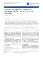
HAdV-14p de Wit strain. A map of the organization of
predicted ORFs within the genome of the emerging
HAdV-14p1 strain is sh own in Figure 1, and is identical
to that of the prototype HAdV-14 de Wit strain.
All HAdV genomes are bounded by inver ted terminal
repeats (ITR) ranging from 100 to 200 bp in size, which
serve as viral replication origins. The representative
ITRs of various HAdV species, such as species A
(HAd V-12, 18, 31), B (HAdV-3, 7, 11), and C (HAdV-1,
2, 5) are available in GenBank. Among t hese HAdVs,
ITRs are highly conserved within species but diverse
between speci es. For example, the ITRs of HAdV-2 and
HAdV-5 (species C) are identical 103 bp sequences.
Similarly conserved ITR patterns are observed for the
137 bp ITRs of species B (HAdV-3, 7 and 11). However,
all three recent US HAdV-14 s share identical inverted
terminal repeat (ITR) sequence of 133 bp, in contrast to
other species B HAdVs. The HAdV-14 prototype deWit
also contains a 133 bp ITR, but this differs from recent
HAdV-14 s of US origin by one substitution at base pair
68 (T68C). This level of polymorphism in ITR
sequences among closely related strains is unusual.
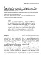
We compared HAdV-14p1 to selected HAdV-B proto-
type strains (HAdV-14, 3, 7, 11, and 21) using the
mVISTA Limited Area Global Alignment of Nucleotides
(LAGAN) tool (MontaVista Software, Inc., Santa Clara,
CA) [35] (Figure 2). With the exception of the hexon
gene of HAdV-11p, HAdV-14p1 showed strong homol-
ogy with both HAdV-14p and -11p. This was expected,
since HAdV-11 and HAdV-14 are both members of
subspecies B2. Comparison of HAdV-14p1 to HAdV-3,
7, and 21, members of subspecies B1, revealed sequence
divergence throughout the genome, especially the pen-
ton, hexon, and fiber genes (Figure 2). These data are
consistent with serological identifi cation of the new
strain as HAdV-14, since the hexon in the primary an ti-
genic determinant a nd the pent on and fiber act as sec-
ondary antigenic determinants.
The nucleotide identity scores for HAdV-14p1 genes
with less than 100% identity with HAdV-14p are shown
in Table 2 . There were 19 nucleotide polymorphisms
Table 1 Summary of Alignment Results
deWit NHRC 30600 1986T
deWit 100.0 99.7 99.7 99.7
NHRC 99.7 100.0 100.0 100.0
30600 99.7 100.0 100.0 100.0
1986T 99.7 100.0 100.0 100.0
Figure 1 Map of apparent open reading frames and their identities in the genome of the emerging North American HAdV-14p1.
Houng et al. Respiratory Research 2010, 11:116
/>Page 4 of 8
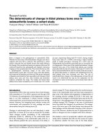
observed. Only two of these aff ected amino acid coding
sequences. The first was a 3-bp insertion in the HAdV-
14p1 sequence, which resulted in an inserted serine at
position #147 of the 25.7 K and 28 K protein sequences
(Figure 3). The proteins encoded by E1A regulate the
transcription of viral as well as cellular genes [36,37].
Thesecondwasa6-bpdeletioninthefibergeneofthe
HAdV-14p1 sequence. This resulted in a two amino
acid deletion in the FG loop of the fiber gene (Figure 4).
The fiber gene is responsible for mediating attachment
of the adenovirus to the host cell [38,39].
Although at the nucleotide level the genomes of
HAdV-14p de Wit and HAdV-14p1 strains were highly
homologous, we wanted to determine whether there was
evidence of recombination. SimPlot bootscan analysis
of HAdV-14p1 with respect to prototypical stra ins of
Figure 2 Global pairwise comparison of multiple species B HAdV genomes.
Houng et al. Respiratory Research 2010, 11:116
/>Page 5 of 8
HAdV-3,4,7,11,14,and21demonstratedthatthis
virus is m ostly closely related to the HAdV-14p proto-
type strain and i s not a recombinant with respect to
other recognized serotyp ic clades (data not shown). As
noted previously, polymorphisms between these two
strains were distributed evenly throughout the genome.
The three sequenced strains of HAdV-14p1 were
almost identical. The two Lackland isolates were exactly
the same, while the San Diego strain differed by the
addition of four ext ra Ts to the TBP polyadenylation
signal repeat, and by a single synonymous base substitu-
tion in the fiber gene.
Conclusions
HAdV-14p1 (strain 303600) appears to be a closely
related direct drift variant of the HAdV-14p (strain de
Wit) prototype seen in the past, differing primarily by
aninsertioninE1A,asmalldeletioninthefibergene,
and a few other coding single nucleotide polymorphisms
(SNPs) in E3 and other genes. The deletion (ΔK250-
E251) in the fiber gene is the most notable genetic dif-
ference between the HAdV-14p and HAdV-14p1 (Figure
4). Despite the observed ΔK250-E 251 deletion, the fiber
gene sequence of HAdV-14p1 shares greater overall
homology with the fiber of HAdV-11a than that o f
HAdV-11p. Whereas, HAdV-11p causes mostly urinary
tract infections and shares very low fiber homology with
HAdV-11p1, HAdV-11a and HAdV-14p all causing
respiratory infections [34]. This is consistent with the
receptor-binding role of the fiber and the close relation-
ship between receptor specificity and organ tropism.
HAdV-14p1 and HAdV-11a both cause upper respira-
tory infections, while HAdV-11p causes mostly urinary
tract and bone marrow infectio ns in transplant patien ts.
Species B viruses are unique in that they use CD46, a
complement protein, as a receptor [38]. Many other
human adenoviruses use the CAR protein [38]. The
deleted amino acids could affect the exposed region of
the FG loop by altering the overall affinity for CD46. A
less likely alternati ve is that t he fiber deletion influenc es
an interac tion with a receptor o ther than CD46, such as
CAR. A third possibility is that this deletion has no
affect at all on the fiber gene. Whether this mutation
affects t he pathogenicity of HAdV-14p1 compared with
HAdV-14p will require further studies.
When HAdV-14p was first identified in Eurasia in the
1950 s and 1960 s, it generated localized, high attack
rate epidemics of FRI similar to those seen with the cur-
rent strain. After a decade of sporadic activity, it disap-
peared and remained almost completely undetected for
4 decades. As a recently emerged virus, HAdV-14p1 has
an increased potential for high rates of transmission and
high attack rates, simply because the vast majority of
North Americans are likely to have never been exposed
(essentially the entire population is susceptible). This is
similar to the situation long recognized for HAdV-4,
which, in the absence of vaccines, has always been the
Table 2 Percent identities of the nucleotide coding
sequences of selected HAdV-14p1 genes to homologous
sequences of the HAdV-14p and HAdV-11p
Protein % nucleotide identity
HAdV-14p HAdV-11p
E1A 28K 98 96
E1A 25.7K 97 97
IVa2 99 98
DNA Polymerase 99 99
pTP 99 99
Penton 99 97
Hexon 99 92
100K 99 98
22K 99 97
CR1-a19895
CR1-b19994
CR1-g19891
RIDb 98 95
Fiber 99 98
ORF6/7 97 97
ORF1 98 95
Figure 3 E1A alignments. Alignment of selected E1A 28K amino acid sequences from HAdV-3, 7, 11, 14p, 14p1, and 21. Black arrow demarcates
the S147 insertion, shared by HAdV-3, 7, and 21.
Houng et al. Respiratory Research 2010, 11:116
/>Page 6 of 8
dominant serotype affecting military r ecruits. Serosur-
veys have generally indicated that a greater proportion
of the young adult population is susceptible to HAdV-4
than to other common respiratory serotypes such as
HAdV-7.
The two sequenced strains from Lackland Air Force
Base,onefromasevere(fatal)pneumoniaandonefrom
a mild case of acute respiratory d isease, were identical.
Therewereonlytwononcodingpolymorphismsdistin-
guishing the Lackland isolates from the San Diego isolate
(another mild case). The poly-T length polymorphism
was studied in a wide rang e of isolates from m ultiple
sites, and found to be a hypervariable and useful source
of geographically specific strain identity information [40].
Neither mutation suggested a significant genetic source
of variation in clinical severity. The results supported
previous observations of a high degree of conservation in
hexon and fiber genes relative to the prototype HAdV-
14p and to the closely related HAdV-11a.
Acknowledgements
The authors acknowledge the Clinic Commanders and medical staff at
Lackland Air Force Base and Wilford Hall Medical Center, San Antonio, TX
(US Air Force) and Marine Corps Recruit Depot, San Diego, CA for the
permissions, access, and assistance necessary to conduct these stud ies. The
authors also acknowledge the administrative support of the Henry M.
Jackson Foundation for Military Medicine and the efforts of the entire
WRAIR, NHRC, LRRI, and DGMC teams, especially the technicians and
collection personnel whose efforts are represented in this work.
Author details
1
Division of Viral Diseases, Walter Reed Army Institute of Research (WRAIR),
503 Robert Grant Avenue, Silver Spring, 20910, USA.
2
Infectious Disease
Program, Lovelace Respiratory Research Institute (LRRI), 2425 Ridgecrest Dr.
SE, Albuquerque, 87108, USA.
3
Clinical Investigation Facility, David Grant
USAF Medical Center (DGMC), 101 Bodin Circle, Travis Air Force Base, 94535,
USA.
4
Advanced Diagnostic Laboratory, Office of the Air Force Surgeon
General, 2460 Pepperrell Dr, Lackland Air Force Base, 78236, USA.
5
Department of Clinical Laboratory, Kaohsiung Medical University, Shih-
Chuan 1st Road, Kaohsiung,80708, Taiwan.
6
Department of Respiratory
Diseases Research, Naval Health Research Center (NHRC), 140 Sylvester Rd
San Diego, 92106, USA.
Authors’ contributions
HG carried out the sequencing of Ad14 genomes. AEK, MSJ, RAK, AL, LL, KL
and DM all participated in the samples collections, sequencing alignment
and draft of manuscript. All authors read and approved the final manuscript
submission.
Competing interests
The authors declare that they have no competing interests.
Received: 5 February 2010 Accepted: 25 August 2010
Published: 25 August 2010
References
1. Hierholzer JC, Stone YO, Broderson JR: Antigenic relationships among the
47 human adenoviruses determined in reference horse serum. Arch Virol
1991, 121:179-197.
2. Wadell G: Adenoviruses. Encyclopedia of virology New York: Academic Press
1994, 1:1-7.
3. Rogers PM, Mashtalir N, Rathod MA, Dubuisson O, Wang ZQ, et al:
Metabolically favorable remodeling of human adipose tissue by human
adenovirus Ad-36. Diabetes 2008, 57(9):2321-2331.
4. Jones MS II, Harrach B, Ganac RD, Gozum MMA, delaCruz WP, Riedel B,
et al: New adenovirus species found in a patient presenting with
gastroenteritis. J Virol 2007, 81(11):5978-5984.
5. Davison AJ, Benkő M, Harrach B: Genetic content and evolution of
adenoviruses. J Gen Virol 2003, 84:2895-2908.
6. Sambrook J, Sleigh M, Engler JA, Broker TR: The evolution of the
adenoviral genome. Ann NY Acad Sci 1980, 354:426-452.
7. Schmitz H, Wigand R, Heinrich W: Worldwide epidemiology of human
adenovirus infections. Am J Epidemiol 1983, 117:455-466.
8. Rubin BA: Clinical picture and epidemiology of adenovirus infections.
Acta Microbiol Hung 1993, 40:303-323.
9. Green M, Mackay JK, Wold WSM, Rigden P: Thirty-one human adenovirus
serotypes (Ad1-Ad31) form five speciess (A-E) based upon DNA genome
homologies. Virology 1979, 93:481-492.
10. Fox JP, Hall CE, Cooney MK: The Seattle virus watch. VII. Observations of
adenovirus infections. Am J Epidemiol 1977, 105:362-386.
11. Metzgar D, Osuna M, Yingst S, Rakha M, Earhart K, et al: PCR analysis of
Egyptian respiratory adenovirus isolates, including identification of
species, serotypes, and coinfections. J Clin Microbiol 2005, 43:5743-5752.
12. Hierholzer J: Adenoviruses. In Diagnostic procedures for viral, rickettsial and
chlamydial infections. Edited by: Lennette EH, Lennette DA, Lennette ET.
Washington, DC: American Public Health Association; , 7 1995:169-188.
13. Metzgar D, Osuna M, Kajon AE, Hawksworth AW, Irvine M, et al: Abrupt
emergence of diverse species B adenoviruses in US military recruit
training centers. J Infect Dis 2007, 196:1465-1473.
14. Brandt CD, Kim HW, Jeffries BC, Pyles G, Christmas EE, Reid JL,
et al:
Infections in 18,000 infants and children in a controlled study of
respiratory tract disease. II. Variation in adenovirus infections by year
and season. Am J Epidemiol 1972, 95:218-227.
Figure 4 Fiber knob binding site alignment. Alignment of the amino acid sequences on the exposed regions of the fiber loops FG, HI, and IJ
that are involved in binding to CD46. Black arrow demarcates the ΔK250-E251 deletion. All sequences were aligned using the ClustalX [40]
alignment method. The following HAdV genomes (GenBank accession numbers) were used: HAdV-14p (AY803294), HAdV-11p (AY163756), HAdV-
3 (NC_011203), HAdV-7 (AC_000018), and HAdV-21 (AY601633).
Houng et al. Respiratory Research 2010, 11:116
/>Page 7 of 8
15. Ryan MA, Gray GC, Smith B, McKeehan JA, Hawksworth AW, et al: Large
epidemic of respiratory illness due to adenovirus types 7 and 3 in
healthy young adults. Clin Infect Dis 2002, 34:577-582.
16. Kajon AE, Moseley JM, Metzgar D, Huong HS, Wadleigh A, et al: Molecular
epidemiology of adenovirus type 4 infections in US military recruits in
the postvaccination era (1997-2003). J Infect Dis 2007, 196:67-75.
17. Kendall EJC, Riddle RW, Tuck HA, Rodan KS, Andrews BE, et al: Pharyngo-
conjunctival fever: school outbreaks in England during the summer of
1955 associated with adenovirus types 3, 7, and 14. BMJ 1957, 2:131-136.
18. Lin KH, Lin YC, Chen HL, Ke GM, Chiang CJ, et al: A two-decade survey of
respiratory adenovirus in Taiwan: the reemergence of adenovirus types
7 and 4. J Med Virol 2004, 73:274-279.
19. van der Veen J, Oei KG, Abarbanel MFW: Patterns of infections with
adenovirus types 4, 7 and 21 in military recruits during a 9-year survey.
J Hyg (Lond) 1969, 67:255-268.
20. Morgan PN, Moses EB, Fody EP, Barron AL: Association of adenovirus type
16 with Reye’s-syndrome-like illness and pneumonia. South Med J 1984,
77:827-830.
21. Mufson MA, Belshe RB: A review of adenoviruses in the etiology of acute
hemorrhagic cystitis. J Urol 1976, 115:191-194.
22. Centers for Disease Control and Prevention: Acute respiratory disease
associated with adenovirus serotype 14 – four states, 2006-2007. MMWR
2007, 56:1181-1184.
23. Allibhai TF, Spinella PC, Meyer MT, Hall BH, Kofos D, et al: Survival after
prolonged pediatric extracorporeal membrane oxygenation support for
adenoviral pneumonia. J Pediatr Surg 2008, 43:E9-E11.
24. Louie JK, Kajon AE, Holodniy M, Guardia-LaBar L, Lee B, Petru AM, et al:
Severe pneumonia due to adenovirus serotype 14: a new respiratory
threat? Clin Infect Dis 2008, 46:421-425.
25. Mevzos LM, Il’ina TS, Makhmudov OS, Zolotarskaia EE, Dreizin RS: An
outbreak of acute respiratory infections among adults caused by
adenovirus serotype 14. Vopr Virusol [Russian] 1966, 11:426-431.
26. Bruj J, Farnik J, Sedmidubsky V: Epidemic of acute respiratory disease due
to adenovirus type 14. Cesk Epidemiol Mikrobiol Imunol [Czech]
1966,
15:165-171.
27. Cooper RJ, Hallett R, Tullo AB, Klapper PE: The epidemiology of adenovirus
infections in Greater Manchester, UK 1982-96. Epidemiol Infect 2000,
125:333-345.
28. van der Veen J, Kok G: Isolation and typing of adenoviruses recovered
from military recruits with acute respiratory disease in the Netherlands.
Am J Hyg 1957, 65:119-129.
29. Chen HL, Chiou SS, Hsiao HP, Ke GM, Lin YC, et al: Respiratory adenoviral
infections in children: a study of hospitalized cases in southern Taiwan
in 2001-2002. J Trop Pediatr 2004, 50:279-284.
30. Zhu Z, Zhang Y, Xu S, Yu P, Tian X, et al: Outbreak of acute respiratory
disease in China caused by B2 species of adenovirus type 11. J Clin
Microbiol 2009, 47(3):697-703.
31. Hierholzer JC, Pumarola A: Antigenic characterization of intermediate
adenovirus 14-11 strains associated with upper respiratory illness in a
military camp. Infect Immun 1976, 13:354-359.
32. Chmielewicz B, Benzler J, Pauli G, Krause G, Bergmann F, et al: Respiratory
disease caused by a species B2 adenovirus in a military camp in Turkey.
J Med Virol 2005, 77:232-237.
33. Li QG, Wadell G: Analysis of 15 different genome types of adenovirus
type 7 isolated on five continents. J Virol 1986, 60:331-335.
34. Kajon AE, Lu X, Erdman DD, Louie J, Schnurr D, George KS, Koopmans MP,
Allibhai T, Metzgar D: Molecular epidemiology and brief history of
emerging adenovirus 14-associated respiratory disease in the United
States. J Infect Dis 2010, 202:93-103.
35. Brudno M, Do CB, Cooper GM, Kim MF, Davydov E, et al: LAGAN and
Multi-LAGAN: efficient tools for large-scale multiple alignment of
genomic DNA. Genome Research 2003, 13(4):721-731.
36. Frisch SM, Mymryk JS: Adenovirus-5 E1A: paradox and paradigm. Nat Rev
Mol Cell Biol 2002, 3:441-452.
37. Gallimore PH, Turnell AS: Adenovirus E1A: remodelling the host cell, a life
or death experience. Oncogene 2001, 20:7824-7835.
38. Gaggar A, Shayakhmetov DM, Liszewski MK, Atkinson JP, Lieber A:
Localization of regions in CD46 that interact with adenovirus. J Virol
2005, 79:7503-7513.
39. Wang H, Tuve S, Erdman DD, Lieber A: Receptor usage of a newly
emergent adenovirus type 14. Virology 2009, 387
:436-441.
40. Houng HSH, Lott L, Gong H, Kuschner RA, Lynch JA, Metzgar D:
Adenovirus microsatellite reveals dynamics of transmission during a
recent HAdV-14 epidemic. J Clin Microbiol 2009, 47:2243-2248.
doi:10.1186/1465-9921-11-116
Cite this article as: Houng et al.: Genome sequences of Human
Adenovirus 14 isolates from mild respiratory cases and a
fatal pneumonia, isolated during 2006-2007 epidemics in North
America. Respiratory Research 2010 11:116.
Submit your next manuscript to BioMed Central
and take full advantage of:
• Convenient online submission
• Thorough peer review
• No space constraints or color figure charges
• Immediate publication on acceptance
• Inclusion in PubMed, CAS, Scopus and Google Scholar
• Research which is freely available for redistribution
Submit your manuscript at
www.biomedcentral.com/submit
Houng et al. Respiratory Research 2010, 11:116
/>Page 8 of 8