Báo cáo y học: " Open Access No evidence of altered alveolar macrophage polarization, but reduced expression of TLR2, in bronchoalveolar lavage cells in sarcoidosis" doc
Bạn đang xem bản rút gọn của tài liệu. Xem và tải ngay bản đầy đủ của tài liệu tại đây (1.19 MB, 13 trang )
RESEARC H Open Access
No evidence of altered alveolar macrophage
polarization, but reduced expression of TLR2,
in bronchoalveolar lavage cells in sarcoidosis
Maria Wikén
1*
, Farah Idali
1,2
, Muntasir Abo Al Hayja
1
, Johan Grunewald
1
, Anders Eklund
1
, Jan Wahlström
1
Abstract
Background: Sarcoidosis is a granulomatous inflammatory disease, possibly of infectious aetiology. We aimed to
investigate whether the degree of functional polarization of alveolar macrophages (AMs), or Toll-like receptor (TLR)
expression, is associated with sarcoidosis or with distinct clinical manifestations of this disease.
Methods: Total BAL cells (cultured four or 24 h in medium, or stimulated 24 h with LPS) from 14 patients and six
healthy subjects, sorted AMs from 22 patients (Löfgren’s syndrome n = 11) and 11 healthy subjects, and sorted
CD4+ T cells from 26 patients (Löfgren’s syndrome n = 13) and seven healthy subjects, were included. Using real-
time PCR, the relative gene expression of IL-10, IL-12p35, IL-12p40, IL-23 p19, CCR2, CCR7, iNOS, CXCL10, CXCL11,
CXCL16, CCL18, CCL20, CD80, and CD86, and innate immune receptors TLR2, TLR4, and TLR9, was quantified in
sorted AMs, and for selected genes in total BAL cells, while IL-17A was quantified in T cells.
Results: We did not find evidence of a difference with regard to alveolar macrophage M1/M2 polarization
between sarcoidosis patients and healthy controls. TLR2 gene expression was significantly lower in sorted AMs
from patients, particular in Löfgren’s patients. CCL18 gene expression in AMs was significantly higher in patients
compared to controls. Additionally, the IL-17A expression was lower in Löfgren’s patients’ CD4+ T cells.
Conclusions: Overall, there was no evidence for alveolar macrophage polarization in sarcoidosis. Howe ver, there
was a reduced TLR2 mRNA expression in patients with Löfgren’s syndrome, which may be of relevance for
macrophage interactions with a postulated sarcoidosis pathogen, and for the characteristics of the ensuing T cell
response.
Introduction
Sarcoidosis is a systemic T helper 1 (Th1) inflammatory
disease [1,2], primarily affecting the lungs. The hallmark of
dise ase is non-caseating granulomas where macrophages
are essential components. These cells are very heteroge-
neous, characterized by plasticity and functional polariza-
tion, with, as here named, M1 and M2 types, at the
extremes of a continuum. Due to micro-environmental
signals, such as cytokines, chemokines and Toll-like recep-
tor (TLR) ligands, macrophages differ in receptor expres-
sion, cytokine and chemokine production, as well as
effector function [3,4]. Classical activation, that is IFNg,
TNF and microbial products (e.g. lipopolysaccharide
(LPS)), elicit the M1 form. This phenotype is characterized
by high capacity to present antigens and high capacity to
produce IL-12 (promoting Th1 responses) and IL-23 (pro-
moting maturation and survival of IL-17 producing
T cells), as well as microbicidal nitric oxide and reactive
oxygen intermediates. In contrast, exposure to IL-4 or
IL-13, immune complexes, and IL-10 induce the alterna-
tive activation leading to an M2 form and a relatively
more Th2 response. High amounts of IL-10, but little IL-
12 and IL-23, and abundant expression of non-opsonic
receptors characterize this phenotype.
In addition to alveolar macrophages (AMs), s arcoido-
sis patients display increased numbers of CD4+ T lym-
phocytes in their lungs. Previously, a study from our
group and others showed that these cells are highly
positive for th e chemokine receptor CXCR3 [5]. Further
more, it has been shown that CXCR3 ligands, that is the
* Correspondence:
1
Respiratory Medicine Unit, Department of Medicine, Karolinska Institutet,
Stockholm, Sweden
Full list of author information is available at the end of the article
Wikén et al. Respiratory Research 2010, 11:121
/>© 2010 Wikén et al; licensee BioMed Central Ltd. This is an Open Access article distributed under the terms of the Creative Commons
Attribution License (http://creativecom mons .org/licenses/by/2.0), which permits unrestricted use, distribution, and reproduction in
any medium, provided the original work is properly cited.
M1 markers CXCL9, CXCL10 and CXCL11, seem
essential in the pathogenesis of pulmonary sarcoidosis
[6,7]. CXCL9 and CXCL1 0 appear to be involved in the
active phase of the granulomatous response, whereas
CXCL11, as well as CXCL10 and CXCL16 [8] and
CCL20 [9], may play a role in the acc umulation of Th1
cells, in the sarcoid lungs. However, the presence of a
recently discovered T cell subset, the IL-17 producing
Th17 cells, has to our knowledge not been investigated
in sarcoidosis. Th17 cells have been implicated in auto-
immune diseases [10] and is also important for combat-
ing extracellular pathogens [11].
The aetiology of sarcoidosis is still unknown. How-
ever, epidemiological studies and findings of DNA from
mycobacteria [12] and propionibacteria [13] and myco-
bacterial antigens [14], in sarcoidosis tissue and lymph
nodes indicate an infectious cause. This is further sup-
ported by the demonstration by us and others that
mycobacterial antigens can elicit adaptive immune
responses [15,16], which suggests a role for pattern-
recognition receptors, such as TLRs, in the pathogenesis.
TLRs are expressed on antigen presenting cells, and as
key mediators of innate host defence these receptors are
involved in recognizing several molecules derived from
microbes of different kinds. For example, mycobacteria
contain ligands for TLR2 and TLR4.
There is a considerable variation in the clinical mani-
festations of sarcoidosis. Patients who present with Löfg-
ren’ s syndrome, i.e. erythema nodosum and/or ankle
arthritis, fever and bilateral hilar lymph adenopathy with
or without parenchymal infiltration, are characterized by
an acute onset and a good prognosis and usually recover
spontaneously within two years. They are often HLA-
DRB1*03 positive. Other patients, here named non-Löfg-
ren’s syndrome patients, often have HLA-DRB1*14 or 15
haplotype, show an insidious disea se onset with dry
cough and fatigue, and are at risk of developing pul-
monary fibrosis [17] . The aim of this study was to eluci-
date if the degree of BAL cell polarization, with regard
to M1 and M2 associated cytokines, chemokines and
chemokine receptors, may be associated with sarcoido-
sis, or related to clinical manifestations of sarcoidosis. In
addition, we studied the expression of the innate
immune receptors TLR2 and TLR4.
Methods
Study subjects
Sarcoidosis patients included in this study were conse-
cutive patients referred to the Respiratory Medicine
Unit (Karolinska University Hospital, Stockholm, Swe-
den) fo r investigation. All patients were diagnosed with
pulmonary sarcoidosis as determined by sym ptoms,
chest radiography and pu lmonary function tests and the
diagnosis was established using the criteria by the
World Association of Sarcoidosis and other Granuloma-
tous Disorders (WASOG) [18]. Written informed con-
sent was obtained from all subjects, and the Regional
Ethical Review Board approved the study.
A total of 36 sarcoidosis patients and 17 healthy sub-
jects participated in this study. Total bronchoalveolar
lavage (BAL) cells of 14 sarcoidosis patients (median age
40 yrs, p25-p75 = 34-67 yrs; nine males and five
females), of which one had Löfgren’s syndrome, and six
healthy subjects (median age 28 yrs, p25-p75 = 26-
39 yrs; two males and four females), were cultured in
medium (four or 24 h), or stimulated with LPS (24 h).
In addition, AM s were sorted (see BAL and preparation
of cells), from a total of 22 sarcoido sis pati ents separate
from the above (median age 38 yrs, p25-p75 = 32-47
yrs; nine males and 13 females), of which 11 had Löfg-
ren’ s syndrome, and 11 healthy subjects (median age
27 yrs, p25-p75 = 26-30 yrs; six males and five females).
Only one patient was undergoing treatment with a non-
steroid anti-inflammatory drug (diclofenac); the rest
were untreated. All patients were fasting before BAL
was performed, and none had signs of infections. All
healthy subjects had a normal chest X-ray. Clinical and
BAL fluid characteristics, of sarcoidosis patients and
healthy subjects, are given in Table 1 and Table 2,
respectively. Significant differences in clinical parameters
between patients and healthy subjects are indicated in
Table 1. There where no significant differences in lung
function parameters or BAL cell differential counts
between patients with or without Löfgren’ s syndrome.
In addition, we used stored cDNA from CD4+ T cells,
from a total of 26 non-smoking sarcoidosis patients, of
which 13 had Löfgren’s syndrome, and seven non-smok-
ing healthy subjects. These CD4+ T cells where pre-
viousl y FACS-sorted to be included in two other studies
from our group, where a detailed patient characteriza-
tion can be found [19,20].
BAL and preparation of cells
BAL was performed as previously described [21]. Briefly,
under local anesthesia, a flexible fiber-optic broncho-
scope (OBF Type 1TR; Olympus Optical Co., Tokyo,
Japan) was passed transo rally and wedged into t he mid-
dle-lobe bronchus. Sterile phosphate-buffered saline
(PBS) solution was at 37°C instilled in five aliquots of 50
ml each. After instillation the fluid was aspirated and
collected in a siliconised plastic bottle kept on ice. The
BAL flu id was strained through a Dacron net (Millipore,
Cork, Ireland) and centrifuged at 400 × g for 10 min at
4°C, and the pellet was resuspended in RPMI-1640 med-
ium (Sigma-Aldrich , Irvin, UK). Cells were counted in a
Bürker chamber and the viability was determined by try-
pan blue exclusion. Ratio of CD4/CD8 T cell were
determined by flow cytometric analysis ( FACSCalibur
Wikén et al. Respiratory Research 2010, 11:121
/>Page 2 of 13
and FACSCanto, Becton Dickinson, Mountain View,
CA, USA) using monoclonal anti-CD3, anti-CD4, and
anti-CD8 antibodies (Dako Cytomation Norden AB,
Solna, Sweden), as previous described [22]. Using FACS-
Vantage (BD Biosciences, Mountain View, CA, USA),
AMs (recovery 0.02-2.8 × 10
6
)weresortedfromtotal
BAL cells, based on cell size (forward scatter) and gran-
ularity (side scatter). CD4+ T cells where sorted as pre-
viously described [20].
In vitro stimulation of total BAL cells
Total BAL cells were pelleted and resuspended in com-
plete medium (10
6
cells/1 ml); RPMI-1640 medium
(Sigma-Aldrich, Irvine, U.K.), s upplemented with 1%
penicillin streptomycin (Invitrogen Corporation, Paisley,
Scotland), 1% L-glutamine (Sigma-Al drich, Irvine, U.K.)
and 2% heat-inactivated human AB serum (Sigma-
Aldrich, Schnelldorf, Germany), for four or 24 h, or sti-
mulated with the classical macrophage activator and
TLR4 l igand LPS (1.6 μg/ml, Salmonella enterica sero-
type abortus equ i, Sigma-Aldrich, Schnelldorf, Germany)
for 24 h [23], in 96-well U-bottom tissue culture pla tes
at 37°C in humidified atmosphere of 5% CO
2
in air.
RNA extraction and cDNA synthesis
Total RNA was extracted via the guanidium thiocyanate
phenol-chloroform technique [24], using RNA Bee
(Nordic Biosite, Stockholm,Sweden).Briefly,1-2×10
6
cultured total BAL cells or AMs were incubated with
300-600 μL RNA Bee for 10 min at room temperature,
and then stored at -70°C until use. After thawing,
60-120 μL chloroform was added to each sample, which
Table 1 Characterization of patients
SORTED ALVEOLAR MACROPHAGES TOTAL BAL CELLS
All sarcoidosis
patients (n = 22)
Löfgren’s
syndrome (n = 11)
Non-Löfgren’s
syndrome (n = 11)
All sarcoidosis
patients (n = 14)
Sex, male/female 9/13 3/8 6/5 9/5
Age, yr 38 (32-47)** 38 (32-39)
#
40 (31-55)
##
40 (34-67)
Smoker (yes/ex/never) 1/7/14 0/3/8 1/4/6 1/4/8 (1 nd)
X-ray stage (0/I/II/III/IV) 0/11/7/3/1 0/9/2/0/0 0/2/5/3/1 2/3/7/1/0 (2 nd)
Oral steroid treatment 0 0 0 0
BAL analyses
% Recovery 73 (61-77) 73 (63-76) 72 (59-79) 68 (59-71)
% Viability 94 (92-96) 94 (92-98) 94 (92-95) 95 (92-97)
Cell concentration (*10
6
/L) 197 (130-250)** 173 (131-225)
#
213 (119-296)
##
203 (153-257)
Total cell number (*10
6
) 30 (24-38)** 31 (25-38)
#
27 (23-45)
#
32 (24-36)
BAL differential cell counts
% Macrophages 74 (62-79)* 74 (62-79) 73 (61-81) 69 (52-76)
% Lymphocytes 24 (18-38)** 23 (19-37)
#
26 (15-39) 31 (22-47)
% Neutrophils 0.9 (0-6-2.5) 1.0 (0.8-3.6) 0.8 (0.5-2.0) 1.0 (0.6-2.0)
% Eosinophils 0.3 (0-1.1) 0.2 (0-1.0) 0.5 (0-1.6) 0.4 (0-1.3)
CD4/CD8 ratio 7.9 (3.6-16) 8.5 (4.7-12) 6.7 (2.0-19) 8.5 (2.4-15)
Pulmonary function tests
VC 89 (81-97) 89 (81-96) 87 (75-98) 87 (79-100)
FEV
1
88 (84-99) 87 (85-100) 94 (82-99) 85 (79-96)
DL
CO
86 (73-94) 86 (73-95) 84 (73-93) 85 (75-99)
Data are shown as median (p25-p75)
nd = not determined
* p < 0.05, ** p < 0.01, versus sorted macrophages healthy subjects
#
p < 0.05,
##
p < 0.01, versus sorted macrophages healthy subjects
p < 0.05, versus total BAL cells healthy subjects
Pulmonary function tests:
VC: Vital capacity, % o f reference value
FEV
1
: Forced expiratory volume in one second
DL
CO
: Diffusing capacity of the lung for carbon monoxide
Values shows % of predicted
Wikén et al. Respiratory Research 2010, 11:121
/>Page 3 of 13
was shaken vigorously and kept on ice for 5-10 min.
After centrifugation at 12.000 × g for 15 min at 4°C, the
upper phase, containing RNA, was t ransferred to a new
tube and at least an equal volume of ice-cold isopropa-
nol was added. After an overnight incubation at -20°C,
the s amples were centrifuged at 12.000 × g for 20 min
at 4°C. The RNA pellet was washed in 75% ice-cold
ethanol (at 7500 × g for 10 min at 4°C), followed by air
drying for 10-15 min. The pellet was thereafter dissolved
in 20 μL autoclaved ultra clean water.
To synthesize cDNA, 1 μg of total RNA was incubated
in the presence of 20 mM random hexamers primers
(Pharmacia Biotech, Uppsala, Sw eden) and 200 units
Superscript™II RNase H
-
Reverse transcriptase (Invitro-
gen, Lidingo, Sweden) for 10 min at room temperature
andthen45minat40°C,followedby5minat95°Cto
inactivate the enzyme. T he cDNA samples were stored
at -20°C until use.
Analysis of gene expression by real-time PCR
By real-time PCR, the relative gene expression of M1
associated markers (IL-12p35, IL-12p40, IL-23p19,
CCR7, iNOS, C XCL10, CXCL11, CXCL16, CCL20,
CD80, CD86), M2 associated markers (IL-10, CCR2,
CCL18), and innate immune receptors (TLR2, TLR4,
TLR9), was quantified, using A BI Prism 7700 Sequence
Detection System (Applied Biosystems, Foster City, CA,
USA), as was the expression of IL-17A.
For human IL-10 and IL-12p40 (both according to
[2]), a PCR reaction was set up in a 25-μl reaction
volume as previously described [2]. Briefly, 1 × Taqman
buffer II, 0.5 U Ampli-Taq gold, MgCl
2
concentration
optimized for each cytokine (Applied Biosystems),
0.5 mM deoxyribonucleoside triphosphate (Amersham
Bio science, Uppsala, Sweden), 5.0 pmol of each forward
and reverse primer and 2.5 pmol probe (all from
Cybergene AB, Stockholm, Sweden). The human assay-
on-demand products f or IL-12p35 (Hs0016840 5_m1),
IL-23p19 (Hs00372324_m1), CCR7 (Hs00171054_m1),
iNOS (Hs00167257_m1), CXCL10 (Hs00171042_m1),
CXCL11 (Hs 00171138_m1), CXCL16 (Hs00222859_m1),
CCL20 (Hs00355476_m1), CD80 (Hs00175478_m1),
CD86 (Hs00199349_m1), CCR2 (Hs00356601_m1),
CCL18 (Hs00268113_m1), TLR2 (Hs00152932_m1),
TLR4 (Hs00152939_m1), TLR9 (Hs00152973_m1), IL-
17A (Hs00174383_m1), and universal master mix were
purchased commercially (Applied Biosystems, Foster
City, CA, USA).
2 μl of the cDNA (diluted in autoclaved u ltra clean
water according to pilot experiments) where put in each
well on an optical 96-well reaction p late (Applied Bio-
systems, Foster City, CA, USA) together with gene mix.
Human b-actin (hs9999903_m1, or according to [2])
was used as a housekeeping gene to normalize the
values of other genes.
The PCR condition was a followed: an initial period of
2 min at 50°C and 10 min at 95°C, followed by 40 cycles
involving denaturation at 95°C for 15 sec and annealing/
extension at 6 0°C for 1 min. All samples were run in
duplicates and the mean values were calculated.
For relative quantification of expression of cytokine
genes in cultured total BAL cells and in sorted macro-
phages, the following arithmetic formula was used: 2
-ΔΔCT
[25]. The amount of target gene was normalized to the
Table 2 Characterization of healthy subjects
SORTED ALVEOLAR MACROPHAGES TOTAL BAL CELLS
Healthy subjects
(n = 11)
Healthy subjects
(n = 6)
Sex, male/female 6/5 2/4
Age, yr 27 (26-30) 28 (26-39)
Smoker (yes/ex/never) 0/1/10 1/0/5
BAL analyses
% Recovery 74 (64-78) 70 (62-82)
% Viability 96 (95-98) 95 (93-98)
Cell concentration (*10
6
/L) 91 (69-131) 127 (86-187)
Total cell number (*10
6
) 18 (12-22) 20 (12-32)
BAL differential cell counts
% Macrophages 84 (79-89) 87 (64-93)
% Lymphocytes 14 (9.0-20) 11 (5.1-33)
% Neutrophils 2.0 (0.7-2.8) 1.9 (1.0-2.1)
% Eosinophils 0.2 (0-1.2) 0.3 (0-0.9)
Data are shown as median (p25-p75)
Wikén et al. Respiratory Research 2010, 11:121
/>Page 4 of 13
housekeeping gene (b-actin) and the relative expression of
targe t genes in cultured total BAL cells was calculated in
relation to the mean values of target gene e xpression in
healthy subjects after 24 h of incubation in medium alone.
The relative expression of target genes in sorted macro-
phages was calculated in relation to the mean values of
targ et gene expression in the healthy subject group. PCR
amplification efficiencies for both the endogenous control
( b-actin) and target genes were tested through serially
diluting a cDNA sample and showing that the CT differ-
ence between the target and endogenous control remained
constant.
Statistical methods and data management
The Mann-Whitn ey U-test was used for comparison of
relative gene expression between sarcoidosis patients
and healthy subjects. One-way ANOVA (Kruskal-
Wallis), was used for comparison of relative gene
expression between patient subgroups (with and without
Löfgren’s syndrome), and healthy subjects. In the case of
a statistically significant result in the ANOVA, statistical
comparisons were made by use of the post-hoc test pro-
posed by Dunn to control for multiplicity. The within
group analysis (cell culture 24 h with or without LPS)
wasmadebyuseoftheWilcoxonSignedRankTest.
The Spearman rank correlation coefficient was used in
order to test independence between variabl es. A p value
of <0.05 was considered as significant. The study
employs multiple hypotheses testing, where each
hypothesis was analyzed separately and the existence of
pattern s in and the consistency of the results were con-
sidered in the analysis. All analyses were carried out by
use of the computer program GraphPad PRISM 4.03
(GraphPad Software Inc., San Diego, CA, USA).
Results
TherelativegeneexpressionofseveralM1andM2
associated markers was quantified in culture d total BAL
cells and sorted AMs from sarcoidosis patients, with or
without Löfgren’ s syndrome, and healthy subjects. In
addition, the expression of selected TLRs was measured
in sorted AMs, and IL-17A was measured in sorted
CD4+ T cells.
Gene expression after culturing and stimulation of total
BAL cells
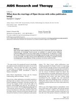
First we studied the relative gene expression of the typi-
cal M1 markers, IL-12p35 (Fig. 1a), IL-23p19 (Fig. 1b),
CCR7(Fig.1c),andM2markers,IL-10(Fig.1d)and
CCR2 (Fig. 1e), in total BAL cells of sarcoidosis patients
and healthy subjects after culturing in medium for four
or 24 h, or after 24 h of LPS stimulation. The number
of markers and stimulating conditions that were
invest igated was limited by the amount of cells available
for the study.
In total BAL cells we found no evidence of different
M1/M2 polarization between patients and controls,
either after 24 h incubation in medium alone or with
LPS stimul ation (Fig. 1). There were only minor differ-
ences in gene expression after 4 h culturing in medium
alone (data not shown) compared to after 24 h culturing
in medium. Due to limitations in the number of cells
available, gene expression after all three culture condi-
tions could not be studied in all individuals. The exact
numbers of included patients or healthy subjects are
indicated under each plot (Fig. 1). Data from the
patients and healthy subjects, where it was possible to
study all three culture conditions, is shown in Figure 2
and 3. LPS upregulated IL-12p3 5 and IL-23p19 in both
groups (this was not statistically significant for IL-12p35
in controls, probably because of too few individuals).
When comparing the magnitude of upregulation (the
differences between relative gene expression after 24 h
incubation in medium alone or with LPS stimulation),
we noted tendencies of lower upregulation in sarcoidosis
patients. In addition, LPS significantly upregulated IL-10
in healthy controls.
It was noted that patients with the highest gene
expression after 24 h of c ulturing of total BAL cells in
medium alone had a large decrease in production after
LPS stimulation. The two patients showing that pattern
had extrathoracic disease, pronounced BAL lymphocyto-
sis, and high BAL CD4/CD8 ratio.
Since there were only total BAL cells from one patient
with Löfgren’ s syndrome, no subgroup comparison was
done.
Screening of M1 and M2 associated markers in alveolar
macrophages
We next freshly isolated AMs, using flow cytometric cell
sorting, and measured the relative gene expression of a
wide range of different M1 and M2 markers.
We did not find any significant differences between
patients and healthy subjects, and no difference between
patients subgroups wit h regard to the M1 markers
CXCL10, C XCL11, CXCL16, CD80 (Fig. 4), CD86 and
CCL20 (data not shown). The expression of IL-12p40,
IL-23p19, CCR7 and iNOS from most of the samples
were below the detection limit (data not shown).
As shown in Figure 5a, the relative gene expression of
CCL18 (an M2 associated marker) was significantly
higher in sarcoidosis patients compared to healthy sub-
jects (p = 0.034). Ye t, there was no difference between
patient subgroups (Fig. 5b). The expression of CCL18
was positively correlated with the percentage of lympho-
cytes in BAL fluid (data not shown). There were no
Wikén et al. Respiratory Research 2010, 11:121
/>Page 5 of 13
differences between patients and healthy subjects
regarding IL-10 (Fig. 5c) or CCR2 (data not shown).
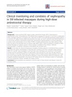
TLR mRNA expression in alveolar macrophages
There was a significantly reduced TLR2 gene expression
in sorted AMs from s arcoido sis patients compare d to
healthy subjects (p = 0.024) (Fig. 6a). Furthermore,
when studying patient subgroups, a significantly lower
TLR2 expression was observed in patients with Löfgren’s
syndrome compared to healthy subjects (p = 0.0058)
(Fig. 6b). The relative gene expression of TLR4 (Fig. 6c,
d), and TLR9 (data not shown), did not differ signifi-
cantly between the groups.
IL-17A mRNA expression in sorted CD4+ T cells
The tendency to a less pronounced upregulation of
IL-23p19 expression in sarcoidosis patients after LPS sti-
mulation, made us believe that this could have an
impact on the induction of Th17 cells. We therefore
analysed IL-17A expression in FACS-sorted CD4+
T cells. As shown in Figure 7a, the relative gene expres-
sion of IL-17A was lower i n patients, statistically signifi-
cant in pa tien ts with Löfgren’ssyndrome.However,this
needs to be interpreted with caution since the signifi-
cance is lost if the outlier with the highest IL-17 mRNA
expression among the healthy controls is omitted.
Furthermore, in patients there was a negative correlation
of IL-17A expression with CD4/CD8 BALF ratio
(Fig. 7b).
Since there is a reciprocal interconnection between
the development of Th17 cells and Treg cells [26], and
Th17 cells can be controlled by Foxp3+ T regulatory
cells [27], we attempted to investigate the balance
between these two subsets by correlating IL-17 mRNA
expression with that of Foxp3, using data for Foxp3
mRNA expression in the same samples of sorted CD4+
BAL T cells already included in a previous study [19].
However, no correlation was found, either in total
patients or in subgroups (Löfgren and non-Löfgren)
(data not shown).
Discussion
Inthepresentstudyweaimedtoinvestigatewhether
functional polarization of alveolar macrophages was
associated with sarcoidosis, or with patient subgroups.
This was found not to be the case, although there was a
higher expression of the fibrosis-associated marker
CCL18 in AMs in the whole group of patients. In addi-
tion, we s tudied the expression of patte rn-recognitio n
receptors TLR2 and TLR4, and found that AMs from
Figure 1 Total BAL cell expression of the M1 markers IL-12p35 (a), IL-23p19 (b) and CCR7 (c), and the M2 markers IL-10 (d) and CCR2
(e) mRNA, in healthy controls (HC) and sarcoidosis patients (Sarc), cultured in medium (24 h), or stimulated with LPS (24 h). Horizontal
lines depict median values. (n: number of individuals analyzed.)
Wikén et al. Respiratory Research 2010, 11:121
/>Page 6 of 13
Figure 2 Intra-individual comparisons of relative gene expression of the M1 markers IL-12p35 (a, b), IL-23p19 (c, d) and CCR7 (e, f) in
total BAL cells cultured in medium (four or 24 h), or stimulated with LPS (24 h), in sarcoidosis patients (left columns) and healthy
controls (right columns).
Wikén et al. Respiratory Research 2010, 11:121
/>Page 7 of 13
patients, in particular those with Löfgren’ ssyndrome,
had a lower expression of TLR2.
In contrast to our negative findings regarding macro-
phage polarization, previous studies have demonstrated
higher expression of the M1 markers CXCL10 [6],
CXCL11 [7] and CXCL16 [8] in sarcoidosis patients
compa red to healthy subjects. However, in those studies
AMs or total BALF cell s were culture d in medium over
night before gene expression were measured. In the pre-
sentstudywefocusedonfreshlyisolatedAMs.These
differences in results may arise because a certain degree
of stimulation is needed to make macrophages express
cytokine genes and reveal their functional potential. E.g.
it has been shown that adherence to plastic can by itsel f
cause macrophage ac tivation and cytokine production
[23]. A lack of such in vitro activation in our study may
also explain why the mRNA levels of some genes such
as IL-2p35 were often below the detection limit, in
agreement with previous studies [28].
Similarlytoapreviousstudy[29],weheredemon-
strate an increased gene expression of CCL18 in AMs of
sarcoidosispatientsascomparedtohealthysubjects.
However, anot her study found no differences in CCL18
mRNA expression between pati ents and controls, yet in
that study total BAL fluid cells and not sorted macro-
phages were studied [30]. Functional studies of CCL18
has indicated a profibrotic role for this chemokine as
part of a positive feedback loop between AMs and fibro-
blasts [29]. CCL18 has been reported to be an indicator
of pulmonary fibrosis since BAL cells of sarcoidosis
patients with X-ray stage IV produce higher levels of
CCL18 compared to a lower X-ray stage [29]. There
have also been findings of high levels of CCL18 in
patients with other fibrosing lung disorders [31,32]. In
addition, plasma levels of CCL18 have been suggested to
be a marker of disease activity [33]. We did not find any
correlations with X-ray stage, or lung function para-
meters in our study. However, the majority of our
patients had X-ray stage I or II, w ith only one at X-ray
stage IV. Neither did we observe any difference in
CCL18 e xpression between patients with L öfgren ’ssyn-
drome or not. We found, however, that the expression
of CCL18 was positively correlated with the percentage
of lymphocytes. Therefore, CCL18 may act mainly as a
T cell attractant, preferentia lly of naïve T cells [34], in
the early stage of disease, whiletheprofibroticroleof
CCL18 may only be imp ortant in more advanced
disease.
We found that AMs of pat ients are characterized by a
lower gene expression of TLR2 compared to healthy
subjects. This is in contrast t o our previo us report o f
higher expression of TLR2 and TLR4 receptors on
Figure 3 Intra-individual comparisons of relative gene expression of the M2 markers IL-10 (a, b) and CCR2 (c, d) in total BAL cells
cultured in medium (four or 24 h), or stimulated with LPS (24 h), in sarcoidosis patients (left columns) and healthy controls (right
columns).
Wikén et al. Respiratory Research 2010, 11:121
/>Page 8 of 13
Figure 4 Alveolar macrophage expression of the M1 markers CXCL10 (a, b), CXCL11, (c, d), CXCL16 (e, f) , and CD80 (g, h) mRN A of
(a, c, e, g) healthy controls (HC) and sarcoidosis patients (Sarc), and (b, d, f, h) HC, Löfgren’s syndrome patients and Non-Löfgren’s
syndrome patients. Horizontal lines depict median values. (n: number of individuals analyzed.)
Wikén et al. Respiratory Research 2010, 11:121
/>Page 9 of 13
blood monocytes [35], although it should be noted that
different cell types w ere studied (monocytes vs.macro-
phages) and different techniques used (cell surface
receptor expression vs. mRNA expression). One expla-
nation for this difference in TLR expression between
lung and blood could be differences in exposure to var-
ious stimuli known to aff ect TLR expression. For exam-
ple, TLR ligands are able to up- or downregulate TLR
mRNA expression depending on dose and time [35-37]
In addition, the cell surface level of TLR2 was found to
be increased by some cytokines, and decreased by others
[38]. E.g. IFNg and TNF downregulated TLR2 levels on
human monocytes, althoug h macrophages were not stu-
died. Therefore, the totality of different inflammatory
mediators p resent in different compartments are likely
to determine local TLR expression. Moreover, the TLR2
down-regulation was mainly seen in patients with Löfg-
ren’s syndrome, possibly indicating that these patients
respond to a particular ligand that specifically binds to
TLR2.
The finding of a tendency to a smaller LPS-induced
increase in IL-23 expression in sarcoidosis patients
raised the question whether Th17 cells may be differ-
ently affected in sarcoidosis patients compared to
healthy subjects. We found that the relative gene expres-
sion of IL-17 in CD4+ T cells was reduced in sarcoido-
sis patients, statistically significant (although weakly so)
in patients with Lö fgren’ s syndro me compared to
healthy subjects. The reason for this is not cle ar, but it
may be related to findings in recent studies reviewed in
[39] showing that the Th17 phenotype is unstable, and
via T-bet induction can be converted to a Th1 pheno-
type. It may be speculated that the Th1-inducing lung
milieu characteristic of sarcoidosis has such an effect on
Th17 cells. Furthermore, we found a negative correla-
tion between BALF CD4+ T cell IL-17A expression and
BALF CD4/CD8 ratio in sarcoidosis, suggesting that the
alveolitis seen in patients is associated with an influx of
CD4 T cells with on average merely little production of
IL-17. The roles of IL-23 and IL-17 in sarcoidosis clearly
merit further investigation.
In a recent publication it was shown that smoking can
affect macrophage polarization in the M2 direction [40].
Although there were very few current smokers among
the patients and controls in our study, the smoking his-
tory differed when ex-smokers were taken into account.
Previous smoking could potentially affect macrophage
polarization, although that was not addressed in the
Figure 5 Alveolar macrophage expression of the M2 markers CCL18 (a, b) and IL-10 (c, d) mRNA in (a, c) he althy controls (HC) and
sarcoidosis patients, and in (b, d) HC, Löfgren’s syndrome patients and Non-Löfgren’s syndrome patients. Horizontal lines depict median
values. * p < 0.05. (n: number of individuals analyzed.)
Wikén et al. Respiratory Research 2010, 11:121
/>Page 10 of 13
above mentioned study. However, considering the diffi-
culties in recruiting healthy controls for bronchoalveolar
lavage obtaining a desired match with patients for fac-
tors such as smoking history and age is not practically
feasible. Also, the above mentioned study was published
after the samples for the present study were collected.
We did not observe any obvious differences with regard
to the expression of the different genes between indivi-
duals with different smoking status. Also, another report
suggests that current, but not previous smoking, i s
important for macrophage gene expression. Using
microarray analysis of human alveolar macrophages, it
was found that despite the inclusion of ex-smokers in
the grou p of non-smokers, there was complete segrega-
tion between non-smokers and smokers after cluster
analysis of the 200 genes with the highest variance
across smokers and non-smokers [41].
In conclusion, we did not find evidence for a diffe r-
ence in alveolar macrophage polarization in patients
compared to healthy controls. The role of IL-23/IL-17
Figure 6 Expression of TLR2 (a, b) and TLR4 (c, d) mRNA in alveolar macrophages in (a, c) heal thy controls (HC) and sarcoidosis
patients, and in (b, d) HC, Löfgren’s syndrome patients and Non-Löfgren’s syndrome patients. Horizontal lines depict median values.
* p < 0.05, ** p < 0.01. (n: number of individuals analyzed.)
Figure 7 Expression of IL-17A mRNA in BAL fluid CD4+ T lymphocytes in healthy subjects (HC), Löfgren’s syndrome patients and Non-
Löfgren’s syndrome patients (a). Horizontal lines depict median values. * p < 0.05. Correlation between IL-17A mRNA expression and
BALF CD4/CD8 ratio (b).
Wikén et al. Respiratory Research 2010, 11:121
/>Page 11 of 13
axis in sarcoidosis merits further study. The reduced
TLR2 expression of AMs from patients with Löfgren’s
syndrome may suggest a distinct innate immune
response to pathogens in these patients.
Acknowledgements
The authors thank Heléne Blomqvist, Margitha Dahl, Benita Dahlberg, Gunnel
de Forest and Lotta Pousette for their excellent technical assistance. We
thank Per Näsman for advice regarding statistical analyses. This study was
supported by the Swedish Heart-Lung Foundation, King Oscar II Jubilee
Foundation, the Swedish Research Council, the U.S. National Institutes of
Health (Grant No. 1 R21 HL077579-01), the Stockholm County Council and
Karolinska Institutet.
Author details
1
Respiratory Medicine Unit, Department of Medicine, Karolinska Institutet,
Stockholm, Sweden.
2
Department of Reproductive Immunology,
Reproductive Biotechnology Research center, Avicenna Research Institute,
Shahid Beheshti University, Evin, Tehran, Iran.
Authors’ contributions
MW participated in study planning, performed patient material collection,
performed experiments and data analysis, and wrote the manuscript. FI
performed patient material collection, performed experiments, and critically
reviewed the manuscript. MH performed experiments. JG participated in
study planning, recruitment of patients, and critically reviewed the
manuscript. AE participated in study planning, recruitment of patients, and
critically reviewed the manuscript. JW conceived the study and its design,
performed data analysis, and supervised the writing of the manuscript. All
authors read and approved the final manuscript.
Competing interests
The authors declare that they have no competing interests.
Received: 22 October 2009 Accepted: 2 September 2010
Published: 2 September 2010
References
1. Moller DR, Forman JD, Liu MC, Noble PW, Greenlee BM, Vyas P, Holden DA,
Forrester JM, Lazarus A, Wysocka M, et al: Enhanced expression of IL-12
associated with Th1 cytokine profiles in active pulmonary sarcoidosis. J
Immunol 1996, 156(12):4952-4960.
2. Idali F, Wiken M, Wahlstrom J, Mellstedt H, Eklund A, Rabbani H,
Grunewald J: Reduced Th1 response in the lungs of HLA-DRB1*0301
patients with pulmonary sarcoidosis. Eur Respir J 2006, 27(3):451-459.
3. Martinez FO, Sica A, Mantovani A, Locati M: Macrophage activation and
polarization. Front Biosci 2008, 13:453-461.
4. Benoit M, Desnues B, Mege JL: Macrophage polarization in bacterial
infections. J Immunol 2008, 181(6):3733-3739.
5. Katchar K, Eklund A, Grunewald J: Expression of Th1 markers by lung
accumulated T cells in pulmonary sarcoidosis. J Intern Med 2003,
254(6):564-571.
6. Agostini C, Cassatella M, Zambello R, Trentin L, Gasperini S, Perin A,
Piazza F, Siviero M, Facco M, Dziejman M, et al: Involvement of the IP-10
chemokine in sarcoid granulomatous reactions. J Immunol 1998,
161(11):6413-6420.
7. Nishioka Y, Manabe K, Kishi J, Wang W, Inayama M, Azuma M, Sone S:
CXCL9 and 11 in patients with pulmonary sarcoidosis: a role of alveolar
macrophages. Clin Exp Immunol 2007, 149(2):317-326.
8. Agostini C, Cabrelle A, Calabrese F, Bortoli M, Scquizzato E, Carraro S,
Miorin M, Beghe B, Trentin L, Zambello R, et al: Role for CXCR6 and its
ligand CXCL16 in the pathogenesis of T-cell alveolitis in sarcoidosis. Am
J Respir Crit Care Med 2005, 172(10):1290-1298.
9. Facco M, Baesso I, Miorin M, Bortoli M, Cabrelle A, Boscaro E, Gurrieri C,
Trentin L, Zambello R, Calabrese F, et al: Expression and role of CCR6/
CCL20 chemokine axis in pulmonary sarcoidosis. J Leukoc Biol 2007,
82(4):946-955.
10. Louten J, Boniface K, de Waal Malefyt R: Development and function of
TH17 cells in health and disease. J Allergy Clin Immunol 2009,
123(5):1004-1011.
11. Iwakura Y, Ishigame H: The IL-23/IL-17 axis in inflammation. J Clin Invest
2006, 116(5):1218-1222.
12. Saboor SA, Johnson NM, McFadden J: Detection of mycobacterial DNA in
sarcoidosis and tuberculosis with polymerase chain reaction. Lancet
1992, 339(8800):1012-1015.
13. Abe C, Iwai K, Mikami R, Hosoda Y: Frequent isolation of
Propionibacterium acnes from sarcoidosis lymph nodes. Zentralbl
Bakteriol Mikrobiol Hyg [A] 1984, 256(4):541-547.
14. Song Z, Marzilli L, Greenlee BM, Chen ES, Silver RF, Askin FB, Teirstein AS,
Zhang Y, Cotter RJ, Moller DR: Mycobacterial catalase-peroxidase is a
tissue antigen and target of the adaptive immune response in systemic
sarcoidosis. J Exp Med 2005, 201(5):755-767.
15. Drake WP, Dhason MS, Nadaf M, Shepherd BE, Vadivelu S, Hajizadeh R,
Newman LS, Kalams SA: Cellular recognition of Mycobacterium
tuberculosis ESAT-6 and KatG peptides in systemic sarcoidosis. Infect
Immun 2007, 75(1):527-530.
16. Chen ES, Wahlstrom J, Song Z, Willett MH, Wiken M, Yung RC, West EE,
McDyer JF, Zhang Y, Eklund A, et al: T cell responses to mycobacterial
catalase-peroxidase profile a pathogenic antigen in systemic sarcoidosis.
J Immunol 2008, 181(12):8784-8796.
17. Berlin MF-HA, Olerup O, Eklund A, Grunewald J: HLA-DR predicts the
prognosis in Scandinavian patients with pulmonary sarcoidosis. Am J
Respir Crit Care Med 1997, 156:1601-1605.
18. Statement on sarcoidosis. Joint Statement of the American Thoracic
Society (ATS), the European Respiratory Society (ERS) and the World
Association of Sarcoidosis and Other Granulomatous Disorders (WASOG)
adopted by the ATS Board of Directors and by the ERS Executive
Committee, February 1999. Am J Respir Crit Care Med 1999,
160(2):736-755.
19. Idali F, Wahlstrom J, Muller-Suur C, Eklund A, Grunewald J: Analysis of
regulatory T cell associated forkhead box P3 expression in the lungs of
patients with sarcoidosis. Clin Exp Immunol 2008, 152(1):127-137.
20. Idali F, Wahlstrom J, Dahlberg B, Khademi M, Olsson T, Eklund A,
Grunewald J: Altered expression of T cell immunoglobulin-mucin (TIM)
molecules in bronchoalveolar lavage CD4+ T cells in sarcoidosis. Respir
Res 2009, 10:42.
21. Eklund A, Blaschke E: Relationship between changed alveolar-capillary
permeability and angiotensin converting enzyme activity in serum in
sarcoidosis. Thorax 1986, 41(8):629-634.
22. Wahlstrom J, Dahlen B, Ihre E, Wigzell H, Grunewald J, Eklund A: Selective
CD8+ T cells accumulate in the lungs of patients with allergic asthma
after allergen bronchoprovocation. Clin Exp Immunol 1998, 112(1):1-9.
23. Krause SW, Kreutz M, Andreesen R: Differential effects of cell adherence
on LPS-stimulated cytokine production by human monocytes and
macrophages. Immunobiology 1996, 196(5):522-534.
24. Chomczynski P, Sacchi N: Single-step method of RNA isolation by acid
guanidinium thiocyanate-phenol-chloroform extraction. Anal Biochem
1987, 162(1):156-159.
25. Perkin-Elmer Instruction manual. User Bulletin no. 2. Foster City, CA,
USA. Perkin-Elmer Applied Biosystems Inc 1997, 1-36.
26. Korn T, Bettelli E, Oukka M, Kuchroo VK: IL-17 and Th17 Cells. Annu Rev
Immunol 2009, 27
:485-517.
27. Chaudhry A, Rudra D, Treuting P, Samstein RM, Liang Y, Kas A,
Rudensky AY: CD4+ regulatory T cells control TH17 responses in a Stat3-
dependent manner. Science 2009, 326(5955):986-991.
28. Shigehara K, Shijubo N, Ohmichi M, Takahashi R, Kon S, Okamura H,
Kurimoto M, Hiraga Y, Tatsuno T, Abe S, et al: IL-12 and IL-18 are
increased and stimulate IFN-gamma production in sarcoid lungs. J
Immunol 2001, 166(1):642-649.
29. Prasse A, Pechkovsky DV, Toews GB, Jungraithmayr W, Kollert F,
Goldmann T, Vollmer E, Muller-Quernheim J, Zissel G: A vicious circle of
alveolar macrophages and fibroblasts perpetuates pulmonary fibrosis via
CCL18. Am J Respir Crit Care Med 2006, 173(7):781-792.
30. Mrazek F, Sekerova V, Drabek J, Kolek V, du Bois RM, Petrek M: Expression
of the chemokine PARC mRNA in bronchoalveolar cells of patients with
sarcoidosis. Immunol Lett 2002, 84(1):17-22.
Wikén et al. Respiratory Research 2010, 11:121
/>Page 12 of 13
31. Prasse A, Pechkovsky DV, Toews GB, Schafer M, Eggeling S, Ludwig C,
Germann M, Kollert F, Zissel G, Muller-Quernheim J: CCL18 as an indicator
of pulmonary fibrotic activity in idiopathic interstitial pneumonias and
systemic sclerosis. Arthritis Rheum 2007, 56(5):1685-1693.
32. Kodera M, Hasegawa M, Komura K, Yanaba K, Takehara K, Sato S: Serum
pulmonary and activation-regulated chemokine/CCL18 levels in patients
with systemic sclerosis: a sensitive indicator of active pulmonary fibrosis.
Arthritis Rheum 2005, 52(9):2889-2896.
33. Boot RG, Hollak CE, Verhoek M, Alberts C, Jonkers RE, Aerts JM: Plasma
chitotriosidase and CCL18 as surrogate markers for granulomatous
macrophages in sarcoidosis. Clin Chim Acta 2010, 411(1-2):31-36.
34. Adema GJ, Hartgers F, Verstraten R, de Vries E, Marland G, Menon S,
Foster J, Xu Y, Nooyen P, McClanahan T, et al: A dendritic-cell-derived C-C
chemokine that preferentially attracts naive T cells. Nature 1997,
387(6634):713-717.
35. Wiken M, Grunewald J, Eklund A, Wahlstrom J: Higher Monocyte
Expression of TLR2 and TLR4, and Enhanced Pro-inflammatory Synergy
of TLR2 with NOD2 Stimulation in Sarcoidosis. J Clin Immunol 2008,
29(1):78-89.
36. Triantafilou M, Manukyan M, Mackie A, Morath S, Hartung T, Heine H,
Triantafilou K: Lipoteichoic acid and toll-like receptor 2 internalization
and targeting to the Golgi are lipid raft-dependent. J Biol Chem 2004,
279(39):40882-40889.
37. Maris NA, Dessing MC, de Vos AF, Bresser P, van der Zee JS, Jansen HM,
Spek CA, van der Poll T: Toll-like receptor mRNA levels in alveolar
macrophages after inhalation of endotoxin. Eur Respir J 2006,
28(3):622-626.
38. Flo TH, Halaas O, Torp S, Ryan L, Lien E, Dybdahl B, Sundan A, Espevik T:
Differential expression of Toll-like receptor 2 in human cells. J Leukoc Biol
2001, 69(3):474-481.
39. Afzali B, Mitchell P, Lechler RI, John S, Lombardi G: Translational mini-
review series on Th17 cells: induction of interleukin-17 production by
regulatory T cells. Clin Exp Immunol 159(2):120-130.
40. Shaykhiev R, Krause A, Salit J, Strulovici-Barel Y, Harvey BG, O’Connor TP,
Crystal RG: Smoking-dependent reprogramming of alveolar macrophage
polarization: implication for pathogenesis of chronic obstructive
pulmonary disease. J Immunol 2009, 183(4):2867-2883.
41. Woodruff PG, Koth LL, Yang YH, Rodriguez MW, Favoreto S, Dolganov GM,
Paquet AC, Erle DJ: A distinctive alveolar macrophage activation state
induced by cigarette smoking. Am J Respir Crit Care Med 2005,
172(11):1383-1392.
doi:10.1186/1465-9921-11-121
Cite this article as: Wikén et al.: No evidence of altered alveolar
macrophage polarization, but reduced expression of TLR2,
in bronchoalveolar lavage cells in sarcoidosis. Respiratory Research 2010
11:121.
Submit your next manuscript to BioMed Central
and take full advantage of:
• Convenient online submission
• Thorough peer review
• No space constraints or color figure charges
• Immediate publication on acceptance
• Inclusion in PubMed, CAS, Scopus and Google Scholar
• Research which is freely available for redistribution
Submit your manuscript at
www.biomedcentral.com/submit
Wikén et al. Respiratory Research 2010, 11:121
/>Page 13 of 13