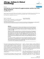
Báo cáo y học: "Detection of Chlamydia trachomatis-DNA in synovial fluid: evaluation of the sensitivity of different DNA extraction methods and amplification systems" pps
Bạn đang xem bản rút gọn của tài liệu. Xem và tải ngay bản đầy đủ của tài liệu tại đây (367.19 KB, 10 trang )
Open Access
Available online />Page 1 of 10
(page number not for citation purposes)
Vol 11 No 6
Research article
Detection of Chlamydia trachomatis-DNA in synovial fluid:
evaluation of the sensitivity of different DNA extraction methods
and amplification systems
Julia Freise
1
, Iris Bernau
2
, Sabine Meier
3
, Henning Zeidler
4
* and Jens G Kuipers
5
*
1
Division of Pneumology, Hannover Medical School, Carl-Neuberg Straße 1, Hannover, 30625, Germany
2
Division of Anaesthesiology, Diako Hospital, Gröpelinger Heerstraße 406 - 408, Bremen, 28239, Germany
3
Division of Immunology and Rheumatology, Hannover Medical School, Carl-Neuberg Straße. 1, Hannover, 30625, Germany
4
Rheumatologikum, Rathenau-Straße 13-14, Hannover, 30159, Germany
5
Division of Rheumatology, Rotes Kreuz Krankenhaus, St Pauli-Deich 24, Bremen, 28199, Germany
* Contributed equally
Corresponding author: Julia Freise,
Received: 16 May 2009 Revisions requested: 18 Jun 2009 Revisions received: 14 Oct 2009 Accepted: 21 Nov 2009 Published: 21 Nov 2009
Arthritis Research & Therapy 2009, 11:R175 (doi:10.1186/ar2864)
This article is online at: />© 2009 Freise et al.; licensee BioMed Central Ltd.
This is an open access article distributed under the terms of the Creative Commons Attribution License ( />),
which permits unrestricted use, distribution, and reproduction in any medium, provided the original work is properly cited.
Abstract
Introduction Polymerase chain reaction (PCR) and ligase chain
reaction (LCR) are used in research for detection of Chlamydia
trachomatis (C. tr.) in synovial fluid (SF). However there is no
standardized system for diagnostic use in clinical practice,
therefore this study aimed at determining the molecular biology
method best suited to detect C. tr. from SF.
Methods SF samples were spiked with C. tr. elementary bodies
(EB) and human peripheral blood monocytes (PBMo)
persistently infected with C. tr. in vitro to evaluate the sensitivity
of different molecular biology methods and assays. Five different
DNA-extraction methods were tested: 1) Alkaline lysis, 2) QIAex
II Gel Extraction Kit
®
+ CTAB, 3) Chelex
®
-extraction, 4) QIAmp
Tissue Kit
®
and 5) QIAmp DNA Stool Kit
®
. All DNA extracts
were subjected to 5 different DNA amplification systems to
detect C. tr DNA in the spiked SF samples: two C. tr. -omp1
directed PCR, one C. tr plasmid-PCR, one C. tr. -16s RNA
directed PCR, and one commercially available LCR (LCX
®
,
Abbott laboratories).
Results In SF samples spiked with C. tr EB and with C. tr
PBMo, alkaline lysis, detecting 1 C. tr EB/ml SF, 0,1 C. tr
PBMo/ml SF and QIAmp gel extraction kit
®
+ CTAB detecting
0,1 C. tr. -EB/ml SF, 1 C. tr PBMo/ml, respectively, allowed
most sensitive detection of the organism in combination with the
C. tr omp1-(152 bp) PCR. Sensitivity decreased in all methods
after storage of the DNA of C. tr dilution series at -20°C for 4
months by at least one log phase.
Conclusions The sensitivity to detect C. tr DNA from SF is
highly dependent on the DNA extraction method and the
detection system applied. Alkaline lysis as well as the QIAmp
Gel extraction kit
®
+ CTAB in combination with C. tr omp1 -
(152 bp) PCR evolved as the most sensitive methods to identify
C. tr. in serial dilutions.
Introduction
Chlamydia-induced arthritis (CIA) is the most frequent form of
reactive arthritis (ReA) in western countries [1]. The hallmark
of CIA is that the synovitis eliciting bacteria persist intraarticu-
larly in very low quantities but cannot be cultured from synovial
fluid (SF) [2,3]. Initially, immunofluorescence studies and RNA
hybridization of synovial specimens were the first methods
demonstrating intra-articularly persisting Chlamydia trachom-
atis [4,5]. Subsequently, from numerous reports PCR
emerged as a very promising tool for the identification of C.
trachomatis in the SF of patients with CIA and related dis-
eases [1,6-15]. Moreover, PCR should overcome the limita-
tions of clinical, urogenital, and serologic diagnosis of this form
of ReA [16].
We previously investigated which DNA extraction methods
provide the best template for PCR analysis of DNA from SF
samples [8,17] as well as for synovial tissue [9]. Our results
bp: base pairs; BSA: bovine serum albumin; CIA: chlamydia induced arthritis; EB: elementary bodies; IFU: infection forming units; LCR: ligase chain
reaction; MOMP: major outer membrane protein; Omp-1: major outer membrane protein 1; PBMO: peripheral blood monocytes; PBS: phosphate
buffered saline; PCR: polymerase chain reaction; ReA: reactive arthritis; SD: standard deviation; SF: synovial fluid.
Arthritis Research & Therapy Vol 11 No 6 Freise et al.
Page 2 of 10
(page number not for citation purposes)
are consistent with those of other groups that noted the rele-
vance of optimized template preparation for SF as well as for
synovial tissue [18]. At present no standardized approach for
Chlamydia-directed PCR has been described.
The aim of the present study was to define a standardized and
optimized test system to evaluate clinical SF samples for C.
trachomatis DNA in routine laboratory analysis. To address
this issue we analyzed SF using spiked SF samples and
human peripheral blood monocytes (PBMO) persistently
infected with C. trachomatis in vitro in serial dilutions to inves-
tigate which template preparation methods provide the best
amplification substrate for each different assay type. We also
tested four different PCR systems and one commercially avail-
able ligase chain reaction (LCR) protocol in use for urogenital
samples in order to determine the most sensitive system to
detect chlamydial DNA from SF. The two systems best suited
for detection of C. trachomatis was applied to clinical samples
of SF (data submitted elsewhere).
Materials and methods
Ethical approval
Before initiation of the study ethical approval was obtained by
the ethical committee of Hannover Medical School, Germany.
Synovial fluid samples
During diagnostic or therapeutic sterile arthrocentesis from
knee effusions of patients with rheumatoid arthritis or osteoar-
thritis, SF was collected without additives. Informed written
consent of each patient was obtained before storage of SF.
SF was tested for the negativity of C. trachomatis DNA prior
to serial dilutions. Samples were stored at -20°C for between
one and two weeks until further processing.
Preparation of Chlamydia
C. trachomatis elementary bodies (EB) (serovar K) were cul-
tured in Hep-2 cells as previously described [19]. Serovar K
was chosen because it causes urogenital tract infection and
has been shown to cause ReA. EB were purified in a discon-
tinuous gradient of Urografin
®
(Schering, Berlin, Germany) by
ultracentrifugation, as described by Schmitz and colleagues
[19]. EB were then resuspended in 1 ml sucrose phosphate
buffer (0.01 M sodium phosphate, 0.25 M sucrose, 5 ml
glutamic acid pH 7.2; Sigma, St. Louis, MO, USA) and stored
at -80°C. Each preparation was analyzed by titration on Hep-2
cells and subsequent indirect immunoperoxidase assay and
then adjusted to a concentration of 2 × 10
7
infection forming
units (IFU)/ml. IFU represent the number of infective Chlamy-
dia given in each sample. The C. trachomatis EB stock was
diluted 100 fold, aliquoted and stored at -80°C. For each
assay one aliquot was thawed and further diluted in 0.9%
NaCl containing 0.5 mg/ml BSA for serial dilutions in C.
trachomatis-negative SF samples.
Serial dilution of Chlamydia in synovial fluid
SF samples were spiked with known numbers of C. trachom-
atis EB as previously described [9,10]. Briefly, aliquots of puri-
fied C. trachomatis EB were thawed and diluted to 20, 30, 40,
60, and 80 IFU/μl. Three slides were made from each dilution
and each was analyzed by immunofluorescence to determine
the number of Chlamydia EB/IFU in each dilution; the murine
anti-major outer membrane protein (MOMP) monoclonal anti-
body used in these determinations was from the Micro-Trak
system (Syva Corp, Palo Alto, CA, USA). Samples were ana-
lyzed using an epifluorescence microscope (Leitz, Wetzlar,
Germany). On average, six particles corresponded to 1 IFU in
each dilution (slope = 6, r
2
= 0.45; P = 0.0001). EB in known
numbers were added to 1 ml SF in 10-fold decreasing num-
bers ranging from 10
3
to 10
-3
C. trachomatis EB/ml SF. One
ml of SF containing no added C. trachomatis EB was proc-
essed in each experiment as a negative control. After addition
of C. trachomatis EB to SF each sample was centrifuged at
60,000 g for 30 minutes at 4°C. The resulting SF cell pellet
was further processed by the different DNA extraction meth-
ods described below.
Serial dilutions of monocytes infected with Chlamydia
Human peripheral monocytes were prepared from healthy vol-
unteer blood samples by the standard method, as previously
described [20,21]. These monocytes were infected with C.
trachomatis serovar K at a multiplicity of infection of 5:1 (i.e. 5
Chlamydia trachomatis EB/1 monocyte). Infected cells were
analyzed by immunofluorescence to determine the number of
infected monocytes in each preparation; the murine MOMP
monoclonal antibodies used in these experiments was again
from the Micro-Trak system. Samples were analyzed with an
epifluorescence microscope. On average, 0.1% (mean
0.0967%, standard deviation (SD) 0.0037) of monocytes was
infected in each preparation analyzed. At 10 days post infec-
tion, the cells were harvested and serially diluted in 10-fold
decreasing steps in C. trachomatis-negative SF in a concen-
tration ranging from 10
3
to 10
-3
C. trachomatis PBMO/ml SF.
After addition of C. trachomatis PBMO to SF each sample
was again centrifuged at 60,000 g for 30 minutes at 4°C. The
resulting SF cell pellet was further processed by the different
DNA extraction methods as described below.
DNA preparation methods
Total DNA was prepared from SF spiked with C. trachomatis
EB and C. trachomatis PBMO by each of five different meth-
ods; 5 μl of each DNA preparation was used for PCR and LCR
analysis, respectively.
Method 1
Alkaline lysis was performed as described by Priem and col-
leagues [22]. Briefly, SF pellets were resuspended in 1 ml 1 M
PBS, pH 7.0 and repelleted. Alkaline lysis was performed by
overlaying the pellets with 75 μl of 50 mM NaOH in a 1.5
Eppendorf reaction tube. Samples were vortexed vigorously,
Available online />Page 3 of 10
(page number not for citation purposes)
spun down briefly and heated at 95°C for 15 minutes. Subse-
quently, neutralization was achieved by adding 12 μl 1 M Tris-
HCl (pH 7.0). A 5 μl sample of the solution was either imme-
diately subjected to PCR or LCR analysis or stored at -20°C
for four months until repetition of analysis.
Method 2
The Qiaex II gel extraction kit
®
+ Cetyltrimethylammoniumbro-
mid (CTAB) was used for method 2. The Qiaex principle is
based on a commercial DNA purification kit with CTAB-modi-
fication supplied by Qiagen (Hilden, Germany); preparations
were performed according to the manufacturer's instructions
and as described by Kuipers and colleagues [17]. SF pellets
were incubated in the supplied digestion buffer (0.1 M NaCl,
1 mM EDTA, 10 mM TRIS HCl, pH 8, 0.5% Tween 20) con-
taining proteinase K (100 μg/ml) and incubated at 56°C over
night. To the samples 20 μl 5 mM NaCl was added and sam-
ples were mixed thoroughly followed by addition of 18 μl
CTAB solution and incubation for 10 minutes at 65°C. Then,
140 μl chloroform (Baker, Deventer, the Netherlands) was
added and samples were mixed for at least 30 seconds and
subsequently centrifuged at 16,000 g for four minutes at room
temperature. DNA was isolated using Qiaex II Gel Extraction
Kit
®
+ CTAB and resuspended in Tris-EDTA buffer. The Qiaex
principle is based on the adsorption of DNA to silica gel parti-
cles in high salt. 5 μl of DNA solution were used immediately
for PCR or LCR analysis and one aliquot of each sample was
stored at -20°C for four months until repetition of PCR or LCR
analysis.
Method 3
Chelex
®
(Biorad, Hemel Hempstead, UK) involved DNA
extraction as previously described by Wilkinson and col-
leagues [23] and according to the manufacturers instructions.
In summary, SF pellets were digested by addition of 50 μl
(150 IU) hyaluronidase (Sigma, St. Louis, MO, USA) over night
at 55°C and then spun to clear. After incubation, 100 μl 10%
Chelex
®
solution was added and thoroughly mixed. Samples
were then centrifuged for 10 minutes at 15,000 g and 5 μl of
the resulting supernatant was used for immediate PCR or LCR
analysis or stored at -20°C for four months until further PCR
analysis.
Methods 4 and 5
QIAmp tissue kit
®
(method 4) and QIAmp DNA Stool kit
®
(method 5) consisted of commercially available DNA extrac-
tion kits supplied by Qiagen (Hilden, Germany); preparations
were performed according to the manufacturer and as
described by Branigan and colleagues [10]. SF pellets were
incubated at 55°C over night in the supplied digestion buffer
containing proteinase K. DNA was isolated by silica columns
supplied according to the manufacturer's protocol and then
eluted. Per sample, 5 μl were used for PCR or LCR analysis
and remaining aliquots were stored at -20°C for four months
until repetition of amplification analysis. Method 4 and 5 differ
in the contents of the added extraction buffers. Exact contents
of the buffers supplied are subject to patent of Qiagen (Hilden,
Germany) and not known to the authors.
Five independent serial SF dilutions of C. trachomatis EB and
C. trachomatis PBMO in 10 fold decreasing C. trachomatis
concentrations ranging from 10
3
to 10
-3
C. trachomatis EB/ml
SF and C. trachomatis PBMO/ml SF, respectively, were per-
formed for each DNA extraction method. Samples were con-
sidered positive when both duplicates were detected to be
positive in the subsequent PCR analysis. In each assay nega-
tive controls containing pure water as well as pure SF in the
spiking assays were analyzed as negative controls. For posi-
tive controls, DNA from pure C. trachomatis EB were used in
each sample analysis round and in each spiking assay at a
concentration of 10
5
C. trachomatis EB/ml SF.
Amplifications using the five different systems described
above were performed immediately after DNA extraction. DNA
aliquots from serial dilution assays extracted by alkaline lysis,
Qiaex gel extraction kit
®
+ CTAB, Qiagen tissue kit
®
and Qia-
gen stool kit
®
were stored at -20°C for four months and were
subjected to the most sensitive amplification system, PCR 1,
again to determine stability of DNA. DNA extraction by
Chelex
®
was the least sensitive method and was therefore not
reanalyzed after storage. PCR analysis was performed in dupli-
cates. A sample was considered positive when both aliquots
were detected to be positive in the subsequent PCR analysis.
PCR and LCR analysis
Template DNA from SF EB and SF C. trachomatis PBMO as
prepared by all previously described DNA extraction methods
was subjected to PCR using four independently developed
PCR primer sets and the commercially available Abbott LCX
®
(Abbott, Abbott Park, IL, USA). Primer system number 1 (Table
1) was first described by Bobo and colleagues [24] and tar-
gets the C. trachomatis major outer membrane protein (omp1)
gene; all assays were performed using the conditions
described by Kuipers and colleagues [17]. Primer system
number 2 targets (Table 1) a different sequence in the C. tra-
chomatis omp1 gene and was developed by Gérard and col-
leagues [9], and the conditions were described in several
papers [4,5,9,17]. Primer set number 3 (Table 1) targets a
sequence within the plasmid genome of C. trachomatis and
conditions were used as first described by Wilkinson and col-
leagues [23]. Primer set number 4 (Table 1) was developed by
Bas and colleagues and targets a 16s RNA sequence within
the chlamydial genome [18]. The LCX
®
system (amplification
system number 5) used for the present studies was the stand-
ard commercial kit supplied by Abbott Laboratories (Abbott
Park, IL, USA) and targets the 7 kbp plasmid sequence in the
C. trachomatis genome; LCR assays were performed accord-
ing to the manufacturer [8].
Arthritis Research & Therapy Vol 11 No 6 Freise et al.
Page 4 of 10
(page number not for citation purposes)
All PCR systems employed are nested PCR systems, for prod-
uct sizes see Table 1.
All PCR amplifications were carried out in an Eppendorf ther-
mal cycler (Eppendorf, Hamburg, Germany) and primers used
were synthesized by MWG Biotech (Ebersberg, Germany).
LCX
®
analysis was performed in an LCR thermal cycler; patent
of (Abbott, Abbott Park, IL, USA). The oligonucleotides used
by the LCR kit were supplied by the manufacturer. Purified
water and C. trachomatis DNA of 10
7
C. trachomatis EB/ml
SF was used for negative and positive controls, respectively.
Visualization of amplification products was performed by 2%
agarose gel electrophoresis and ethidium bromide staining
under ultraviolet light. A sample was considered positive if
there was a visible amplification product of correct length, with
correct negative and positive controls. The product identity of
PCR 1 was confirmed by hybridization using the digoxigenin
hybridization protocol from Boehringer (Ingelheim, Germany)
in combination with Dyna Beads (Dynal, Hamburg, Germany)
for all analyzed samples. Hybridization was performed accord-
ing to the manufacturer's protocol.
Figure 1 summarizes the above described algorithm of sample
analysis.
Statistical analysis
Definition of the number of EB relative to IFU was conducted
by standard regression analysis. The number of C. trachomatis
EBs and C. trachomatis PBMOs measured by immunofluores-
cence were the basis for determining sensitivity. For the PCR
and LCR assays, sensitivity was defined as reproducibly
detected lowest number of measured C. trachomatis EB/ml
SF and C. trachomatis PBMO/ml SF. For comparison, sensi-
tivity is given for each method as the number of C. trachomatis
EB/ml SF and C. trachomatis PBMO/ml SF. Determination of
statistical significant difference between the sensitivities
determined for the different extraction methods using the five
amplification methods was performed by the Kruskal-Wallis
test, followed by the Mann-Whitney U test. A value of P ≤ 0.05
was considered significant in all such analyses.
Results
Sensitivity of Chlamydia-directed PCR and LCR testing
for C. trachomatis EB DNA as a function of template
preparation
Highest sensitivity (0.1 C. trachomatis EB/ml SF) was
achieved with the Qiaex II Gel Extraction Kit
®
+ CTAB fol-
lowed by alkaline lysis, Qiagen Tissue Kit
®
and QIAmp DNA
Stool Kit
®
, which detected repeatedly 1 C. trachomatis EB/ml
SF in combination with PCR 1. The Chelex
®
DNA extraction
method was least sensitive, detecting repeatedly 100 C. tra-
chomatis EB/ml SF in combination with PCR 1. Figure 2 visu-
alizes the raw data of alkaline lysis as the most and Chelex
®
as
the least sensitive DNA extraction method. All other detection
systems achieved equal or lower sensitivities in combination
with the five DNA extraction methods investigated (Table 2). In
particular, PCR 3 achieved equal detection limits as PCR 1 in
combination with DNA extraction by the QIAmp tissue kit
®
(1
C. trachomatis EB/ml SF) and alkaline lysis (1 C. trachomatis
EB/ml SF). PCR 4 also detected equal C. trachomatis EB/ml
SF in combination with alkaline lysis (1 C. trachomatis EB/ml
SF).
PCR 1 gave constantly most sensitive detection of C. tracho-
matis EB DNA in combination with all DNA extraction methods
applied. None of the other amplification systems allowed
higher sensitivity than PCR 1 regardless of the extraction
method employed. The DNA extraction methods alkaline lysis
and Qiaex II Gel Extraction Kit
®
+ CTAB allowed almost equal
sensitivity limits according to our definition of sensitivity (low-
est reproducible detection limit). Because PCR 1 allowed the
highest sensitivity with several DNA extraction systems in con-
trast to the other PCR systems evaluated, in further analysis
we restricted comparative detection of different sample prep-
aration procedures based on the results of PCR 1. All internal
controls remained negative during PCR analysis.
Sensitivity of C. trachomatis-directed PCR and LCR
testing for infected monocytes as a function of template
preparation method
C. trachomatis EB are the extracellular form of the organism
and they possess an extremely durable cell wall. During infec-
Table 1
Summary of evaluated amplification methods, target on chlamydial genome, product size and references of primer sequences
Target Product size Primer sequences Application in Germany
PCR 1 omp-1 (152 bp) 152 bp Bobo and colleagues [24] Kuipers and colleagues [17]
PCR 2 omp-1 (739 bp) 739 bp Gérard and colleagues [27] Freise and colleagues [9]
PCR 3 Plasmid 402 bp Wilkinson and colleagues [23] M. Rudwaleit, Benjamin Franklin Hsp., Berlin
PCR 4 16sRNA 141 bp Bas and colleagues [18]
LCR Plasmid LCX
®
Abbott Kuipers and colleagues [17]
LCR = ligase chain reaction; omp-1 = major outer membrane protein 1.
Available online />Page 5 of 10
(page number not for citation purposes)
tion of the joint, the organism is present in the SF and synovial
tissue in the intracellular, aberrant body form, which lacks a
particular cell wall. To determine whether the methods used
for DNA extraction for EB are equally effective in template
preparation from intracellular persisting Chlamydia, human
PBMO persistently infected with C. trachomatis were serially
diluted in SF. These SF samples spiked with infected PBMO
were processed with each of the above listed DNA extraction
methods as in the EB studies. The most sensitive detection of
chlamydial DNA was performed by DNA extraction by alkaline
lysis which repeatedly detected 0.1 C. trachomatis PBMO/ml
SF. DNA prepared by Qiaex II Gel Extraction Kit
®
+ CTAB and
DNA prepared by the Qiagen tissue kit
®
allowed detection of
10 C. trachomatis PBMO/ml SF in combination with number
1 PCR system. The QIAmp DNA Stool Kit
®
detected 1 C. tra-
chomatis PBMO/ml SF together with PCR system number 1
Figure 1
Algorithm of sample analysisAlgorithm of sample analysis. bp = base pairs; C. tr. = Chlamydia trachomatis; EB = elementary bodies; LCR = ligase chain reaction; PBMO =
peripheral blood monocytes; PCR = polymerase chain reaction; SF = synovial fluid.
Arthritis Research & Therapy Vol 11 No 6 Freise et al.
Page 6 of 10
(page number not for citation purposes)
(Table 3). Chelex
®
did not achieve sufficient sensitivity in C.
trachomatis EB serial dilutions and was therefore not per-
formed in the C. trachomatis PBMO assays.
Alkaline lysis and the Qiagen Stool Kit
®
allowed significantly
lower detection limits of C. trachomatis PBMO compared with
the Qiagen Tissue Kit
®
(P < 0.05). All controls remained neg-
ative during PCR and LCR analysis.
Influence of storage of DNA on sensitivity of detection
limits of C. trachomatis EB and C. trachomatis PBMO
DNA
In routine diagnostic settings, it might become necessary to
postpone analysis or reevaluate previously evaluated samples
of SF DNA in order to reconfirm or simply repeat results. We
therefore addressed the question of how detection limits of
chlamydial DNA might change after storage of DNA depend-
ing on the different DNA extraction methods applied. To our
knowledge, some laboratories performing PCR analysis for
routine diagnostic procedures store the extracted DNA at -
20°C [25]. Therefore, DNA was stored at -20°C for four
months and subjected to the PCR system 1, which was iden-
tified as the most sensitive detection system. Detection limits
for C. trachomatis EB and C. trachomatis PBMO decreased
dramatically after storage by 10- to 1000-fold. Highest loss of
sensitivity was observed after DNA extraction using Qiaex II
Gel Extraction Kit
®
+ CTAB dropping from initial detection lim-
its of 0.1 C. trachomatis EB/ml SF and 10 C. trachomatis
PBMO/ml SF to 1000 C. trachomatis EB/ml SF and 1000 C.
trachomatis PBMO/ml SF after storage of DNA. Detection lim-
its of alkaline lysis dropped from an initial detection of 0.1 C.
trachomatis EB/ml SF 100 fold to 10 detected C. trachomatis
EB/ml SF and from 0.1 C. trachomatis PBMO/ml SF in imme-
diate analysis to a 100-fold decreased detection rate to 1000
C. trachomatis PBMO/ml SF for stored samples (Table 4).
Discussion
In previous studies we showed that sensitivity of PCR and
LCR for C. trachomatis in SF and synovial tissue basically
depends on the sample preparation as well as the amplifica-
tion process itself [6,8,9,17]. However, testing for detection of
C. trachomatis DNA in SF has not yet been standardized
accordingly for use in clinical practice. Laboratories employ
different in-house methods for preparation of template DNA as
well as different amplification systems. This diversity most
likely contributes to the variability of positive testing for C. tra-
chomatis in clinical SF samples. No national or international
reference standards for in-house tests nor commercially avail-
able test systems exist to test for C. trachomatis DNA in SF.
Moreover, the existing in-house laboratory test systems have
not yet been evaluated for their feasibility and sensitivity to
detect C. trachomatis DNA in SF in clinical practice. We
therefore analyzed five previously published DNA extraction
methods and five amplification systems - four PCR systems
and one commercially available LCR - currently used in differ-
ent laboratories in Europe and the USA for C. trachomatis in
SF in order to develop a test procedure that would be applica-
ble in the routine diagnostic setting [6,9,17,18,23] The Ampli-
cor Roche
®
PCR, which when performed in previous studies
was less sensitive [8,26] than all other systems, was not
included in this study.
We initially compared sensitivities to detect C. trachomatis EB
DNA serially diluted in SF using the five DNA extractions in
combination with the five amplification systems. C. trachoma-
tis EB represent the extracellular infectious form of C. tracho-
Figure 2
Results of PCR analysis of Chlamydia trachomatis EB in synovial samples following DNA extraction by (a) alkaline lysis and (b) Chelex
®
Results of PCR analysis of Chlamydia trachomatis EB in synovial samples following DNA extraction by (a) alkaline lysis and (b) Chelex
®
. On the y-
axes concentration of Chlamydia trachomatis elementary bodies (EB)/ml are given. Each point on the graph indicates the detection limit of one serial
dilution analysis. Numbers in boxes represent lowest reproducible detection limit of Chlamydia trachomatis EB/ml in synovial fluid.
Available online />Page 7 of 10
(page number not for citation purposes)
matis This approach was chosen because C. trachomatis EB
can be quantified accurately and easily diluted in SF. The
Qiaex II Gel Extraction Kit
®
+ CTAB gave the highest sensitiv-
ity to detect C. trachomatis EB DNA from SF in combination
with the C. trachomatis omp1 152 bp PCR. Lower, but still
reasonable, sensitivities to detect C. trachomatis EB DNA
were achieved using alkaline lysis, QIAmp Tissue Kit
®
and
QIAmp Stool Kit
®
in combination with the same amplification
system. The same detection limits were observed using alka-
line lysis in combination with the plasmid PCR and the 16s
RNA PCR as well as using the Qiagen tissue kit
®
in combina-
tion with the plasmid PCR. All other combinations of DNA
extraction methods and amplification systems resulted in
lower, non-acceptable sensitivities. C. trachomatis EB are
known to have a strong cell wall. Therefore, we speculate that
the decreased sensitivity to detect C. trachomatis EB DNA
applying alkaline lysis is due to the fact that the chlamydial cell
wall is not easily degraded by this method.
In previous studies we already investigated the sensitivity of
the Qiaex II gel extraction kit
®
in combination with the C. tra-
chomatis omp1 152 bp PCR and have demonstrated that the
DNA extraction method prior to PCR analysis influences the
sensitivity to detect C. trachomatis DNA in synovial tissue [9]
as well as in SF [17]. In a step further we now evaluated for the
first time in a more extensive systematic approach five different
DNA extraction methods in combination with five different
amplification systems for their sensitivity to detect C. trachom-
atis in SF. In the inflamed joint, Chlamydia persists intracellu-
larly in monocytes [4,27], which is the reason why the analysis
of C. trachomatis EB is not fully comparable with the clinical in
vivo situation. In order to approach more appropriately the in
vivo situation we also analyzed for the first time persistently C.
Table 2
Sensitivity of PCR for the detection of Chlamydia trachomatis in synovial fluid depending on DNA extraction method and primer
system used
Amplification
system
1 Alkaline Lysis 2 Qiaex II gel extraction kit
®
3 Chelex
®
4 Qiagen Tissue kit
®
5 Qiagen Stool kit
®
(C. trachomatis EB/ml SF) (C. trachomatis EB/ml SF) (C. trachomatis EB/ml SF) (C. trachomatis EB/ml SF) (C. trachomatis EB/ml SF)
PCR 1 1 0.1 100 1 1
C. trachomatis (0.1-10) (1-100) (10-100) (1-10) (1-100)
omp 1 M1 M 1 M 100 M 1 M 100
Statistical analysis * 2,5 * 1,4 * 2 * 1
PCR 2 10 1000 1000 10 10
C. trachomatis (10) (1-1000) (100-1000) (0.1-10) (10-1000)
omp 1 M 10 M 1000 M 1000 M10 M 10
Statistical analysis * 2 * 1,4,5 * 2 * 2
PCR 3 1 100 1000 1 100
Plasmid (0.1-10) (10-1000) (1000) (1-10) (10-1000)
M 1 M 100 M 1000 M 1 M 100
Statistical analysis * 2,3,5 * 1,3,4,5 * 1,2 * 2,3 * 1,2,4
PCR 4 1 10 1000 10 100
16 sRNA (0.1-10) (10-1000) (10-1000) (0.1-10) (10-100)
M 1 M 100 M 1000 M 10 M 10
Statistical analysis * 2,3,5 * 1,4,5 * 1 * 2,3 * 1,2
LCR 10 1 10000 10 100
Plasmid (0.1-100) (1-100) (100-1000) (10- 100) (1-1000)
M 10 M 100 M 1000 M 10 M 100
Statistical analysis * 2 * 1,3,4,5 * 2 * 2 * 2
Synovial fluid was spiked with isolated C. trachomatis- elementary bodies (C. trachomatis EB) per ml synovial fluid (SF) ranging from 10,000 C.
trachomatis EB/ml SF to 0.1 C. trachomatis-EB/ml SF in 10-fold decreasing concentrations. Five independent repeats of each serial dilution was
performed (n = 5) for each DNA extraction method and amplification system evaluated. The range of detection limits of each method is given in
brackets, median (M) of serial dilutions given below. Sensitivity was defined as reproducibly detected lowest number of detected C. trachomatis
EB/ml SF. Statistical analysis: significant results are indicated in order of method compared. Statistical significant results are indicated by *,
method compared with is indicated by number (P < 0.05). LCR = ligase chain reaction; omp-1 = major outer membrane protein.
Arthritis Research & Therapy Vol 11 No 6 Freise et al.
Page 8 of 10
(page number not for citation purposes)
trachomatis-infected monocytes diluted in SF. PCR and LCR
results were thought to give higher sensitivity in these assays
because the persisting chlamydial cells in the C. trachomatis
PBMO are undergoing active, intracellular vegetative growth
and lack the strong cell wall characteristics of C. trachomatis
EB [4,21,27]. Moreover, some monocytes were observed to
be infected with more than one C. trachomatis (data not
shown). But, only DNA extracted by alkaline lysis resulted in
higher sensitivity than with isolated EBs. This might be due to
the fact that intracellular persisting Chlamydia are showing an
aberrant gene expression profile [27], which may influence the
ease with which DNA extraction methods can release chlamy-
dial DNA. Therefore, the alkaline lysis is superior to other DNA
methods to extract DNA from intracellularly persisting C.
trachomatis.
Altogether, alkaline lysis and Qiaex II gel extraction kit
®
+
CTAB gave reproducibly the highest detection rates in the C.
trachomatis EB as well as in the C. trachomatis PBMO serial
dilution analysis. However, the DNA extracted by either
Table 3
Sensitivity of PCR for the detection of intracellular persisting Chlamydia trachomatis in synovial fluid depending on DNA extraction
method
Number of serial dilution 1 Alkaline lysis 2 Qiaex II + CTAB gel extraction kit
®
3 Qiagen Tissue Kit
®
5 Qiagen Stool Kit
®
11 0.1 101
20.1 10 1001
3 0.1 100 1 1
41 1 101
50.1 10 10 1
Sensitivity 0.1 M 0.1 10 M 10 10 M 10 1 M 1
Statistical analysis * 2, 4 * 1 * 2, 5 * 4
Synovial fluid was spiked with C. trachomatis persistently infected peripheral blood monocytes (C. trachomatis PBMO) per ml synovial fluid (SF)
ranging from 10,000 C. trachomatis PBMO/ml SF to 0.1 C. trachomatis PBMO/ml SF in 10-fold decreasing numbers. Five independent repeats
of each serial dilution were performed (n = 5) for each DNA extraction method. Amplification was performed using system number 1 (C.
trachomatis-omp1 directed PCR). The median (M) of serial dilutions is given below. Sensitivity was defined as reproducibly detected lowest
number of detected C. trachomatis PBMO/ml SF. Statistical significant results are indicated by *, the method compared with is indicated by
number (P < 0.05). omp-1 = major outer membrane protein.
Table 4
PCR sensitivities of the different DNA extraction methods detection Chlamydia trachomatis EB and C. trachomatis PBMO DNA/ml
SF using PCR-system 1 for amplification immediately after extraction and post storage at -20°C for four months
Sensitivity
achieved
with PCR
amplification
system 1
1 Alkaline
lysis
immediately
1 Alkaline
lysis ps
2 Qiaex II gel
extraction
kit
®
+CTAB
immediately
2 Qiaex II gel
extraction
kit
®
+ CTAB
ps
4 Qiagen
Tissue Kit
®
immediately
4 Qiagen
Tissue Kit
®
Ps
5 Qiagen
Stool Kit
®
immediately
5Qiagen
Stool Kit
®
ps
(C.
trachomatis
EB/ml SF)
(0.1 -10)
M 1
10 (1-10)
M 10
0.1 1000 (1000)
M 1000
1 (1-10)
M 1
10 (1-10)
M 10
1 (1-100)
M 100
1 (1-10)
M 10
Statistical
analysis ps
See Table 3 * See Table 3 * See Table 3 See Table 3
(C.
trachomatis
PBMO/ml
SF)
(0.1-1)
M 0.1
1000 (1000)
M 1000
10 (0.1 -
100)
M 10
1000 (1000)
M 1000
10 (1-100)
M 10
1000 (1000)
M 1000
1 (0.1-1)
M 1
1000 (1000)
M 1000
Statistical
analysis ps
See Table 4 * See Table 4 * See Table 4 * See Table 4 *
Statistical analysis post storage (ps) compared with sensitivity results compared with sensitivity achieved after immediate PCR analysis are
indicated by *. EB = elementary bodies; M = median; PBMO = peripheral blood monocytes; SF = synovial fluid.
Available online />Page 9 of 10
(page number not for citation purposes)
method should be amplified without storage of DNA at a tem-
perature of -20°C because this leads to loss of sensitivity to
detect the organism. A storage temperature of -20°C was cho-
sen due to practicability reasons. In our and other laboratories
[20,23,27,28], the extracted DNA is stored at this temperature
to possibly reamplify the sample DNA in order to confirm
results. This observation implies that storage of DNA should
be avoided in order to maintain the high sensitivity rates that
molecular technology techniques such as PCR and LCR
allow. However, future studies have to investigate if storage at
different temperatures, i.e. -80°C, or with nitric oxide can pre-
serve the high detection rate.
Conclusions
In summary, alkaline lysis and the QIAmp gel extraction kit
®
+
CTAB in combination with the most sensitive C. trachomatis -
omp1- 152 bp - PCR are the most sensitive test systems for
detection of chlamydial DNA in C. trachomatis SF serial dilu-
tions. However, analysis of SF samples from patients with var-
ious rheumatological diseases showed that alkaline lysis has a
higher sensitivity to detect C. trachomatis DNA from clinical
SF samples (data submitted elsewhere). Given its high sensi-
tivity, simplicity, reliability, cost-effectiveness and no require-
ment of toxic chemicals, the alkaline lysis should to our mind
be considered the most feasible detection system of C. tra-
chomatis in SF for standardized testing in a clinical practice
and to advance the diagnosis of CIA.
Competing interests
The authors declare that they have no competing interests.
Authors' contributions
JF was responsible for organizational aspects of the study, col-
lection of clinical samples, culture of C. trachomatis and per-
formed DNA extraction, PCR analysis and drafted the
manuscript. IB and SM performed parts of DNA extraction as
well as parts of PCR analysis. Additionally IB performed trans-
portation of samples. HZ and JK conceived of the study and
participated in its design and coordination and helped to draft
the manuscript. All authors read and approved the manuscript.
Acknowledgements
The authors acknowledge M. Rihl, MD, Hannover, Germany for assist-
ance with statistical calculations. This work was supported by grant
BMBF rheumatology competence network No. 01 GI 9950; project
number C-3.4. The Qiagen products used for evaluation were supplied
by courtesy of the Qiagen Company.
References
1. Zeidler H, Kuipers JG, Köhler L: Chlamydia-induced arthritis.
Curr Opin Rheumatol 2004, 16:380-392.
2. Hammer M, Nettelnbreker E, Hopf S, Schmitz E, Pörschke K, Zei-
dler H: Chlamydial rRNA in the joint of patients with Chlamydia
induced arthritis and indifferentiated arthritis. Clin Exp
Rheumatol 1992, 10:63-66.
3. Keat A, Dixey J, Soonex C, Thomas B, Osdorne M, Taylor-Robin-
son D: Chlamydia trachomatis and reactive arthritis: the miss-
ing link. Lancet 1987, 1:72-74.
4. Nanagara R, Li F, Beutler A, Hudson A, Schumacher H: Alteration
of Chlamydia trachomatis biologic behavior in synovial
membranes. Arthritis Rheum 1995, 38:1410-1417.
5. Nanagara R, Duray PH, Schumacher HR Jr: Ultrastructural dem-
onstration of spirochetal antigens in synovial fluid and syno-
vial membrane in chronic Lyme disease: possible factors
contributing to persistence of organisms. Hum Pathol 1996,
27:1025-1034.
6. Kuipers JG, Scharmann K, Wollenhaupt J, Nettelnbreker E, Hopf S,
Zeidler H: Sensitivities of PCR, MicroTrak, ChlamydiaEIA,
IDEIA, and PACE 2 for purified Chlamydia trachomatis ele-
mentary bodies in urine, peripheral blood, peripheral blood
leukocytes, and synovial fluid. J Clin Microbiol 1995,
33:3186-3190.
7. Taylor-Robinson D, Gilroy CB, Thomas BJ, Keat AC: Detection of
Chlamydia trachomatis DNA in joints of reactive arthritis
patients by polymerase chain reaction. Lancet 1992,
340:81-82.
8. Kuipers JG, Andresen J, Kohler L, Schnarr S, Putschky N, Zeidler
H, Wollenhaupt J: Evaluation of amplicor chlamydia PCR and
LCX chlamydia LCR to detect Chlamydia trachomatis in syno-
vial fluid. Clin Exp Rheumatol 2002, 20:185-192.
9. Freise J, Gerard HC, Bunke T, Whittum-Hudson JA, Zeidler H,
Kohler L, Hudson AP, Kuipers JG: Optimized sample DNA prep-
aration for detection of Chlamydia trachomatis in synovial tis-
sue by polymerase chain reaction and ligase chain reaction.
Ann Rheum Dis 2001, 60:140-145.
10. Branigan PJ, Gerard HC, Hudson AP, Schumacher HR Jr, Pando
J: Comparison of synovial tissue and synovial fluid as the
source of nucleic acids for detection of Chlamydia trachomatis
by polymerase chain reaction. Arthritis Rheum 1996,
39:1740-1746.
11. Li F, Bulbul R, Schumacher HR Jr, Kieber-Emmons T, Callegari PE,
Von Feldt JM, Norden D, Freundlich B, Wang B, Imonitie V, Chang
CP, Nachamkin I, Weiner DB, Williams WV:
Molecular detection
of bacterial DNA in venereal-associated arthritis. Arthritis
Rheum 1996, 39:950-958.
12. Nikkari S, Puolakkainen M, Yli-Kerttula U, Luukkainen R, Lehtonen
OP, Toivanen P: Ligase chain reaction in detection of Chlamy-
dia DNA in synovial fluid cells. Br J Rheumatol 1997,
36:763-765.
13. Pacheco-Tena C, Alvarado DLB, Lopez-Vidal Y, Vazquez-Mellado
J, Richaud-Patin Y, Amieva RI, Llorente L, Martínez A, Zúñiga J,
Cifuentes-Alvarado M, Burgos-Vargas R: Bacterial DNA in syno-
vial fluid cells of patients with juvenile onset
spondyloarthropathies. Rheumatology 2001, 40:920-927.
14. Schnarr S, Putschky N, Jendro MC, Zeidler H, Hammer M, Kuipers
JG, Wollenhaupt J: Chlamydia and Borrelia DNA in synovial
fluid of patients with early undifferentiated oligoarthritis:
results of a prospective study. Arthritis Rheum 2001,
44:2679-2685.
15. Taylor-Robinson D, Gilroy CB, Thomas BJ, Keat AC: Detection of
Chlamydia trachomatis DNA in joints of reactive arthritis
patients by polymerase chain reaction. Lancet 1992,
340:81-82.
16. Wollenhaupt J, Schnarr S, Kuipers JG: Bacterial antigens in reac-
tive arthritis and spondarthritis. Rational use of laboratory test-
ing in diagnosis and follow-up. Baillieres Clin Rheumatol 1998,
12:627-647.
17. Kuipers JG, Nietfeld L, Dreses-Werringloer U, Koehler L, Wollen-
haupt J, Zeidler H: Optimized sample preparation of synovial
fluid for detection of Chlamydia trachomatis DNA by polymer-
ase chain reaction. Ann Rheum Dis 1999, 58:103-108.
18. Bas S, Griffais R, Kvien TK, Glennas A, Melby K, Vischer TL:
Amplification on plasmid and chromosome Chlamydia DNA in
synovial fluid of patients with reactive arthritis and undifferen-
tiated seronegative ologoarthropathies. Arthritis Rheum 1995,
38:1005-1013.
19. Schmitz E, Nettelnbreker E, Zeidler H, Hammer M, Manor E, Wol-
lenhaupt J: Intracellular persistence of chlamydial major outer-
membrane protein, lipopolysaccharide and ribosomal RNA
after non-productive infection of human monocytes with
Chlamydia trachomatis serovar K. J Med Microbiol 1993,
38:278-285.
20. Gérard H, Köhler L, Branigan P, Zeidler H, Schumacher H, Hudson
A: Vialbility and gene expression in Chlamydia trachomatis
Arthritis Research & Therapy Vol 11 No 6 Freise et al.
Page 10 of 10
(page number not for citation purposes)
during persitent infection of cultured human monocytes. Med
Microbiol Immunol 1998, 187:115-120.
21. Köhler L, Nettelnbreker E, Hudson A, Ott N, Gérard H, Branigan P,
Schumacher HR, Drommer W, Zeidler H: Ultrastructual and
molecular analysis of the persistence of Chlamydia trachoma-
tis (serovar K) in human monocytes. Microb Pathog 1997,
22:133-142.
22. Priem S, Rittig MG, Kamradt T, Burmester GR, Krause A: An opti-
mized PCR leads to rapid and highly sensitive detection of
Borrelia burgdorferi in patients with Lyme borreliosis. J Clin
Microbiol 1997, 35:685-690.
23. Wilkinson N, Kingsley G, Jones H, Sieper J, Braun J, Ward M:
Detection of DNA from a range of bacterial species in the
joints of patients with a variety of arthitides using a nested
broad- range polymerase chain reaction. Rheumatology
(Oxford) 1999, 38:260-266.
24. Bobo L, Coutlee F, Yolken R, Quinn T, Viscidi R: Diagnosis of
Chlamydia trachomatis cervical infection by detection of
amplified DNA with an enzyme immunoassay. J Clin Microbiol
1990, 28:1968-1973.
25. Jensen JS, Björnelius E, Dohn B, Lidbrink P: Comparison of first
void urin and urogenital swab specimens for detetcion of Myc-
oplasma genitalium and Chlamydia trachomatis by polymer-
ase chain reaction in patients attending a sexually transmitted
disease clinic. Sex transm dis 2004, 31:499-507.
26. Bas S, Ninet B, Delaspre O, Vischer TL: Evaluation of commer-
cially available tests for Chlamydia nucleic acid detection in
synovial fluid of patients. Br J Rheumatol 1997, 36:198-202.
27. Gérard H, Branigan P, Schumacher H, Hudson A: Synovial
Chlamydia trachomatis in patients with reactive arthritis/
Reiter's syndrome are viable but show aberant gene
expression. J Rheumatol 1998, 25:734-742.
28. Gérard H, Whittum-Hudson J, Hudson A: Genes required for
assembly of the protein synthetic system in Chlamydia tracho-
matis are expressed early in elementary to reticulate body
transformation. Mol Gen Genet 1997, 255:637-643.
29. Zeidler H, Schumacher HR:
Chlamydia-induced arthritis. In The
spondylarthritides Edited by: Calin A, Taurog JD. Oxford, New
York, Tokyo: Oxford University Press; 1998:69-96.
30. Schumacher HR Jr, Magge S, Cherian PV, Sleckman J, Rothfuss
S, Clayburne G, Sieck M: Light and electron microscopic stud-
ies on the synovial membrane in Reiter's syndrome. Immuno-
cytochemical identification of chlamydial antigen in patients
with early disease. Arthritis Rheum 1988, 31:937-946.