Báo cáo y học: " Immunoglobulin G galactosylation and sialylation are associated with pregnancy-induced improvement of rheumatoid arthritis and the postpartum flare: results from a large prospective cohort study" pot
Bạn đang xem bản rút gọn của tài liệu. Xem và tải ngay bản đầy đủ của tài liệu tại đây (813.66 KB, 10 trang )
Open Access
Available online />Page 1 of 10
(page number not for citation purposes)
Vol 11 No 6
Research article
Immunoglobulin G galactosylation and sialylation are associated
with pregnancy-induced improvement of rheumatoid arthritis and
the postpartum flare: results from a large prospective cohort study
Fleur E van de Geijn
1
, Manfred Wuhrer
2
, Maurice HJ Selman
2
, Sten P Willemsen
3
, Yaël A de Man
1
,
André M Deelder
2
, Johanna MW Hazes
1
and Radboud JEM Dolhain
1
1
Department of Rheumatology, Erasmus MC University Medical Center Rotterdam, Dr. Molewaterplein 50, 3015 GE Rotterdam, The Netherlands
2
Biomolecular Mass Spectrometry Unit, Department of Parasitology, Leiden University Medical Center, Albinusdreef 2, PB 9503, Leiden, The
Netherlands
3
Department of Biostatistics, Erasmus MC University Medical Center Rotterdam, Dr. Molewaterplein 50, 3015 GE Rotterdam, The Netherlands
Corresponding author: Fleur E van de Geijn,
Received: 21 Sep 2009 Revisions requested: 3 Nov 2009 Revisions received: 1 Dec 2009 Accepted: 16 Dec 2009 Published: 16 Dec 2009
Arthritis Research & Therapy 2009, 11:R193 (doi:10.1186/ar2892)
This article is online at: />© 2009 van de Geijn et al.; licensee BioMed Central Ltd.
This is an open access article distributed under the terms of the Creative Commons Attribution License ( />),
which permits unrestricted use, distribution, and reproduction in any medium, provided the original work is properly cited.
Abstract
Introduction Improvement of rheumatoid arthritis (RA) during
pregnancy has been causatively associated with increased
galactosylation of immunoglobulin G (IgG) N-glycans. Since
previous studies were small, did not include the postpartum flare
and did not study sialylation, these issues were addressed in the
present study.
Methods Serum from 148 RA cases and 32 healthy controls
was collected at several time points before, during and after
pregnancy. Improvement during pregnancy and postpartum
flare were determined according to the European League
Against Rheumatism (EULAR) response criteria.
Galactosylation and sialylation of Immunoglobulin G (IgG) and
the presence of bisecting N-acetylglucosamine (GlcNAc) were
analyzed by matrix-assisted laser desorption/ionization - time of
flight - mass spectrometry (MALDI-TOF-MS).
Results IgG1 and IgG2 galactosylation of the cases and
controls increased during pregnancy with a maximum in the third
trimester. Galactosylation decreased directly postpartum. IgG
galactosylation of controls was at a higher level than cases (P <
0.001 at all time points) and a similar pattern was observed for
sialylation. Moreover, there was a good association between
galactosylation and sialylation. The increase in galactosylation
was significantly more pronounced for cases with improvement
than cases without improvement during pregnancy. The reverse
was true for deteriorators and non-deteriorators postpartum.
The presence of bisecting GlcNAc was not significantly
influenced by pregnancy or postpartum for cases and controls.
Conclusions This large cohort study demonstrates the
association of changes in galactosylation with both pregnancy-
induced improvement and postpartum flare in RA-patients,
suggesting a role for changes in glycosylation in the pregnancy-
induced improvement of RA.
Introduction
Pregnancy is the only natural situation that results in spontane-
ous improvement of rheumatoid arthritis (RA) and a flare after
delivery. Insight into this mechanism may not only enlarge our
knowledge on pregnancy-induced improvement of RA, but
may also contribute to a better understanding of pathogenic
factors involved in RA in general. It has been suggested that
pregnancy-related changes in the glycosylation of immu-
noglobulins might mediate these changes in disease severity
[1,2].
AcN: acetonitrile; ACR: American College of Rheumatology; CCP: cyclic citrullinated peptide; CRP: C-reactive protein; DAS28: Disease activity
score 28; EULAR: European League Against Rheumatism; Fab: Fragment antigen-binding; Fc: Fragment crystalizable; Gal: galactose; GlcNAc: N-
Acetylglucosamine; IgG: immunoglobulin G; IgG-G0: agalactosyl IgG, Gal-0, no galactose; IgG-G1: Gal-1, galactose on one arm; IgG-G2: Gal-2,
galactose on both arms; MALDI-TOF-MS: matrix-assisted laser desorption/ionization - time of flight - mass spectrometer; MBL: mannose-binding lec-
tin; Mon: months; MTP: microtitre plate; PARA-study: Pregnancy-induced Amelioration of Rheumatoid Arthritis-study; PP: postpartum; RA: rheuma-
toid arthritis; SA: sialic acid; SAS: Statistical Analysis Software; SD: standard deviation; SPE: solid phase extraction; SPSS: statistical package for
the social sciences; TFA: trifluoroacetic acid; Trim: trimester of pregnancy; Wk: weeks.
Arthritis Research & Therapy Vol 11 No 6 van de Geijn et al.
Page 2 of 10
(page number not for citation purposes)
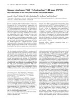
For immunoglobulin G (IgG) multiple glycoforms can be iden-
tified due to the presence of a single N-glycan chain attached
to each IgG fragment crystallizable (Fc) portion [3]. This N-gly-
can shows heterogeneity due to the presence or absence of
fucose, galactose or sialic acid residues and bisecting N-
acetylglucosamine (GlcNAc) (Figure 1) [4,5]. Regarding
galactosylation, three subfamilies called either galactose-0
(Gal-0) (agalactosyl IgG, no galactose), Gal-1 (galactose on
one arm) or Gal-2 (galactoses on both arms) have been
defined [6]. On the Gal-1 and Gal-2 glycans one terminal sialic
acid residue can be present.
In RA-patients higher levels of agalactosyl IgG are found com-
pared to controls and this is associated with increased dis-
ease activity and more disease progression [4,7]. Moreover, in
two small studies increased galactosylation during pregnancy
has been associated with the pregnancy-induced improve-
ment of RA [1,2]. Due to the small sample size and limited fol-
low-up period these studies could not provide detailed
description of the changes in galactosylation during preg-
nancy and postpartum. Fucosylation seems not to be related
to RA or pregnancy [8], whereas sialylation and the presence
of the bisecting GlcNAc have not yet been studied in these
settings. Moreover these studies applied the lectin analysis
method or the GN7 antibody ELISA to detect the galactosyla-
tion level, however, both of which could not analyze the Frag-
ment crystalizable (Fc) and Fragment antigen-binding (Fab)
glycosylation separately and its accuracy was questioned.
Now, the MALDI-TOF-MS method which is now applied can
investigate the Fc fragment galactosylation and the position of
bisecting GlcNAc with great accuracy and reproducibility.
The aim of the present study is to investigate the changes in
IgG glycosylation in detail (galactosylation, sialylation and the
presence of the bisecting GlcNAc) in a large cohort of 148
RA-patients and 32 controls from pre-pregnancy onwards
until six months postpartum, together with associations with
disease activity and medication use as well as other factors.
Materials and methods
Study population
The present study is embedded in the PARA-study (Preg-
nancy-induced Amelioration of Rheumatoid Arthritis), a pro-
spective cohort study on pregnancy and RA [9]. Data of the
first 148 Caucasian RA-patients (cases) are included. Thirty-
two healthy pregnant Caucasian volunteers without adverse
obstetric history served as controls. All participants gave
informed consent. The study is in compliance with the Helsinki
Figure 1
MALDI-TOF-MS analysis of tryptic glycopeptides of IgG1 and IgG2MALDI-TOF-MS analysis of tryptic glycopeptides of IgG1 and IgG2. A representative sample of an RA-patient before pregnancy (a) and in the third
trimester (b) is shown. Glycopeptides derived from IgG1 and IgG2 were analyzed for galactosylation and sialylation in the reflectron positive mode.
Glycopeptides of IgG 1 are indicated by continued arrows, while glycopeptides of IgG2 are indicated by striated arrows. Three glycoforms of IgG1
have been found to be below the detection limit of the MALDI-TOF-MS method in this sample as well as in several other samples.
2600 2700 2800 2900 3000
m/z
3100 3200 3300 2600 2700 2800 2900 3000
m/z
3100 3200 3300
N-acetylglucosamine
AB
galactose
IgG1 tryptic
Fc-glycopeptide
E
293
EQ
Y
NST
Y
R
301
mannose
fucose
peptide moiety
sialic acid
pep
IgG2 tryptic
Fc-glycopeptide
E
293
EQFNSTFR
301
pep pep
- 2633.9
- 2796.2
- 2926.2
- 2958.0
- 2999.5
- 3054.3
- 3128.6
- 3249.1
- 3217.5
- 2763.9
- 2764.4
2602.1
- 2796.8
- 2634.4
- 2957.9
- 2999.8
- 3055.2
- 3129.7
- 3216.9
- 2925.9
2601.9
pep
pep pep
pep peppep pep
pep pep pep pep pep pep pep
Available online />Page 3 of 10
(page number not for citation purposes)
Declaration and approved by the Ethics Review Board at the
Erasmus MC University Medical Center Rotterdam.
Data collection
N = 57 cases were followed from pre-pregnancy, n = 65
cases from the first trimester, n = 14 cases from the second
trimester and n = 12 cases from the third trimester and
onwards, all controls from first trimester and onwards.
Disease activity was scored using a disease activity score
(DAS28) with three variables (swollen joint count, tender joint
count and a C-reactive protein (CRP) level) [10,11].
Categorization of disease activity and clinical response
According to the EULAR criteria, remission of RA was defined
as DAS28<2.6 and intermediate and high disease activity as
DAS28>3.2 [12]. Improvement during pregnancy was defined
according to the EULAR criteria as good, moderate (com-
bined to responders) or non-responders [12]. The response
criteria can only be applied to those patients with an initial
DAS28>3.2 at first trimester (n = 75). A postpartum flare was
defined according to so called reversed EULAR criteria [9,12].
Since there is no baseline DAS28 requirement for these crite-
ria, this classification was applied to all cases. An early flare
was defined when deterioration began between six weeks and
three months postpartum, a late flare with deterioration
between three to six months postpartum.
IgG glycosylation analysis
IgG was purified from sera using Protein A-Sepharose beads
(GE Healthcare, Eindhoven, The Netherlands) followed by
trypsinisation as described previously with minor alterations
[13]. These beads bind IgG1, IgG2 and IgG4, but not IgG3.
Then the resulting glycopeptides were purified by reverse
phase- solid phase extraction (SPE) (Supelco DSC-18 plate
SPE-96 (Sigma, Zwijndrecht, The Netherlands)) and eluted
into a V-bottom 96-well microtitration plate (Nunc, Roskilde,
Denmark) using 200 μl 18% acetonitrile (AcN) containing
0.1% trifluoroacetic (TFA). Glycopeptide samples were dried
by vacuum centrifugation and dissolved in water.
Galactosylation of IgG1 and IgG2 and the incidence of bisect-
ing GlcNAc were analyzed for all samples: aliquots of the glyc-
opeptide samples after reverse phase purification were
spotted on a polished steel 384-positions MALDI-TOF-MS
target plate and allowed to dry. Sample spots were overlaid
with α-cyanocinnamic acid matrix (5 mg/ml in 50% AcN) and
allowed to dry, resulting in a microcristalline sample prepara-
tion. Glycopeptides were analyzed in the reflectron positive
mode on an Ultraflex II MALDI-TOF-MS (Bruker Daltonics,
Bremen, Germany). N = 100 shots were acquired per posi-
tion, and spectra were acquired from n = 30 different positions
per spot, resulting in a sumspectrum obtained by accumula-
tion from 3,000 spectra per sample spot.
For the analysis of sialylation, aliquots of the glycopeptide
samples were spotted on a mitrotiter plate (MTP) AnchorChip
600/384 plate (Bruker Daltonics) and allowed to dry. Sample
spots were overlaid with 2,5-dihydroxybenzoic acid matrix (5
mg/ml in 50% AcN with 0.1% TFA) and allowed to dry, result-
ing in a macrocristalline sample preparation. Glycopeptides
were analyzed by MALDI-TOF-MS in the linear positive mode.
Per sample spot 2,000 spectra were accumulated. These
analyses were performed in three subgroups: first, n = 10
cases and n = 10 controls randomly selected from our cohort
at every timepoint before (cases), during and after pregnancy.
Second, sialylation was determined in n = 15 responders and
n = 15 non-responders selected upon the most pronounced
and the least-pronounced changes in disease activity during
pregnancy. Third, sialylation was determined in n = 15 cases
with a flare early postpartum and n = 15 cases without a flare
selected upon the most pronounced and the least-pro-
nounced changes in disease activity postpartum.
Mass spectra were processed in FlexAnalysis (Bruker Dalton-
ics) with baseline subtraction and peak detection of the IgG1
and IgG2 glycopeptide signals. Peak lists were imported into
Excel.
To determine the inter- and intra-day variation on every plate
one or more standard sera were added and measured.
From the MALDI-TOF-MS measurements in the reflectron pos-
itive mode, IgG1 and IgG2 signals for six glycoforms were ana-
lyzed: Gal-0 without bisecting GlcNAc, Gal-1 without
bisecting GlcNAc, Gal-2 without bisecting GlcNAc and Gal-
0+bisecting GlcNAc (G0+N), Gal-1+bisecting GlcNAc
(G1+N), Gal-2+bisecting GlcNAc (G2+N).
All analyzed glycopeptides contain fucose residues. Due to
relative low incidence (approximately 5%) and overlap with
IgG4 glycoforms, the applied analytical approach did not allow
the analysis of non-fucosylated glycopeptides.
The levels of galactosylation of IgG1 and IgG2 without bisect-
ing GlcNAc were calculated on the basis of signal heights
observed in MALDI-TOF-MS. Based upon these signal
heights a percentage of galactosylation was determined. This
percentage represents the actual number of galactoses
present on the outer arms of the N-glycan chain (1 Gal on Gal-
1 and 2 Gal on Gal-2) divided by the total number of available
antenna positions for galactosylation on the outer arms of the
N-glycan (two available antenna positions both on Gal-0, Gal-
1 as well as on Gal-2). It was calculated using the following
term:
The incidence of bisecting GlcNAc on IgG1 and IgG2 was
also calculated on the basis of the signal heights observed in
()/
()%
112 2
2 0 2 1 2 2 100
×−+×−
×−+×−+×−×
Gal Gal
Gal Gal Gal
Arthritis Research & Therapy Vol 11 No 6 van de Geijn et al.
Page 4 of 10
(page number not for citation purposes)
MALDI-TOF-MS. On the basis of these signal heights a per-
centage of the presence of GlcNAc (N) was determined. This
percentage represents all signal heights with GlcNAc present
on the N-glycan with or without galactoses (Gal-0+N, Gal-
1+N or Gal-2+N) divided by all available signal heights with
and without GlcNAc using the following term:
Based on the MALDI-TOF-MS measurements in the linear
positive mode, the incidence of sialic acid (SA) per galactose
moiety on IgG1 and IgG2 was calculated on the basis of signal
heights observed in MALDI-TOF-MS. Based upon these sig-
nal heights a percentage of sialylation was determined. This
percentage represents the actual number of sialic acid sugar
moieties present on the galactose sugar moieties on outer
arms of the N-glycan chain (one SA on Gal-1 and one or two
SA on Gal-2) divided by the total number of available antenna
positions for sialic acid on the galactose sugar moieties on the
outer arms of the N-glycan (two available positions on Gal-2
with or without SA and one available position on Gal-1 with or
without SA). It was calculated using the following term:
Statistical analysis
Statistical analysis was performed using the Statistical Pack-
age for the Social Sciences (SPSS) 15.0 and Statistical Anal-
ysis Software (SAS) 9.1. A two-sided P-value ≤ 0.05 was
considered statistically significant.
A Linear Mixed Model (LMM) with unstructured residual corre-
lation was used to test for differences in the galactosylation
and sialylation at each timepoint and for changes in time
between cases and controls as well as responders versus
non-responders and flare versus no-flare postpartum. Pearson
and Spearman rank tests were used to determine possible
associations.
A multivariate analysis, conditional on the timepoint of visit,
was performed to investigate which covariates determine the
level of galactosylation. A constant effect in time was
assumed. Only covariates with a P- value < 0.20 in the univar-
iate analysis were introduced in the multivariate analysis. The
following covariates were tested: use of salazopyrine, pred-
nisone, methotrexate or biologicals, DAS28, presence of joint
erosions, rheumatoid factor (RF) positivity and anti-cyclic cit-
rullinated peptide (anti-CCP) positivity, breast feeding and
maternal age at delivery.
Finally, to determine whether changes in galactosylation pre-
cede changes in disease activity, for every timepoint interval
the change in IgG galactosylation was divided by the total
change in galactosylation. The change in disease activity was
also calculated per interval and divided by the total change in
disease activity. This resulted in a percentage of change per
timepoint interval. A paired sample t-test tested for equality.
Results
Description of study cohort
All cases (n = 148) fulfilled the American College of Rheuma-
tology (ACR) 1987 revised criteria for RA (Table 1). Medica-
tion use of this cohort was described before [14]. In more
detail: the use of sulfasalazine and prednisone through preg-
nancy and postpartum was documented as below. For
sulfasalazine use 23 of 57 cases (40.4%) at pre-pregnancy,
37/118 (31.4%) in first trimester, 42/133 (31.6%) in second
trimester, 43/146 (29.5%) in third trimester, 43/144 (29.9%)
six weeks postpartum, 48/144 (33.3%) three months postpar-
tum, 49/142 (34.5%) six months postpartum.
For prednisone use 19 of 57 cases (33.3%) at pre-pregnancy,
43/118 (36.4%) in first trimester, 48/133 (36.1%) in second
trimester, 50/146 (34.2%) in third trimester, 49/144 (34.0%)
six weeks postpartum, 48/144 (33.3%) three months postpar-
tum, 46/142 (32.4%) six months postpartum.
The use of methotrexate and biologicals postpartum was doc-
umented as below: For methotrexate use postpartum 24 of
144 cases (16.7%) at six weeks postpartum, 40/144 (27.8%)
three months postpartum, 56/142 (39.4%) six months
postpartum.
For use of biologicals postpartum 7 of 144 cases (4.9%) at six
weeks postpartum, 13/144 (9.0%) three months postpartum,
14/142 (9.9%) six months postpartum. Other DMARDs were
only used by a very limited number of participants in this cohort
both during (≤ 2.3%) and after pregnancy (≤ 7.6%).
MALDI-TOF-MS measurements accuracy and
reproducibility
The intraday and interday variability for the analyzed glycopep-
tides of IgG1 and IgG2 was below 4% and 6%, respectively.
The IgG4 glycopeptides were not analyzed due to their low
abundance.
Galactosylation profiles during pregnancy and
postpartum of cases and controls
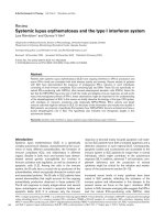
For cases an increase in IgG1 galactosylation was observed
during pregnancy from preconception (mean 43.4% (standard
deviation (SD) 8.3%) to first trimester (mean 48.4% (SD
8.4%) until the third trimester (53.7%, (SD 8.3%), P <
0.0001). After pregnancy a significant decrease in galactos-
ylation was observed with lowest levels at six months postpar-
tum (44.9% (SD 7.7%), P < 0.0001, Figure 2a). IgG2
galactosylation profiles show a similar pattern as IgG1 (Figure
2b). In the controls, IgG1 and IgG2 galactosylation profiles
were at a significantly higher level than in cases (P < 0.001),
()/
(
Gal N Gal N Gal N
Gal N Gal N Gal N Gal G
−+ + −+ + −+
−++−++−++−+
012
0120aal Gal−+ − ×1 2 100)%
()/
()
Gal SA Gal SA
Gal Gal SA Gal Gal SA
−− + − −
−+ −− + × − + × − −
12
112222
Available online />Page 5 of 10
(page number not for citation purposes)
and changes were less pronounced than in the cases (Figure
2).
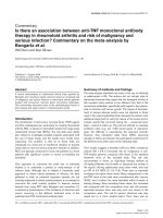
Galactosylation and disease activity levels
IgG1 and IgG2 galactosylation levels are associated with dis-
ease activity at every timepoint (Pearson correlation
0.35<R<0.49, P < 0.005). Lower disease activity levels show
higher galactosylation levels, and resemble more the levels of
the controls. Both IgG1 (Figure 3) and IgG2 (data not shown)
galactosylation levels which are associated with disease
remission (DAS28<2.6) depend on the timepoint of
measurement.
Changes in IgG galactosylation are associated with
improvement of disease activity in responders and non-
responders
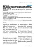
The change in galactosylation from the first to the third trimes-
ter was significantly different between responders (n = 37)
and non-responders (n = 38) for IgG1 (6.8% (SD 0.80%) ver-
sus 4.2% (SD 0.79%), respectively, P < 0.02), whereas for
IgG2 a trend could be observed (5.6% (SD 0.51%) versus
4.5% (SD 0.50%), respectively, P < 0.11, Figure 4a).
Changes in IgG galactosylation are associated with flare
postpartum
Cases with a late flare may also have experienced an early flare
(n = 9). The change in galactosylation from six weeks to three
months postpartum was significantly different between the
cases with an early flare (n = 35) and without flare (n = 106)
Table 1
Cohort characteristics
Cases
(n = 148)
Controls
(n = 32)
Mean age at delivery in years (SD) 32.3 (3.8) 32.0 (4.4)
Median disease duration at delivery in years (range) 8.0 (0.7 to 29.7) -
Number of nulliparous women, n (%) 70/147 (47.6) 14/32 (43.8%)
Mean gestational age at delivery, weeks (range) 39.3 (31.4 to 42.1) 40.4 (34.0 to 42.0)
Breastfeeding (six weeks postpartum), n (%) 62/148 (41.9) 27/32 (84.4)
Anti-CCP positive, n (%) 93/147 (63.3) -
Rheumatoid Factor (IgM) positive, n (%) 108/148 (73.0) -
Erosive disease, n (%) 43/147 (70.7)
DAS28-CRP3 >3.2 in first trimester, n (%) 75 (61.5) -
Classification of disease activity during pregnancy
good response/moderate response 37/75 (49.3) -
no response 38/75 (50.7) -
Classification of disease activity during postpartum period
(early flare)
severe deterioration/moderate deterioration (n, %) 35/141* (24.8) -
no deterioration (n, %) 106/141* (75.2) -
Classification of disease activity during postpartum period
(late flare)
severe deterioration/moderate deterioration (n, %) 29/141* (20.6) -
no deterioration (n, %) 112/141* (79.4) -
Median number of DMARDs (incl prednisone)
prior to conceive (min-max)
2 (0-7) -
No DMARD** use prior to conceive, n (%) 7/147 (4.8)
Use of methotrexate prior to conceive, n (%) 75/147 (51.0) -
*n = 7 cases are missing, since a small proportion of DAS scores are missing.
** DMARDs: disease modifying anti-rheumatic drugs.
Arthritis Research & Therapy Vol 11 No 6 van de Geijn et al.
Page 6 of 10
(page number not for citation purposes)
(-3.3% (SD 0.58%) versus -1.3% (SD 0.33%), respectively,
for IgG1; -2.7% (SD 0.42%) versus -1.3% (SD 0.24%),
respectively, for IgG2, both P < 0.004, Figure 4b). The change
in galactosylation between three and six months postpartum
was also significantly different between the cases with a late
flare (n = 29) and without late flare (n = 112) (-1.2% (SD
0.49%) versus +0.58% (SD 0.33%), respectively, for IgG1, P
< 0.0001 and -1.1% (SD 0.30%) versus +0.16% (SD
0.20%), respectively, for IgG2, P < 0.0004, Figure 4c).
Galactosylation changes do not precede disease activity
changes
When changes in galactosylation and changes in disease
activity were tested for equality, this could not be rejected,
indicating that galactosylation and DAS28 may change syn-
chronically in time.
IgG sialylation during pregnancy and postpartum
IgG sialylation was, like galactosylation, determined as total
percentage of sialic acid (SA) residues per N-glycan. The
presence of SA on IgG1 and IgG2 is low in the serum for
cases and controls (all measurements taken together: mean
5.8%, SD 2.3% SA per N-glycan for IgG1 for cases (controls
7.1%, SD 2.7%) and 6.6%, SD 2.6% per N-glycan for IgG2
for cases (controls 7.9%, SD 2.5%)). In RA-cases N-glycan
sialylation levels and IgG galactosylation were significantly
correlated (Spearman rho 0.57 and 0.69 for IgG1 and IgG2,
respectively, both P = 0.0001). In controls, the correlation
between sialylation and IgG galactosylation was 0.77 and
0.72 for IgG1 and IgG2, respectively, both P = 0.0001).
The mean sialylation levels per N-glycan for IgG1 and IgG2
increased during pregnancy and decreased postpartum for
cases (n = 10) and controls (n = 10). In cases, for IgG1, an
increase in sialylation was observed during pregnancy from
preconception (mean 5.01% (SD 0.83%) to first trimester
(mean 6.47% (SD 0.69%) until second trimester (7.45% (SD
0.75%), P < 0.049). Third trimester: 7.27% (SD 0.82%). After
pregnancy a decrease in sialylation was observed with lowest
levels at six months postpartum (4.61% (SD 0.47%), P <
0.056). For IgG2, an increase in sialylation was observed dur-
ing pregnancy from preconception (mean 5.45% (SD 0.11%)
to first trimester (mean 6.59% (SD 0.64%) until second tri-
mester (8.81% (SD 0.75%), P < 0.022). Third trimester:
8.52% (SD 0.77%). After pregnancy a decrease in sialylation
was observed with lowest levels at six months postpartum
(5.90% (SD 0.44%), P < 0.550). The controls showed a
higher level of sialylation than the cases, but this difference
was not significantly different (P < 0.280). The increase in N-
glycan sialylation was larger in responders than in non-
Figure 2
Mean galactosylation of IgG1 and IgG2 in cases and controls during pregnancy and postpartumMean galactosylation of IgG1 and IgG2 in cases and controls during pregnancy and postpartum. IgG1 (a) and IgG2 (b) galactosylation levels (in
percentages) increase during pregnancy and decline postpartum. IgG1 and IgG2 galactosylation profiles of controls are at a significantly higher
level than cases (P < 0.001, Linear Mixed Model at all timepoints). The vertical bars illustrate the 95% confidence intervals. Abbreviations: trim = tri-
mester of pregnancy; wk = weeks; PP = postpartum; mon = months.
Available online />Page 7 of 10
(page number not for citation purposes)
responders during pregnancy (for IgG1: within responders
+1.8%, SD 0.42%, P = 0.0007; within non-responders
+0.34%, SD 0.42%, p < 0.216; for IgG2: within responders
+1.8%, SD 0.50%, P = 0.0008; within non-responders
+1.0%, SD 0.42%, P < 0.052).
In the postpartum period no significant changes were
observed for IgG1 sialylation between cases with or without
flare. The change in IgG1 sialylation was for cases with early
flare: +0.03% (SD 0.64%), P < 0.957, and for cases without
early flare: -0.20% (SD 0.56%), P < 0.728%; for cases with a
late flare: -0.81% (SD 0.73%), P < 0.271, and for cases with-
out late flare -0.49% (SD 0.67%), P < 0.471.
For IgG2 sialylation a decrease in N-glycan sialylation could be
observed in cases with an early flare (-0.95% (SD 0.40%), P
= 0.024) and cases without an early flare ( 68%, (SD 0.40%),
P < 0.095). For the late postpartum flare in IgG2 sialylation
non-significant results were seen: -0.01% (SD 0.92%), P <
0.99, and for cases without late flare -0.02% (SD 0.92%), P <
0.98.
Dependent variables of galactosylation
To investigate which factors determine the level of galactosyla-
tion multivariate analyses were performed. In the multivariate
analyses only DAS28 and sulfasalazine (P = 0.06) for IgG1
and DAS28 and prednisone use for IgG2 had a significant
negative effect on galactosylation.
Presence of bisecting GlcNAc and its relation to
galactosylation
The presence of IgG with a bisecting GlcNAc is low in the
serum (first trimester mean 13.7%, SD 2.8% for IgG1 (both
cases and controls) and mean 13.3%, SD 3.2% or 13.5%, SD
3.5% (cases and controls, respectively) for IgG2. The pres-
ence of bisecting GlcNAc was not influenced by pregnancy or
postpartum and was similar in cases (range min-max IgG1
13.7 to 14.7%, range min-max IgG2 13.2 to 14.3%) and con-
trols (range min-max IgG1 13.7 to 14.4%, range min-max
IgG2 13.0 to 14.0%). Moreover uni- and multivariate analyses
did not reveal any effect of the previously mentioned covari-
ates on the presence of bisecting GlcNAc.
The presence of the bisecting GlcNAc is related to IgG galac-
tosylation. The levels of galactosylation of IgG1 or IgG2 with
bisecting GlcNAc were at a significant lower level than IgG1
or IgG2 without bisecting GlcNAc (range min-max
IgG1+bisecting GlcNAc 38.2 to 44.8%, range min-max
IgG2+bisecting GlcNAc 31.8 to 38.9%) at every timepoint (P
< 0.0001), but showed a similar pattern in time.
Ethics committee approval
All participants gave informed consent. The study is in compli-
ance with the Helsinki Declaration and approved by the Ethics
Review Board at the Erasmus MC University Medical Center
Rotterdam, The Netherlands.
Discussion
This study demonstrates the association between changes in
IgG galactosylation and RA-disease activity during pregnancy
and postpartum. The most prominent increase in galactosyla-
tion was observed in RA-patients that spontaneously improved
during pregnancy, whereas the reverse was observed for the
flare postpartum. Finally, a good correlation between IgG N-
glycan galactosylation and sialylation was demonstrated.
IgG galactosylation and RA in relation to pregnancy have been
studied previously. Our results are in line with a previous study
in which galactosylation levels in RA are described during
pregnancy using the lectin analysis method [1]. The applica-
tion of the MALDI-TOF-MS allowed us to analyze IgG1 and
IgG2 separately and to analyze specifically the Fc-fragment
glycosylation (and galactosylation). This was in contrast to the
lectin method which cannot distinguish between IgGs and
determines a combined value for the Fc and Fab fragment gly-
cosylation. Compared to previous literature, we studied a
larger cohort with a longer follow-up time postpartum. This
Figure 3
Mean IgG1 galactosylation levels in relation to rheumatoid arthritis dis-ease activity levelsMean IgG1 galactosylation levels in relation to rheumatoid arthritis dis-
ease activity levels. For this purpose at every timepoint all cases were
divided in two categories; that is, those with a DAS28>3.2 or
DAS28<2.6. Please note that each timepoint may include different RA-
cases. For comparison controls are added to the graph. The IgG1
galactosylation level which is associated with disease remission
(DAS28 <2.6) is dependent on the timepoint of measurement. Similar
data were observed for IgG2 (data not shown). The vertical bars illus-
trate the 95% confidence intervals. Abbreviations: DAS28 = disease
activity score; trim = trimester of pregnancy; wk = weeks; PP = post-
partum; mon = months.
Arthritis Research & Therapy Vol 11 No 6 van de Geijn et al.
Page 8 of 10
(page number not for citation purposes)
enabled the description of the postpartum flares and identifi-
cation of factors that influence galactosylation. Moreover a
control group was added. Based upon studies in one patient
it has been suggested that pregnancy-induced remission is
associated with a fixed galactosylation level [2]. In contrast we
demonstrated that the level of clinical remission was associ-
ated with a different level of galactosylation per timepoint dur-
ing pregnancy and postpartum.
We have shown that the IgG galactosylation changes take
place simultaneously with the changes in RA disease activity.
Therefore one could argue that changes in IgG galactosylation
are a mere epiphenomenon accompanying changes in dis-
ease activity. However, the strongest argument that galactos-
ylation of IgG, and in particular agalactosyl IgG itself, indeed
plays a pathogenic role is derived from animal studies. In these
studies arthritis could only be transferred by infusion of agalac-
tosyl IgG [15].
The pro-inflammatory role of agalactosyl IgG may be explained
in multiple ways: first, IgG can act as an auto-antigen itself in
RA. Since RF preferentially binds to agalactosyl IgG, this
would result in more pronounced RF-agalactosyl IgG interac-
tion and hence more inflammation [16,17]. Secondly, the path-
ogenicity of agalactosyl IgG is thought to be associated with
its ability to activate the complement pathway via binding to
mannose-binding lectin (MBL) [18]. This hypothesis has been
questioned recently based upon studies in MBL-deficient
mice [3]. As a result of the absence of galactose, agalactosyl
IgG antibodies also lack terminal sialic acid residues. These
terminal sialic acid residues have recently been implicated in
suppressing inflammation via the induction of inhibitory FcãRI-
Ib expression in mice [19,20]. Our analyses revealed a good
correlation between IgG N-glycan galactosylation and sialyla-
tion. However, IgG sialylation levels were low and did not
exceed 10%. Whether the effect of galactosylation is medi-
ated in humans through the presence of increased sialylation
of IgG still needs to be elucidated. Nevertheless, sialylation
Figure 4
Mean change in IgG1 and IgG2 galactosylation during pregnancy and early or late postpartumMean change in IgG1 and IgG2 galactosylation during pregnancy and early or late postpartum. (a) Mean change in IgG1 and IgG2 galactosylation
(×100%) during pregnancy in (good and moderate) responders according to the EULAR response criteria (cases that improved during pregnancy,
n = 37) and non-responders (cases that did not improve during pregnancy, n = 38). The change in IgG galactosylation was significantly different
between responders and non-responders for IgG1 (P < 0.02), whereas for IgG2 a trend towards significance could be observed (P = 0.11). (b)
Mean change in IgG1 and IgG2 galactosylation (×100%) in the postpartum period in cases with an early flare between six weeks and three months
postpartum (deterioration, n = 35) and cases without an early flare (no deterioration, n = 106). The change in galactosylation was significantly differ-
ent between early flare and no early flare for IgG1 and IgG2 (P < 0.004). (c) Mean change in IgG1 and IgG2 galactosylation (×100%) in the post-
partum period in cases with a late flare from three to six months postpartum (deterioration, n = 29) and cases without a late flare (no deterioration, n
= 112). The change in galactosylation was significantly different between late flare and no late flare for IgG1 and IgG2 (P < 0.0001 and P < 0.0004,
respectively). The vertical bars illustrate the 95% confidence intervals.
Available online />Page 9 of 10
(page number not for citation purposes)
seems to be an additional important modification of IgG during
pregnancy.
Since we have shown that changes in IgG galactosylation lev-
els are associated with improvement of RA during pregnancy
and the flare postpartum, identification of factors that influence
galactosylation might give insight into pathogenic mecha-
nisms underlying RA and might be a lead for the development
of future therapies. For this purpose multivariate analyses were
performed. These revealed that mainly disease activity and
timepoint in pregnancy remained as an explanatory parameter
for galactosylation. However, in the multivariate analysis also
use of prednisone (for IgG2) and sulfasalazine (for IgG1) were
associated with decreased galactosylation of IgG. Since
decreased IgG galactosylation has been shown to be associ-
ated with more severe disease activity it is unlikely that this
association is a direct consequence of the mode of action of
these effective medications for RA. Although speculative, it is
more likely that this association is related to the fact that both
medications are only used during pregnancy by patients with
a more aggressive RA and hence serve as markers for those
patients with more severe RA.
Pregnancy-induced changes in cytokine or hormonal levels
could be a possible explanation for the changes in galactosyla-
tion during pregnancy and postpartum. It has been suggested
that IL-6 [21] or pregnancy-associated hormones like estro-
gen [17] or prolactin [22] could induce altered glycosyltrans-
ferase (or other (iso)enzyme) activity in B-cells that could result
in immunoglobulins with different glycoforms.
For the first time it has been shown that the levels of bisecting
GlcNAc are not influenced by pregnancy. Pekelharing et al
found no changes in the presence of GlcNAc during preg-
nancy using gas-liquid chromatography [8], not distinguishing
between antenna GlcNAc and the bisecting GlcNAc on other
positions. The clinical relevance of bisecting GlcNAc is still
unknown. Interestingly, the levels of IgG galactosylation with
bisecting GlcNAc were significantly lower than the levels of
IgG without bisecting GlcNAc.
Conclusions
This large prospective cohort study demonstrates the associ-
ation between IgG galactosylation changes with pregnancy-
induced improvement and postpartum flare in RA-patients.
Since IgG galactosylation was associated with sialylation, also
sialylation seems to be an additional important modification
during pregnancy. The levels of IgG galactosylation largely
depend on the trimester of pregnancy or the timepoint of visit
postpartum and disease activity, even after correction for med-
ication use.
Future studies should focus on unraveling the exact mecha-
nism behind the changes in IgG galactosylation and sialylation
and on the consequences of these changes on the function of
IgG itself during pregnancy and postpartum.
Competing interests
The authors declare that they have no competing interests.
Authors' contributions
FG and RD had full access to all of the data in the study and
take responsibility for the integrity of the data and the accuracy
of the data analysis. FG, MW, MS, AD, MH and RD designed
the study. FG, MW, MS and YM were involved in acquisition
of the data. FG, MW, MS, SW, MH and RD analyzed the data
of the MALDI-TOF-MS and interpreted the data. The manu-
script was prepared by FG, MW, SW, MS, YM, AD, MH and
RD. FG and SW did the statistical analyses. All authors read
and approved the final manuscript.
Acknowledgements
We would like to acknowledge Prof Dr E Steegers (Department of
Obstetrics and Gynaecology, Erasmus MC, University Medical Center
Rotterdam, The Netherlands) and Dr C de Groot (Department of
Obstetrics and Gynaecology, Medical Center Haaglanden, The Hague,
The Netherlands) for their valuable advice. This research is financed by
the Dutch Arthritis Association (Reumafonds).
References
1. Alavi A, Arden N, Spector TD, Axford JS: Immunoglobulin G gly-
cosylation and clinical outcome in rheumatoid arthritis during
pregnancy. J Rheumatol 2000, 27:1379-1385.
2. Rook GA, Steele J, Brealey R, Whyte A, Isenberg D, Sumar N, Nel-
son JL, Bodman KB, Young A, Roitt IM, et al.: Changes in IgG gly-
coform levels are associated with remission of arthritis during
pregnancy. Journal of autoimmunity 1991, 4:779-794.
3. Nimmerjahn F, Anthony RM, Ravetch JV: Agalactosylated IgG
antibodies depend on cellular Fc receptors for in vivo activity.
Proceedings of the National Academy of Sciences of the United
States of America 2007, 104:8433-8437.
4. Arnold JN, Wormald MR, Sim RB, Rudd PM, Dwek RA: The
impact of glycosylation on the biological function and struc-
ture of human immunoglobulins. Annual review of immunology
2007, 25:21-50.
5. Wuhrer M, Stam JC, Geijn FE van de, Koeleman CA, Verrips CT,
Dolhain RJ, Hokke CH, Deelder AM: Glycosylation profiling of
immunoglobulin G (IgG) subclasses from human serum. Pro-
teomics 2007, 7:4070-4081.
6. Nimmerjahn F, Ravetch JV: Divergent immunoglobulin g sub-
class activity through selective Fc receptor binding. Science
2005, 310:1510-1512.
7. van Zeben D, Rook GA, Hazes JM, Zwinderman AH, Zhang Y,
Ghelani S, Rademacher TW, Breedveld FC: Early agalactosyla-
tion of IgG is associated with a more progressive disease
course in patients with rheumatoid arthritis: results of a fol-
low-up study. British journal of rheumatology 1994, 33:36-43.
8. Pekelharing JM, Hepp E, Kamerling JP, Gerwig GJ, Leijnse B:
Alterations in carbohydrate composition of serum IgG from
patients with rheumatoid arthritis and from pregnant women.
Annals of the rheumatic diseases 1988, 47:91-95.
9. de Man YA, Dolhain RJ, Geijn FE van de, Willemsen SP, Hazes JM:
Disease activity of rheumatoid arthritis during pregnancy:
Results from a nationwide prospective study. Arthritis Rheum
2008, 59:1241-1248.
10. Disease activity score in rheumatoid arthritis. Nijmegen (The
Netherlands) [ />index.html]
11. De Man YA, Hazes JM, Geijn FE Van de, Krommenhoek C, Dolhain
RJ: Measuring disease activity and functionality during preg-
nancy in patients with rheumatoid arthritis. Arthritis Rheum
2007, 57:716-722.
Arthritis Research & Therapy Vol 11 No 6 van de Geijn et al.
Page 10 of 10
(page number not for citation purposes)
12. Van Riel PLVGA, Scott DG: Interpreting disease course. In
EULAR handbook of clinical assessments in rheumatoid arthritis
Edited by: Van Riel PL, van Gestel AM, Scott DG. Alphen aan den
Rijn: Van Zuiden Communications; 2000:39-43.
13. Wuhrer M, Porcelijn L, Kapur R, Koeleman CA, Deelder A, de Haas
M, Vidarsson G: Regulated glycosylation patterns of IgG during
alloimmune responses against human platelet antigens. J
Proteome Res 2009, 8:450-456.
14. de Man YA, Hazes JM, Heide H van der, Willemsen SP, de Groot
CJ, Steegers EA, Dolhain RJ: Association of higher rheumatoid
arthritis disease activity during pregnancy with lower birth
weight: Results of a national prospective study. Arthritis
Rheum 2009, 60:3196-3206.
15. Rademacher TW, Williams P, Dwek RA: Agalactosyl glycoforms
of IgG autoantibodies are pathogenic. Proceedings of the
National Academy of Sciences of the United States of America
1994, 91:6123-6127.
16. Imafuku Y, Yoshida H, Yamada Y: Reactivity of agalactosyl IgG
with rheumatoid factor. Clinica chimica acta; international jour-
nal of clinical chemistry 2003, 334:217-223.
17. Axford JS: Glycosylation and rheumatic disease. Biochimica et
biophysica acta 1999, 1455:219-229.
18. Malhotra R, Wormald MR, Rudd PM, Fischer PB, Dwek RA, Sim
RB: Glycosylation changes of IgG associated with rheumatoid
arthritis can activate complement via the mannose-binding
protein. Nat Med 1995, 1:237-243.
19. Bruhns P, Samuelsson A, Pollard JW, Ravetch JV: Colony-stimu-
lating factor-1-dependent macrophages are responsible for
IVIG protection in antibody-induced autoimmune disease.
Immunity 2003, 18:573-581.
20. Kaneko Y, Nimmerjahn F, Ravetch JV: Anti-inflammatory activity
of immunoglobulin G resulting from Fc sialylation. Science
2006, 313:670-673.
21. Van Dijk W, Mackiewicz A: Interleukin-6-type cytokine-induced
changes in acute phase protein glycosylation. Annals of the
New York Academy of Sciences 1995, 762:319-330.
22. Bond A, Ratkay LG, Waterfield JD, Hay FC: Post-partum flare in
MRL-lpr/lpr mice is associated with a parallel increase of N-
acetylglucosamine on serum IgG. British journal of
rheumatology 1997, 36:174-177.