Báo cáo y học: " CD8 positive T cells express IL-17 in patients with chronic obstructive pulmonary disease" ppsx
Bạn đang xem bản rút gọn của tài liệu. Xem và tải ngay bản đầy đủ của tài liệu tại đây (7.46 MB, 10 trang )
RESEARCH Open Access
CD8 positive T cells express IL-17 in patients with
chronic obstructive pulmonary disease
Ying Chang
1
, Jessica Nadigel
1
, Nicholas Boulais
1
, Jean Bourbeau
2
, François Maltais
3
, David H Eidelman
1
and
Qutayba Hamid
1*
Abstract
Background: Chronic obstructive pulmonary disease (COPD) is a progressive and irreversible chronic inflammatory
disease of the lung. The nature of the immune reaction in COPD raises the possibility that IL-17 and related
cytokines may contribute to this disorder. This study analyzed the expression of IL-17A and IL-17F as well as the
phenotype of cells producing them in bronchial biopsies from COPD patients.
Methods: Bronchoscopic biopsies of the airway were obtained from 16 COPD subjects (GOLD stage 1-4) and 15
control subjects. Paraffin sections were used for the investigation of IL-17A and IL-17F expression in the airways by
immunohistochemistry, and frozen sections were used for the immunofluorescence double staining of IL-17A or IL-
17F paired with CD4 or CD8. In order to confirm the expression of IL-17A and IL-17F at the mRNA level, a
quantitative RT-PCR was performed on the total mRNA extracted from entire section or CD8 positive cells selected
by laser capture microdissection.
Results: IL-17F immunoreactivity was significantly higher in the bronchial biopsies of COPD patients compared to
control subjects (P < 0.0001). In the submucosa, the absolute number of both IL-17A and IL-17F positive cells was
higher in COPD patients (P < 0.0001). After adjusting for the total number of cells in the submucosa, we still found
that more cells were positive for both IL-17A (P < 0.0001) and IL-17F (P < 0.0001) in COPD patients compared to
controls. The mRNA expression of IL-17A and IL-17F in airways of COPD patients was confirmed by RT-PCR. The
expression of IL-17A and IL-17F was co-localized with not only CD4 but also CD8, which was further confirmed by
RT-PCR on laser capture microdissection selected CD8 positive cells.
Conclusion: These findings support the notion that Th17 cytokines could play important roles in the pathogenesis
of COP D, raising the possibility of using this mechanism as the basis for novel therapeutic approaches.
Keywords: Chronic Obstructive Pulmonary Disease IL-17, Tc17 cells
Introduction
Chronic obstructive pulmonary disease (COPD), a pro-
gressive and irreversible chronic inflammatory disease of
the lung caused predominantly by cigarette smoking, is
one of the most important causes of mortality globally
[1]. The i nflammatory response in the lungs of COPD
patients has been found to be strongly linked to tissue
destruction and alveolar airspace enlargement, which
lead to disease progression [2].
The inflammatory response reflects both the innate
immune response to cigarette smoke exposure in the
form of cellular infiltration by neutrophils and macro-
phages, as well as the adaptive immune response invol-
ving B and T cells, which is intimately linked wit h
innate immunity [3]. COPD is marked by the accumula-
tion of both CD4
+
and CD8
+
T cells in the alveolar
walls, with CD8
+
cells predominating [4]. Recent find-
ings concerning the innate and acquired immune
responses in COPD have led to the suggestion that
there is an autoimmune component to its pathogenesis.
This notion is supported by the similarity of pathophy-
siological characteristics between COPD and several
autoimmune diseases, including rheumatoid arthritis
* Correspondence:
1
Meakins-Christie Laboratories and Respiratory Division, Department of
Medicine McGill University, 3626 rue St. Urbain, Montreal, QC, H2X 2P2
Canada
Full list of author information is available at the end of the article
Chang et al. Respiratory Research 2011, 12:43
/>© 2011 Chang et al; licensee BioMed Central Ltd. This is an Open Access articl e distributed under the te rms of the Creative Commons
Attribution License ( s/by/2.0), which permits unrestricted use, distribution, and reproduction in
any medium, provided the original work is properly cited.
(RA), defects in phagocytosis and other modes of clear-
ance of necrotic cells and s ubcellular particles, a defi-
ciency of regulatory T cells and the pr esence of
autoantibodies and autoreactive T cells [5].
The nature of the immune reaction in COPD raises
the possibility that IL-17 and related cytokines may
contribute to this disorder. Th17, a newly described
subsetofTcells,weresuggestedtoplayaroleinRA
and psoriasis. To date six IL-17 family members
(IL-17A, IL-17B, IL-17C, IL-17D, I L-17E/IL -25 and IL-
17F) and five receptors (IL-17RA, IL-17R B, IL-17RC,
IL-17RD and IL-17RE) have been identified, which are
conserved in rodents and humans [6]. IL-17A and IL-
17F display high sequence homology and can be
secreted as homodimers, as well as IL-17A/F heterodi-
mers, by b oth mouse and human cells [7,8]. Although
IL-17 has been closely associated with a subset of T
helper cells known as Th17 cells, gδ T cells, natural
killer [9] T cells and neutrophils have also been shown
to produce IL-1 7A in the lung [10]. IL-17 secretion
triggers production of numerous chemokines, resulting
in neutrophil and macrophage recruitment and subse-
quent pathogen clearance, thus IL-17 mediates cross-
talk between the adaptive and innate immune systems,
allowing for orchestration of an effective immune
response [10,11].
Numerous studies demonstrated the importance of IL-
17 in the context of autoimmunity [10], however little is
known about IL-17 production in COPD. A recent
study showed that IL-17A could induce production of
mucin (MUC)5AC in human bronchial epi thelial cells
[12], s upporting the potential invo lvement of IL-17A in
the phenotypic manifestations of COPD. In addition,
transgenic over expression of Il-17 in the alveoli of mur-
ine lung induces lung inflammation with a COPD-like
phenotype [13]. Aside from IL-17A, IL-17F mediated
pathways might also provide a link between local activa-
tion of T cells and sustained accumulation of neutro-
phils in inflamed airways [14]. A case-control study
demonstrates an association between an IL-17F gene
polymorphism and chronic inflammatory lung diseases,
including bronchial asthma and COPD, suggesting that
IL-17F may be critically involved in the pathogenesis o f
chronic inflammatory lung diseases [15].
A well-known hallmark ofCOPDisthatitisrela-
tively unresponsive to treatment with steroids. Corti-
costeroids alone have little impact on the cellular
inflammation or increased protease burden observed in
COPD [16]. In addition, whereas exogenous steroids
are able to suppress cytokine production in cells col-
lected from non-diseased airways, the same cell types
from patients with COPD are resistant to steroid treat-
ment [17]. In this regard, it is of interest that IL-17
expression has been associated with diminished steroid
responsiveness [15]. Moreover, there has been a recent
suggestion that autoimmunity plays a role in the
pathogenesis of COPD [5] and given the increased
expression of IL-17 in certain autoimmune diseases
[10], this further raises the possibility o f its involve-
ment in the pathogenesis of COPD.
In the present study, we analyzed the expression of IL-
17A and IL-17F as well as the phenotype of cells produ-
cing them in the bronchial biopsies from COPD patients
using immunohistochemistry, immunofluorescence
staining, laser capture microdissection and quantitative
reverse transcription-PCR. For the first time, we demon-
strated the IL-17A and IL-17F expression in CD4
+
and
especially in C D8
+
T cells in the airways of COPD
patients. We also showed h igher expression of these
cytokines in COPD patients compar ed to control sub-
jects. This study supports the notion that IL-17 is a
pathogenetic element of COPD and suggests the possi-
bility that a strategy of targeting IL-17 as a therapeutic
target may be of value in this disease.
Methods
Subjects
Bronchoscopic biopsies from the subsegmental bronchi
were obtained from 16 clinical diagnosis of COPD
patients (GOLD stage 1-4) and 15 control subjects using
published techniques [18] at the Montreal Chest Insti-
tute of the McGill University Health Centre and Laval
Hospital, C anada. The COPD patients were eligible for
this study if they met the following criteria: age ≥ 40
and ≤ 75 years; smoking history (≥ 10 pack-years); post-
bronchodilator FEV
1
≥ 25% o f predicted value and post-
bronchodilator FEV
1
/forced vital capacity (FVC) ≤ 0.70;
no history o f asthma, atopy (as assessed by an allergy
skin prick test during screening) or any other active
lung disease. Patients on home oxygen or with raised
carbon dioxide tension (>44 mmHg), a
1
-antitrypsin defi-
ciency, recent exacerbation (in the last 4 weeks), uncon-
trolled medical condition or hypersensitivity to inhaled
cortic osteroids and bronchodilators were not eligible for
the study. The experimental procedures were performed
with ethical approval from the Research Ethics Boards
of the McGill University Health centre and Laval Uni-
versity (Table 1).
Processing of airway biopsies
Duplicate biopsy specimens from each case were imme-
diately fixed in 4% paraformaldehyde for 4 h, and t hen
treated in PBS/DEPC for overnight at 4°C. One speci-
men was dehydrated in alcohol and xylol and embedded
in paraffin for immunohistochemistry, which was carried
out on 5 μm thick sections. The second was snap-frozen
in liquid nitrogen-cooled isopentane for immunofluores-
cence (6 μm thick), laser capture microdissection and
Chang et al. Respiratory Research 2011, 12:43
/>Page 2 of 10
quantitative reverse transcription-PCR studies (10 μm
thick).
Immunohistochemistry
Paraffin-embedded specimens were deparaffinized in
xylene, rehydrated through a decreasing ethanol gradi-
ent, and rinsed in P BS. Antigen unmasking was per-
formed with 10 Mm citrate buffer p H 6 and following
with 0.2% Trit on X100 in PBS. Endogenous per oxidase
activity was blocked with 6% hydrogen peroxide for 30
min at room temperature. The slides were washed and
pretreated with universal blocking solution (Dako, Car-
pinteria, USA). Slides were incubated overnight at 4°C
using diluted goat anti-human IL-17A (AF317-NA, R&D
Systems) or IL-17F (AF1335 , R&D Systems) polyclonal
antibodies or relevant isotype contr ols (AB-108-C, R&D
Systems). The slides were rinsed and incubated with a
biotinylated secondary antibody for 30 min at room
temperature. After washing in PBS, the complex Strepta-
vidin/HorseRadish Peroxidase ( Vector) was applied for
30 min at room temperature. The reaction result was
visualized with DAB/hydrogen peroxide (DAB Kit,
Dako). The sections were finally rinsed in distilled
water, lightly stained with hematoxylin, dehydrated,
cleared, and cover slipped. Sample processed the same
isotypes as primary antibody served as negative control.
Immunofluorescence double staining
After permeabilization in PBS -Triton X100 0.2% for 10
min at room temperature, the sec tions were blocked
with the universal blocking solution (Dako) for 30 min.
The sections for double labeling with IL-17A or IL-17F
paired with CD4 and CD8 respectively were incubated
with diluted goat anti-human IL-17A antibody (1:100)
or IL-17F antibody (1:200) (R&D Systems) paired with
mouse anti-human CD4 antibody (1:40) (VP-C319, Vec-
tor), CD8 an tibody (1:120) (M7103, Dako) or relevant
isotype controls (MAB002, R&D Systems) for overnight
at 4°C. Af ter rinsing with PB S, the sections w ere then
reacted with Alexa 488-conjugated rabbit anti-goat IgG
and Alexa 555-conjugated rabbit anti-mouse IgG (Mole-
cular probes Inc., Eugene, OR), diluted together at 1:300
in PBS for 30 min. The sections were then cover slipped
with PermaFluor Aqueous mounting medium (Thermo;
Pittsburgh, PA). F luorescence immunolabeling signals
were detected by a fluorescence microscope (Olympus
BX51TF, Japan).
Laser Capture Microdissection
Laser capture microdissection (LCM) was performed
using the PixCell II apparatus (Arcturus Biosciences,
Moutain View, CA) in accordance with the manufac-
turer’ s instructions. A fast immunohistochemistry
staining was performed on frozen tissue sections (10
μm). Briefly, after treated with blocking solution
(Dako) and 0.5% Triton-X100, the sections were incu-
bated with mouse anti-human CD8 (Ced) for 10 min
and following with biotinylated rabbit anti-mouse IgG
(Dako) for 10 m in. Then the sections were incubated
with streptavidin-HRP for 8 min and visualized with
DAB/hydrogen peroxide. After counterstaining with
haematoxyline, sections were dehydrated in increasing
ethanol gradient and 100% xylene immediately before
performing LCM. The labeled cells were captured by
LCM. For each sample, LCM was performed on 8 to
10 tissue sections y ielding approximately 300 to 500
cells per section. The sections were pooled to yield
approximate ly 3000 to 5000 cells per sample. As CD4
+
cells were already known to ex press IL-17 [17,19], this
part of study was done to confirm the expression o f
IL-17 i n CD8
+
cells .
RNA Isolation and Quantitative Reverse Transcription-PCR
Total RNA was isolated using the RNeasy Micro RNA
isolation kit (Qiagen) from LCM samples or RLT lysis
buffer (Qiagen) with 1% b-mercaptoethanol treated air-
way tissues from entire frozen sectio ns. Complementary
DNA was synthesized by reverse transcription (RT) o f
total isolated RNA (Superscript II First Strand Synthesis,
Invitrogen, Carlsbad, CA). Quantitative RT-PCR for IL-
17A, IL-17F an d glyceraldehyde-3-phosphate dehydro-
genase (GAPDH) as performed using a Step One Plus
Thermal Cycler (Applied Biosystems, Foster City, CA)
with Power SYBR Green PCR Master Mix (Applied
Table 1 Clinical characteristics of COPD and control
subjects
COPD Controls
Number 16 15
Age 53 ± 6 48 ± 9
Male/Female 10/6 11/4
Current/ex-smokers 7/8 0/3
Post-BD FEV1% predicted 60 ± 18 95 ± 12
TLCO% 60 ± 15 100 ± 20
GOLD Stage
I2-
II 8 -
III-IV 6 -
Respiratory Medication
SABD 13 -
LABD 4 -
ICS 5 -
Combination (LABD+ICS) 3 -
Theophylline 0 -
Data are presented as mean ± SD. BD, bronchodilator; FEV1, forced expiratory
volume in 1s; TLCO, Transfer Factor of the Lung for Carbon Monoxide; SABD,
short-acting bronchodilators; LABD, long-acting bronchodilators; ICS, inhaled
corticosteroids.
Chang et al. Respiratory Research 2011, 12:43
/>Page 3 of 10
Biosystems). The primers used fo r the specific amplified
gen es of IL-17A (174 bp), IL-17F (200 bp) and GAPDH
(139 bp) are as follows:
IL-17A forward: 5’ -CATCCATAACCGGAATAC-
CAATA-3’; IL-17A reverse: 5’ -TAGTCCACGTTCC-
CATCAGC-3’ ; IL-17F forward: 5’ -GTGCCAGGAGG
TAGTATGAAGC-3’; IL-17F reverse: 5’-ATGTCTTCC
TTTCCTTGAGCATT-3’ ;GAPDHforward:5’ -AGT-
CAACGGATTTGGTCGTATT-3’; GAPDH reverse: 5’-
ATGGGTGGAATCATATTGGAAC-3’;
Analysis for immunohistochemistry
Immunostained cells in the airway submucosa were
counted at a magnification of 400. The area of submu-
cosa was measured by using the software Image Pro 6 .2
(MediaCybernetics, Bethesda, USA). The final result was
expressed as the number of positive cells/mm
2
.The
number of cells was corrected for the total number of
cells by c ounting the number of nuclei in the submu-
cosa. The positive staining area of IL-17F in airway
epithelium was measured and the results were presented
as the percentage of positive area in total epithelium
area.
Statistical analysis
Data were expresse d as median (range). The mean value
of IL-17A (in positive cells/mm
2
) and IL-17F (in positive
cells/mm
2
or positive area percentage) in the COPD
patients and in th e normal controls were analyzed using
Mann-Whitney U test. Probability values of P <0.05
were considered significant. Data analysis was performed
by using the Graphpad Instat 3 software (GraphPad
Software, La Jolla, California).
Results
IL-17A and IL-17F expression is increased in airways of
COPD patients
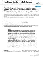
We observed IL-17A and IL-17F expression in the air-
ways of control subjects and COPD patients by immu-
nohistochemistry.Wewereonlyabletodetect
occasional immunoreactivity of IL-17A expression in
epithelium. In contrast, we observed considerable
staining for IL-17F in airway epithelium ( Figure 1A),
which was greater in the a irways of COPD subjects
compared to controls (25 (5-65) % vs. 11 (0-32) %, P <
0.0001) (Figure 1B). In the submucosa, both IL-17A
and IL-17F positive cells were observed (Figure 1A),
and the absolute number of cells expressing both of
these cytokines was higher in COPD subjects than in
control subjects (IL-17A
+
: 199 (70-310) vs. 49 (0-150);
IL-17F
+
: 287 (195-501) vs. 67 (0-203); P < 0.0001) (Fig-
ure 1C). There was also some immunoreactivity of IL-
17A and F in the endothelial site of few blood vessels
in some sections.
More submucosal cells expressed IL-17A and IL-17F in
airways of COPD patients
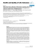
As expected, we found that the number of submucosal
cells in the airways of COPD subjects was greater than in
control subjects (2371 (509-5011) vs. 1025 (391-4087),
P < 0.001) (Figure 2A). We therefore evaluated the rela-
tive number of IL-17A
+
and IL-17F
+
cells taking in
consideration the total number of submucosal cells. Simi-
larly there was greater number of cells positive for both
IL-17AandIL-17FinCOPDsubjectscomparedtocon-
trols (IL-17A: 8 (6-14) % vs. 3 (0-7) %, P < 0.0001; IL-
17F: 10 (4-28) % vs. 4 (0-14) %, P < 0.0001) (Figure 2B).
IL-17A and IL-17F expression is not regulated on
transcriptional level
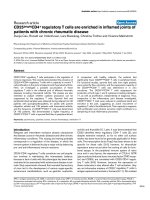
To further investigate the expr ession of IL-17A and IL-
17F in COPD, we performed quantitative RT-PCR on
frozen airways sections of COPD patients. As with pro-
tein expression, the expression of IL-17A and IL-17F
mRNA was also detected in airways of COPD patients
(Figure 3A). Although there was trend for IL-17F to be
more increased in COPD patients compared to control,
the quantification of IL-17A and IL-17F mRNA in
COPD patients was not statistically higher compared to
control (Figure 3B).
IL-17A and IL-17F expressed in CD4
+
and CD8
+
T cells
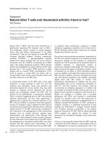
To investigate the relationship of IL-17A&F to T cells,
we used double immu nofluores cenc e staining with anti-
bodies to IL-17A or IL-17F and antibodies to CD4
+
or
CD8
+
T cells. In the airways of COPD subjects, both
CD4
+
and CD8
+
T cells expressed IL-17A and IL -17F
(Figure 4A). To our knowledge, this is the first demon-
stration that CD8
+
cells produce IL-17A and IL-17F in
COPD. Furthermore, we estimated the percentage of
CD4
+
and CD8
+
T cells that express IL-17 A and IL-17F
as well as the percentage of IL-17A
+
and IL-17F
+
cells
that co-express T cell markers. In COPD patien ts, simi-
lar percentage of CD4
+
and CD8
+
T cells that express
IL-17A and IL-17F was observed (Figure 4B). While in
total IL-17A
+
cells, the percentage of IL-17A positive
cells that co-express CD8 immunoreactivity was signifi-
cantly higher than that expressing CD4
+
T cells (16.0 ±
4.3% vs. 3.4 ± 2.0%, P < 0.05, Figure 4C). A similar
trend was also observed in total IL-17F
+
cells (15.8 ±
8.8% vs. 6.6 ± 4.3%, Figure 4C). We further confirmed
this finding using LCM to select CD8
+
T cells for the
detection of expression of IL-17 mRNA by RT-PCR
(Figure 4D).
Discussion
This study aimed to investigate the possibility that the
Th17 cytokines including IL-17A and IL-17F are
involved in the pathogenesis of COPD. Using bronchial
Chang et al. Respiratory Research 2011, 12:43
/>Page 4 of 10
biopsies from COPD patients, we found evidence that
the expression of both IL-17A and IL-17F is increased
in the airways of COPD subjects in both inflammatory
cells as well as the airway epithelium. These observa-
tions add to the growing evidence, which suggests that
Th17 cytokines play a significant role in this disease.
Using immunocytochemistry, we co nsistentl y detected
increased expression of both IL-17A and IL-17F in the
airways of COPD patients compared to controls. Both
cytokines were present to a much greater extent than in
controls (Figure 1). The pattern of expression appeared
to differ between the two cytokines in that we detected
Figure 1 IL-17A and IL-17F expression in COPD patients. (A) Immunohistochemistry, positive staining appears brown color. Magnification, 100 ×.
Scale bar = 50 μm. (B) IL-17F expression in epithelium of airways of COPD patients. IL-17F positive area in epithelium was measured as outlined in
text. (C) IL-17A and IL-17F expression in submucosa of airways of COPD patients. Absolute IL-17A
+
and IL-17F
+
positive cells in submucosa were
counted. Results are expressed as median (range), n = 15 and 16 subjects for controls and COPD patients respectively. ***P < 0.0001.
Chang et al. Respiratory Research 2011, 12:43
/>Page 5 of 10
IL-17A and F in the epithelium of COPD patients but
very little in controls. However, the best control group
is smokers without COPD, but we were unable to obtain
such a group.
The detection of considerable level of IL-17F in the
epithelium is of interest given the potential importance
of the epithelium in the inflammatory process of COPD
[20]. When stimulated with pro-inflammatory mediators,
the airway epithelium releases chemoatt ract ants CXCL1
(GRO-a), CXCL5 (ENA-78), CXCL6 (GCP-2), CXCL8
(IL-8) and CCL5 (RANTES) [21,22]. Overexpression of
IL-17F predominantly expressed in bronchial epithelial
cells has also been reported in ovalbumin challenged
mice [23]. In addition, overexpression of IL-17F in mur-
ine lung epithelium leads to infiltration of lymphocytes
and macrophages and mucus hyperplasia [24]. Taken
together, these observations suggest the possibility that
IL-17F contributes to amplification of the ongoing
inflammatory processes not only through the recr uit-
ment and activation of specific subset of inflammatory
cells, but by prolonging their survival in the airway.
Our results contrast to some degree with the recent
report of Di Stefano et al [25] who found evidence of
increased production of IL-17A but not IL-17F in the
bronchial submucosa of COPD patients. Furthermore,
they detected expression of both IL-17A and IL-17F in
the epithelium but failed to detect a difference between
controls and COPD p atients. The discrepancy between
their results and ours may reflect differences in patient
selection or technique. Notwithstanding these differ-
ences, reports to date consistently support the notion
that there is increased expression of IL-17A and IL-17F
Figure 2 Percentage of IL-17A
+
and IL-17F
+
cells in airway submucosal cells of COPD patients. (A) Subm ucosal cells in airways of COPD
patients. (B) Percentage of IL-17A
+
and IL-17F
+
cells in airway submucosal cells of COPD patients. Results are expressed as median (range), n =
15 and 16 subjects for controls and COPD patients respectively. **P < 0.001, ***P < 0.0001.
Chang et al. Respiratory Research 2011, 12:43
/>Page 6 of 10
in COPD patients, underscoring the potential impor-
tance of Th17 cytokines in this disease.
A potential explanation of increased expression of IL-
17 in COPD airways is that this may be simply a ref lec-
tion of the presence of greater numbers of submucosal
cells. Indeed, consistent with previous studies, we
detected increased cell number in the airway submucosa
of COPD patients (Figure 2). However, even after
accounting for this, we still detected significant differ-
ences between COPD and control, as the proportion of
submucosal cells expressing IL-17A and IL-17F in
COPD subjects was greater than that in controls. To
further explore the basis for this increased expression by
submucosal cells, we undertook studies of cytokine
expression at the mRNA level. As expected, we were
able to consistently detect evidence of IL-17A and IL-
17F mRNA in the airways of COPD subjects (Figure 3).
However the quantification results showed that the
mRNA e xpression of IL-17A and F was not statistically
different between COPD patients and controls, suggest-
ing that there is a discrepancy between mRNA and pro-
tein expression for IL-17A and F in COPD patients and
that increased IL-17A expression in COPD patients is
regulated at translational level. To refine this
observation, we employed a combination of immunocy-
tochemistry and laser capture microscopy. Double
immunostaining demonstrated detection of IL-17A and
IL-17F not only in CD4
+
cells as expected, but also in
CD8
+
cells (Figure 4). The high percentage of IL-17A
and IL-17F expressing CD immunoreactivity suggested
that CD8
+
T cells are major source of these cytokines
particularly in COPD [4]. We then used laser capture
microscopy to select regions of the airway that were
positive for either CD4 or CD8 by immunostaining from
which we extracted the RNA to confirm that both CD4
+
and CD8
+
cells expres s IL-17A and IL-17F mRNA (Fig-
ure 4). To our knowledge, this is the first definitive
demonstration that both CD4
+
and C D8
+
cells are cap-
able of expressing Th17 cytokines in COPD. COPD is
marked by increased number of T cells in lung parench-
yma and both peripheral and central airways, with a
greater increase in CD8
+
cells relative to CD4
+
T cells
[4]. A num ber of studies have attempted to characterize
the pattern of lymphocyte cytokine production in
COPD, but the results are conflicting [18,26]. Neverthe-
less, in the context of this observation it is noteworthy
that a recent study has reported that CD8
+
T cells are
activated in the presence of the cytokines IL-6 or IL-21
Figure 3 IL-17A and IL-17F mRNA express ion in airways of COPD patients. (A) Quantitative RT-PCR was performed from frozen airways
sections of COPD patients. One representative example from 7 subjects with similar results is shown. (B) Quantification of IL-17A and IL-17F
mRNA expression in airways of control subjects and COPD patients. Results are expressed as means ± SEM. N = 7 for both control subjects and
COPD patients.
Chang et al. Respiratory Research 2011, 12:43
/>Page 7 of 10
Figure 4 Double immunofluorescence staining for detection of IL-17A and IL-17F expression in CD4
+
and CD8
+
T cells in airways of
COPD patients. (A) Double immunofluorescence staining was performed. Scale bar = 5 μm. (B) Percentage of CD4
+
and CD8
+
T cells that
express IL-17A and IL-17F. Results are expressed as means ± SEM. (C) Percentage of CD4
+
and CD8
+
T cells that express IL-17A and IL-17F in
total IL-17A
+
and IL-17F
+
cells. Results are expressed as means ± SEM. *P < 0.05. N = 3 COPD patients. (D) IL-17A and IL-17F mRNA expression in
CD8
+
T cells in airways of COPD patients. Immunohistochemistry determined CD8
+
T cells were selected by LCM, and then RT-PCR was
performed to detect the mRNA expression of IL-17A and IL-17F. One representative result from 3 subjects is shown.
Chang et al. Respiratory Research 2011, 12:43
/>Page 8 of 10
plus TGF-b, develop into IL-17-producing (Tc17) cells.
Our findings also need to be t aken seen in the context
of reports of Tc17 cells in a variety of immunological
diseases. For example, Tc17 have also been found in
cutaneous inflammatory diseases like psoriasis vulgaris
[27] and allergic contac t dermatiti s [28]. Tc17 cells may
also be important in defense against viruses [29,30].
The observation that expression of Th17 cytokines is
increased in COPD raises questions as to how this may
come about. The combination of IL-6 and TGF-b is
reported to sk ew the balance of T helper cells toward
Th17 cell differentiation [31]. In this regard, it is of
interest that increased production of IL-6 and TGF-b
has been reported in COPD patients [32], raising the
possibility that IL-6 and T GF-b may enable the promo-
tion of Th17 cells differentiation in COPD. Regardless
of the mechanism, Th17 cytokines have the potential to
contribute to COPD in various ways. IL-17A acts
directly on epithelial cells and on airway fibr oblasts and
smooth muscle cells to induce the secretion of neutro-
phil-recruiting chemokines, such as CXCL8 [31].
Although a comprehensive comparative analysis of IL-
17F and IL-17A has not been performed, IL-17F appears
to have biological actions similar to IL-17A both in vitro
and in vivo [14]. Therefore it is possible that with acti -
vation of IL-17A and IL-17F mediated pathways, a
crosstalk between local activation of T cells and sus-
tained accumulation of neutrophils in inflamed ai rways
could be established. Zhu et al [33] have suggested that
biopsies from patients with chronic bronchitis have
more inflammation compared to patients with COPD
but without chorionic bronchitis. This group of patients
mighthavemoreIL-17expression. However in our
study w e did not group our subjects and presented the
data of our patients as one group according to GOLD
classification.
In summary, in bronchial biopsies we detected clear
evidence that the expression of the cytokines IL-17A
and IL-17 F is increased in COPD compared to control.
In the case of IL-17F, this increased expression extends
to the epithelium and is no t simply restricted to the
submucosa. Most importantly, we detected increased
expression of these cytokines in both CD4
+
and CD8
+
cells, suggesting that the inflammatory process in COPD
may resemble that in other disorders where Tc17 cells
are active. These findings contribute to the growing
body of information that supports the importance of
investigating t he role of IL-17 and related cytokines in
COPD, potentially providing novel therapeutic targets in
this important chronic disease.
Acknowledgements
This study was supported by a grant from the CIRF program.
Author details
1
Meakins-Christie Laboratories and Respiratory Division, Department of
Medicine McGill University, 3626 rue St. Urbain, Montreal, QC, H2X 2P2
Canada.
2
Respiratory Division, Research Institute of McGill University Health
Centre, 2155 Guy Street, Suite 900 Montreal, QC, H3H 2R9 Canada.
3
Respiratory Division, Laval University, 2325 rue de l’Université, Québec, QC,
G1V0A6 Canada.
Authors’ contributions
YC carried out the cell counting and data analysis and drafted the
manuscript. JN performed the RT-PCR. NB carried out the
immunohistochemistry staining and laser capture. JB and FM participated in
the sample collection and did the immunocytochemiostry. DHE participated
in the design of the study and corrected the manuscript. QH supervised of
the study. All authors read and approved the final manuscript.
Competing interests
The authors declare that they have no competing interests.
Received: 26 August 2010 Accepted: 10 April 2011
Published: 10 April 2011
References
1. Pauwels RA, Rabe KF: Burden and clinical features of chronic obstructive
pulmonary disease (COPD). Lancet 2004, 364(9434):613-620.
2. Cosio MG, Saetta M, Agusti A: Immunologic aspects of chronic
obstructive pulmonary disease. N Engl J Med 2009, 360(23):2445-2454.
3. Roth M: Pathogenesis of COPD. Part III. Inflammation in COPD. Int J
Tuberc Lung Dis 2008, 12(4):375-380.
4. Cosio MG, Majo J: Inflammation of the airways and lung parenchyma in
COPD: role of T cells. Chest 2002, 121(5 Suppl):160S-165S.
5. Agusti A, MacNee W, Donaldson K, Cosio M: Hypothesis: does COPD have
an autoimmune component? Thorax 2003, 58(10):832-834.
6. Kolls JK, Linden A: Interleukin-17 family members and inflammation.
Immunity 2004, 21(4):467-476.
7. Liang SC, Long AJ, Bennett F, Whitters MJ, Karim R, Collins M, Goldman SJ,
Dunussi-Joannopoulos K, Williams CM, Wright JF, Fouser LA: An IL-17F/A
heterodimer protein is produced by mouse Th17 cells and induces
airway neutrophil recruitment. J Immunol 2007, 179(11):7791-7799.
8. Wright JF, Guo Y, Quazi A, Luxenberg DP, Bennett F, Ross JF, Qiu Y,
Whitters MJ, Tomkinson KN, Dunussi-Joannopoulos K, Carreno BM,
Collins M, Wolfman NM: Identification of an interleukin 17F/17A
heterodimer in activated human CD4+ T cells. J Biol Chem 2007,
282(18):13447-13455.
9. Huber M, Heink S, Grothe H, Guralnik A, Reinhard K, Elflein K, Hunig T,
Mittrucker HW, Brustle A, Kamradt T, Lohoff M: A Th17-like developmental
process leads to CD8(+) Tc17 cells with reduced cytotoxic activity. Eur J
Immunol 2009, 39(7):1716-1725.
10. Kramer JM, Gaffen SL: Interleukin-17: a new paradigm in inflammation,
autoimmunity, and therapy. J Periodontol 2007, 78(6):1083-1093.
11. Miossec P, Korn T, Kuchroo VK: Interleukin-17 and type 17 helper T cells.
N Engl J Med 2009, 361(9):888-898.
12. Fujisawa T, Velichko S, Thai P, Hung LY, Huang F, Wu R: Regulation of
airway MUC5AC expression by IL-1beta and IL-17A; the NF-kappaB
paradigm. J Immunol 2009, 183(10):6236-6243.
13. Park H, Li Z, Yang XO, Chang SH, Nurieva R, Wang YH, Wang Y, Hood L,
Zhu Z, Tian Q, Dong C: A distinct lineage of CD4 T cells regulates tissue
inflammation by producing interleukin 17. Nat Immunol 2005,
6(11):1133-1141.
14. Hizawa N, Kawaguchi M, Huang SK, Nishimura M: Role of interleukin-17F
in chronic inflammatory and allergic lung disease. Clin Exp Allergy 2006,
36(9):1109-1114.
15.
McKinley L, Alcorn JF, Peterson A, Dupont RB, Kapadia S, Logar A, Henry A,
Irvin CG, Piganelli JD, Ray A, Kolls JK: TH17 cells mediate steroid-resistant
airway inflammation and airway hyperresponsiveness in mice. J Immunol
2008, 181(6):4089-4097.
16. Hattotuwa KL, Gizycki MJ, Ansari TW, Jeffery PK, Barnes NC: The effects of
inhaled fluticasone on airway inflammation in chronic obstructive
pulmonary disease: a double-blind, placebo-controlled biopsy study. Am
J Respir Crit Care Med 2002, 165(12):1592-1596.
Chang et al. Respiratory Research 2011, 12:43
/>Page 9 of 10
17. Al-Ramli W, Prefontaine D, Chouiali F, Martin JG, Olivenstein R, Lemiere C,
Hamid Q: T(H)17-associated cytokines (IL-17A and IL-17F) in severe
asthma. J Allergy Clin Immunol 2009, 123(5):1185-1187.
18. Hodge G, Nairn J, Holmes M, Reynolds PN, Hodge S: Increased intracellular
T helper 1 proinflammatory cytokine production in peripheral blood,
bronchoalveolar lavage and intraepithelial T cells of COPD subjects. Clin
Exp Immunol 2007, 150(1):22-29.
19. Lane N, Robins RA, Corne J, Fairclough L: Regulation in chronic
obstructive pulmonary disease: the role of regulatory T-cells and Th17
cells. Clin Sci (Lond) 119(2):75-86.
20. Larsson K: Aspects on pathophysiological mechanisms in COPD. J Intern
Med 2007, 262(3):311-340.
21. Prause O, Laan M, Lotvall J, Linden A: Pharmacological modulation of
interleukin-17-induced GCP-2-, GRO-alpha- and interleukin-8 release in
human bronchial epithelial cells. Eur J Pharmacol 2003, 462(1-3):193-198.
22. Wang JH, Devalia JL, Xia C, Sapsford RJ, Davies RJ: Expression of RANTES
by human bronchial epithelial cells in vitro and in vivo and the effect of
corticosteroids. Am J Respir Cell Mol Biol 1996, 14(1):27-35.
23. Suzuki S, Kokubu F, Kawaguchi M, Homma T, Odaka M, Watanabe S, Ieki K,
Matsukura S, Kurokawa M, Takeuchi H, Sasaki Y, Huang SK, Adachi M, Ota H:
Expression of interleukin-17F in a mouse model of allergic asthma. Int
Arch Allergy Immunol 2007, 143(Suppl 1):89-94.
24. Yang XO, Chang SH, Park H, Nurieva R, Shah B, Acero L, Wang YH,
Schluns KS, Broaddus RR, Zhu Z, Dong C: Regulation of inflammatory
responses by IL-17F. J Exp Med 2008, 205(5):1063-1075.
25. Di Stefano A, Caramori G, Gnemmi I, Contoli M, Vicari C, Capelli A, Magno F,
D’Anna SE, Zanini A, Brun P, Casolari P, Chung KF, Barnes PJ, Papi A,
Adcock I, Balbi B: T helper type 17-related cytokine expression is
increased in the bronchial mucosa of stable chronic obstructive
pulmonary disease patients. Clin Exp Immunol 2009, 157(2):316-324.
26. Zhu X, Gadgil AS, Givelber R, George MP, Stoner MW, Sciurba FC,
Duncan SR: Peripheral T cell functions correlate with the severity of
chronic obstructive pulmonary disease. J Immunol 2009, 182(5):3270-3277.
27. Ortega C, Fernandez AS, Carrillo JM, Romero P, Molina IJ, Moreno JC,
Santamaria M: IL-17-producing CD8+ T lymphocytes from psoriasis skin
plaques are cytotoxic effector cells that secrete Th17-related cytokines.
J Leukoc Biol 2009, 86(2):435-443.
28. Zhao Y, Balato A, Fishelevich R, Chapoval A, Mann DL, Gaspari AA: Th17/
Tc17 infiltration and associated cytokine gene expression in elicitation
phase of allergic contact dermatitis. Br J Dermatol 2009, 161(6):1301-1306.
29. Hamada H, Garcia-Hernandez Mde L, Reome JB, Misra SK, Strutt TM,
McKinstry KK, Cooper AM, Swain SL, Dutton RW: Tc17, a unique subset of
CD8 T cells that can protect against lethal influenza challenge. J
Immunol 2009, 182(6)
:3469-3481.
30. Kader M, Bixler S, Piatak M, Lifson J, Mattapallil JJ: Anti-retroviral therapy
fails to restore the severe Th-17: Tc-17 imbalance observed in peripheral
blood during simian immunodeficiency virus infection. J Med Primatol
2009, 38(Suppl 1):32-38.
31. Nembrini C, Marsland BJ, Kopf M: IL-17-producing T cells in lung
immunity and inflammation. J Allergy Clin Immunol 2009, 123(5):986-994,
quiz 995-986.
32. Kim V, Rogers TJ, Criner GJ: New concepts in the pathobiology of chronic
obstructive pulmonary disease. Proc Am Thorac Soc 2008, 5(4):478-485.
33. Zhu J, Qiu Y, Valobra M, Qiu S, Majumdar S, Matin D, De Rose V, Jeffery PK:
Plasma cells and IL-4 in chronic bronchitis and chronic obstructive
pulmonary disease. Am J Respir Crit Care Med 2007, 175(11):1125-1133.
doi:10.1186/1465-9921-12-43
Cite this article as: Chang et al.: CD8 positive T cells express IL-17 in
patients with chronic obstructive pulmonary disease. Respiratory Research
2011 12:43.
Submit your next manuscript to BioMed Central
and take full advantage of:
• Convenient online submission
• Thorough peer review
• No space constraints or color figure charges
• Immediate publication on acceptance
• Inclusion in PubMed, CAS, Scopus and Google Scholar
• Research which is freely available for redistribution
Submit your manuscript at
www.biomedcentral.com/submit
Chang et al. Respiratory Research 2011, 12:43
/>Page 10 of 10