Báo cáo y học: " In vivo imaging of the airway wall in asthma: fibered confocal fluorescence microscopy in relation to histology and lung function" potx
Bạn đang xem bản rút gọn của tài liệu. Xem và tải ngay bản đầy đủ của tài liệu tại đây (2.56 MB, 9 trang )
RESEARCH Open Access
In vivo imaging of the airway wall in asthma:
fibered confocal fluorescence microscopy in
relation to histology and lung function
Ching Yong Yick
1*
, Jan H von der Thüsen
2,3
, Elisabeth H Bel
1
, Peter J Sterk
1
and Peter W Kunst
1
Abstract
Background: Airway remodelling is a feature of asthma including fragmentation of elastic fibres observed in the
superficial elastin network of the airway wall. Fibered confocal fluorescence microscopy (FCFM) is a new and non-
invasive imaging technique performed during bronchoscopy that may visualize elastic fibres, as shown by in vitro
spectral analysis of elastin powder. We hypothesized that FCFM images capture in vivo elastic fibre patterns within
the airway wall and that such patterns correspond with airway histolog y. We aimed to establish the concordance
between the bronchial elastic fibre pattern in histology and FCFM. Second, we examined whether elastic fibre
patterns in histology and FCFM were different between asthmatic subjects and heal thy controls. Finally, the
association between these patterns and lung function parameters was investigated.
Methods: In a cross-sectional study comprising 16 subjects (8 atopic asthmatic patients with controlled disease and
8 healthy controls) spirometry and bronchoscopy were performed, with recording of FCFM images followed by
endobronchial biopsy at the airway main carina. Elastic fibre patterns in histological sections and FCFM images were
scored semi-quantitatively. Agreement between histology and FCFM was analysed using linearly weighted kappa
w
.
Results: The patterns observed in histological sections and FCFM images could be divided into 3 distinct groups.
There was good agreement between elastic fibre patterns in histology and FCFM patterns (
w
0.744). The semi-
quantitative pattern scores were not different between as thmatic patients and controls. Notably, there was a
significant difference in post-bronchodilator FEV
1
%predicted between the different patterns by histology (p =
0.001) and FCFM (p = 0.048), regardless of asthma or atopy.
Conclusion: FCFM captures the elastic fibre pattern within the airway wall in humans in vivo. The association
between post-bronchodilator FEV
1
%predicted and both histological and FCFM elastic fibre patterns points towards
a structure-function relationship between extracellular matrix in the airway wall and lung function.
Trial registration: Netherlands Trial Register NTR1306
Keywords: Asthma, Confocal Laser Scanning Microscopy, Extracellular Matrix, Respiratory Function Tests, Smooth
muscle
Background
Asthma is characterized by episodic symptoms, variable
airway obstruction and airway hyperresponsiveness to a
variety of inhaled stimuli [ 1-3], and impairment of
bronchodilation following deep inspiration [4,5]. The
underlying pathophysiological mechanisms that lead to
the observed functional changes in asthma have only
partly been resolved. Airway remodelling, a process of
structural changes of the airway wall seen in asthma
likely impairs lung function [6,7]. This includes an
increased deposition and altered organization of extracel-
lular matrix (ECM) proteins in the lamina propria,
smooth muscle layer and adventitial layer [8-10].
Evidence shows that the elastic fibres, which represent a
major component of the ECM in the airway wall, are
disrupted and fragmented in asthmatic patients as
* Correspondence:
1
Department of Respiratory Medicine, Academic Medical Centre,
Meibergdreef 9, Amsterdam, The Netherlands
Full list of author information is available at the end of the article
Yick et al. Respiratory Research 2011, 12:85
/>© 2011 Yick et al; licensee BioMed Central Ltd. This is an Open A ccess article distributed under the terms of the Creative Commons
Attribution License ( which pe rmits unrestricted use, distribution, and reproduction in
any medium, provided the origin al work is properly cited.
compared to healthy control subjects [11]. Additionally,
fragmentation and a decreased amount of elastic fibres
have been observed in the superficial elastin network of
central airways in fatal asthma [12].
The presence and extent of airway remodelling in
asthma can be visualized by histology in endobronchial
biopsy specimens. However, this is not real-time and
requires careful and time-consuming histological techni-
ques. Fibered confocal fluorescence microscopy (FCFM) is
a new imaging modality, representing a non-invasive
method that can be used to image the microscopic struc-
ture of airway wall tissue in vivo during a bronchoscopic
procedure [13]. T he principle of this imaging method is
based on the autofluorescence of endogenous or exogen-
ous fluorophores inside the tissues after excitation by an
external laser light source. The laser light is guided
through a bundle of optical microfibres to the tip of the
miniprobe of 1 mm in diameter, which can be inserted
into the working channel o f a fibreoptic bronchoscope
[14]. High-quality and real-time in vivo morphological
images or ‘optical biopsies’ of the airway wall are obtained
by placing the tip of the miniprobe onto the airway wall
surface. Another advantage of FCFM and its miniprobe is
the ability to reach and therefore visualize the alveoli
in vivo [15].
It has been shown in vitro t hat the aut ofluorescen ce
spectra of proximal bronchial mucosa and elastin pow-
der extracted from healthy human lung at an e xcitation
laser light wavelength of 488 nm were very similar,
whereas this w as not the case with bronchial mucosa
and collagen I gel [13]. Therefore, the autofluorescence
in FCFM images at 488 nm excitation wavelength is
likely to originate from the elastic fibres present in the
airway wall. However, the patterns obtained by FCFM
have not yet been compared with the ‘gold standard’,
which is histology of the airway wall.
We hypothesized that FCFM images capture elastic
fibre patterns withi n the airway wall and that these pat-
terns are comparable to those observed by airway histol-
ogy. Therefore, the first aim of the study was to
investigate the agreement between semi-quantitative
pattern scores between histological sections and FCFM
images. The second aim was to investigate whether the
patterns seen at the airway main carina in histological
sections and FCFM are different between asthmatic and
healthy control subjects. Finally, we exa mined whether
these patterns are associated with lung function.
Methods
Design and subjects
This study had a cross-sectiona l design and included 16
subjects: atopic asthmatic patients (n = 8 ) and healthy
controls (n = 8). All subjects were recruited by the
department of Respiratory Medicine of the Academic
Medical Centre Amsterdam using public advertisements.
The study consisted of 2 visits. During the first visit,
subjects were screened for eligibility to participate
according to the in- and exclusion criteria (see below).
Spirometry and a methacholine bronchoprovocation
test were performed. At visit 2, b ronchoscopy was car-
ried out with real-time digital recording of FCFM
images and collection of endobronchial biopsy
specimens.
Asthmatic subjects had controlled disease according to
GINA guidelines [1]. Furthermore, they met all of the fol-
lowing criteria: aged 18 to 50 years; non-smok ing or hav-
ing stopped smoking > 12 months with ≤ 5 pack years;
no exacerbations within the last 6 weeks prior to partici-
pation; steroid-naïve or having stopped steroids by any
dosing route ≥ 8 weeks prior to participation; no other
medication for treating asthma than the use of inhaled
short-acting b2-agonists as rescue medication; airway
hyperresponsiveness defined by a methacholine broncho-
provocation test with PC
20
≤ 8 mg/mL; post-bronchodila-
tor FEV
1
> 70% of predicted; and atopy defined by a
positive skin prick test.
Healthy control subjects met all of the following criteria:
aged 18 to 50 years; non-smoking or having stopped
smoking > 12 months with ≤ 5 pack years; steroid-naïve;
no airway hyperresponsiveness defined by a methacholine
bronchoprovocation test with PC
20
> 8 mg/mL; and post-
bronchodilator FEV
1
> 70% of predicted. Skin prick test
was performed on all healthy control subjects, but the out-
come was no selection criterion.
Participants with pulmonary diseases other than
asthma were excluded, as w ell as pregnant females. All
subjects gave written informed consent prior to enrol-
ment. This study was approved by the Medical Ethics
Committee of the Academic Medical Centre Amsterdam
and is registered at the Netherlands Trial Register
(NTR1306).
Lung function and allergy
Spirometry was performed using a daily calibrated spi-
rometer according to European Respiratory Society
(ERS) recommendations [16]. The methacholine bronch-
oprovocation test was performed according to the stan-
dardized tidal volume m ethod [3]. All but one of the
healthy con trol subjects didn’t reach a PC
20
at the maxi-
mum m ethacholine dose of 16 mg/mL, so that we also
used the linear model of the dose-response slope as pro-
posed and validated by O’Connor et al. [17]. Skin prick
tests were performed using 12 common aeroallergen
extracts according to the position paper by the Eur-
opean Academy of Allergology and Clinical Immunology
(EAACI) [18].
Yick et al. Respiratory Research 2011, 12:85
/>Page 2 of 9
Bronchoscopic procedure
Fibreoptic bronchoscopy was perfor med according to the
recommendations made by the National Heart, Lung, and
Blood Institute (NHLBI) and the National Institute of
Allergy and Infectious Diseases (NIAID) [19]. Participants
received local anaesthetic by Lignocaine 1% and 10% spray
in the nose and throat. Instillation of Lignocaine 1% solu-
tion in separate lung segments was ap plied to further
dampen the cough reflex. The oxygen saturation and heart
rate of the participant were monitored continuously dur-
ing the bronchoscopic procedure. Additional oxygen
through an intranasal catheter was given to the participant
when n ecessary.
Immediately a fter adequate local anaesthetic the bron-
chial tree was inspected with an autofluorescence
bronchoscope (SAFE 3000, Pentax, Japan). Next, the
Alveoflex miniprobe of the FCFM system (Cellvizio,
Mauna Kea Technologies, France [20]) was inserted
through the working channel of the bronchoscope and
placed on the main carina of the airways. Special care
was taken to position the miniprobe perpendicularly to
the surface of the main carina as much as possible in
order to get good quality FCFM images (Figure 1a, c).
Real-time digital video recordings of 9 frames per second
at 488 nm laser light excitation wavelength were made
during sever al seconds and stored digitally. After record-
ing the FCFM images, one endo bronchial biopsy speci-
men was taken with a cup forceps (Pentax KW2411S) at
the exact same location where the miniprobe had been
placed before (Figure 1b, d). Directly after collection, the
biopsy specimen was fixed in 4% buffered formaldehyde
and embedded in paraffin.
Elastic fibres and FCFM pattern analysis
Biopsy specimens were processed as described previously
[21]. Briefly, biopsy specimens were cut into 4 μmsections
and stained with haematoxylin and eosin for initial analysis
of the bronchial morphology. One representative slide per
biopsy specimen with good morphologic quality including
epithelial cells, an intact reticular basement membrane,
and submucosa without crushing artefacts was stained for
elastin with Elastica-van Gieson (EVG). Next, images of
the histological sections were captured with a digital cam-
era coupled to a light microscope (Leica Microsystems,
Germany) and analysed at 20 × magnification using
Image-Pro Plus 5 (Media Cybernetics, Bethesda, MD,
USA). For eac h slide, a repre se ntative area with positive
stai ning for elastin in the subepithelial layer was selected
for elastic fibre pattern analysis.
FCFM digital video recordings were analysed using the
image analysis software MedViewer (Mauna Kea Tech-
nologies, France). This software allows detailed analysis
of recordings frame by frame. Additionally, video mosai-
cing techniques were applied to reconstruct a FCFM
digital video recor ding compensated for the rigid and
non-rigid deformations due to motion and irregular
contact of the miniprobe with the tissue surface respec-
tively [22]. A representative image frame for each FCFM
recording, in which the FCFM pattern did not change in
several consecutive frames and without imaging artefacts
or overexposure, was selected for pattern analysis.
The elastic fibre pattern in the histological sections and
the FCFM patterns of the selected frames from the same
subject were scored semi-quantitatively by two separate
observers, who were blinded for the study groups. To
define the scoring, slides stained for initial analysis of mor-
phologic quality, were analysed. Three distinct patterns of
elastic fibres were distingui shed based on their thickne ss
and organisation: wispy (score 1), mixed (score 2), and
lamellar (score 3) (Figure 2). Histological sections with the
classification ‘wispy’ contained typically thin and loosely
organised elastic fibres in the subepithelial area, whereas
those observed for ‘lamellar’ were thick and linearly orga-
nized. Additionally, the thickened fibres of the latter group
were abundantly present and organized into a distinctive
layer beneath the epithelium compared to the ‘ wispy’
group. The ‘ mixed’ group contained a mix of thin and
thick elasti c fibres, partly loosely and partly linearly
organized.
Data analysis
Demographic data of the study groups with a normal dis-
tribution were compared using unpaired t-tests. When
normality was not achieved, data were compared using
Mann-Whitney U tests. Chi-square tests were performed
to analyze the distribution of the classification scores in
asthmatics and controls. To analyze the agreement
between semi-quantitative classification scores of the elas-
tic fibre pattern in histology and FCFM pattern, a
weight ed ka ppa
w
with linear weight s was calculated. A
p-value of < 0.05 was considered statistically significant.
Thesamplesizeofthepresentstudywasbasedonthe
estimation that a kappa of 0.8 could be detected with a
power of 80% at the 5% level of significance with a study
population of 14 subjects [23,24]. Statistical analyses were
performed using SPSS version 18 (IBM Corporation, Som-
ers, NY, USA).
Results
Subjects
Fourteen out of the total 16 test subjects were included for
pattern analysis by histology and FCFM. Two subjects,
including 1 asthmatic patient and 1 control subject, were
excluded from analysis due to instability and/or overexpo-
sure of the FCFM images.
Subject characteristics of the study groups can be found
in Table 1. The FEV
1
/FVC ratio was significantly lower
and the methacholine dose-response slope significantly
Yick et al. Respiratory Research 2011, 12:85
/>Page 3 of 9
higher in asthmatic subjects when compared to healthy
controls, as expected.
Elastic fibre pattern in histology
Elastic fibres were c learly visible in the EVG-stained his-
tological sections from all subjects. Two asthmatics and 1
healthy control subject were scored as ‘wispy’,whereas2
asthmatics and 3 healthy control subjects as ‘ mixed’.The
‘lamellar’ group consisted of 3 asthmatics and 3 healthy
control subjects (Figure 3a). There was no difference in
histological elastic fibre patterns between asthmatic
patients and healthy control subjects (p > 0.05).
FCFM pattern
FCFM digital video recordings were classified into the
three groups as described in the Methods section. In the
FCFM images that were scored as ‘wispy’, no specific pat-
tern could be distinguished (Figure 2). Occasionally,
some indefinite and thin lines without a specific orienta-
tion could be identified. In contrast, a line arly orientated
pattern composed of clearly discernible light-coloured
and thick individual lines could be discriminated in
FCFM images with score 3 (‘lamellar’). These lines were
organized into layers similar to the histological sections.
The linear and orientated pattern was less clearly defined
in the ‘ mixed’ group and was a combination of the pat-
ternsseeninthe‘wispy’ and ‘lamellar’ groups. Further-
more, this ‘ mixed’ pattern showed inter-individual
variability with some resembling the ‘ wispy’ patt ern,
whereas others the ‘lamellar’ pattern. Two asthmatics
and 1 healthy control subject were scored as ‘ wispy’ ,
whereas 2 asthmatics and 2 healthy control subjects as
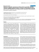
Figure 1 FCFM and biopsy during bronchoscopy. The FCFM probe was placed perpendicularly to the surface of the main carina (a) followed by
endobronchial biopsy at the same location (b). Figure 1c and 1d gives a lateral view of the probe and biopsy location respectively. * = main carina.
Yick et al. Respiratory Research 2011, 12:85
/>Page 4 of 9
‘mixed’ .The‘lamellar’ group consisted of 3 asthmatics
and 4 healthy control subjects (Figure 3b). There was no
difference in FCFM patterns between asthmatic patients
and healthy controls (p > 0.05).
Agreement between histology and FCFM
Of the 14 subjects that were inclu ded for p attern anal y-
sis, 11 subjects were scored consistently for elastic fibre
pattern between histology and FCFM (weighted kappa
w
0.744, Table 2 and 3). There was only discrepancy
between histology and FCFM in the classification of the
patterns into scores 2 or 3 (’mixed’ or ‘lamellar’).
Association between patterns and lung function
Post-bronchodilator FEV
1
%predicted was significantly
lower for FCFM score 3 (’la mellar’)ascomparedto
FCFM score 1 (’wispy’) (p = 0.048, Figure 4b), regardless
of asthma or atopy. This was confirmed and extended
by histology, showing a significantly lower post-bronch-
odilator FEV
1
%predicted in the ‘lamellar’ as compared
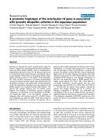
Figure 2 Representative histological sections (20 × magnification) and corresponding FCFM images.Fibreswerethinandloosely
organized in the ‘wispy’ group (a), whereas these were thick and linearly organized into a layer in the ‘lamellar’ group (c). No specific pattern
was present in the FCFM images of the ‘wispy’ group (d). Individual thick lines in layer-form were observed in FCFM images of the ‘lamellar’
group (f). Patterns of the ‘mixed’ group were a combination of those seen in the ‘wispy’ and ‘lamellar’ groups (b, e).
Table 1 Subject characteristics of asthma (A) and healthy (H) subjects
AH
Total HA HNA
Subjects (n) 8853
Male/Female (n) 2/6 4/4 2/3 2/1
Age (years)
a
24 (2) 28 (10) 30 (12) 23 (2)
FEV
1
/FVC (%pred.)
a
,* 96 (9) 108 (7) 105 (7) 113 (4)
Post-bronchodilator FEV
1
(%pred.)
a
108 (11) 115 (6) 115 (6) 114 (6)
Dose response slope (% decline FEV
1
/μmol methacholine)
b
,** 6.49
(3.47-31.30)
0.20
(0.04-0.30)
0.23
(0.10-0.49)
0.07
(0.03-0.23)
Total = all healthy control subjects; HA = healthy, atopic; HNA = healthy, non-atopic
a
Mean (SD)
b
Median (P25-P75)
* A vs. Total p = 0.012; A vs. HNA p = 0.016
** A vs. Total p = 0.001; A vs. HA p = 0.003; A vs. HNA p = 0.014
Yick et al. Respiratory Research 2011, 12:85
/>Page 5 of 9
to the ‘wispy’ (p = 0.001) and ‘mixed’ (p = 0.021) groups
(Figure 4a).
Discussion
The present study shows that elastic fibres in the airway
wall can be visualized by FCFM, a novel b ronchoscopic
imaging modality, and that a laminar pattern of these
fibres is associated with reduced lung fu nction. The
elastic fibres in histology and FCFM images exhibited 3
distinct patterns. There was good agreement in semi-
quantitative pattern score between histology and FCFM,
but there were no differences in such patterns between
asthma patients and cont rols. These findings indicate
that FCFM can be used to capture structural changes in
the airway wall in humans in vivo, and might become a
real-time imaging tool to estimate the type and degree
of airway remodelling in chronic airway disease s such as
asthma in the near future. Since FCFM has the ability to
visualize the airway wall of the whole bronchial tree in
vivo during bronchoscopy, it has complementary advan-
tages as compared to taking snapshot biopsies at several
locations.
There are some study data and case reports in literature
examining the association between FCFM and histology of
e.g. preinvasive bronchial lesions and sarcoidosis [13,25].
However, to our knowledge this is the first study to inves-
tigate the histological substrate of autofluorescence in 488
nm FCFM images using in vivo human endobronchial
biopsy specimens obtained from asthmatic patients and
health y control subjects with histology as ‘gold standard’.
The agreement of elastic fibre patterns between histology
and FCFM is a novel finding and extends previous in vitro
observations showing that bronchial mucosa and elastin
powder extracted from human lung had similar autofluor-
escence spectra, suggesting that the autofluorescence in
FCFM mainly originates from the elastic fibres present in
the airway wall [13]. We did not observe differences in
elastic fibre patterns between asthmatics and controls.
This contrasts previous studies using histology [11,12],
which is likely explained by differences in disease severity
of the asthmatic patients, which in our study included
mild disease.
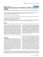
Figure 3 Pattern grading by histology (a) and FCFM (b). A = asthma; Total = all healthy control subjects; HA = healthy, atopic; HNA =
healthy, non-atopic. Chi-square test: p > 0.05.
Table 2 Elastic fibre pattern grading: Paired comparison
histology and FCFM
Subject Study group Histology FCFM
1 Asthma Wispy Wispy
2 Asthma Wispy Wispy
3 Asthma Mixed Lamellar
4 Asthma Mixed Mixed
5 Asthma Lamellar Lamellar
6 Asthma Lamellar Mixed
7 Asthma Lamellar Lamellar
8 Healthy, atopic Mixed Mixed
9 Healthy, atopic Mixed Lamellar
10 Healthy, atopic Lamellar Lamellar
11 Healthy, atopic Lamellar Lamellar
12 Healthy, non-atopic Wispy Wispy
13 Healthy, non-atopic Mixed Mixed
14 Healthy, non-atopic Lamellar Lamellar
Subjects with discrepancy between pattern scores by histology and FCFM in
Italic.
Table 3 Elastic fibre pattern grading: Agreement
histology and FCFM
Histology
Wispy Mixed Lamellar
Wispy 3 - -
FCFM Mixed - 3 1
Lamellar - 2 5
w
(SE, 95% CI) = 0.744 (0.145, 0.46-1)
Yick et al. Respiratory Research 2011, 12:85
/>Page 6 of 9
The asso ciatio n between the elastic fibre patterns and
lung function is a novel finding and adds to the validity
of our histological and FCFM scoring. A plausible expla-
nation for the lower FEV
1
%predicted in the ‘lamellar’
group as compared to the other groups is that the paral-
lel organisation of the thickened elastic fibres in a layer
just beneath the epithelium changes airway wall
mechanics and thereby FEV
1
. Airway wall mechanics are
different in asthma as compared to controls [26], but
additional ECM components e.g. collagen, proteogly-
cans, and glycoproteins are likely to contribute to this as
well.EventhoughECMmaythickentheairwaywall
and thereby promoting luminal narrowing, any accom-
panying stiffening can stabilize the airways from collapse
[27]. It is still unknown how elastic f ibre patterns could
influence either of the above mechanisms. Hence, our
current struct ure-function observations should be con-
sidered as hypothesis-generating.
The present results suggest that FCFM is an adequate
method to examine bronchial elastic fibre morphology
in vivo and might be an important tool to detect asth-
matic patients who are prone to loss of lung function, at
an early stage enabling timely interventi on. Based on
data found in literature we would expect that the degree
of airway remodelling differs with asthma severity,
including a varying amount or organization of elastic
fibres [10,12,28-30]. Therefore, subsequent studies
including larger numbers of subjects with varying
asthma severity and FCFM images of multiple locations
in the bronchial tree are needed. These will give a mor e
detailed insight into the association between histology,
FCFM, and airway function.
The strength of our study is that we applied strict
patient selection criteria and that histology and FCFM
were obtained from the same endobronchial sites. The
bronchial main carina was chosen as the location for
FCFM and biopsy to minimize imaging artefacts result-
ing from inadequate positioning of the miniprobe super-
imposed on the movement of the airways due to tidal
breathing. However, there are potential limitations that
need to be addressed. First, the number of 16 subjects
was relatively low when using 3 semi-quantitative scores.
Although this study was powere d on kappa, power esti-
mation b ased on this value is not firmly developed yet.
Thi s is due to the fact that this estimation is dependen t
on the kappa expected to be found and the marginal
frequencies, which are the proportions of test subjects
in each semi-quantitative category of histology and
FCFM. Second, FCFM images during bronchoscopy
were captured by placi ng the miniprobe perpendicu larly
tothesurfaceoftheairwaymaincarinafollowedbya
biopsy from the same location. It was technically impos-
sible to orientate the small biopsy specimens in such a
way that the cutting plane was identical to the plane of
view during the FCFM recordings. This may have intro-
duced bias in the semi-quantitati ve scoring o f the histo-
logical sections. However, this bias seems to be limited
as only the superficially located elastic fibres in the sub-
epithelial layer were graded.
Our findings show a good agreement between pattern
scores by histology and FCFM. The FCFM miniprobe
has a fixed depth of view of 50 μm and therefore cap-
tures images at the level of the subepithelial layer.
Accordingly, elastic fibre patt erns in t he subepithelial
layer were scored in the histological sections. Pattern
scores by histology and FCFM proved to be close in
resemblance. By analysing both the autofluorescence
patterns and the surrounding darker areas in FCFM
images, this imaging modality also has the potential to
visualize airway remodelling in general, which today is
Figure 4 Post-bronchodilato r FEV
1
%predicted of the three classification scores in histology (a) and FCFM (b). Data presented as post-
bronchodilator FEV
1
%predicted of individual subjects and the mean per classification score. ● = asthma, ■ = healthy.
Yick et al. Respiratory Research 2011, 12:85
/>Page 7 of 9
only possibly by histology of biopsy specimens. Other
bronchoscopic real-time imaging modalities visualizing
airway wall structures have recently been introduced
including anatomical optical coherence tomography
(aOCT) and endobronchial ultrasonography (EBUS)
[31-34]. While aOCT and EBUS may visualize the dif-
ferent layers of the airway wall, FCFM can image a spe-
cific airway structural component in microscopic detail.
Nevertheless, all three imaging techniques are no t suita-
ble to replace histology in the clinical setting yet. The
technical part has to be further improved to acquire
even higher quality images with minimal imaging
artefacts.
Conclusions
In the current study, we observed good agreement
between elastic fibre pattern scores of the bronchial wall
by histology and fibered confocal fluorescence microscopy,
suggesting that this imaging technique is suitable to
capture the morphology of bronchial elastic fibres non-
invasively in humans in vivo. Post-bronchodilator FEV
1
%
predicted was associated with elastic fibre patterns, point-
ing towards a structure-function relationship between
extracellular matrix and lung function. The results of our
study suggest that fibered confocal fluorescence micro-
scopy might become a real-time imaging tool to estimate
the type and degree of airway remodelling in chronic air-
way diseases such as asthma.
List of abbreviations
EAACI: European Academy of Allergology and Clinical Immunology; EBUS:
endobronchial ultrasonography; ECM: extracellular matrix; ERS: European
Respiratory Society; EVG: Elastica-van Gieson; FCFM: fibered confocal
fluorescence microscopy; FEV
1
: forced expiratory volume in 1 second; FVC:
forced vital capacity; GINA: Global Initiative for Asthma; NHLBI: National
Heart, Lung, and Blood Institute; NIAID: National Institute of Allergy and
Infectious Diseases; aOCT: anatomical optical coherence tomography; PC
20
:
provocative concentration of methacholine causing a 20% drop in forced
expiratory volume in 1 second;
Acknowledgements
This study was supported by a research-grant from the Netherlands Asthma
Foundation (project number 3.2.09.065).
Author details
1
Department of Respiratory Medicine, Academic Medical Centre,
Meibergdreef 9, Amsterdam, The Netherlands.
2
Department of Pathology,
Academic Medical Centre, Meibergdreef 9, Amsterdam, The Netherlands.
3
Department of Histopathology, Royal Brompton and Harefield NHS
Foundation Trust, Sydney Street, London, UK.
Authors’ contributions
CYY carried out the study procedures and spirometry measurements,
participated in the design of the study, and wrote the manuscript. JHVDT
carried out the sectioning, staining, and analysis of the biopsy specimens,
and helped to draft the manuscript. EHB participated in the design of the
study and its coordination, and helped to draft the manuscript. PJS
participated in the design of the study and its coordination, and helped to
draft the manuscript. PWK conceived the study, participated in its design
and coordination, performed all bronchoscopic procedures, and helped to
draft the manuscript. All authors read and approved the final manuscript.
Competing interests
The authors declare that they have no competing interests.
Received: 9 March 2011 Accepted: 23 June 2011
Published: 23 June 2011
References
1. Global Initiative for Asthma. [].
2. Reddel H, Jenkins C, Woolcock A: Diurnal variability - time to change
asthma guidelines? BMJ 1999, 319:45-47.
3. Sterk PJ, Fabbri LM, Quanjer PhH, Cockcroft DW, O’Byrne PM, Anderson SD,
Juniper EF, Malo J-L: Airway responsiveness. Standardized challenge
testing with pharmacological, physical and sensitizing stimuli in adults.
Report Working Party Standardization of Lung Function Tests, European
Community for Steel and Coal. Official Statement of the European
Respiratory Society. Eur Respir J Suppl 1993, 16:53-83.
4. Kapsali T, Permutt S, Laube B, Scichilone N, Togias A: Potent
bronchoprotective effect of deep inspiration and its absence in asthma.
J Appl Physiol 2000, 89:711-720.
5. Slats AM, Janssen K, van Schadewijk A, van der Plas DT, Schot R, van den
Aardweg JG, de Jongste JC, Hiemstra PS, Mauad T, Rabe KF, Sterk PJ:
Bronchial inflammation and airway responses to deep inspiration in
asthma and chronic obstructive pulmonary disease. Am J Respir Crit Care
Med 2007, 176:121-128.
6. Mauad T, Bel EH, Sterk PJ: Asthma therapy and airway remodeling. J
Allergy Clin Immunol 2007, 120:997-1009.
7. Sumi Y, Hamid Q: Airway remodelling in asthma. Allergol Int 2007, 56:341-348.
8. Fixman ED, Stewart A, Martin JG: Basic mechanisms of development of
airway structural changes in asthma. Eur Respir J 2007, 29:379-389.
9. Pini L, Hamid Q, Shannon J, Lemelin L, Olivenstein R, Ernst P, Lemière C,
Martin JG, Ludwig MS: Differences in proteoglycan deposition in the
airways of moderate and severe asthmatics. Eur Respir J 2007, 29:71-77.
10. Araujo BB, Dolhnikoff M, Silva LF, Elliot J, Lindeman JH, Ferreira DS,
Mulder A, Gomes HA, Fernezlian SM, James A, Mauad T: Extracellular
matrix components and regulators in the airway smooth muscle in
asthma. Eur Respir J 2008, 32:61-69.
11. Bousquet J, Lacoste JY, Chanez P, Vic P, Godard P, Michel FB: Bronchial
elastic fibers in normal subjects and asthmatic patients. Am J Respir Crit
Care Med 1996, 153:1648-1654.
12. Mauad T, Xavier AC, Saldiva PH, Dolhnikoff M: Elastosis and fragmentation
of fibers of the elastic system in fatal asthma. Am J Respir Crit Care Med
1999, 160:968-975.
13. Thiberville L, Moreno-Swirc S, Vercauteren T, Peltier E, Cavé C, Bourg
Heckly G: In vivo imaging of the bronchial wall microstructure using
fibered confocal fluorescence microscopy. Am J Respir Crit Care Med 2007,
175:22-31.
14. Thiberville L, Salaün M, Lachkar S, Dominique S, Moreno-Swirc S, Vever-
Bizet C, Bourg-Heckly G: Confocal fluorescence endomicroscopy of the
human airways. Proc Am Thorac Soc
2009, 6:444-449.
15.
Thiberville L, Salaün M, Lachkar S, Dominique S, Moreno-Swirc S, Vever-
Bizet C, Bourg-Heckly G: Human in vivo fluorescence microimaging of the
alveolar ducts and sacs during bronchoscopy. Eur Respir J 2009,
33:974-985.
16. Miller MR, Hankinson J, Brusasco V, Burgos F, Casaburi R, Coates A, Crapo R,
Enright P, van der Grinten CP, Gustafsson P, Jensen R, Johnson DC,
MacIntyre N, McKay R, Navajas D, Pedersen OF, Pellegrino R, Viegi G,
Wanger J, ATS/ERS Task Force: Standardisation of spirometry. Eur Respir J
2005, 26:319-338.
17. O’Connor G, Sparrow D, Taylor D, Segal M, Weiss S: Analysis of dose-
response curves to methacholine. An approach suitable for population
studies. Am Rev Respir Dis 1987, 136:1412-1417.
18. Allergen standardization and skin tests. Position paper. The European
Academy of Allergology and Clinical Immunology. Allergy 1993, 48(Suppl
14):48-82.
19. Busse WW, Wanner A, Adams K, Reynolds HY, Castro M, Chowdhury B,
Kraft M, Levine RJ, Peters SP, Sullivan EJ: Investigative bronchoprovocation
and bronchoscopy in airway diseases. Am J Respir Crit Care Med 2005,
172:807-816.
20. Mauna Kea Technologies. [].
21. Borensztajn K, Bresser P, van der Loos C, Bot I, van den Blink B, den
Bakker MA, Daalhuisen J, Groot AP, Peppelenbosch MP, von der Thüsen JH,
Yick et al. Respiratory Research 2011, 12:85
/>Page 8 of 9
Spek CA: Protease-activated receptor-2 induces myofibroblast
differentiation and tissue factor up-regulation during bleomycin-induced
lung injury: potential role in pulmonary fibrosis. Am J Pathol 2010,
177:2753-2764.
22. Vercauteren T, Perchant A, Malandain G, Pennec X, Ayache N: Robust
mosaicing with correction of motion distortions and tissue deformations
for in vivo fibered microscopy. Med Image Anal 2006, 10:673-692.
23. Flack VF, Afifi AA, Lachenbruch PA: Sample size determinations for the
two rater kappa statistic. Psychometrika 1988, 53:321-325.
24. Hadzi-Pavlovic D: Sample size for kappa. Acta Neuropsychiatrica 2010,
22:199-201.
25. Newton R, Kemp S, Zoumot Z, Yang GZ, Darzi A, Shah PL: An unusual case
of haemoptysis. Thorax 2010, 65:309-353.
26. Brackel HJ, Pedersen OF, Mulder PG, Overbeek SE, Kerrebijn KF, Bogaard JM:
Central airways behave more stiffly during forced expiration in patients
with asthma. Am J Respir Crit Care Med 2000, 162:896-904.
27. McParland BE, Macklem PT, Pare PD: Airway wall remodeling: friend of
foe? J Appl Physiol 2003, 95:426-434.
28. Carroll N, Elliot J, Morton A, James A: The structure of large and small
airways in nonfatal and fatal asthma. Am Rev Respir Dis 1993, 147:405-410.
29. Benayoun L, Druilhe A, Dombret MC, Aubier M, Pretolani M: Airway
structural alterations selectively associated with severe asthma. Am J
Respir Crit Care Med 2003, 167:1360-1368.
30. Pepe C, Foley S, Shannon J, Lemiere C, Olivenstein R, Ernst P, Ludwig MS,
Martin JG, Hamid Q: Differences in airway remodeling between subjects
with severe and moderate asthma. J Allergy Clin Immunol 2005,
116:544-549.
31. Armstrong JJ, Leigh MS, Sampson DD, Walsh JH, Hillman DR, Eastwood PR:
Quantitative upper airway imaging with anatomical optical coherence
tomography. Am J Respir Crit Care Med 2006, 173:226-233.
32. Coxson HO, Lam S: Quantitative assessment of the airway wall using
computed tomography and optical coherence tomography. Proc Am
Thorac Soc 2009, 6:439-443.
33. Williamson JP, McLaughlin RA, Noffsinger WJ, James AL, Baker VA,
Curatolo A, Armstrong JJ, Regli A, Shepherd KL, Marks GB, Sampson DD,
Hillman DR, Eastwood PR: Elastic properties of the central airways in
obstructive lung diseases measured using anatomical optical coherence
tomography. Am J Respir Crit Care Med 2011, 183:612-619.
34. Soja J, Grzanka P, Sladek K, Okon K, Cmiel A, Mikos M, Mikrut S, Pulka G,
Gross-Sondej I, Nizankowska-Mogilnicka E, Szczeklik A: The use of
endobronchial ultrasonography in assessment of bronchial wall
remodeling in patients with asthma. Chest
2009, 136:797-804.
doi:10.1186/1465-9921-12-85
Cite this article as: Yick et al.: In vivo imaging of the airway wall in
asthma: fibered confocal fluorescence microscopy in relation to
histology and lung function. Respiratory Research 2011 12:85.
Submit your next manuscript to BioMed Central
and take full advantage of:
• Convenient online submission
• Thorough peer review
• No space constraints or color figure charges
• Immediate publication on acceptance
• Inclusion in PubMed, CAS, Scopus and Google Scholar
• Research which is freely available for redistribution
Submit your manuscript at
www.biomedcentral.com/submit
Yick et al. Respiratory Research 2011, 12:85
/>Page 9 of 9