Báo cáo y học: "Low bone mineral density in men with chronic obstructive pulmonary disease" pot
Bạn đang xem bản rút gọn của tài liệu. Xem và tải ngay bản đầy đủ của tài liệu tại đây (318.92 KB, 8 trang )
RESEARC H Open Access
Low bone mineral density in men with chronic
obstructive pulmonary disease
James M Duckers
1*
, Bronwen AJ Evans
2
, William D Fraser
3
, Michael D Stone
4
, Charlotte E Bolton
1,5†
and
Dennis J Shale
1†
Abstract
Background: Osteoporosis is common in patients with COPD but the likely multi-factorial causes contributing to
this cond ition (e.g. sex, age, smoking, therapy) mask the potential contribution from elements related to COPD. In
order to study osteoporosis and bone mineral density (BMD) related to COPD, we studied a well -defined group of
patients and controls.
Methods: BMD, forced expiratory volume in one secon d (FEV
1
), circulating bone biomarkers and biochemistry
were determined in 30 clinically stable male ex-smokers with confirmed COPD and 15 age matched “ex-smoker”
male controls. None of the patients were on inhaled corticosteroids or received more than one short course of
steroids.
Results: Mean (SD) FEV
1
% predicted of patients was 64(6)%, the majority having Global Initiative for Chronic
Obstructive Lung Disease (GOLD) II airflow obstruction. There were 5/30 patients and 1/15 controls who were
osteoporotic, while a further 17 patients and 5 controls were osteopenic. The BMD at the hip was lower in patients
than controls, but not at the lumbar spine. Mean values of procollagen type 1 amino-terminal propeptide and
osteocalcin, both markers of bone formation, and Type 1 collagen b C-telopeptide, a marker of bone resorption,
were similar between patients and controls. However, all bone biomarkers were inversely related to hip BMD in
patients (r = -0.51, r = -0.67, r = -0.57, p < 0.05) but did not relate to lumbar spine BMD. 25-OH Vitamin D was
lower in patients.
Conclusions: Men with COPD had a greater prevalence of osteoporosis and osteopenia than age matched male
controls, with a marked difference in BMD at the hip. Bone biomarkers suggest increased bone turnover.
Keywords: bone biomarkers, bone mineral density, chronic obstructive pulmonary disease, osteoporosis
Background
Chronic obstructive pulmonary disease (COPD) is a
major cause of mortality wor ldwide [1]. In addition to
progressive loss of lung function, there is an increasing
awareness of the development of extra-pulmonary co-
morbidities, and these include osteoporosis, cardiovascu-
lar disease and low skeletal muscle mass and function
with an adverse effect on health outcomes [2].
A low bone mineral density (BMD), leading to osteo-
porosisiscommoninCOPDwithpreviousstudies
reporting osteoporosis in 24-44% of patients with COPD
[3-7]. The aetiology of this loss is likely to be due to
multiple factors including female sex, corticosteroid
(CS) therapy, smoking, physical de-conditioning, vitamin
D deficiency, hypogonadism and chronic systemic
inflammation [3,4]. Although a low BMD is often
asymptomatic, subsequent vertebral fractures may
further compromise lung function [8,9], while hip frac-
tures decrease mobility and increase the mortality risk
[10].
Traditionally, loss of BMD, and osteoporosis in parti-
cular, have been considered “late manifestations” related
to cumulative oral CS treatment of airways disease
[11,12]. However, significant loss of BMD occurs in
mild airways obstruction [4] and vertebral fractures have
* Correspondence:
† Contributed equally
1
Section of Respiratory Medicine, Wales Heart Research Institute, School of
Medicine, Cardiff University, University Hospital of Wales, Heath Park, Cardiff,
CF14 4XN, UK
Full list of author information is available at the end of the article
Duckers et al. Respiratory Research 2011, 12:101
/>© 2011 Duckers et al; licensee BioMed Central Ltd. This is an Open Access article distributed under the terms of the Creative Commons
Attribution License (htt p://creativecommons.org/licenses/by/2.0), which permits unrestricted use, distribution, and reproduction in
any medium, provided the original work is properly cited.
been reported in a high proportion of CS naive men
withCOPD[13].Thatsaid,BMDisonlyone,albeit
important, contributory cause of vertebral fractures, and
other factors for e.g. heavy lifting, may play important
roles. The impact of inhaled CS on bone status is
unclear with conflicting findings in terms o f the rate of
loss of BMD, the risk of osteoporosis and the risk of
fractures [14-20]. Many studies on the effect of inhaled
CSareconfoundedbydifficulties in quantifying the
varying and of ten intermittent use of oral CS and few
take in to account the potential for a disease specific
component in BMD loss.
We hypothesised that osteoporosis would be present
in men with COPD of mild to moderate severity airways
obstruction and that this would be related to disease
factors such a s the persisting chronic systemic inflam-
matory state. We explored this hypothesis by determin-
ing BMD and circulating bone biomarkers in men with
COPD and minimal/no CS exposure and no other sec-
ondary cause for osteoporosis.
Method
Study Subjects
Male ex-smokers with confirmed COPD were recruited
at clinical stability; defined as no requirement for anti-
biotics or oral CS therapy and no change in respiratory
symptoms beyond normal day to day variation in the
preceding month [21,22].
Exclusion criteria for all subjects included a known
diagnosis of or receiving treatment for osteoporosis,
neoplastic disea se or any disorder with an inflammator y
or metabolic component, cardiac failure or requiring
long-term oxygen therapy or on inhaled CS. Given that
short course oral CS are occasionally given prior to
diagnosis of COPD for “acute bronchitis”, we pre-agreed
and incorporated into the ethics proposal that one short
course (< 1 week in duration) of oral CS would be
allowed in a lifetime, which could be confirmed against
primary care records. There were no other known sec-
ondary cause of osteoporosis.
Patients were recruited from 4 Cardiff and Vale GP
surgeries covering a population of over 42,000 between
them and 625 on their COPD registers. From the
patients with COPD , only 53 (8.5%) met the study cri-
teria and 23 of these were recruited. Patients were addi-
tionally approached at diagnosis of COPD during the
study p eriod from these surgeries (n = 5) and opportu-
nistically from respiratory out-patients at University
Hospital Llandough (n = 2) when the study criteria were
met.
Healthy, sedentary, e x-smoker male control subjects
free from respiratory symptoms and other exclusion cri-
teria but with a minimum of 10 pack year exposure
were also recruited from a database of past volunteers
(n = 5) who had expressed a willingness to participate
in future studies, as a spouse of out-patients (n = 5) and
subjects attending smoking cessation clinics (n = 5).
All subjects gave written, informed cons ent and the
study had Local Research Ethics Committee approval.
Anthropometry, Lung Function and Incremental Shuttle
Walk Test
Height and weight (Seca; Vogel and Halke, Hamburg,
Germany) were determined barefoot and in lightweight
indoor clothing and the body mass index (BMI) calcu-
lated. A low BMI was defined as < 20 kg/m
2
[4].
All subjects performed spirometry (FEV
1
, Forced Vital
Capacity [FVC], and FEV1/FVC ratio), Vitalograph Ltd
Bucks UK having withheld short acting and long acting
bronchodilators for six and twelve hours respectively in
accordance with ATS/ERS guidance [22]. Arterialised
ear lobe gases were determined in patients seated at rest
prior to exertion breathing air. Subjects also p erf ormed
an incremental shuttle walk test to determine a distance
(ISWD) following a practice attempt [23].
Dual-Energy X-ray Absorptiometry (DXA)
Whol e body composition and BMD at the lumbar spine
and hip were determined by DXA (Hological Discovery,
Hologic, Bedford , MA). The coefficient of variation (CV)
waslessthan2.2%forthelumbarspine,hipBMD,and
fat-free mass (FFM). The FFM was expressed as a ratio
to height squared to give an index: FFMI [4]. A low
FFMI was defined as less than the lower 5
th
percentile
for the controls recruited f or the study [4]. The BMD
was expressed as an absolute value and as a T score
(standard deviations from a young, sex-s pecific reference
mean BMD) [24]. Osteoporosis was defined as a T score
less than -2.5 for either the total lumbar spine, the total
hip or each of the 3 hip subregions; osteopenia as T
score less than -1 but greater than -2.5 [24].
Bone turnover marker and biochemistry assays
An early morning, fasted venous blood sample was
collected.
Bone turnover markers
Plasma bioc hemical markers of bone turnover were mea-
sured and all had inter and intra assay coefficients of var-
iation of < 6.0% across the working range of the assays.
Osteoprotegerin (OPG) was measured using a commer-
cial enzyme linked immunosorbent assay (ELISA) (IDS
Ltd Boldon UK) (detection limit 0.4 pmol/L).
Bone formation markers Procollagen type 1 amino-
terminal propeptide (P1NP): was measured using an
ECLIA (Roche Diagnostics) (detection limit of 4 μg/L).
Osteocalcin (OC) was measured using an electrochemi-
luminescence immunoassayECLIAN-MID-OC(Roche
Diagnostics Lewes UK)(detection limit of 0.6 ug/L).
Duckers et al. Respiratory Research 2011, 12:101
/>Page 2 of 8
Bone resorption marker Plasma concentrations of b-C-
telopeptides of type I collagen (bCTX) were measured
using an ECLIA (Roche Diagnostics) (detection limits of
0.01 μg/L).
Biochemistry
Total testosterone, free thyroxine (fT4), thyroid stimu-
lating hormone (TSH), insulin, total 25-OH Vitamin D
(25 OH D) and parathyroid hormone (PTH) were all
measured using direct competitive immunoassay
(ADVIA Centaur and Diasorin Liason Analysers). Cal-
cium (Ca), creatinine (Cr) and fasting glucose (FBG),
total cholesterol (TC), high density lipoprotein (HDL),
low density lipoprotein (LDL) and triglycerides were
measured by standard methodology on the Abbott
Aeroset (Abbott Diagnostics Berkshire).
Inflammatory mediators
Interleukin-6 (IL-6) was determined by immunoassay
(Quantikine, R&D Systems Inc, MN, USA). Both intra-
and interassay variation was < 10%, with a minimum
detection limit of 0.70 pg/ml.
Data Analysis
Data analysis was performed using the Statistical Pack-
age for the Social Scien ces (SPSS, Chicago, IL), version
12.0. Log
10
transformation was used where data was not
normally distributed. Results are presented as arithmetic
or geometric mean (for non-norm ally distributed) and
standard deviation. Analyses included c
2
test, indepen-
dent t test, Pearson’s correlations, one-way analysis of
variance with post hoc Tukey analysis, and stepwise
multiple re gression analysis. A p < 0.05 was considered
significant.
Results
Subject characteristics
The male patients (n = 30) and controls (n = 15) were
matched 2:1 for age, Table 1. The patients had a greater
pack year tobacco exposure, though the controls had
between 10 and 80 pack years exposure. As expected,
the patients had a lower FEV
1
and shorter ISWD than
the controls. Based on the Global Initiative for Chronic
Obstructive Lung Disease (GOLD) severity criteria, [21]
the patients comprised GOLD Stage I (n = 5), Stage II
(n = 20) and Stage III (n = 5). No patients met the UK
long-term oxygen therapy criteria (PaO
2
<7.3kPa).
Two patients had rec eived a one week course of oral CS
at 30 mg/day. No patients had been g iven inhaled CS; 6
were on long acting b
2
agonist inhaler.
Body Composition
The BMI was less in the patients than controls and
three of the 30 patients had a low BMI compared wit h
none of the controls, Table 1. The patients with a low
BMI also had low FFMI and six other patients had a
low FFMI with normal BMI-hidden loss of FFM. Only
one control subject had a low FFMI.
Bone Mineral Density
Total BMD at the hip and also at the three hip sub
regions was lower in patients than controls (p < 0.05)
while the BMD at the lumbar spine was not diff erent,
Table 1. In patients both FFMI (r = 0.51, p = 0.004) and
FEV
1
(r = 0.51, p = 0.004) were related to the hip BMD
but not to lumbar spine BMD.
Within the patient group, multiple regression analyses
were performed with either total BMD at the hip or
lumbar spine as the dependent variab le, and age, smok-
ing pack years, FEV
1
, and FFMI as independent vari-
ables. The FEV
1
and FFMI (p < 0.05) were both
predictive for hip BMD with an adjusted R
2
= 0.35,
Table 2. At the lumbar site, smoking pack year history
(p < 0.05) was predictive for BMD with an adjusted
R
2
= 0.12.
Osteoporosis and Osteopenia
Five patients (17%) had osteoporosis at either site - two
had osteoporosis at the to tal hip or a sub region site
and five had osteoporosis at the lumbar spine, Figure 1a
and 1 b. Importantly, the 2 patients who had previously
received a one week course of oral CS in their lifetime
were not osteoporotic.
One (7%) control subject had osteoporosis at the lum-
bar spine. Seventeen (57%) patients and five (33%) con -
trols had osteopenia - predominantly in the h ip in both
subject groups.
Bone Biochemistry
Circulating biochemical ma rkers of bone formation
(P1NP and OC) and resorption (bCTX) were similar
between patients and con trols, Table 3. There was no
association between any biochemical bone turnover
marker and smoking pack year history or FFMI. In
the whole group there was an association between
log
10
P1NP and log
10
OC (r = 0.81, p < 0.001) and log
10
bCTX (r = 0.85, p < 0.001).
The hip BMD in patients was inversely related to
log
10
P1NP (r = -0.67), log
10
OC (r = -0.51), and log
10
bCTX (r = -0.57), all p < 0.05, Figure 2a &2b. Further,
hip BMD was related to log
10
OPG (r = -0.41). All of
these associations persisted if adjusted for age and
smoking pack yea rs. Similar associations remained if the
hip T score was substituted for absolute hip BMD, but
there were no relationships with either measure at the
lumbar spine. Circulating biomarkers of bone formation
and resorption were not related to BMD at the hip or
lumbar spine in the controls.
OPG was greater in osteoporotic than non-osteoporo-
tic patients (p < 0.05) when corrected for age. Other
Duckers et al. Respiratory Research 2011, 12:101
/>Page 3 of 8
bone markers were not different between osteoporotic
and non-osteoporotic patients.
Mean 25-OH Vitamin D were lower in patients,
Table 3. Insufficient total 25-OH Vitamin D levels (< 20
μg/L) were recorded in 24 (80%) of the patients and
nine (60%) of the controls, c
2
= 0.26 [25]. Of these, nine
patients and one control had significantly l ow total
25-OH vitamin D levels (< 8 μg/L).
PTH above the reference range, all with normal
adjusted calcium and creatinine levels, were recorded in
13 (43%) patients and three (20%) con trols. Elevated
PTH levels in conjunction with significantly low total
25-OH Vitamin D, but normal adjusted calcium levels
were seen in four (13%) patients and one (7%) control.
Neither log
10
total 25-OH Vitamin D or log
10
PTH
were associated with BMD at the hip or lumbar spine or
any of the b iochemical markers of bone turnover in the
whole group or patient subset (p > 0.05).
Table 1 Pulmonary Characteristics and Body Composition between Patients and Controls
Controls (n = 15) Patients (n = 30) P value
Age, yr 63.5 (5.7) 66.0 (8.5) 0.25
Smoking pack-years, median (range) 34.3 (10-80) 53.3 (10-150) 0.04
Long acting b
2
agonist n 06
FEV
1
(% predicted) 92.9 (10.6) 63.7 (17.9) < 0.001
FEV
1
(l) 2.88 (0.48) 1.95 (0.63) < 0.001
FVC (l) 3.80 (0.71) 3.41 (0.84) 0.12
O
2
saturations at room air (%) 96.3 (1.7) 95.4 (1.7) 0.08
pO
2
(kPA) ND 9.64 (1.5)
ISWD (m) 563 (221) 424 (171) 0.03
BMI (kg/m2) 29.7 (3.5) 26.4 (5.1) 0.03
Total FFMI (kg/m
2
) 20.4 (1.7) 18.3 (2.4) 0.004
BMD Total Hip (g/cm
2
) 1.05 (0.17) 0.93 (0.14) 0.01
BMD Femoral neck (g/cm
2
) 0.85 (0.14) 0.74 (0.11) 0.01
BMD Total lumbar spine (g/cm
2
) 1.13 (0.23) 1.03 (0.20) 0.11
Data pr esented as means (SD) unless stated otherwise
Definition of abbreviations: FEV
1
= Forced expiratory volume 1 second, FVC = Forced vital capacity, RA = Room Air, ISWD = Incremental Shuttle Walk distance,
BMI = Body Mass Index, BMD = Bone Mineral Density, FFMI = fat-free mass index, FMI = fat mass index, O
2
= oxygen, ND = Not determined
Table 2 Multiple Regressions for Bone Mineral Density in
Patients
R
2
b SE P
BMD total Hip
Constant 0.331 0.166 0.056
FEV
1
0.23 0.087 0.035 0.019
FFMI 0.12 0.023 0.009 0.02
BMD total Lumbar spine
Constant 1.159 0.068 < 0.001
Smoking pack years 0.12 -0.002 0.001 0.035
Definitions of abbreviations:- BMD = Bone Mineral Density; FEV
1
= Forced
expiratory volume 1 second; FFMI fat-free mass index;
a)
1
2
-3
-2
-1
0
1
2
T Score Total Hip
b)
-4
-2
0
2
4
T score Lumbar Spine
Patients
Controls
Patients
Controls
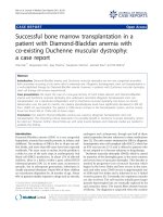
Figure 1 The T Score at the Hip and Lumbar Spine.Figure1a
T score Total Hip. Figure 1b T score Total Lumbar Spine. Shaded
grey area represents osteopenia (T score -1.0 to -2.5). Osteoporosis
below T score -2.5
Duckers et al. Respiratory Research 2011, 12:101
/>Page 4 of 8
Other Biochemistry and Systemic inflammation
Thyroid function tests were norma l in all subjects and
there was no difference in fT4 and TSH between
patients and controls. Total testosterone levels was not
different between patients and controls, however four
(13%) patients and two (13%) controls had low early
morning levels (< 8.0 nmol/L). Circulating IL-6 was
similar in patients: 4.8 (2.8) and controls: 3.1 (2.2) pg/
ml, p = 0.28. There was no difference in IL-6 between
those patients with and without osteoporosis, p = 0.45.
Nor was log
10
IL-6 rel ated to BMD at the hip or lumbar
spine.
Discussion
Men with COPD had a low BMD, with a greater preva-
lence of osteoporosis at the hip and lumbar spine, com-
pared with age matched, ex-smoker, sedent ary, male
controls. This highly select group of males were pre-
defined to remove the possible confounding effects of
the female post-menopausal loss of BMD and addition-
ally with no/minimal oral CS to eliminate the contribu-
tion of these agents to bone thinning. Further, most had
mild severity airways obstruction with over 80% of the
men with COPD being in GOLD class I or II. Thus, our
findings in this group of patients, suggest that the tradi-
tional view of bone thinni ng and osteoporosis occurring
largely as a result of CS use is simplistic. Our findings
could be interpreted as indicating a disease specific
component involved in the loss of BMD in COPD.
We confirmed the previous relationships between both
BMI and FFMI with BMD in COPD [4,26]. The FFM is
a surrogate of peripheral skeletal muscle mass and was
reduced in nine of our patients, including six with a
normal BMI. This suggests preferential skeletal muscle
mass loss in 20% of our patients, which is in keeping
with levels reported in patients with more severe lung
disease. The importance of the link between FFMI and
BMD status was emphasised by the predic tive nature of
this variable along with airways obstruction for hip
BMD[4].Thissuggeststhateveninmilderseverity
Table 3 Biochemistry and Markers of Bone Metabolism
Controls
(n = 15)
Patients
(n = 30)
P value
25 OH Vitamin D (ug/l) # 16.1 (1.4) 11.4 (1.9) 0.03
PTH (pmol/l)# 6.71 (1.36) 6.12 (1.74) 0.58
Adjusted Calcium (mmol/l) 2.32 (0.09) 2.37 (0.11) 0.14
Creatinine (μmol/l) 100 (14) 95 (19) 0.36
Fasting glucose (mmol/l) 5.7 (0.7) 5.4 (0.6) 0.07
Testosterone (nmol/l) 13.9 (4.1) 14.8 (6.2) 0.62
T4 (pmol/l) 14.99 (1.97) 15.37 (2.47) 0.60
TSH (mU/l) 1.72 (0.91) 1.76 (0.81) 0.89
IL-6 (pg/ml) # 3.1 (2.2) 4.8 (2.8) 0.28
OPG (pmol/l) # 6.5 (1.9) 8.5 (1.4) 0.06
Osteocalcin (μg/l) # 23.0 (1.4) 22.7 (1.5) 0.93
P1NP (μg/l) # 40.9 (1.4) 45.6 (1.9) 0.47
CTX (μg/l) # 0.4 (1.5) 0.4 (1.9) 0.69
Data pr esented as mean (SD), unless # Geometric Mean (SD)
Definition of abbreviations:- PTH = parathyroid hormone, T4 = free thyroxine,
TSH = thyroid stimulating hormone, IL-6 = Interleukin -6, OPG =
osteoprotogerin, P1NP = Procollagen type 1 amino-terminal propeptide,
bCTX = b-C-telopeptides of type I collagen
a)
0.600 0.700 0.800 0.900 1.000 1.100 1.200 1.300
BMD total hip region (g/cm2)
1.20
1.40
1.60
1.80
2.00
2.20
2.40
logP1NP
b)
0.600 0.700 0.800 0.900 1.000 1.100 1.200 1.300
BMD total hip re
g
ion
(g
/cm2
)
-1.00
-0.80
-0.60
-0.40
-0.20
0.00
0.20
logCTX
Figure 2 The relationship of the BMD at the hip to bone
biomarkers in Patients. Figure 2a The Marker of Bone Formation
P1NP. Figure 2b The Marker of Bone Resorption CTX.
Duckers et al. Respiratory Research 2011, 12:101
/>Page 5 of 8
airways obstruction there are the same relationships
between muscle mass loss and bone thinning as pre-
viously report ed in more severe status patients [4,26]. It
is unclear from this study what the causative link is, but
we previously demonstrated a parallel increased excre-
tion of cellular protein and bone collagen breakdown
products in patients wit h a low FFM and BMD indicat-
ing a protein catabolic state in COPD, which m ay be a
factor linking bone and skeletal muscle mass loss [4].
Further, the association of severity of emphysema on
CT scan to low BMD was emphasised in 2 recent stu-
dies, one of men with COPD and the other comprising
of tobacco exposed individuals, 60% having COPD
[27,28], again suggesting a systemic proteolytic effect.
Within our patient group there are other possible
causes of bone thinning including physical inactivity.
TheshorterISWDinthepatie nts indicates a reduced
functional capacity for exercise [23] despite their airways
obstruction being relatively mild. This reduction even in
milder airw ays obstruction, an interpretation supported
by a similar observation [29], may contribute to physica l
deconditioning, which might exert a greater effect on
hip BMD than on the l umbar spine and possibly expla in
the differences we report between the two sites. Differ-
ences between the hip and lumbar spine sites may also
be due to loss o f skeletal muscle mass from the lower
limbs and reduction in weight bearing activity as well as
differences in bone composition between the hip and
lumbar spine.
Other potential factors in the loss of BMD are
changes in bone homeostasis due to alterations in hor-
mone and vitamin activity and systemic inflammation.
In health, bone is metabolically active with continuous
remodelling as an adaptation to changes in distribution
of mechanical force and to repair damage. Bone resorp-
tion and formation are normally tightly coupled, but
loss may occur if this balance is disturbed. Currently
bone homeostasis in COPD is not fully understood. We
therefore explored the use of circulating biomarkers of
bone formation and resorption in this study as they
have been widely used in non-COPD osteoporosis as
indicators of bone turnover [30]. They provide insight
into bone physiology and pathophysiology, and have
been used to monitor the response to the treatment of
osteoporosis in post-menopausal women [31] . Howe ver,
to date there is little experience of their use in COPD,
having only been studied in a small pre-transplant popu-
lation [32]. We found no difference between the mean
levels of any of the bone biomarkers in patients and
controls. However, in patients, the greatest levels of
both bone formation and resorption markers were asso-
ciated with a low BMD at the hip. This may i ndicate
that increased bone turnover accounts for altered BMD
at the hip. This is similar to the pattern of bone
biomarkers seen in the majority of post-menopausal
women with osteoporosis [33].
Chronic systemic inflammation has been postulated as
amechanisminthelossofBMDinCOPD,butthere
was no difference between patient and controls for IL-6,
which has been implicated with TNF-a in post-meno-
pausal osteoporosis and which in vitro stimulates osteo-
clasts and bone resorption [34]. However, systemic
levels are unlikely to reflect this tissue level and we have
previously been unable to r elate BMD to systemic levels
in other patients with COPD. Interestingly, OPG was
greater in osteoporotic patients than those without
osteoporosis and related inversely to hip BMD. It could
be considered as a pro-formation marker due to its
pot ential to act as a decoy for receptor activator for N F
kappa B ligand (RANKL) and thus act as a local anti-
inflammatory agent. However, caution is needed in
interpreting the OPG level and the rat io of OPG to
RANK-Lmaybemoreinformativebutatpresentthere
is debate about the sensitivity of the RANK-L assays
available.
In pre-transplant patients with COPD Forli et al [35]
reported a direct relationship between TNF-a receptor
II and the resorption marker bCTX, whilst Bon et al
analysed 27 inflammatory mediators in relation to sev-
eral serological markers of b one turnover i n patients
with severe COPD pre tr ansplant, encompassing patien t
on OCS and osteoporosis treatment [32]. The lack of a
relationship between IL-6 and bone turnover biomarkers
in our study mirrors previous s tudies [4] b ut may b e a
result of the relatively mild airways obstruction of our
patients and the small population size.
The PTH, Cr, adjusted Ca and total testosterone
levels, and thyroid function, all of whi ch are involved in
bone homeostasis we re not diff erent between patients
and controls, though, the patients had a lower total 25-
OHVitaminDlevelthancontrols confirming previous
findings [36]. Insufficient total 25-OH Vitamin D levels
were seen in 80% of patients with around a third having
significantly low levels. Low levels may reflect both a
disease related component, such as decreased physical
activity [29] and lower sunlight exposure, and the gener-
ally low levels reported in aging populations. Indeed,
60% of the control subjects had insufficient 25-OH Vita-
min D levels . In four patients with insufficient 25-OH
Vitamin D and increased PTH there may have been
some osteomalacia, but we did not differentiate osteo-
malacia from osteoporosis [37].
In this study, we did not quantitatively assess for ver-
tebral fractures in COPD, though marked distortion on
the DXA images excluded that specific vertebra from
evaluation - in keeping with standard clinical evaluation
of lumbar vertebrae, n = 5 individual vertebrae. A recent
paper retrospectively reviewed men diagnosed with
Duckers et al. Respiratory Research 2011, 12:101
/>Page 6 of 8
COPD, picking up a high rate of fragility fracture but a
low proportion having a DXA or being treated with
anti-resorptive therapy[38]. The EOLO study reported
an increased risk of fracture with increasing severity of
airflow obstruction in addition to the contribution of
daily inhaled steroid use [20]; with McEvoy demonstrat-
ing a high fracture rate in steroid naïve men [13].
The l imitation of this study is the modest size of the
study popula tion. Recruiting men with COPD who met
the rigorous study entry criteria proved challenging
despite working with across several primary care sur-
geries and scre ening large numbers of patients. The pro-
portion of male controls with osteoporo sis mirror ed our
previous research in two different male control popula-
tions of a similar age that we have studied, though osteo-
porosis was not reported in these publications accordi ng
to gender [4,5]. The controls a nd patient s were not
matched for smoking history despite attempts, though the
controls had a median 34 pack year tobacco exposure.
The measurement of BMD using DXA may be con-
founded by several factors [39]. In particular, measure-
ment of the lumbar spine BMD using DXA becomes
less useful with increasing age due to confounding
effects such as vertebral collapse, osteophytes and aortic
calci fication all leading to a “ pseu do-normalising” of the
BMD [40].
Conclusion
A low BMD occurs in males with COPD of mild to
moderate severity airways obstruction, with minimal/no
CS exposure compared to ex smoker controls and
osteoporosis is common. This study suggests a disease
related causative component. As in previous studies, a
low BMD was more prominent at the hip compared
with the lumbar spine, which may reflect physical
deconditioning or different bone composition. The
mechanism of BMD loss in COPD remains unclear but
our results suggest increased bone turnover. Our find-
ings highlight the potential value of studying milder
severity patients free from potential causative confoun-
ders and reinforce the need for earlier identification and
targeting of risk factors for osteoporosis as part of the
management of COPD.
Funding
Dr J Duck ers was supported by a Cardiff and Vale NHS
Trust Clinical Research Fellowship. D r C Bolton is cur-
rently funded by the NIHR Nottingham Respiratory Bio-
medical Research Unit.
Abbreviations
BMD: Bone mineral density; P1NP: Procollagen type 1 amino-terminal
propeptide; CTX: β-C-telopeptides of type I collagen
Acknowledgements
The authors thank Mrs C Elford and Mr G Dunseath for work with
biochemical assays and Dr W Evans and Mrs R Pettit for Medial Physics
input. We also thank Consultant Respiratory Physicians at University Hospital
Llandough; Drs P Edwards, K Holgate, C Allanby, J Prichard; Srs T Faulkner, R
Arthurs, C Oxenham, G Prichard and Mrs S Edwards for their collaboration
and recruitment of patients in this study.
Author details
1
Section of Respiratory Medicine, Wales Heart Research Institute, School of
Medicine, Cardiff University, University Hospital of Wales, Heath Park, Cardiff,
CF14 4XN, UK.
2
Child Health, School of Medicine, Cardiff University, Heath
Park, Cardiff CF14 4XN, UK.
3
Unit of Clinical Chemistry, School of Clinical
Sciences, Liverpool University, Liverpool, UK.
4
Bone Research Unit, School of
Medicine, Cardiff University, Academic Centre, University Hospital Llandough,
Penlan Road, Penarth, Vale of Glamorgan, CF64 2XX UK.
5
NIHR Nottingham
Respiratory Biomedical Research Unit, University of Nottingham, Clinical
Sciences, City Hospital, Hucknall road, Nottingham. NG5 1PB. UK.
Authors’ contributions
JD helped design the study, conducted the clinical assessments, analysed
and interpreted data and wrote the first draft; BE helped design the study
and contributed to the interpretation and writing; WF contributed to the
interpretation and writing; MS contributed to the interpre tation and writing;
CB helped design the study and contributed to the interpretation and
writing; DS helped design the study and contributed to the interpretation
and writing. All authors have read and approved the final manuscript.
Competing interests
The authors declare that they have no competing interests.
Received: 13 May 2011 Accepted: 3 August 2011
Published: 3 August 2011
References
1. Murray CJ, Lopez AD: Mortality by cause for eight regions of the world:
Global Burden of Disease Study. Lancet 1997, 349:1269-76.
2. Sin DD, Anthonisen NR, Soriano JB, Agusti AG: Mortality in COPD: Role of
comorbidities. Eur Respir J 2006, 28(6):1245-57.
3. Biskobing DM: COPD and osteoporosis. Chest 2002, 121(2):609-20.
4. Bolton CE, Ionescu AA, Shiels KM, Pettit RJ, Edwards PH, Stone MD,
Nixon LS, Evans WD, Griffiths TL, Shale DJ: Associated loss of fat-free mass
and bone mineral density in chronic obstructive pulmonary disease. Am
J Respir Crit Care Med 2004, 170(12):1286-93.
5. Sabit R, Bolton CE, Edwards PH, Pettit RJ, Evans WD, McEniery CM,
Wilkinson IB, Cockcroft JR, Shale DJ: Arterial stiffness and osteoporosis in
chronic obstructive pulmonary disease. Am J Respir Crit Care Med 2007,
175(12):1259-65.
6. Dimai HP, Domej W, Leb G, Lau KH: Bone loss in patients with untreated
chronic obstructive pulmonary disease is mediated by an increase in
bone resorption associated with hypercapnia. J Bone Miner Res 2001,
16(11):2132-41.
7. Jorgensen NR, Schwarz P, Holme I, Henriksen BM, Petersen LJ, Backer V: The
prevalence of osteoporosis in patients with chronic obstructive
pulmonary disease: a cross sectional study. Respir Med 2007,
101(1):177-85.
8. Leech JA, Dulberg C, Kellie S, Pattee L, Gay J: Relationship of lung function
to severity of osteoporosis in women. Am Rev Respir Dis 1990,
141(1):68-71.
9. Lisboa C, Moreno R, Fava M, Ferretti R, Cruz E: Inspiratory muscle function
in patients with severe kyphoscoliosis. Am Rev Respir Dis 1985,
132(1):48-52.
10. Myers AH, Robinson EG, Van Natta ML, Michelson JD, Collins K, Baker SP:
Hip fractures among the elderly: factors associated with in-hospital
mortality. Am J Epidemiol 1991, 134(10):1128-37.
11. Adinoff AD, Hollister JR: Steroid-induced fractures and bone loss in
patients with asthma. N Engl J Med 1983, 309(5):265-8.
12. Goldstein MF, Fallon JJ, Harning R: Chronic glucocorticoid therapy-
induced osteoporosis in patients with obstructive lung disease. Chest
1999, 116(6):1733-49.
Duckers et al. Respiratory Research 2011, 12:101
/>Page 7 of 8
13. McEvoy CE, Ensrud KE, Bender E, Genant HK, Yu W, Griffith JM,
Niewoehner DE: Association between corticosteroid use and vertebral
fractures in older men with chronic obstructive pulmonary disease. Am J
Respir Crit Care Med 1998, 157(3 Pt 1):704-9.
14. Calverley PM, Anderson JA, Celli B, Ferguson GT, Jenkins C, Jones PW,
Yates JC, Vestbo J: Salmeterol and fluticasone propionate and survival in
chronic obstructive pulmonary disease. N Engl J Med 2007, 356(8):775-89.
15. Lee TA, Weiss KB: Fracture risk associated with inhaled corticosteroid use
in chronic obstructive pulmonary disease. Am J Respir Crit Care Med 2004,
169(7):855-9.
16. Etminan M, Sadatsafavi M, Ganjizadeh Zavareh S, Takkouche B,
FitzGerald JM: Inhaled corticosteroids and the risk of fractures in older
adults: a systematic review and meta-analysis. Drug Saf 2008,
31(5):409-14.
17. Yang IA, Fong KM, Sim EH, Black PN, Lasserson TJ: Inhaled corticosteroids
for stable chronic obstructive pulmonary disease. Cochrane Database Syst
Rev 2007, 2:CD002991.
18. Hubbard R, Tattersfield A, Smith C, West J, Smeeth L, Fletcher A: Use of
inhaled corticosteroids and the risk of fracture. Chest 2006, 130(4):1082-8.
19. Loke YK, Cavallazzi R, Singh S: Risk of fractures with inhaled
corticosteroids in COPD: systematic review and meta-analysis of
randomised controlled trials and observational studies. Thorax 2011, E
published.
20. Gonnelli S, Caffarelli C, Maggi S, Guglielmi G, Siviero P, Rossi S, Crepaldi G,
Nuti R, EOLO study group: Effect of inhaled glucocorticoids and beta(2)
agonists on vertebral fracture risk in COPD patients: the EOLO study.
Calcif Tissue Int 2010, 87(2):137-43.
21. Fabbri LM, Hurd SS: Global Strategy for the Diagnosis, Management and
Prevention of COPD: 2003 update. Eur Respir J 2003, 22(1):1-2.
22. Celli BR, MacNee W: Standards for the diagnosis and treatment of
patients with COPD: a summary of the ATS/ERS position paper. Eur Respir
J 2004, 23(6):932-46.
23. Singh SJ, Morgan MD, Scott S, Walters D, Hardman AE: Development of a
shuttle walking test of disability in patients with chronic airways
obstruction. Thorax 1992, 47(12):1019-24.
24. World Health Organisation: Assessment of fracture risk and its application
to screening for postmenopausal osteoporosis. WHO Technical Report
Series 1984, 84.
25. Holick MF: High prevalence of vitamin D inadequacy and implications for
health. Mayo Clin Proc 2006, 81(3):353-73.
26. Iqbal F, Michaelson J, Thaler L, Rubin J, Roman J, Nanes MS: Declining
bone mass in men with chronic pulmonary disease: contribution of
glucocorticoid treatment, body mass index, and gonadal function. Chest
1999,
116(6):1616-24.
27. Ohara T, Hirai T, Muro S, Haruna A, Terada K, Kinose D, Marumo S, Ogawa E,
Hoshino Y, Niimi A, Chin K, Mishima M: Relationship between pulmonary
emphysema and osteoporosis assessed by CT in patients with COPD.
Chest 2008, 134(6):1244-9.
28. Bon J, Fuhrman CR, Weissfeld JL, Duncan SR, Branch RA, Chang CC,
Zhang Y, Leader JK, Gur D, Greenspan SL, Sciurba FC: Radiographic
Emphysema Predicts Low Bone Mineral Density in a Tobacco-exposed
Cohort. Am J Respir Crit Care Med 2011, 183(7):885-90.
29. Pitta F, Troosters T, Spruit MA, Decramer M, Gosselink R: Activity
monitoring for assessment of physical activities in daily life in patients
with chronic obstructive pulmonary disease. Arch Phys Med Rehabil 2005,
86(10):1979-85.
30. Seibel MJ: Biochemical Markers of Bone Turnover Part II: Clinical
Applications in the Management of Osteoporosis. Clin Biochem Rev 2006,
27(3):123-38.
31. Eastell R, Baumann M, Hoyle NR, Wieczorek L: Bone markers: Biochemical
and Clinical Perspectives. Martin Dunitz, London; 2001.
32. Bon JM, Zhang Y, Duncan SR, Pilewski JM, Zaldonis D, Zeevi A, McCurry KR,
Greenspan SL, Sciurba FC: Plasma inflammatory mediators associated
with bone metabolism in COPD. COPD 2010, 7(3):186-91.
33. Garnero P, Sornay-Rendu E, Chapuy MC, Delmas PD: Increased bone
turnover in late postmenopausal women is a major determinant of
osteoporosis. J Bone Miner Res 1996, 11(3):337-49.
34. Gianni W, Ricci A, Gazzaniga P, Brama M, Pietropaolo M, Votano S, Patane F,
Agliano AM, Spera G, Marigliano V, Ammendola S, Agnusdei D, Migliaccio S,
Scandurra R: Raloxifene modulates interleukin-6 and tumor necrosis
factor-alpha synthesis in vivo: results from a pilot clinical study. J Clin
Endocrinol Metab 2004, 89(12):6097-9.
35. Forli L, Mellbye OJ, Halse J, Bjortuft O, Vatn M, Boe J: Cytokines, bone
turnover markers and weight change in candidates for lung
transplantation. Pulm Pharmacol Ther 2008, 21(1):188-95.
36. Riancho JA, Gonzalez Macias J, Del Arco C, Amado JA, Freijanes J,
Anton MA: Vertebral compression fractures and mineral metabolism in
chronic obstructive lung disease. Thorax 1987, 42(12):962-6.
37. Bingham CT, Fitzpatrick LA: Noninvasive testing in the diagnosis of
osteomalacia. Am J Med 1993, 95(5):519-23.
38. Morden NE, Sullivan SD, Bartle B, Lee TA: Skeletal health in men with
chronic lung disease: rates of testing, treatment, and fractures.
Osteoporos Int 2011, 22(6):1855-62.
39. Bolotin HH: DXA in vivo BMD methodology: an erroneous and
misleading research and clinical gauge of bone mineral status, bone
fragility, and bone remodelling. Bone 2007, 41(1):138-54.
40. Bolton CE, McEniery CM, Raj V, McDonnell BJ, Dixon AK, Munnery M,
Sabit R, Screaton N, Stone MD, Wilkinson IB, Shale DJ, Cockcroft JR: Aortic
Calcification, Bone Mineral Density and Arterial Stiffness in Patients with
COPD. Artery Research J 2011, 5(1):30-36.
doi:10.1186/1465-9921-12-101
Cite this article as: Duckers et al.: Low bone mineral density in men
with chronic obstructive pulmonary disease. Respiratory Research 2011
12:101.
Submit your next manuscript to BioMed Central
and take full advantage of:
• Convenient online submission
• Thorough peer review
• No space constraints or color figure charges
• Immediate publication on acceptance
• Inclusion in PubMed, CAS, Scopus and Google Scholar
• Research which is freely available for redistribution
Submit your manuscript at
www.biomedcentral.com/submit
Duckers et al. Respiratory Research 2011, 12:101
/>Page 8 of 8