Báo cáo y học: " Cyclic hydrostatic pressure and cotton particles stimulate synthesis by human lung macrophages of cytokines in vitro" ppsx
Bạn đang xem bản rút gọn của tài liệu. Xem và tải ngay bản đầy đủ của tài liệu tại đây (1.1 MB, 14 trang )
BioMed Central
Page 1 of 14
(page number not for citation purposes)
Respiratory Research
Open Access
Research
Long-term activation of TLR3 by Poly(I:C) induces inflammation
and impairs lung function in mice
Nicole C Stowell*
1
, Jonathan Seideman
1
, Holly A Raymond
1
,
Karen A Smalley
1
, Roberta J Lamb
1
, Devon D Egenolf
1
, Peter J Bugelski
1
,
Lynne A Murray
1
, Paul A Marsters
1
, Rachel A Bunting
1
, Richard A Flavell
2
,
Lena Alexopoulou
3
, Lani R San Mateo
1
, Don E Griswold
1
, Robert T Sarisky
1
,
M Lamine Mbow
1,4
and Anuk M Das
1
Address:
1
Discovery Research, Centocor Research & Development, Inc, Radnor, Pennsylvania, USA,
2
Department of Immunobiology, Yale
University School of Medicine and Howard Hughes Medical Institute, New Haven, Connecticut, USA,
3
Centre d'Immunologie de Marseille-
Luminy, CNRS-INSERM-Universite de la Mediterranee, Campus de Luminy, Case 906, Marseille Cedex 13288, France and
4
Genomics Institute of
the Novartis Research Foundation, San Diego, California, USA
Email: Nicole C Stowell* - ; Jonathan Seideman - ;
Holly A Raymond - ; Karen A Smalley - ; Roberta J Lamb - ;
Devon D Egenolf - ; Peter J Bugelski - ; Lynne A Murray - ;
Paul A Marsters - ; Rachel A Bunting - ; Richard A Flavell - ;
Lena Alexopoulou - ; Lani R San Mateo - ; Don E Griswold - ;
Robert T Sarisky - ; M Lamine Mbow - ; Anuk M Das -
* Corresponding author
Abstract
Background: The immune mechanisms associated with infection-induced disease exacerbations in asthma and COPD
are not fully understood. Toll-like receptor (TLR) 3 has an important role in recognition of double-stranded viral RNA,
which leads to the production of various inflammatory mediators. Thus, an understanding of TLR3 activation should
provide insight into the mechanisms underlying virus-induced exacerbations of pulmonary diseases.
Methods: TLR3 knock-out (KO) mice and C57B6 (WT) mice were intranasally administered repeated doses of the
synthetic double stranded RNA analog poly(I:C).
Results: There was a significant increase in total cells, especially neutrophils, in BALF samples from poly(I:C)-treated
mice. In addition, IL-6, CXCL10, JE, KC, mGCSF, CCL3, CCL5, and TNF were up regulated. Histological analyses of
the lungs revealed a cellular infiltrate in the interstitium and epithelial cell hypertrophy in small bronchioles. Associated
with the pro-inflammatory effects of poly(I:C), the mice exhibited significant impairment of lung function both at baseline
and in response to methacholine challenge as measured by whole body plethysmography and an invasive measure of
airway resistance. Importantly, TLR3 KO mice were protected from poly(I:C)-induced changes in lung function at
baseline, which correlated with milder inflammation in the lung, and significantly reduced epithelial cell hypertrophy.
Conclusion: These findings demonstrate that TLR3 activation by poly(I:C) modulates the local inflammatory response
in the lung and suggest a critical role of TLR3 activation in driving lung function impairment. Thus, TLR3 activation may
be one mechanism through which viral infections contribute toward exacerbation of respiratory disease.
Published: 1 June 2009
Respiratory Research 2009, 10:43 doi:10.1186/1465-9921-10-43
Received: 3 March 2008
Accepted: 1 June 2009
This article is available from: />© 2009 Stowell et al; licensee BioMed Central Ltd.
This is an Open Access article distributed under the terms of the Creative Commons Attribution License ( />),
which permits unrestricted use, distribution, and reproduction in any medium, provided the original work is properly cited.
Respiratory Research 2009, 10:43 />Page 2 of 14
(page number not for citation purposes)
Background
The activation of Toll-Like Receptors (TLRs), a family of
innate immune receptors, is believed to be an important
step in the initiation of the inflammatory response raised
against numerous pathogens. TLR3 is a mammalian pat-
tern recognition receptor that recognizes double-stranded
(ds) RNA as well as the synthetic ds RNA analog poly-
riboinosinic-ribocytidylic acid (poly(I:C)) [1]. Activation
of TLR3 by poly(I:C) or by endogenous mRNA ligands,
such as those released from necrotic cells [2], induces
secretion of pro-inflammatory cytokines and chemokines,
a finding that suggests that TLR3 agonists modulate dis-
ease outcome during infection-associated inflammation
[3]. Thus, long-term activation of TLR3 in vivo is thought
to occur in the context of viral infection [4] or necrosis
associated with inflammation [2].
In vitro studies have demonstrated that stimulation of
lung epithelial cells with poly(I:C) elicited the secretion of
multiple cytokines, chemokines, the induction of tran-
scription factors and increased expression of TLRs [3]. It
has also been demonstrated that poly(I:C) enhanced
bradykinin- and [des-Arg
9
]-bradykinin-induced contrac-
tions of tracheal explants in vitro, an effect mediated by C-
jun-amino-terminal kinase (JNK) and nuclear factor
kappa B (NF-kB) signaling pathways [5]. Taken together,
these data suggest that TLR3 activation may have a physi-
ological consequence in the lung. Further, these data dem-
onstrate that ligation of TLR3 initiates cascades of
phosphorylation and transcriptional activation events
that result in the production of numerous inflammatory
cytokines that are thought to contribute to innate immu-
nity [5]. Overall, these data suggest that sustained TLR3
activation can be a critical component in the modulation
of infection-associated inflammatory diseases.
Exacerbations in respiratory diseases such as asthma and
chronic obstructive pulmonary disease (COPD) are char-
acterized by the worsening of symptoms and a decline in
lung function. Viral infections are associated with respira-
tory disease exacerbations [6] and may be associated with
progression of disease. Secretion of pro-inflammatory
cytokines in the lungs following viral infection represents
a crucial step in promoting the inflammatory response in
various lung diseases [7,8]. A better understanding of the
effects of TLR3 activation may provide insight into the
mechanisms underlying virally-induced respiratory dis-
ease exacerbations.
In the current study we examined the effects of TLR3 acti-
vation in vivo. We sought to induce long term activation of
TLR3 to mimic the physiologic disease state associated
with virally-induced disease exacerbations. Administra-
tion of poly(I:C) to the lungs of mice induced a marked
impairment of lung function that was accompanied by the
production of pro-inflammatory mediators and inflam-
matory cell recruitment into the airways. TLR3 appears to
play a role in the effects of poly(I:C) since TLR3 KO mice
were partially protected. Taken together, our data suggest
an important role for TLR3 activation in impairment of
lung function.
Methods
Poly(I:C) induced cytokine secretion in BEAS-2B cells
The SV-40-transformed normal human bronchial epithe-
lial cell line, BEAS-2B (ATCC, VA) was cultured in LHC-9
media without additional supplements. (Biosource, CA).
1 × 10
6
cells were seeded in collagen type I-coated T75
flasks (BD, NJ) and split every 2–3 days using 0.25%
trypsin/ethylenediaminetetraacetic acid (EDTA) (Gibco,
CA). Poly(I:C) (Amersham, NJ) was dissolved in phos-
phate-buffered saline (10 mM phosphate, 150 mM NaCl,
pH 7.4; phosphate buffered saline (PBS)) at a concentra-
tion of 2 mg/ml and aliquots were stored at -20°C. For
poly(I:C) stimulation, cells were incubated at 37°C with
different concentrations of poly(I:C). Supernatants were
collected after 24 hours and stored at -20°C or assayed
immediately for cytokine secretion using a multi-plex
bead assay (Biosource, CA) for detection of interferon-
alpha (IFN), interferon-gamma (IFN), interleukin-1-
beta (IL-1), interleukin-10 (IL10), interleukin-12p70
(IL12p70), tumor necrosis factor-alpha (TNF), Chemok-
ine (C-C motif) ligand 3 (CCL3), interleukin-6 (IL-6),
interleukin-8 (IL-8), Chemokine (C-C motif) ligand 2
(CCL2), Chemokine (C-C motif) ligand 5 (CCL5), and
Chemokine (C-X-C motif) ligand 3 (CXCL10). Limits of
detection for the analytes range from 3 – 20 pg/ml. Sam-
ple acquisition and analysis was performed using the
Luminex 100S with StarStation software (Applied Cytom-
etry Systems).
Administration of Poly(I:C) to the lungs of mice
Female C57BL/6 mice wild-type (WT) (12 weeks old) or
female TLR3 knock-out (KO) mice (C57BL/6; 12 weeks
old, ACE animals, PA) were anesthetized with isoflurane
and different doses (10–100 g) of poly(I:C) in 50 l ster-
ile PBS, or PBS alone, were administered intranasally
(I.N.) Mice received three administrations of poly(I:C) (or
PBS) with a 24 hour rest period between each administra-
tion. KO mice were fully backcrossed to C57BL/6 back-
ground to at least N10.
All animal care was performed according to the Guide for
the Care and Use of Laboratory animals and the Institu-
tional Animal Care and Use Committee approved all stud-
ies.
Whole Body Plethysmography
Twenty-four hours following the last poly(I:C) (or PBS)
administration, lung function without provocation (base-
Respiratory Research 2009, 10:43 />Page 3 of 14
(page number not for citation purposes)
line) and airway hyperresponsiveness (AHR) to metha-
choline were measured using whole body
plethysmography (BUXCO system). The mice were placed
into the whole body plethysmograph chamber and
allowed to acclimate for at least 5 minutes. Following
baseline readings, mice were exposed to increasing doses
of nebulized methacholine (Sigma, MO). The nebulized
methacholine was administered for 2 minutes, followed
by a 5-minute data collection period, followed by a 10-
minute rest period before subsequent increasing-dose
methacholine challenges. The increased airflow resistance
was measured as Enhanced Pause (Penh) and is repre-
sented as the average penh value over the 5-minute
recording period.
Invasive measures of lung function
Twenty-four hours following the last poly(I:C) (or PBS)
administration, lung function and increased lung resist-
ance in response to methacholine were measured using
invasive measures of lung function (BUXCO system).
Mice were anesthetized with 50 mg/kg sodium pentobar-
bital (Nembutal, Abbot Labs, IL). The trachea was cannu-
lated with a 19 gauge cannula and the mouse was
connected to a mechanical ventilator, with breath fre-
quency of 120 and stroke volume of 0.3 mL. The mouse
was connected to the plethysmograph for lung function
measurements. After establishing a stable baseline of lung
resistance, methacholine was administered I.V. through
the tail vein (240 g/kg). The peak resistance measured
over 3 minutes was recorded.
Measurement of lung inflammation
Following lung function measurements, mice were sacri-
ficed by CO
2
asphyxiation and the lungs were cannulated.
Bronchoalveolar lavages (BAL) were performed by inject-
ing 1 mL of PBS into the lungs and retrieving the effluent.
The lung tissues were removed and frozen. The BALs were
centrifuged (1200 rpm, 10 minutes) and the cell-free
supernatants were collected and stored at -80°C until
analysis. The cell pellet was resuspended in 200 l PBS for
total and differential cell counts using a hemacytometer
(on Wright's – Giemsa-stained cytospin preparations).
Measurement of proteins in bronchoalveolar lavage
samples
The cell-free supernatants were collected and stored at -
80°C until used for analyses. The multiplex assay was per-
formed following the manufacturer's protocol and the
LINCOplex Multiplex Immunoassay Kit (LINCO
Research, St. Charles, MO). Analytes included in the anal-
ysis were MIP1, Granulocyte Macrophage Colony Stim-
ulating Factor (GMCSF), JE, KC, RANTES, IFN, IL-1, IL-
1, Granulocyte Colony Stimulating Factor (GCSF),
CXCL10, IL-2, IL-4, IL-5, IL-6, IL-7, IL-9, IL-10, IL-
12(p70), IL-13, IL-15, IL-17 and TNF. Limits of detection
for the analytes range from 3 – 20 pg/ml.
Measurement of lung mRNA expression
Following collection of BAL samples, the right lobes of the
lung were removed and placed in Trizol total RNA isola-
tion reagent (Life Technologies, Gaithersburg, MD). RNA
was isolated using manufacturer's instructions of the Qia-
gen Rneasy Mini kit (Qiagen, Valencia, CA). Total RNA (2
g) from pooled groups was then reverse transcribed
using the OmniScript RT kit (Qiagen, Valencia, CA)
according to the manufacturer's protocol. One hundred
nanograms of cDNA was then amplified using both the
TaqMan
®
Low Density Immune Profiling Array cards
(Applied Biosystems, Foster City, CA), or microfluidic
cards, and custom Low Density Array cards. Primer-probes
with genes of interest were plated in a 384 well format fol-
lowing the manufacturer's protocol for Real-Time PCR.
Data are normalized to 18s rRNA and represent fold
change over PBS treated mice.
Histological Analysis
Following BAL collection, the left lobes were inflated with
10% neutral buffered formalin under constant pressure
then immersed in additional fixative, the right lobes were
clamped with hemostats and ligated. Tissue was processed
by routine methods, oriented so as to provide coronal sec-
tions and 5 micron mid-coronal sections cut and stained
with hematoxylin and eosin.
Morphometric analysis
A Nikon Eclipse E800 (Nikon Corporation, Tokyo, Japan)
microscope was equipped with an Evolution™ MP 5.0 RTV
color camera (Media Cybernetics, Inc. Silver Spring, MD).
Images were captured and analyzed using Image-Pro Plus
software version 5.1 (Media Cybernetics, Inc. Silver
Spring, MD). GraphPad Prism version 4.03 (GraphPad
Software, Inc. San Diego, CA) was used to interpret, ana-
lyze and graph the raw data. SigmaStat Statistical Software
version 2.03 (SPSS, Inc. Chicago, IL) was used to perform
statistical analysis on the collected data. Using the Auto-
Pro tool within the Image-Pro Plus software, custom writ-
ten macros were used to perform the analysis. Six TLR3
KO mice treated with poly(I:C), six WT mice treated with
poly(I:C), four TLR3 KO mice treated with PBS and six WT
mice treated with PBS were imaged and analyzed. No
imaging or analysis was performed on areas of the lung
that were torn, damaged, or folded.
Tissue Density
From each lung, five fields were randomly selected and
imaged using a 20× objective lens. The total area of the tis-
sue was measured and the ratio of total area of tissue to
total area of field calculated.
Respiratory Research 2009, 10:43 />Page 4 of 14
(page number not for citation purposes)
Tissue Cellularity
From each lung, five fields were randomly selected and
imaged using a 20× objective lens. The total area of the
nuclei was measured and the ratio of total area of nuclei
to total area of field calculated.
Airway Cellularity
From each lung, five airways were chosen and imaged
using a 40× objective lens. A line of 100 m in length was
superimposed on the airway at a random location. The
number of nuclei within the fixed distance were counted
and recorded.
Airway Mucosal Height
From each lung, five airways were chosen and imaged
using a 40× objective lens. The image was segmented so as
to include only the airway mucosa and the average thick-
ness of the airway mucosa was measured using the curve
thickness algorithm built into ImagePro. This algorithm
parses the mucosa into 30,000 arc segments, measures the
thickness of the mucosa at each arc segment and calcu-
lated the average thickness for the mucosa.
Statistical analysis
Specific statistical methods are described in the figure leg-
ends. Graphs and summary statistics were also used to
assess the results. All statistical tests were 2-sided. Except
for where noted, all p-values presented are unadjusted for
multiple comparisons.
Results
Poly(I:C) induces a marked inflammatory response in the
lungs of mice
Intranasal administration of three once-daily doses of
poly(I:C) resulted in a dose-dependent inflammatory cell
influx into the lung. There was a significant increase in
total cells in the BAL samples at 50 and 100 g poly(I:C)
compared to PBS treated mice (Figure 1A). This increase
in total cellularity in the BAL samples was partially due to
a significant influx of neutrophils (Figure 1B) and mono-
nuclear cells (Figure 1C). Due to the robust response at 50
and 100 g, these doses of poly(I:C) were used in our sub-
sequent studies.
In an effort to understand the responses to poly(I:C) treat-
ment in the lung at a molecular level, Taqman real-time
PCR analyses of the lung tissues was performed. Multiple
administrations of poly(I:C) elicited up regulation of a
number of pro-inflammatory genes, TLRs and their asso-
ciated intracellular signaling molecules (Table 1). TLR
genes that were up regulated at the mRNA level as a result
of TLR3 stimulation included TLR2, TLR3, TLR7, and
TLR9 with approximately 7, 5, 11, and 56 fold increases
respectively. In addition there was dramatic increase in
CXCL10, TNF, CCL2, CCL3, and CCL7 gene expression
as well as interferon regulatory factor 7 (IRF7), interferon-
stimulated transcription factor 3 (ISGF3G), 2'-5'-oligoad-
enylate synthetase 2 (OAS2), and protein kinase-R (PKR.)
Poly(I:C) administration also induced elevated protein
levels of cytokines, chemokines, and growth factors in the
lavage including significant increases of IFN, IL-1, IL-6,
TNF, CXCL10, JE, KC, MIP-1, RANTES, GCSF and
GMCSF (Table 2). There were no changes in IL-1, IL-2,
IL-4, IL-5, IL-7, IL-9, IL-10, IL-12(p70), IL-13, IL-15, or IL-
17 (data not shown) among the groups. These data dem-
onstrate that poly(I:C) administered I.N. elicits a cascade
of events resulting in the expression and secretion of mul-
tiple pro-inflammatory cytokines, and chemokines as well
as the up regulation of TLR gene expression.
Histological analyses of the lungs were performed to bet-
ter understand the pathology induced by poly(I:C)
administration. Representative micrographs from H&E
stained lung sections are shown (Figure 2). The histology
of the control lungs was unremarkable in that the lungs
exhibited normal pulmonary architecture and resident
cells. The most remarkable changes induced by poly(I:C)
were a marked perivascular and a moderate peribronchi-
olar interstitial inflammatory infiltrate. There were also
signs of pulmonary edema as evidenced by a widening of
the interstitial space surrounding the airways and vascula-
ture in the poly(I:C) treated mice. The alveolar septa were
thickened and contained numerous inflammatory cells,
consistent with an interstitial pneumonitis. Few inflam-
matory cells were observed in the alveolar spaces, but as
the bronchoalveolar fluids were collected, most of the
cells in the alveoli were probably lost from analysis. The
other remarkable changes observed were thickening of the
bronchiolar epithelium consistent with hypertrophy. The
hypertrophy was accompanied by an increase in the gran-
ularity of the cytoplasm of the bronchiolar epithelium,
however, there was no evidence for increased mucus pro-
duction by PAS staining. There was no notable increase in
goblet cells.
The results of the morphometric analysis are shown in
Table 3. Reflecting the increase in interstitial penumonitis
there was a 1.7 fold increase in tissue density and a 2 fold
increase in overall tissue cellularity. In the small airways,
there was a 1.7 fold increase in the mucosal height, reflect-
ing the mucosal hypertrophy and no change in cellularity
(data not shown).
Poly(I:C) activates BEAS2B epithelial cells
The morphometric data identified the induction of
mucosal hypertrophy in WT mice following poly(I:C)
challenge. To further elucidate the effects of poly(I:C) on
epithelial cells, the response of the normal human lung
epithelial cell line, BEAS-2B, to poly(I:C) was investigated.
Respiratory Research 2009, 10:43 />Page 5 of 14
(page number not for citation purposes)
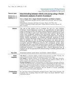
Poly(I:C) induces a dose dependent influx of inflammatory cells into the airways of miceFigure 1
Poly(I:C) induces a dose dependent influx of inflammatory cells into the airways of mice. Mice were administered
PBS or, 10, 20, 50 or 100 g poly(I:C) (I.N.) every 24 h for three days. 24 hours after the last administration, mice were eutha-
nized and BALs were performed. The total number of cells (1A), neutrophils (1B) and mononuclear cells (1C) were measured
in the BAL. Data are the mean ± SEM of 6–15 mice from two separate experiments. The Kruskal-Wallace test was used to
compare the treatment groups. When this test showed a difference among the treatment groups, selected pairs of treatments
were compared using Dunn's multiple comparison test. ** p < 0.001 when compared to PBS-treated mice.
90
Total Cells
80
70
60
50
40
30
20
10
0
PBS
1
0
20
50
1
00
Poly(I:C) (Pg)
**
**
A
Total Cells 10^4
300
Total Neutrophils
200
100
0
P
B
S
10
20
50
10
0
PolyI:C (Pg)
**
**
B
Neutrophils 10^3
700
Total Mononuclear Cells
600
500
400
300
200
100
0
P
B
S
10
20
5
0
1
0
0
PolyI:C (Pg)
**
**
C
Mononuclear Cells 10^3
Respiratory Research 2009, 10:43 />Page 6 of 14
(page number not for citation purposes)
Similar to the mouse in vivo data, where analysis was per-
formed 24 hours post final poly(I:C) challenge, BEAS-2B
cells responded to a range of poly(I:C) concentrations (16
to 1000 ng/ml) in a dose-dependent manner by secreting
a number of cytokines observed in the mouse lungs
including IL-6, IL-8, CCL2, CCL5, and CXCL10 (Fig. 3),
consistent with previous findings [9-11]. There was no
change in response to poly(I:C) in the other analytes
included in the multiplex (data not shown), nor was there
any obvious change in morphometric parameters of the
stimulated cells.
TLR3 stimulation leads to impairment of pulmonary
function
In order to investigate the functional consequences of
TLR3 ligation, we measured lung function in poly(I:C)-
treated mice. Airway hyperresponsiveness to increasing
doses of methacholine was measured using whole body
plethysmography (WBP) (Figure 4A). Poly(I:C)-chal-
lenged mice exhibited greater airway hyperresponsiveness
to methacholine. Poly(I:C)-challenged mice also exhib-
ited an increase in baseline penh in the absence of provo-
cation, measured using WBP (Figure 4B). To confirm the
effects of poly(I:C) on lung function, invasive lung func-
tion measurements were also performed and the results
confirmed those obtained using WBP (Fig 4C).
Poly(I:C)-induced inflammatory cell influx is attenuated in
TLR3 KO mice
In order to elucidate whether the effects induced by
poly(I:C) were mediated through TLR3, we treated TLR3
KO and age-matched WT control mice with three repeated
doses of 100 g poly(I:C) I.N. 24 hours after the third
dose, mice were euthanized and bronchoalveolar lavage
samples were collected. There was a significant increase in
total cells, including both neutrophils and mononuclear
cells in the bronchoalveolar lavage samples harvested
from WT mice administered 3 doses of 100 g poly(I:C)
compared to PBS treated mice (Figure 5A–C). In contrast,
TLR3 KO mice displayed a reduced influx of inflammatory
cells compared to WT mice. The increase in total cells,
neutrophils, and mononuclear cells in poly(I:C)-treated
WT mice was 18, 70, and 15 fold over PBS treated mice
respectively. In contrast, poly(I:C)-treated TLR3 KO mice
had increases of 3, 6, and 3 fold in total cells, neutrophils,
and mononuclear cells over PBS treated TLR3 KO mice.
TLR3 KO mice are protected from poly(I:C)-induced
bronchial epithelial cell hypertrophy
Representative micrographs from H&E stained lung sec-
tions from control and poly(I:C)-treated TLR3 KO mice
are shown in Figure 2. The histology of the control lungs
was largely unremarkable. However, focal eosinophilic
mixed inflammatory infiltrates were observed in 2 of 4
TLR3 KO mice examined. The ranges of changes observed
in the TLR3 KO mice treated with poly(I:C) was similar to
that observed in wild type mice (described above).
Perivascular and peribronchiolar interstitial chronic
inflammatory infiltrates were present in these mice but
Table 1: Poly(I: C) induces up regulation of gene expression of
cytokines, chemokines, signaling molecules and TLRs in the
lungs of mice.
Cytokines/Chemokines Fold Increase
CXCL10 357.38
TNF 78.45
CCL2 76.62
CCL3 30.49
CCL7 48.38
TLRs
TLR9 55.78
TLR7 10.86
TLR3 5.41
TLR2 6.96
Transcription Factors
IRF7 22.92
ISGF3G 4.45
Enzymes
OAS2 10.76
PKR 9.32
Mice were administered PBS or 100 g poly(I:C) I.N. every 24 h for
three days. 24 h following the last poly(I:C) administration, lungs were
lavaged, excised and frozen. RNA was isolated from the tissue and
real-Time PCR was then performed. Data are expressed as fold
change in mRNA expression over PBS-treated animals and represent
pooled cDNA from 6 – 8 mice.
Table 2: Poly(I: C) induces the secretion of cytokines,
chemokines, and growth factors into the airways.
Treatment
Protein (pg/ml) PBS 100 g Poly(I:C)
IFN 11.0 +/- 1.6 52.2 +/- 11.2 **
IL-1 16.5 +/- 1.2 21.8 +/- 1.4 *
IL-6 8.8 +/- 1.5 879.0 +/- 171.2 **
CXCL10 30.3 +/- 5.9 411.3 +/- 34.9 **
JE 11.7 +/- 1.2 798.7 +/- 182.6 **
KC 6.2 +/- 1.3 55.4 +/- 6.5 **
GCSF 5.2 +/- 0.7 60.6 +/- 6.8 **
MIP1 37.7 +/- 6.3 441.1 +/- 61.6 **
RANTES 0.5 +/- 0.04 155.8 +/- 41.6 **
TNF 2.3 +/- 0.33 81.2 +/- 13.7 **
GMCSF 19.1 +/- 2.1 33.5 +/- 4.5 *
Mice were administered PBS or 100 g polyI:C (I.N.) every 24 h for
three days. 24 h following the last polyI:C administration, BALs were
performed. Analyte levels in BAL were determined. Data are
expressed as mean pg/ml ± SEM from 6 – 8 mice. Statistical
significance was determined using the Mann-Whitney test. * p < 0.05,
**p < 0.01 when compared to PBS-treated mice. There was no
measureable change in the following cytokines (data not shown): IL-
1, IL-2, IL-4, IL-5, IL-7, IL-10, IL-12(p70), IL-9, IL-13, IL-15, or IL-17.
Respiratory Research 2009, 10:43 />Page 7 of 14
(page number not for citation purposes)
were somewhat less extensive. The pulmonary edema and
interstitial pneumonitis were modestly attenuated and the
bronchiolar epithelial hypertrophy observed in the wild
type mice treated with Poly(I:C) was markedly attenuated
in the TLR3 KO mice.
The attenuation of the effects of poly(I:C) is corroborated
by the morphometric analysis (Table 3). Although there
was only a slight change in tissue density in the KO mice
compared to WT, the bronchiolar epithelial hypertrophy
was decreased substantially.
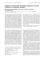
TLR3 KO mice are partially protected from poly(I:C)-induced inflammation in lung interstitiumFigure 2
TLR3 KO mice are partially protected from poly(I:C)-induced inflammation in lung interstitium. Representative
H&E-stained lung sections from WT- PBS treated (A,E, I)WT poly(I:C)-treated (B, F, J), TLR3 KO PBS treated mice (C ,G, K)
and TLR3 KO poly(I:C)-treated (D, H, L). Figures A-L are representative images from each group. Figure A-D are at 10×, Fig-
ures E-H are at 40 × and Figures I-L are at 60 ×.
A
B
C
D
E
F
G
H
I
J
K
L
Respiratory Research 2009, 10:43 />Page 8 of 14
(page number not for citation purposes)
TLR3 KO mice are protected from poly(I:C)-induced
changes in lung function at baseline
In order to investigate whether TLR3 plays a role in
poly(I:C)-induced lung function impairment, lung func-
tion was measured following poly(I:C) treatment of TLR3
KO mice and WT age-matched controls. As shown in Fig-
ure 6B, TLR3 KO mice were protected from poly(I:C)-
induced changes at baseline. The increase in penh
observed at baseline following poly(I:C) administration
was significantly reduced in TLR3 KO mice.
Discussion
Exacerbations of respiratory diseases such as asthma and
COPD are often associated with concomitant respiratory
viral infections. Since TLR3 is activated by viral dsRNA,
the purpose of the current study was to better understand
the functional consequences of TLR3 activation in vivo.
Administration of poly(I:C), a synthetic TLR3 ligand, to
the lungs of mice induced marked inflammation accom-
panied by impaired lung function. TLR3 KO mice were
partially protected from the effects of poly(I:C) demon-
strating the involvement of TLR3. These data provide fur-
Table 3: Morphometric analysis of lungs from WT PBS control and poly(I:C)-treated, and TLR-3 KO PBS control and poly(I:C)-treated
mice.
Group Tissue Density
%
Tissue Cellularity
%
Airway Mucosal Height
m
WT PBS 32 ± 2 8 ± 1 15 ± 1
WT Poly(I:C) 50 ± 5* 16 ± 2* 26 ± 4*
Fold Increase (Compared to WT PBS) 1.7 2 1.7
KO PBS 36 ± 7 9 ± 1 18 ± 3
KO Poly(I:C) 49 ± 6* 14 ± 3* 19 ± 2**
Fold Increase (Compared to KO PBS) 1.4 1.5 NC
NC = No change, * Different from respective PBS control. ** Different from poly(I:C)-treated WT. p < 0.01 using T-test to compare groups.
Poly(I:C) induces cytokine secretion from BEAS-2B cellsFigure 3
Poly(I:C) induces cytokine secretion from BEAS-2B cells. BEAS-2B cells were incubated for 24 hours at 37°C with
serial dilutions of polyI:C. Supernatants were collected after 24 hours and assayed for cytokine levels of IL-6 (A), IL-8 (B),
CCL2 (C), CCL5 (D), and CXCL10 (E). Data is representative of 2 different experiments.
IL6
0 16 31 63 125 250 500 100
0
0
5000
10000
15000
Poly(I:C) [ng/ml]
CONC [ pg /ml]
IL 8
0 16 31 63 125 250 500 100
0
0
500
1000
1500
2000
2500
Poly(I:C) [ng/ml]
CONC [pg/ml]
CCL2
0 16 31 63 125 250 500 100
0
0
200
400
600
800
1000
1200
1400
Poly(I:C) [ng/ml]
CONC [pg/ml]
CCL5
0 16 31 63 125 250 500 1000
0
300
600
900
Poly(I:C) [ng /ml]
CO NC [pg /m l ]
CXCL10
0 16 31 63 125 250 500 100
0
0
500
1000
1500
2000
Poly(I:C) [ng/ml]
CONC [pg/ml]
ABC
DE
Respiratory Research 2009, 10:43 />Page 9 of 14
(page number not for citation purposes)
Figure 4
Poly(I:C) induces impairment of lung function and AHR. Mice were administered PBS or 10, 20, 50 or 100 g polyI:C
(I.N.) every 24 h for three days. 24 h after the last poly(I:C) administration, baseline lung function and AHR to increasing doses
of methacholine was measured by whole body plethysmography (A & B). The 100 ug poly I:C group had higher penh levels than
the PBS, 10, and 20 ug groups, p < 0.05 (B). Methacholine challenge resulted in a larger increase from baseline in the poly(I:C)-
treated groups than in the PBS group, p < 0.001 for each methacholine dose. Invasive measurements of lung function were per-
formed 24 h following three administrations (24 h apart) of 100 g poly(I:C) (C). Peak airway resistance after i.v. injection of
methacholine at 240 ug/kg are shown. Methacholine challenge resulted in a larger increase from baseline in the poly(I:C)-
treated group than in the PBS group, p = 0.015. Repeated measures ANOVA was used to assess the Penh values over increas-
ing methacholine doses as well as to compare increases in resistance in response to methacholine from baseline among the
groups. Data are the mean ± SEM of 5–7 mice.
Respiratory Research 2009, 10:43 />Page 10 of 14
(page number not for citation purposes)
TLR3 KO mice are partially protected from poly(I:C)-induced inflammatory cell influx in the airwaysFigure 5
TLR3 KO mice are partially protected from poly(I:C)-induced inflammatory cell influx in the airways. Mice were
administered PBS or 100 g poly(I:C) I.N. every 24 h for three days. 24 hours after the last poly(I:C) administration, mice were
euthanized and the lungs were lavaged. The total number of cells (5A), neutrophils (5B) and mononuclear cells(5C) were meas-
ured in the BAL. Data are the mean ± SEM of 6 mice. Treatment groups (PBS or 100 g poly(I:C)) and mouse types were com-
pared using 2-way ANOVA, including an interaction term. *p < 0.05, **p < 0.01 compared to PBS-treated mice. When
comparing the impact of poly(I:C) treatment on cell populations in the lavage, there was a significantly larger increase in the
response of wild type mice than knockout mice, with respect to total cells and mononuclear cells alone, **p < 0.01 in each
case. Similar trends were observed in neutrophils alone but failed to reach statistical significance (p = 0.056).
Total Cells
PBS 100 g Poly(I:C)
0
10
20
30
40
50
60
KO
WT
**p<0.01
A
*p<0.05
Total Cells X10
4
Neutrophils
125
KO
WT
**p<0.01
B
100
75
50
25
**p<0.01
Total Cells X10
3
0
PBS 100
g Poly(I:C)
Mononuclear Cells
PBS 100 g Poly(I:C)
0
50
100
150
200
250
300
350
400
450
KO
WT
**p<0.01
C
ns
Total Cells X10
3
Respiratory Research 2009, 10:43 />Page 11 of 14
(page number not for citation purposes)
ther support for a role of TLR3 in respiratory diseases and
suggest a potential mechanisitic pathway for viral exacer-
bations.
Upon activation, TLR3 recruits a Toll-IL-1 receptor (TIR) –
related adaptor protein inducing interferon (TRIF), which
activates both IFN-regulatory factor 3 (IRF3) and NF-kB
[12] and [13]. In our model, following poly(I:C) admin-
istration to the lungs, there was an up regulation of TLR3,
-2, -7, and 9 gene expression and their associated signaling
molecules. Previous in vitro studies have demonstrated
that activation of TLR3 with poly(I:C) induces up regula-
TLR3 KO mice are partially protected from poly(I:C)-induced impairment of lung function and AHRFigure 6
TLR3 KO mice are partially protected from poly(I:C)-induced impairment of lung function and AHR. Mice were
exposed to three doses of 100 mg poly(I:C) (I.N.; 24 h apart). Baseline lung function and AHR to increasing doses of metha-
choline was measured by whole body plethysmography 24 hours following the last dose of poly(I:C). Data are the mean ± SEM
of 6 mice. Prior to challenge, the groups given poly(I:C) had higher Penh values than those given PBS, p < 0.001. This difference
was greater in the WT mice than in the KO mice, p = 0.047. Increasing methacholine challenges lead to higher mean penh val-
ues for the Poly I:C treated groups than for the PBS groups, p < 0.001, but there was not a statistically significant difference
between the poly I:C-treated KO and WT groups p = 0.115. A repeated measure ANOVA was used to assess the change from
pre-challenge penh values over increasing methacholine doses. ANOVA was used to compare the peak resistance levels at
baseline among the groups.
Respiratory Research 2009, 10:43 />Page 12 of 14
(page number not for citation purposes)
tion of its own expression as well as the expression of
other TLRs. For example, poly(I:C) up regulates mRNA for
TLR2, 3 and 4 in airway smooth muscle cells [14] and
TLR2, 3, 6 and 10 in lung epithelial cells [3]. In vivo, the
up regulation of TLR mRNA expression may have
occurred as a result of expression of TLRs on infiltrating
cells or through up regulation on resident lung cells.
Indeed, monocytes express all of the known TLRs [15]. In
contrast, neutrophils have been shown to express all the
TLRs except TLR3 [16]. Within the lung, all of the known
TLRs have been found to be expressed by human primary
bronchial epithelial [3] and smooth muscle cells [14]. The
up regulation of multiple members of the TLR family, as a
consequence of activation of one TLR, may indicate the
creation of an environment of hyper-responsiveness to
pathogen insult whereby, an exacerbation event could be
triggered in the event that the lung is exposed to other toll-
ligands. In support of this hypothesis, it has been shown
that infection of airway epithelial cells with Hemophilus
influenza induced the secretion of CXCL-8, up regulated
TLR3 expression and increased the responsiveness to a
secondary challenge of Rhinovirus. Interestingly, inhibi-
tion of TLR3 with small interfering RNA, inhibited the
Rhinovirus-induced CXCL-8 production [17]. In addition
this same group demonstrated that pretreatment with Rhi-
novirus resulted in delayed bacterial clearance when a sec-
ondary infection was induced using nontypeable
Hemophilus influenza. Sajjan et al. showed that this may be
the result of decreases in transepithelial resistance or com-
promised tight junctions and loss of zona occludins-1 and
junctional adhesion molecule-1 [18]. Taken together
these studies suggest that activation of TLRs, such as TLR3
can result in a perturbation of the local environment, spe-
cifically dysregulation of the airway epithelium thereby
supporting an environment primed for an exacerbation.
We are currently focusing efforts in our laboratory toward
identifying the composition of the mononuclear cell pop-
ulations in this model including the activation state of
various cell types including dendritic cells. In a review by
Fe et. al. it is summarized that TLR3 can induce a variety
of cytokines in human dendritic cells including IFN, and
CXCL10 [19].
In vivo TLR3 agonism by synthetic dsRNA also resulted in
a profound up regulation of the expression and secretion
of multiple pro-inflammatory cytokines, chemokines,
and growth factors. In vitro studies have demonstrated
that activation of TLR3 by dsRNA on different cell types
including natural killer cells [20], epithelial cells
[3,21,22], and smooth muscle cells [14] results in
increased expression and/or secretion of pro-inflamma-
tory cytokines including IL-6, CXCL-8, CCL-2, CCL-5,
CXCL-10, GM-CSF, TNF and IFN. A likely source of
cytokines following poly(I:C) administration may be the
airway epithelium since activation of BEAS-2B cells in vitro
induced a profile of pro-inflammatory cytokines similar
to that observed following in vivo poly(I:C) challenge.
TLR3 has been identified and functionally characterized
in mouse tracheal muscle [23] and in primary human
small airway epithelial cells [21,3,22]. Previous in vitro
studies have also demonstrated the secretion of inflam-
matory mediators following TLR3 activation of epithelial
cells[3,3,21]. The up regulation of pro-inflammatory
cytokines and chemokines provides an inflammatory
milieu supporting the infiltration of inflammatory cells
into the airways and lung interstitium. Accompanying the
inflammation-rich pathology was the presence of bron-
chial epithelial cell hypertrophy. The hypertrophic cells
extended into the secondary and tertiary airways. Epithe-
lial cell hypertrophy is normally associated with increased
mucus production [23]. However, in the current study,
there was no evidence for increased mucus production by
PAS staining. Given the distribution of goblet cells in nor-
mal mouse airways, which is restricted to the main bron-
chi and primary bronchioles, the data suggest that the
hypertrophic epithelial cells are not mucus-producing
goblet cells.
Along with the demonstration that poly(I:C), acting as a
TLR3 ligand, results in an inflammatory response in vivo,
the study presents a novel finding that stimulation of
TLR3 results in a measurable impairment of lung function
both without provocation and characterized by increased
AHR to methacholine. Similar changes in baseline lung
function have also been described in mice exposed to Res-
piratory Syncytial virus (RSV) [24]. Recent studies have
demonstrated that pre-exposure of mouse tracheas to
poly(I:C) in vitro increases the expression of bradykinin
B1 and B2 receptors on the smooth muscle and confers
AHR to bradykinin [25]. Notably, inhibition of the brady-
kinin B1 receptor confers protection from acetylcholine-
induced AHR following allergen sensitization and chal-
lenge [26]. In contrast, AHR to histamine following
parainfluenza-3 infection in guinea pigs was inhibited by a
bradykinin B2 receptor antagonist [27]. Taken together
these data suggest a role for bradykinin in TLR3-induced
airway dysfunction. In the current study some, but not all,
functional responses were protected in TLR3 KO mice fol-
lowing multiple administrations of poly(I:C). Specifi-
cally, they were protected from baseline lung function
changes in response to poly(I:C), however protection
from AHR in response to provocation with methacholine
did not result in significant protection. Further, the pro-
inflammatory mediators produced following poly(I:C)
administration were not modulated in TLR3 KO mice.
Unpublished data from our laboratory has shown that
TLR3 KO mice were significantly protected from a single
administration of poly(I:C) with respect to pro-inflamma-
tory mediators in the bronchoalveolar lavage (data not
shown), indicating that mediators released in response to
Respiratory Research 2009, 10:43 />Page 13 of 14
(page number not for citation purposes)
acute activation with poly(I:C) may be more TLR3
dependent. This data suggests that another receptor for
poly(I:C) may be available. Indeed, since a percentage of
TLR3 KO mice succumb to poly(I:C)-induced shock, it
suggests that poly(I:C) may still signal in the absence of
TLR3 [1]. Indeed, dsRNA can also signal through dsRNA-
dependent protein kinase (PKR) [28], RIGI [29] and
MDA-5 [30]. The potential redundancy in the dsRNA
downstream pathways may be an explanation for the
incomplete protection observed in TLR3 KO mice.
Understanding the different signaling pathways involved
in recognition of dsRNA by the host has been a major area
of focus by many researchers. Le Goffic et al. demon-
strated that sensing of influenza A virus by TLR3 and RIG-I
regulates a pro-inflammatory response. In contrast, RIG-I
but not MDA-5 also mediates type I IFN-dependent anti-
viral signaling response[31]. Use of non-poly(I:C) TLR3
ligands is necessary to further define the impact of TLR3-
specific signaling on pulmonary pathophysiology. Inter-
estingly, TLR3 KO mice demonstrate protection from
influenza A virus-induced lung function impairment
accompanied by reduced inflammation and improved
survival [32].
These data taken along with the inflammatory conse-
quences of TLR3 activation suggest that sustained TLR3
activation may also contribute to severe exacerbations of
chronic pulmonary diseases. In summary, the data pre-
sented in this study suggest that sustained TLR3 activation
may play an important role in respiratory disease patho-
genesis. A better understanding of the effects of TLR3 acti-
vation will provide additional insight into the
mechanisms underlying virus-induced exacerbations
associated with respiratory diseases. Additionally, these
studies provide an opportunity to identify suitable targets
for therapeutic intervention for respiratory disease exacer-
bations.
Competing interests
NCS, JS, HAR, KAS, RJL, DDE, PJB, LAM, PA M, RAB, LRS,
DEG, RTS, MLM, and AMD are current or former employ-
ees of Centocor Research & Development, Inc. RAF and LA
declare that they have no competing interests.
Authors' contributions
NCS conceived of the study and participated in its design
and coordination as well as all analysis. JS, LAM, LRS,
DEG, RTS, MLM, and AMD participated in the design and
coordination of the studies. HAR, and KAS executed the
in-life portion of the studies. RJL carried out the BEAS2B
studies. DDE and PJB carried out the histopath analysis of
the lungs. PAM carried out the statistical analysis of all
data sets. RAB carried out the analysis of cellular infiltrates
in the lung. RAF and LA made the TLR3 KO mice and gave
input on the design of the studies and the manuscript. All
authors read and approved the final manuscript.
Acknowledgements
The authors would like to thank Cory M. Hogaboam, Ph.D. Associate Pro-
fessor, Immunology Program, Department of Pathology, University of
Michigan Medical School, for assistance in guiding the invasive measure-
ments of lung function.
References
1. Alexopoulou L, Holt AC, Medzhitov R, Flavell RA: Recognition of
double-stranded rna and activation of nf-kappab by toll-like
receptor 3. Nature 2001, 413(6857):732-738.
2. Kariko K, Ni H, Capodici J, Lamphier M, Weissman D: Mrna is an
endogenous ligand for toll-like receptor 3. J Biol Chem 2004,
279(13):12542-12550. Epub 12004 Jan 12516
3. Sha Q, Truong-Tran AQ, Plitt JR, Beck LA, Schleimer RP: Activation
of airway epithelial cells by toll-like receptor agonists. Am J
Respir Cell Mol Biol 2004, 31(3):358-364.
4. Tabeta K, Georgel P, Janssen E, Du X, Hoebe K, Crozat K, Mudd S,
Shamel L, Sovath S, Goode J, et al.: Toll-like receptors 9 and 3 as
essential components of innate immune defense against
mouse cytomegalovirus infection. Proc Natl Acad Sci USA 2004,
101(10):3516-3521. Epub 2004 Mar 3511
5. Takeda K, Akira S: Microbial recognition by toll-like receptors.
J Dermatol Sci 2004, 34(2):73-82.
6. Johnston SL: Natural and experimental rhinovirus infections of
the lower respiratory tract. Am J Respir Crit Care Med 1995,
152(4 Pt 2):S46-52.
7. Gern JE, French DA, Grindle KA, Brockman-Schneider RA, Konno S,
Busse WW: Double-stranded rna induces the synthesis of spe-
cific chemokines by bronchial epithelial cells. Am J Respir Cell
Mol Biol 2003, 28(6):731-737.
8. Panina-Bordignon P, D'Ambrosio D: Chemokines and their
receptors in asthma and chronic obstructive pulmonary dis-
ease. Curr Opin Pulm Med 2003, 9(2):104-110.
9. Guillot L, Le Goffic R, Bloch S, Escriou N, Akira S, Chignard M, Si-
Tahar M: Involvement of toll-like receptor 3 in the immune
response of lung epithelial cells to double-stranded rna and
influenza a virus. J Biol Chem 2005, 280(7):5571-5580. Epub 2004
Dec 5573
10. Hewson CA, Jardine A, Edwards MR, Laza-Stanca V, Johnston SL:
Toll-like receptor 3 is induced by and mediates antiviral
activity against rhinovirus infection of human bronchial epi-
thelial cells. J Virol 2005, 79(19):12273-12279.
11. Matsukura S, Kokubu F, Kurokawa M, Kawaguchi M, Ieki K, Kuga H,
Odaka M, Suzuki S, Watanabe S, Takeuchi H, et al.: Synthetic dou-
ble-stranded rna induces multiple genes related to inflam-
mation through toll-like receptor 3 depending on nf-kappab
and/or irf-3 in airway epithelial cells. Clin Exp Allergy 2006,
36(8):1049-1062.
12. Yamamoto M, Sato S, Hemmi H, Hoshino K, Kaisho T, Sanjo H,
Takeuchi O, Sugiyama M, Okabe M, Takeda K, et al.: Role of adaptor
trif in the myd88-independent toll-like receptor signaling
pathway. Science 2003, 301(5633):640-643. Epub 2003 Jul 2010
13. Yamamoto M, Sato S, Mori K, Hoshino K, Takeuchi O, Takeda K,
Akira S: Cutting edge: A novel toll/il-1 receptor domain-con-
taining adapter that preferentially activates the ifn-beta pro-
moter in the toll-like receptor signaling. J Immunol 2002,
169(12):6668-6672.
14. Sukkar MB, Xie S, Khorasani NM, Kon OM, Stanbridge R, Issa R,
Chung KF: Toll-like receptor 2, 3, and 4 expression and func-
tion in human airway smooth muscle. J Allergy Clin Immunol
2006, 118(3):641-648.
15. Zarember KA, Godowski PJ: Tissue expression of human toll-
like receptors and differential regulation of toll-like receptor
mrnas in leukocytes in response to microbes, their products,
and cytokines. J Immunol 2002, 168(2):554-561.
16. Hayashi F, Means TK, Luster AD: Toll-like receptors stimulate
human neutrophil function. Blood 2003, 102(7):2660-2669.
17. Sajjan US, Jia Y, Newcomb DC, Bentley JK, Lukacs NW, LiPuma JJ,
Hershenson MB: H. Influenzae potentiates airway epithelial
Publish with BioMed Central and every
scientist can read your work free of charge
"BioMed Central will be the most significant development for
disseminating the results of biomedical research in our lifetime."
Sir Paul Nurse, Cancer Research UK
Your research papers will be:
available free of charge to the entire biomedical community
peer reviewed and published immediately upon acceptance
cited in PubMed and archived on PubMed Central
yours — you keep the copyright
Submit your manuscript here:
/>BioMedcentral
Respiratory Research 2009, 10:43 />Page 14 of 14
(page number not for citation purposes)
cell responses to rhinovirus by increasing icam-1 and tlr3
expression. Faseb J 2006, 20(12):2121-2123.
18. Sajjan US, Wang Q, Jia Y, LiPuma J, Hershenson MB: Rhinovirus
compromises tight junctions in differentiated airway epithe-
lial cells and predisposes mice to secondary bacterial infec-
tions. Am J Respir Crit Care Med 2007, 2007(175):A774.
19. Re F, Strominger JL: Heterogeneity of tlr-induced responses in
dendritic cells: From innate to adaptive immunity. Immunob-
iology 2004, 209(1–2):191-198.
20. Schmidt KN, Leung B, Kwong M, Zarember KA, Satyal S, Navas TA,
Wang F, Godowski PJ: Apc-independent activation of nk cells by
the toll-like receptor 3 agonist double-stranded rna. J Immu-
nol 2004, 172(1):138-143.
21. Ritter M, Mennerich D, Weith A, Seither P: Characterization of
toll-like receptors in primary lung epithelial cells: Strong
impact of the tlr3 ligand poly(i:C) on the regulation of toll-
like receptors, adaptor proteins and inflammatory response.
J Inflamm (Lond) 2005, 2(1):16.
22. Ieki K, Matsukura S, Kokubu F, Kimura T, Kuga H, Kawaguchi M,
Odaka M, Suzuki S, Watanabe S, Takeuchi H, et al.: Double-
stranded rna activates rantes gene transcription through co-
operation of nuclear factor-kappab and interferon regula-
tory factors in human airway epithelial cells. Clin Exp Allergy
2004, 34(5):745-752.
23. Rogers DF: Airway mucus hypersecretion in asthma: An
undervalued pathology? Curr Opin Pharmacol 2004, 4(3):241-250.
24. Schwarze J, Schauer U: Enhanced virulence, airway inflamma-
tion and impaired lung function induced by respiratory syn-
cytial virus deficient in secreted g protein. Thorax 2004,
59(6):517-521.
25. Bachar O, Adner M, Uddman R, Cardell LO: Toll-like receptor
stimulation induces airway hyper-responsiveness to bradyki-
nin, an effect mediated by jnk and nf-kappa b signaling path-
ways. Eur J Immunol 2004, 34(4):1196-1207.
26. Huang TJ, Haddad EB, Fox AJ, Salmon M, Jones C, Burgess G, Chung
KF: Contribution of bradykinin b(1) and b(2) receptors in
allergen-induced bronchial hyperresponsiveness.
Am J Respir
Crit Care Med 1999, 160(5 Pt 1):1717-1723.
27. Folkerts G, Vlieger JW, de Vries A, Faas S, Linde H van Der, Engels F,
de Jong JC, Verheyen FA, Van Heuven-Nolsen D, Nijkamp FP: Virus-
and bradykinin-induced airway hyperresponsiveness in
guinea pigs. Am J Respir Crit Care Med 2000, 161(5):1666-1671.
28. Clemens MJ, Elia A: The double-stranded rna-dependent pro-
tein kinase pkr: Structure and function. J Interferon Cytokine Res
1997, 17(9):503-524.
29. Yoneyama M, Kikuchi M, Natsukawa T, Shinobu N, Imaizumi T, Miy-
agishi M, Taira K, Akira S, Fujita T: The rna helicase rig-i has an
essential function in double-stranded rna-induced innate
antiviral responses. Nat Immunol 2004, 5(7):730-737.
30. Gitlin L, Barchet W, Gilfillan S, Cella M, Beutler B, Flavell RA, Dia-
mond MS, Colonna M: Essential role of mda-5 in type i ifn
responses to polyriboinosinic:Polyribocytidylic acid and
encephalomyocarditis picornavirus. Proc Natl Acad Sci USA 2006,
103(22):8459-8464.
31. Le Goffic R, Pothlichet J, Vitour D, Fujita T, Meurs E, Chignard M, Si-
Tahar M: Cutting edge: Influenza a virus activates tlr3-
dependent inflammatory and rig-i-dependent antiviral
responses in human lung epithelial cells. J Immunol 2007,
178(6):3368-3372.
32. Le Goffic R, Balloy V, Lagranderie M, Alexopoulou L, Escriou N, Fla-
vell R, Chignard M, Si-Tahar M: Detrimental contribution of the
toll-like receptor (tlr)3 to influenza a virus-induced acute
pneumonia. PLoS Pathog 2006, 2(6):e53.