Báo cáo y học: " Cyclic hydrostatic pressure and cotton particles stimulate synthesis by human lung macrophages of cytokines in vitro" pptx
Bạn đang xem bản rút gọn của tài liệu. Xem và tải ngay bản đầy đủ của tài liệu tại đây (838.97 KB, 11 trang )
BioMed Central
Page 1 of 11
(page number not for citation purposes)
Respiratory Research
Open Access
Research
Cyclic hydrostatic pressure and cotton particles stimulate synthesis
by human lung macrophages of cytokines in vitro
Sarah Lewis
1
, Dave Singh
2
and Carol E Evans*
1
Address:
1
Tissue Injury and Repair Group, School of Clinical and Laboratory Sciences, Faculty of Medical and Human Sciences, University of
Manchester, Stopford Building , Oxford Road, Manchester M13 9PT, UK and
2
NIHR Translational Research Facility, University Of Manchester,
University Hospital Of South Manchester Foundation Trust, UK
Email: Sarah Lewis - ; Dave Singh - ; Carol E Evans* -
* Corresponding author
Abstract
Background: Inhalation of particulates is a leading cause of the development of lung diseases and
current understanding of the complex relationship between lung metabolism and airborne
particulates is incomplete. It is well established that mechanical load is important in the
development of the lung and in lung cell differentiation. The interaction between particle exposure
and physical forces on alveolar macrophages is a physiologically relevant issue, but as yet
understudied. This study examines the effect of cyclic hydrostatic pressure and cotton particles on
synthesis of cytokines by human alveolar macrophages.
Methods: Alveolar macrophages were obtained from patients with lung disease, either from lavage
samples or from lung tissue resection. The commonly used cell line THP-1 was included in the
experiments. Cell cultures were exposed to cotton particles and/cyclic hydrostatic pressure (3 or
5 psi); control cultures were exposed to medium only. TNFα, IL-1β and IL-6 were assayed in the
culture media using specific ELISAs. Cells were characterized using morphology and markers
specific for macrophages (Jenner/Giemsa staining, CD14 and CD68).
Results: Exposure to cotton particles stimulated cytokine synthesis by macrophages from all three
sources; exposure to cyclic hydrostatic pressure alone did not stimulate cytokine synthesis
significantly. However, the combination of both particles and cyclic hydrostatic pressure increased
the simulation of cytokine synthesis still further. Cell characterization demonstrated that the large
majority of cells had a macrophage morphology and were positive for CD14 and CD68.
Conclusion: These data suggest an interaction between cyclic hydrostatic pressure and particulate
exposure, which increases alveolar macrophage cytokine production. This interaction was only
observed at the higher cyclic hydrostatic pressure. However, in patient samples, there was
considerable variation in the amount by which secretion of an individual cytokine increased and
there was also variation in the mechanosensitivity of cells from the three different sources. Cyclic
hydrostatic pressure, therefore, may be an important modulator of the response of alveolar
macrophages to cotton particles, but the source of the cells may be a confounding factor which
demands further investigation.
Published: 2 June 2009
Respiratory Research 2009, 10:44 doi:10.1186/1465-9921-10-44
Received: 8 August 2007
Accepted: 2 June 2009
This article is available from: />© 2009 Lewis et al; licensee BioMed Central Ltd.
This is an Open Access article distributed under the terms of the Creative Commons Attribution License ( />),
which permits unrestricted use, distribution, and reproduction in any medium, provided the original work is properly cited.
Respiratory Research 2009, 10:44 />Page 2 of 11
(page number not for citation purposes)
Introduction
The lungs are continually subject to mechanical load, in
the form of hydrostatic pressure and strain generated dur-
ing inspiration and expiration. In this context, hydrostatic
pressure is a load which deforms the tissue and cells by
compression, whereas strain may be described as a load
which causes elongation of the tissue and hence the cells
within that tissue. The role of mechanical load in lung
development [1,2] and lung cell differentiation [3] is now
well established. However, although there have been sev-
eral interesting studies on the effect of strain on lung cells
[4-7], there have been few similar studies on the effect of
load on lung cells [8]. Hydrostatic pressure may be ele-
vated during increased ventilation, including forced venti-
lation, or pulmonary oedema. A recent publication by
Garcia et al [9] described how physical forces affected the
function and phenotype of cells in the lung. This review
described the stimulation of cytokine synthesis by strain,
by macrophages and lung epithelial cells and examines
possible signalling pathways for such mechanotransduc-
tion.
Alveolar macrophages play a role in pulmonary inflam-
mation in a variety of lung diseases. These cells are contin-
ually subject to mechanical load, but our knowledge of
the response of these cells to such forces is sparse. We have
previously shown macrophages from peripheral blood to
be mechanoresponsive, causing a profound induction of
the synthesis of proinflammatory mediators [10-14]. Fur-
thermore, the pro-inflammatory effects of mechanical
load forces on peripheral blood macrophages are
enhanced by particulates.
Chronic environmental exposure to particulate matter can
result in upregulation of the pro-inflammatory activity of
alveolar macrophages. Examples of increased alveolar
macrophage pro-inflammatory activity include chronic
obstructive pulmonary disease (COPD) caused by ciga-
rette smoking, and occupational cotton dust exposure
which can cause byssinosis, chronic bronchitis or airflow
obstruction. The interaction between particle exposure
and physical forces on alveolar macrophages is a physio-
logically relevant issue, but as yet understudied.
The study reported here examined the effect of cyclic
hydrostatic pressure (CHP) cotton particles or a combina-
tion of the two, on alveolar macrophages. We have evalu-
ated the potential for CHP to modulate macrophage pro-
inflammatory cytokine production, and the interaction
between CHP and cotton particle exposure.
Methods
Patient samples
Five patients who were undergoing clinical investigational
bronchoscopies were recruited, as well as 6 patients
undergoing surgical resection for suspected or confirmed
lung cancer. COPD was diagnosed based on a history of
smoking for at least 10 pack years, typical symptoms (pro-
ductive cough, breathlessness or wheeze), and airflow
obstruction defined as FEV
1
< 80% predicted, and FEV
1
/
FVC ratio < 0.7. All subjects gave written informed con-
sent. The study was approved by the local research ethics
committee.
The 5 subjects undergoing bronchoscopy were all male
and aged from 43–64 years. Three subjects were current
smokers with normal lung function, while 2 were ex-
smokers (1 with COPD).
The 6 subjects undergoing lung surgery were aged from 53
to 77 years; 5 male and one female. Four were current
smokers (3 with COPD and 1 with normal lung function)
and 2 were ex-smokers (both with normal lung function).
Alveolar Macrophage Isolation
Broncho-alveolar lavage (BAL) was collected from the
right lower lobe, or a lobe not affected by radiographic or
endobronchial abnormalities: The bronchoscope was
wedged in the right middle lobe and a maximum of 4 × 60
ml aliquots of prewarmed sterile 0.9% NaCl solution were
instilled. The aspirated fluid was stored on ice before fil-
tration (100 μm filter, Becton Dickenson). The filtrate was
centrifuged (400 g/10 min at 4°C) and the cell pellet
washed in RPMI 1640 medium supplemented with 2 mM
L-glutamine, 100 U/ml penicillin, and 100 μg/ml strepto-
mycin. BAL samples were collected and kept on ice to pre-
vent cells sticking to the sample tube. Samples were
filtered through a 100 μm cell sieve to remove debris then
centrifuged at 1500 rpm (400 g) at 4°C for 10 minutes.
The supernatant was discarded and cell count performed
on the cell pellet.
Resected lung tissue was obtained from areas distant from
the tumour, and perfused with 0.1 M NaCl to isolate mac-
rophages. Lung tissue was perfused with 0.1 M Na Cl to
isolate the cells before filtering and centrifuging as with
the BAL samples. The supernatant was then discarded and
cell counts performed on the cell pellet.
Macrophages were isolated from the mixed cell popula-
tions by the property of adherence. Cells were incubated
in 20% Dulbecco's modified Eagles medium (DMEM,
Invitrogen UK) + 1% Glutamine + 1% Penicillin/Strepto-
mycin (Invitrogen UK) for 1 hour at 37°C in 5% CO
2
.
Cell cultures were washed gently with phosphate-buffered
saline (PBS) to remove any non-adherent cells. Approxi-
mately 80% of the white cells were found to be macro-
phages by this technique. The culture medium was
replenished and the macrophages cultured for 24 hours
before being exposed to experimental conditions.
Respiratory Research 2009, 10:44 />Page 3 of 11
(page number not for citation purposes)
THP-1 Cell line
As this alveolar macrophage cell line is used extensively in
research into the lung, we also performed loading experi-
ments on THP-1 cells. The experimental protocol was the
same as that used for the patient cells, except that, because
of their increased sensitivity (vis-a-vis patient cells); THP-
1 cells were seeded at a much lower density.
Cotton Particulates
To examine the effect of typical cotton dust particles on
these cells, Standard Cotton Dust used in all experiments.
This is produced by the Cotton Incorporated company
from crude cotton dust collected in a West Texas cotton
mill between 1981 and 1983. This single source cotton
dust allows the comparison of data and hypotheses from
different scientific groups. The dust was analysed for
endotoxin contamination using the Charles River
Endosafe
®
Portable Test System. This standard technique
allows the quantitative detection of endotoxin by a kinetic
chromagenic method, and involves the interaction of
Limulus Amebocyte Lysate (LAL) and synthetic colour-
producing substrate. This technique was performed at our
laboratory under the guidance of a Charles River repre-
sentative, using endotoxin free solutions and equipment.
Before being used in any experiments, 100 mg samples of
cotton dust were sterilized by autoclaving and then sus-
pended in 10 mls of the usual culture medium. This was
filtered through a 40 μm cell sieve and the resulting fil-
trate, containing the smaller particles was used in the
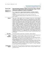
experiments. The size distribution of the filtered cotton
particles was measured using image analysis and it was
found that 22% of the measured particles had a diameter
≤2 μm and 94% ≤8 μm (Fig 1). The cotton particles used
were of a size which has been shown previously to be the
range phagocytosed by alveolar macrophages, evoking an
inflammatory response [15,16].
A frequency diagram of size of cotton particlesFigure 1
A frequency diagram of size of cotton particles.
Cotton Particle Size
0
5
10
15
20
25
30
35
123456789101112131415161718192021222324
Particle Size (um)
Percentage of Particles
Respiratory Research 2009, 10:44 />Page 4 of 11
(page number not for citation purposes)
Cell characterization
The cells used in these studies were characterized using
markers specific for macrophages. Cells were washed in
PBS and fixed for 2 minutes in ice-cold ethanol (BDH,
UK) prior to staining.
Histological staining, using the Jenner/Giemsa technique,
was performed on bronchial lavage in order to establish
the percentage of macrophages present in the samples.
Briefly, lavage cell cytospins were immersed in Jenner
solution (Raymond Lamb Ltd., UK, 0.3% in 100% meth-
anol) for 2 minutes before immersing in Giemsa solution
(Raymond Lamb ltd., UK, 1% in pH 6.4 buffer) for a fur-
ther 20 minutes. Cytospins were then rinsed in pH 6.4
buffer, air-dried and mounted with Pertex. Leucocyte mor-
phology and identification is clear using this technique,
with macrophage nuclei staining purple and cytoplasm
blue.
In addition, immunohistochemistry was performed using
a commercially available antibody specific for CD68
(mouse anti-human CD68 diluted 1 in 100, Serotec Ltd.
UK) and visualized using DAB (3,3 diamino bezidine,
Sigma UK). CD68 is a glycoprotein found on the surface
of macrophages, so cells staining positive for CD68 will
therefore be macrophages.
Cell culture and Pressurization
Macrophages from BAL or lung biopsies were seeded at 5
× 10
5
/ml into 24 well plates (1 ml/well) and incubated for
24 hours; THP-1 cells were seeded at 1 × 10
5
/ml. Culture
media were then removed and 1 ml of fresh medium or 1
ml of the cotton dust/medium suspension was added to
each well. The cultures were exposed to the cotton parti-
cles for 24 hours before pressurization and control cul-
tures were exposed to medium only.
BAL, lung surgery macrophage and THP-1 cultures were
loaded into our novel loading jig [10,11] and subjected to
cyclic hydrostatic pressure (CHP). The pressure regime
was a load of 3 psi, at a frequency of 2 seconds on/off for
1 hour. This load was in addition to atmospheric pressure
psi [14.69]. Macrophages from the lung surgery samples
and THP-1 cells were also exposed to 3 psi pressure (20.7
KPa) and/or cotton dust (<40 μm). Cultures were also
exposed to a higher pressure of 5 psi (34.5 KPa) and/or
cotton dust (<40 μm). The cultures were then returned to
the incubator for 23 hours prior to analysis and the con-
trol (unloaded) cultures remained in the incubator
throughout the experiment.
Culture media were removed from the cultures 23 hours
post-pressure and cytokine levels were analysed by the
ELISA technique. This was performed using commercially
available ELISA kits from Diaclone (purchased from IDS
Ltd Boldon, Tyne and Wear, UK). TNFα, IL-1β and IL-6
were assayed in the culture media from the patient's sam-
ples and TNFα was assayed in the culture media from the
THP-1 cells.
As our previous research into CHP used peripheral blood
macrophages, we compared the response of these cells
and BAL macrophages to cotton dust exposure, collecting
paired blood and lavage samples from our first few
patients. However, it soon became apparent that periph-
eral blood macrophages underwent apoptosis on contact
with the cotton dust particles, making any comparison
impossible (data not shown).
Statistical Analysis
Statistical analysis was performed on the results of both
BAL macrophages and lung surgery macrophages using
the non-parametric Friedman Test with Dunn Post Test.
The parametric paired ANOVA with Bonferroni Post Test
was performed on the results of the THP-1 experiments.
Significance was defined as p < 0.05. Analyses were per-
formed using Graphpad Instat 3.
Results
BAL macrophages
In cultures of BAL macrophages, synthesis of the cytokines
TNFα, IL-1β and IL-6 was increased by exposure to cotton
particles (Fig 2a–c). However, there was no significant
increase in cytokine production caused by CHP alone at 3
psi. Cytokine levels after exposure to cotton particles and
CHP were similar to particles alone.
Lung Resection Macrophages
Lung resection alveolar macrophages released signifi-
cantly greater levels of the cytokines TNFα, IL-1β and IL-6
after exposure to cotton particles (p < 0.001) (Fig 3a–f).
CHP at either 3 psi or 5 psi had no statistically significant
effect on the levels of these cytokines. (p > 0.05) although
a trend towards increase in synthesis of cytokines was
seen, when compared with unpressurized controls (TNFα
17%; IL-1β 6%; IL-6 26%).
Exposure to both pressure and cotton particles produced
numerically the greatest response from the cultures, which
reached statistical significance for IL-6 synthesis at 5 psi
compared to pressure alone or particles alone (p < 0.01).
Comparison of the results from these two cell types
showed that there was a significant difference in response
between the two cell types when exposed to particles or
both stimuli together (p < 0.01 in both cases) (Fig 3g).
There was no significant difference between the two cell
types when exposed to pressure alone; neither was there
any difference to the control.
Respiratory Research 2009, 10:44 />Page 5 of 11
(page number not for citation purposes)
THP-1 Cell line
Cultures exposed to cotton particles showed a significant
increase in synthesis of TNFα when compared to the con-
trols (p < 0.001). Synthesis of TNFα was not increased
when THP-1 cells were exposed to either level of CHP (3
psi or 5 psi) (Fig 4a, b).
Exposure to both CHP and cotton particles produced
numerically the greatest response from the cultures. At 5
psi, TNFα production was significantly increased com-
pared to particle exposure alone or pressure alone (p <
0.05).
Cotton Particulates
Standard Cotton Dust was found to contain 17.37 EU/mg
dust ≡ 1.74 ng endotoxin/ml dust.
Cell characterization
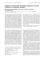
Using the Jenner/Giemsa stain to visualize cell morphol-
ogy, it was found that approximately 85% of cells
obtained by bronchial lavage exhibited macrophage mor-
phology (Figure 5). They displayed a large, reniform, pur-
ple nucleus and granular free, blue cytoplasm Fig 5). This
percentage is in agreement with previously published data
[17].
Immunocytochemistry of the BAL cell cultures using anti-
bodies to the macrophage cell surface markers CD68 and
CD14, demonstrated that all the cells in the preparation
stained positively for CD68 (Figure 6) and many stained
positive for CD14 (Figure 7). It is of interest that there was
an apparent increase in the depth of staining for both of
these markers when cells were exposed to both CHP and
particles (Fig 6, 7c), compared to exposure to CHP alone
(Fig 6, 7b).
Discussion
The main findings of this study were that alveolar macro-
phages from patients with a history of smoking responded
to cotton particulate exposure by increasing the produc-
tion of pro-inflammatory cytokines. CHP at 3 psi and 5
psi had no effect on cytokine production from these mac-
rophages, but cells exposed to CHP at 5 psi and cotton
particulates displayed increased cytokine production,
most notably of IL-6. This suggests an interaction between
CHP and particulate exposure in increasing alveolar mac-
rophage cytokine production. This interaction was
dependent on the pressure used, as it was only observed at
the higher CHP. These observations were supported by
experiments in THP-1 cells, where an interaction was
observed for TNF production only at 5 psi and not 3 psi.
CHP may be an important modulator of the response of
alveolar macrophages to cotton particles, but the source of
the cells may be a confounding factor which demands fur-
ther investigation.
(a-c) Effect of pressure and particles on synthesis of cytokines by BAL macrophages; data expressed as box and whisker plots with median lineFigure 2
(a-c) Effect of pressure and particles on synthesis of
cytokines by BAL macrophages; data expressed as
box and whisker plots with median line. Comparison
with unpressurized controls; ***p < 0.001, **p < 0.01, *p <
0.05
Į
BAL TNFa
C
o
nt
rol
Partic
l
es
Pres
s
ure 3
p
si
3
psi
+
P
art
i
c
l
es
0
5000
10000
15000
20000
25000
Regime
TNFa pg/ml
ȕ
BAL IL-1B
C
o
ntrol
Pa
r
ti
c
l
e
s
Pressure 3psi
3
p
s
i + Pa
r
ti
c
l
e
s
0
25
50
75
Regime
IL-1B pg/ml
Respiratory Research 2009, 10:44 />Page 6 of 11
(page number not for citation purposes)
Particles of standard cotton dust were used in this study to
investigate their effect, together with CHP, on alveolar
macrophages. The problem of exposure of cotton workers
to the particulates in organic dusts is well documented
[18-20]. There is a large body of work which examines the
effect of a variety of different particulates on lungs and
lung cells but standardization of such particulates can be
problematic and the variation in the materials could well
be a confounding factor in the data obtained.
This study demonstrates the importance of particles of
cotton dust and mechanical load, as CHP, to cytokine syn-
thesis by lung macrophages from three different sources.
It also highlights the different responses by these cells to
the stimuli used, using the outcome measure of an
increase in secretion of three pro-inflammatory cytokines,
TNFα, IL-1β and IL-6.
It should be noted that in vitro alveolar macrophage stud-
ies are usually conducted at constant atmospheric pres-
sure, which may not reflect the natural alveolar
environment which is prone to pressure changes during
ventilation. In order to study the effects of pressure
changes on these cells, we used the same principle as in
many previous studies [10-14]; cells were exposed to
atmospheric pressure (14.69 psi), and a cyclical extra pres-
sure (3 – 5 psi) was applied. Therefore, this model allows
the hypothesis that small cyclical increments in pressure
may have an effect on cytokine production to be tested.
These pressures are higher than observed within normal
humans lungs, as the alveolar pressure is lower than
atmospheric pressure. However, lungs undergoing posi-
tive pressure ventilation are exposed to higher cyclical
pressures, and our study shows that cyclical pressure may
augment particle induced cytokine production. The effect
(a-g) Effect of pressure and particles on synthesis of cytokines by Lung Surgery macrophages; data expressed as box and whisker plots with median lineFigure 3
(a-g) Effect of pressure and particles on synthesis of cytokines by Lung Surgery macrophages; data expressed
as box and whisker plots with median line. Comparison with unpressurized controls; ***p < 0.001, **p < 0.01, *p < 0.05
Figure 3 Synthesis of Cytokines by Lung Surgery Macrophages
3a TNF Synthesis (pg/ml) After Exposure to 3psi Pressure (n=6)
Lung Surgery TNF Alpha 3psi
Con
t
rol
P
articles
Pres
s
ure
3
ps
i
3p
s
i+P
a
r
tic
l
es
0
5000
10000
15000
20000
25000
Regime
TNF a pg/ml
**
***
** **
3b TNF Synthesis (pg/ml) After Exposure to 5psi Pressure (n=6)
Lung Surgery TNF Alpha 5psi
Con
t
rol
P
articles
Pres
s
ure
5
ps
i
5p
s
i+P
a
r
tic
l
es
0
10000
20000
30000
***
***
** **
Regime
TNF a pg/ml
3c IL-1 Synthesis (pg/ml) After Exposure to 3psi Pressure (n=6)
Lung Surgery IL-1B 3psi
C
o
n
t
r
o
l
P
a
r
t
i
c
l
e
s
P
r
e
s
s
u
r
e
3
p
s
i
3
p
s
i
+
P
a
r
t
i
c
l
e
s
0
100
200
300
400
Regime
IL-1B pg/ml
***
**
** **
3d IL-1 Synthesis (pg/ml) After Exposure to 5psi Pressure (n=6)
Lung Surgery IL-1B 5psi
C
o
n
t
r
ol
P
a
r
ti
c
les
Pre
s
s
u
r
e
5psi
5p
s
i
+P
a
r
ticles
0
100
200
300
400
Regime
IL-1B pg/ml
** ***
***
**
3e IL-6 Synthesis (pg/ml) After Exposure to 3psi Pressure n=6
Lung Surgery IL-6 3psi
Con
tr
ol
P
ar
tic
le
s
P
re
ss
u
re 3ps
i
3psi + Particles
0
500
1000
1500
2000
Regime
IL-6 pg/ml
**
***
***
3f IL-6 Synthesis (pg/ml) After Exposure to 5psi Pressure n=6
Lung Surgery IL-6 5psi
C
on
t
ro
l
P
artic
l
es
Pre
s
s
u
re
5psi
5
p
s
i+Part
ic
le
s
0
1000
2000
3000
Regime
IL-6 pg/ml
***
***
**
**
Statistically significant comparisons: ***p<0.001, **p<0.01, *p<0.05
All other comparisons: p>0.05
Respiratory Research 2009, 10:44 />Page 7 of 11
(page number not for citation purposes)
of CHP was relatively small, although statistically signifi-
cant.
Unlike our previous research [10-14], the effect of CHP on
cytokine synthesis by the lung macrophages was small
and did not attain statistical significance when compared
to unpressurized cultures. This was found for all three
types of alveolar macrophage tested.
However, exposure to cotton particles did cause statisti-
cally significant increases in cytokine synthesis in all three
types of macrophage. In addition, there appears to be a
trend in the data, whereby cultures exposed to both CHP
and particles secrete more cytokine than cultures exposed
to particles alone, but statistical significance was not
found.
There was considerable variation in the amount by which
secretion of an individual cytokine increased. In cultures
of BAL macrophages, when we compared the levels of
cytokines for cultures exposed to CHP and particles with
control cultures, TNFα increased by nearly 500× and IL-6
by more than 300×, whereas the increase in IL-1β was very
much lower (4×). The results from the macrophages iso-
lated from biopsies were slightly different; whilst the
greatest increase was still seen in TNFα (160×), IL-1β was
increased more than IL-6 (16× compared to 2.5×).
It is of interest that, in cultures of BAL macrophages, when
the ratios of the three cytokines are compared, a relation-
ship of TNFα: IL-1β: IL-6 of approximately 1: 0.4: 0.5 is
seen for endogenous synthesis (no stimulus) and also
with exposure to pressure alone (data not shown). When
cotton particles are included, this ratio is disturbed in a
random fashion, suggesting disruption of a feedback
mechanism. However, this ratio is not found with macro-
phages cultured from lung biopsies, demonstrating the
differential response of the two cell types to CHP.
There has been extensive research (reviewed by
Thorn)[21], on the response of macrophages from a vari-
ety of sources to cotton or dust particulates and the con-
founding effect of LPS, or the endotoxin often found on
ambient samples of such materials [22-24]. These studies
(a-b) Effect of pressure and particles on synthesis of TNF-α by THP-1 macrophages; data expressed as scatter plots with mean lineFigure 4
(a-b) Effect of pressure and particles on synthesis of
TNF-α by THP-1 macrophages; data expressed as
scatter plots with mean line. Comparison with unpressu-
rized controls; ***p < 0.001, **p < 0.01, *p < 0.05
4a Exposure to 3psi Pressure n=4
THP-1 3psi
C
on
tr
o
l
Particles
Pr
es
su
re
3p
s
i
3psi & Particles
0
1000
2000
3000
4000
TNFa pg/ml
***
***
***
***
4b Exposure to 5psi Pressure n=4
THP-1 5psi
C
on
tr
o
l
Particles
Pr
es
su
re
5p
s
i
5psi & Particles
0
500
1000
1500
2000
2500
3000
3500
TNFa pg/ml
***
***
*
***
***
Statistically significant comparisons: ***p<0.001, **p<0.01, *p<0.05
All other comparisons: p>0.05
BAL samples stained with Jenner/Giemsa stain to visualize cell morphologyFigure 5
BAL samples stained with Jenner/Giemsa stain to vis-
ualize cell morphology.
Respiratory Research 2009, 10:44 />Page 8 of 11
(page number not for citation purposes)
Immunocytochemistry of BAL cell cultures using antibody to the macrophage marker CD68; images show a) control; b) cells which have been exposed to CHP at 3 psi, no particles; c) cells which have been exposed to CHP at 3 psi, + particles; d) nega-tive control (no primary antibody)Figure 6
Immunocytochemistry of BAL cell cultures using antibody to the macrophage marker CD68; images show a)
control; b) cells which have been exposed to CHP at 3 psi, no particles; c) cells which have been exposed to
CHP at 3 psi, + particles; d) negative control (no primary antibody).
a) b)
c) d)
Macrophages cultured and immunostained for CD68 a) control ; b) cells which have
been exposed to CHP at 3psi, no particles; c) cells which have been exposed to CHP
at 3 psi, + particles; d) negative control (no primary antibody).
Respiratory Research 2009, 10:44 />Page 9 of 11
(page number not for citation purposes)
Immunocytochemistry of BAL cell cultures using antibody to the macrophage marker CD14; images show a) control; b) cells which have been exposed to CHP at 3 psi, no particles; c) cells which have been exposed to CHP at 3 psi, + particles; d) nega-tive control (no primary antibody)Figure 7
Immunocytochemistry of BAL cell cultures using antibody to the macrophage marker CD14; images show a)
control; b) cells which have been exposed to CHP at 3 psi, no particles; c) cells which have been exposed to
CHP at 3 psi, + particles; d) negative control (no primary antibody).
a) b)
c) d)
Macrophages cultured and immunostained for CD14. a) control ; b) cells which have
been exposed to CHP at 3psi, no particles; c) cells which have been exposed to CHP
at 3 psi, + particles; d) negative control (no primary antibody).
Respiratory Research 2009, 10:44 />Page 10 of 11
(page number not for citation purposes)
all show greater activation of macrophages when exposed
to particulates and endotoxin. The cotton dust particles
used here were selected to be of a size which macrophages
can phagocytose (30–60% < 2 μm; 70–90% < 8 μm). In
addition, they are coated with endotoxin, so these two
properties together act to activate the macrophages in our
study. In addition, these particles also mimic the in vivo
contaminants to which people are exposed.
Whilst macrophages isolated from BAL had some sensitiv-
ity to mechanical load in the form of CHP, macrophages
isolated from lung biopsies or from the cell line THP-1,
appeared to exhibit comparatively less mechanosensitiv-
ity and their responses, whilst evident from the data, did
not attain statistical significance. It is of interest that cells
which did not show statistically significant mechanosen-
sitivity in response to CHP, showed an increased response
with the further addition of particles, which appeared to
be dependent upon the level of CHP experienced by the
cell. This result is not unexpected as we have shown previ-
ously, using peripheral blood macrophages, that secretion
of cytokines exhibited an increased response to increased
CHP [11]. However, the findings reported here suggest
that CHP might sensitize macrophages to the effects of
particulates. Whilst other workers have examined the
effect of pressure on tissues ex vivo, such as pulmonary
artery strips [7,25-27] ] this is, to the authors' knowledge,
the first study of the combined effect of particulates and
CHP on alveolar macrophages.
This research used only lung macrophages and does not
attempt to examine the effect of CHP on other cells of the
lung. However, it is likely that they may also be mechano-
sensitive and interaction between the cell types will be a
confounding factor in any conclusions drawn from fur-
ther studies. In addition, it is well established that there
are complex interactions between the cytokines measured,
but such activity is outwith the scope of this study.
The cell line THP-1 is commonly used in research into the
lung as it has many of the characteristics of lung macro-
phages [28-31] and we wished to compare its response to
CHP in relation to the primary lung macrophages we had
used previously. In addition, as it is often difficult to
obtain sufficient numbers of primary macrophages from
patients for meaningful studies, we wished to see whether
THP-1 cells would be a suitable alternative. However, the
results show that, in our hands, THP-1 cells were less
mechanosensitive than primary cells and their responses
did not attain statistical significance. They were also less
sensitive to particles of standard cotton dust than are pri-
mary cells.
The variation in responsiveness to particles between the
different macrophages types leads one to speculate about
the adapatability of macrophages to their environment. At
the start of this study, we compared the response of BAL
macrophages and peripheral blood macrophages to cot-
ton dust exposure, demonstrating that peripheral blood
macrophages rapidly underwent apoptosis on contact
with the cotton dust particles, making any comparison
impossible (data not shown). The apoptosis of peripheral
blood macrophages and the apparent ability of alveolar
macrophages to engulf cotton particles may indicate
adaptation of a cell type to the different conditions found
in different areas of the body. Other workers have shown
that MP from three different sources showed different
responses to particle exposure [32] but, to our knowledge,
this is the first study to compare the response to CHP of
MP from different sources. In addition, the data demon-
strate that lung macrophages, which are continuously
exposed to CHP, are much less sensitive to it than are
blood macrophages, which are not normally exposed to
this stimulus.
Finally, the data emphasise the differential sensitivity to
both CHP and particulates of macrophages from different
individuals. At present it is unclear why such variability
exists and we aim to investigate the role of CD antigens in
this complex response.
Conclusion
This study shows that alveolar macrophages from patients
with a history of smoking increased their production of
pro-inflammatory cytokines in response to exposure to
cotton particulates. It also demonstrated that CHP alone
had little effect on cytokine production, but that CHP
caused a small increase in cotton particulate stimulation
of cytokine production, most notably of IL-6. This sug-
gests an interaction between CHP and particulate expo-
sure in increasing alveolar macrophage cytokine
production. The study also highlighted that alveolar mac-
rophages from different sources responded differently and
that even cells from the same source showed some varia-
tion between individuals.
Competing interests
The authors declare that they have no competing interests.
Authors' contributions
SL performed all the laboratory techniques and experi-
ments, prepared, analysed and helped to interpret the
data and helped to prepare the manuscript. She also par-
ticipated in the design of the study. DS helped with the
design of the study, supplied the patient samples and also
participated in the preparation of the manuscript. CEE
conceived and designed the study, co-ordinated it, inter-
preted the data and drafted the manuscript. All authors
read and approved the final manuscript.
Publish with BioMed Central and every
scientist can read your work free of charge
"BioMed Central will be the most significant development for
disseminating the results of biomedical researc h in our lifetime."
Sir Paul Nurse, Cancer Research UK
Your research papers will be:
available free of charge to the entire biomedical community
peer reviewed and published immediately upon acceptance
cited in PubMed and archived on PubMed Central
yours — you keep the copyright
Submit your manuscript here:
/>BioMedcentral
Respiratory Research 2009, 10:44 />Page 11 of 11
(page number not for citation purposes)
Acknowledgements
Funding: The study was funded by the British Cotton Growers Association,
who also paid the salary for SL. CEE is an academic at the University of Man-
chester and is funded by HEFCE, as is DS.
BAL Sample Collection: Northwest Lung Centre, Wythenshawe Hospital,
Manchester. Dr Dave Singh, Dr Cerys Starkey, Dr Lucy Smyth.
Standard Cotton Dust: Welsh School of Pharmacy, Cardiff University. Dr
Bob Sewell.
References
1. Sanchez-Esteban J, Tsai SW, Sang JB, Qin J, Torday JS, Rubin LP:
Effects of mechanical force on lung-specific gene expression.
Am J Med Sci 1998, 3:200-04.
2. Torday JS, Sanchez-Esteban J, Rubin LP: Paracrine mediators of
mechanotransduction in lung development. Am J Med Sci 1998,
316:205-8.
3. Torday JS, Rehan VK: Mechanotransduction determines the
structure and function of lung and bone – A theoretical
model for the pathophysiology of chronic disease. Cell Biochem
Biophys 2003, 37:235-46.
4. Liu MA, Souza P, Tanswell B, Tanswell AK, Post M: The effect of
mechanical strain on fetal rat lung cell proliferation: Com-
parison of two- and three-dimensional culture systems. In
Vitro Cell Dev Biol Anim. 1995, 31(11):858-866.
5. Liu MA, Qin Y, Liu J, Tanswell AK, Post M: Mechanical strain
induces pp60
src
activation and translocation to cytoskeleton
in fetal rat lung cells. J Biol Chem 1996, 271:7066-71.
6. Mourgeon E, Isowa N, Keshavjee S, Keshavjee S, Zhang XM, Slutsky
AS, Liu MY: Mechanical stretch stimulates macrophage
inflammatory protein-2 secretion from fetal rat lung cells.
Am J Physiol Lung Cell Mol Physiol. 2000, 279(4):L699-L706.
7. Wirtz HR, Dobbs LG: The effects of mechanical forces on lung
functions. Resp Physiol 2000, 119:1-17.
8. Kuebler WM, Ying XY, Bhattacharya J: Mechanotransduction in
the lung – Pressure-induced endothelial Ca
2+
oscillations in
lung capillaries. Am J Physiol 2002, 282:L917-23.
9. Garcia CSNB, Prota LFM, Morales MM, Romero PV, Zin WA, Rocco
PRM: Understanding the mechanisms of lung mechanical
stress. Braz J Med Biol Res 2006, 39(6):697-706.
10. Ferrier GM, McEvoy A, Evans CE, Andrew JG: The effect of cyclical
pressure on human monocyte-derived macrophages in vitro.
J Bone Joint Surg 2000, 82-B:755-9.
11. McEvoy A, Jeyam M, Ferrier G, Evans CE, Andrew JG: Synergistic
effect of particles and cyclic pressure on cytokine production
in human monocyte/macrophages: Proposed role in
periprosthetic osteolysis. Bone 2002, 30:171-177.
12. Sampathkumar S, Evans CE, Andrew JG: Role of cyclical pressure
in release of M-CSF, chemokines and PGE
2
and their role in
implant loosening. JBJS 2003, 85-B(2):288-091.
13. Evans CE, Mylchreest S, Andrew JG: Age of donor alters the
effect of cyclic hydrostatic pressure on production by human
macrophages and osteoblasts of sRANKL, OPG and RANK.
BMC Musculoskeletal Disorders 2006, 7:21.
14. Evans CE, Mylchreest S, Mee AP, Berry JL, Andrew JG: Cyclic hydro-
static pressure and UHMWPE particles modulate synthesis
of 1,25-dihydroxyvitamin D
3
by human peripheral blood
monocytes. International Journal Biochemistry & Cell Biology 2006,
38:1540-46.
15. Tabak S, Broday DM, Tabak I, Manor G: Occupational exposure
to cotton dust in cottonseed oil mills. Appl Occup Environ Hyg.
2002, 17(2):1221-1230.
16. Becker S, Fenton MJ, Soukup JM: Involvement of microbial com-
ponents and toll-like receptors 2 and 4 in cytokine responses
to air pollution particles. Am J Respir Cell Mol Biol 2002,
27(5):611-618.
17. BAL Cooperative Group: Bronchoalveolar lavage constituents
in healthy individuals, idiopathic pulmonary fibrosis, and
selected comparison groups. The BAL Cooperative Group
Steering Committee. Am Rev Respir Dis 1990, 141:S169.
18. Niven R, Fletcher AM, Pickering CAC, Fishwick D, Warburton CJ,
Simpson JCG, Francis H, Oldham LA: Chronic bronchitis in textile
workers. Thorax 1997, 52:22-27.
19. Simpson JCG, Niven RM, Pickering CAC, Fletcher AM, Oldham LA,
Francis HM: Prevalence and predictors of work related respi-
ratory symptoms in workers exposed to organic dusts. Occ.
Environ. Med 1998:668-672.
20. Raza SN, Fletcher AM, Pickering CAC, Niven RM, Faragher EB: Res-
piratory symptoms in Lancashire textile weavers. Occ. Envi-
ron. Med 1999, 56:514-519.
21. Thorn J: The inflammatory response in humans after inhala-
tion of bacterial endotoxin: a review. Inflamm Res 2001,
50:254-61.
22. Daniels AU, Barnes FH, Charlebois SJ, Smith RA: Macrophage
cytokine response to particles and lipopolysaccharide in
vitro. J Biomed Mater Res 2000, 49:469-78.
23. Imrich A, Ning YY, Kobzick L: Insoluble compounds of concen-
trated air particles mediate alveolar macrophage responses
in vitro. Toxicol Appl Pharmacol 2000, 167:140-150.
24. Becker S, Soukup JM, Sioutas C, Cassee FR: Response of human
alveolar macrophages to ultrafine, fine and coarse urban air
pollution particles. Exp Lung Res 2003, 29:29-44.
25. Tozzi CA, Poiani GJ, Harangozo AM, Boyd CD, Riley DJ: Pressure-
induced connective tissue aynthesis in pulmonary artery seg-
ments is dependent on intact endothelium. J Clin Invest 1989,
84:1005-12.
26. Acvedo AD, Bowser SS, Gerritsen ME, Bizios R: Morphological and
proliferative responses of endothelial cells to hydrostatic
prerssure: role of fibroblast growth factor. J Cell Physiol 1993,
157:603-14.
27. Kolpakov V, Rekhter MD, Gordon D, Wang WH, Kulik TJ: Effect of
mechanical forces on growth and matrix proteins in the in
vitro pulmonary artery. Analysis of the role of individual
types. Circ Res 1995, 77:823-31.
28. Hino M, J Kohchi C, Nishizawa T, Yoshida A, Nakata K, Inagawa H,
Hori H, Makino K, Terada H, Sona GI: Innate-immune therapy for
lung carcinoma based on tissue-macrophage activation with
lipopolysaccharide. Anticancer Res 2005, 25:3747-54.
29. Janic B, Umstead TM, Phelps DS, Floros J: Modulatory effectso f
ozone on THP-1 cells in response to SP-A stimulation. Am J
Physiol 2005, 288:L317-25.
30. Yao PL, Tsai MF, Lin YC, Wang CH, Liao WY, Chen JJW, Yang PC:
Global expression profiling of theophylline response genes in
macrophages: evidence of airway anti-inflammatory regula-
tion. Respir Res 2005, 6:89.
31. Birrell MA, Wong S, McCLuskie K, Catley MC, Hardaker EL, Haj-
Yahia S, Belvisi MG: Second-generation inhibitors demonstrate
the involvement of p38 mitogen-activated protein kinase in
post-transcriptional modulation of inflammatory mediator
production in human and rodent airways. J Pharmacol Exp Ther
2006, 316:1318-27.
32. Glant TT, Jacobs JJ: Response of three murine macrophage
populations to particulate debris: bone resorption in organ
cultures. J Orthop Res 1994, 12:720-31.