Báo cáo y học: " The angiogenic factor midkine is regulated by dexamethasone and retinoic acid during alveolarization and in alveolar epithelial cells" potx
Bạn đang xem bản rút gọn của tài liệu. Xem và tải ngay bản đầy đủ của tài liệu tại đây (1.25 MB, 10 trang )
BioMed Central
Page 1 of 10
(page number not for citation purposes)
Respiratory Research
Open Access
Research
The angiogenic factor midkine is regulated by dexamethasone and
retinoic acid during alveolarization and in alveolar epithelial cells
Huayan Zhang
1
, Samuel J Garber
1
, Zheng Cui
1
, Joseph P Foley
1
,
Gopi S Mohan
1
, Minesh Jobanputra
1
, Feige Kaplan
2
, Neil B Sweezey
3
,
Linda W Gonzales
1
and Rashmin C Savani*
1,4
Address:
1
Division of Neonatology, Department of Pediatrics, Children's Hospital of Philadelphia, University of Pennsylvania School of Medicine,
Philadelphia, PA, USA,
2
Departments of Human Genetics and Pediatrics, McGill University, Montreal, Canada,
3
Division of Respiratory Medicine,
Departments of Pediatrics and Physiology, The Hospital for Sick Children, University of Toronto, Toronto, Canada and
4
Divisions of Pulmonary
& Vascular Biology and Neonatal-Perinatal Medicine, Department of Pediatrics, University of Texas Southwestern Medical Center, Dallas, TX, USA
Email: Huayan Zhang - ; Samuel J Garber - ; Zheng Cui - ;
Joseph P Foley - ; Gopi S Mohan - ; Minesh Jobanputra - ;
Feige Kaplan - ; Neil B Sweezey - ; Linda W Gonzales - ;
Rashmin C Savani* -
* Corresponding author
Abstract
Background: A precise balance exists between the actions of endogenous glucocorticoids (GC)
and retinoids to promote normal lung development, in particular during alveolarization. The
mechanisms controlling this balance are largely unknown, but recent evidence suggests that
midkine (MK), a retinoic acid-regulated, pro-angiogenic growth factor, may function as a critical
regulator. The purpose of this study was to examine regulation of MK by GC and RA during
postnatal alveolar formation in rats.
Methods: Newborn rats were treated with dexamethasone (DEX) and/or all-trans-retinoic acid
(RA) during the first two weeks of life. Lung morphology was assessed by light microscopy and
radial alveolar counts. MK mRNA and protein expression in response to different treatment were
determined by Northern and Western blots. In addition, MK protein expression in cultured human
alveolar type 2-like cells treated with DEX and RA was also determined.
Results: Lung histology confirmed that DEX treatment inhibited and RA treatment stimulated
alveolar formation, whereas concurrent administration of RA with DEX prevented the DEX
effects. During normal development, MK expression was maximal during the period of
alveolarization from postnatal day 5 (PN5) to PN15. DEX treatment of rat pups decreased, and RA
treatment increased lung MK expression, whereas concurrent DEX+RA treatment prevented the
DEX-induced decrease in MK expression. Using human alveolar type 2 (AT2)-like cells
differentiated in culture, we confirmed that DEX and cAMP decreased, and RA increased MK
expression.
Conclusion: We conclude that MK is expressed by AT2 cells, and is differentially regulated by
corticosteroid and retinoid treatment in a manner consistent with hormonal effects on
alveolarization during postnatal lung development.
Published: 21 August 2009
Respiratory Research 2009, 10:77 doi:10.1186/1465-9921-10-77
Received: 11 January 2009
Accepted: 21 August 2009
This article is available from: />© 2009 Zhang et al; licensee BioMed Central Ltd.
This is an Open Access article distributed under the terms of the Creative Commons Attribution License ( />),
which permits unrestricted use, distribution, and reproduction in any medium, provided the original work is properly cited.
Respiratory Research 2009, 10:77 />Page 2 of 10
(page number not for citation purposes)
Background
Lung development consists of embryonic, pseudoglandu-
lar, canalicular, saccular, and alveolar stages that define a
dynamic progression from a rudimentary lung bud to a
saccule with a completely developed respiratory tree. The
formation of alveoli involves mesenchymal thinning and
the development of crests, or secondary septae, at precise
sites of the saccular wall. These crests protrude into the
saccular air space, include the inner layer of the capillary
bilayer, and further subdivide the saccule into subsaccules
that later become mature alveoli. The end result is the for-
mation of a complex distal airway structure with a dra-
matic increase in the surface area available for gas
exchange. While not fully understood, the mechanisms
regulating secondary septation involve several cell types
including endothelial cells, myofibroblasts, and epithelial
cells as well as growth factors, hormones, and environ-
mental conditions that either inhibit or stimulate alveolar
growth [1].
Lung development in humans reaches its final stage
around 35 weeks of gestation, with alveolarization and
microvascular maturation continuing postnatally for at
least three years if not longer. Lung development in
rodents matches that in humans except that alveolariza-
tion is entirely a postnatal event, occurring in the first
three weeks of life [2,3]. This process is associated with
decreased plasma corticosteroid concentrations [4], and
administration of corticosteroids during this period
inhibits alveolarization [5]. Using a neonatal rat model,
Massaro and others have demonstrated the effects of dex-
amethasone (DEX) and all-trans-retinoic acid (RA) treat-
ment on alveolar development. DEX-treated animals
develop a simplified architecture with impaired secondary
septation and large terminal air sacs, whereas RA-treated
animals develop smaller, more numerous alveoli. DEX-
induced changes are ameliorated in animals that receive
concomitant DEX+RA administration [6].
In rodent models, a precise balance exists between the
actions of endogenous GC and retinoids to promote nor-
mal lung development, in particular during alveolariza-
tion. The mechanisms controlling this balance are largely
unknown, but recent evidence suggests that midkine
(MK) may function as a critical regulator. MK, a 13 kDa
heparin-binding growth factor, is a RA-responsive gene
involved in numerous processes including cell migration,
tumor progression, inflammation, and angiogenesis. Dur-
ing murine development, MK expression is widespread
early in gestation and becomes restricted to specific sites
by late gestation [7]. Further, in the normal developing
lung, MK expression increases from PN2, peaks at PN4,
and decreases thereafter [8]. In addition, MK has been
implicated in mesenchymal thinning in a lung explant
culture system [9]. Not affected, however, was branching
morphogenesis, a process known to play a key role in the
earlier pseudoglandular stage of lung development [9].
Lastly, we have previously shown that MK is upregulated
in glucocorticoid receptor knockout mice, and that GC
and RA differentially regulate MK in vitro [10]. Collec-
tively, these data suggest that MK is normally decreased in
late gestation, corresponding to increased GC and
decreased RA signals.
The purpose of this study was to examine regulation of
MK expression by GC and RA during postnatal alveolar
formation in neonatal rat pups. We hypothesized that MK
expression in both lungs and in isolated AT2 cells would
be decreased by corticosteroids and increased by RA.
Methods
Reagents
Cell culture media, antibiotics and fetal calf serum were
obtained from Invitrogen Inc. (Carlsbad, California).
Restriction enzymes, modifying enzymes and other
molecular biology reagents were purchased from Promega
(Madison, WI), Roche Applied Sciences (Indianapolis,
IN) and New England Biolabs Inc. (Beverly, MA). Dexam-
ethasone and 8-bromo-cAMP were purchased from Sigma
Chemical Company and
35
S-methionine was purchased
from Perkin-Elmer Inc. (Boston, MA). All other chemicals
were obtained from either Sigma Chemical Company (St.
Louis, MO) or Fisher Scientific Inc. (Pittsburgh, PA)
unless otherwise specified. H441 and A549 cells were
obtained from American Type Culture Collection (Rock-
ville, MD).
Fetal Lung Epithelial Cell and Fibroblast Isolation and
Culture
We isolated enriched populations of epithelial cells from
second trimester (1420 wk) human fetal lung tissue
obtained from Advanced Bioscience Resources, Inc.
(Alameda, CA) under IRB-approved protocols of the Chil-
dren's Hospital of Philadelphia (CHOP). Epithelial cells
were isolated and cultured as previously described [11].
Briefly, after overnight culture as explants [12], the tissue
was digested with trypsin, collagenase and DNase, and
fibroblasts were removed by differential adherence as
described [13]. Non-adherent cells were plated on 60 mm
plastic culture dishes in Waymouth's medium containing
10% fetal calf serum. After overnight culture (d1),
attached cells were cultured an additional 2 days or 4 days
in 1 ml of serum-free Waymouth's medium alone (con-
trol), or with dexamethasone (DEX, 10 nM), plus 8-Br-
cAMP (0.1 mM) and isobutylmethylxanthine (IBMX, 0.1
mM), a combination referred to as DCI, or with DEX or 8-
Br-cAMP/isobutylmethylxanthine (cAMP) separately. In
addition, cultured cells were treated with all-trans-retinoic
acid (RA, 5 μM) with or without concomitant DEX, or
with RA+cAMP, or with RA+DCI. In previous studies, we
Respiratory Research 2009, 10:77 />Page 3 of 10
(page number not for citation purposes)
showed that DCI promotes differentiation of the isolated
fetal lung epithelial cells toward a type II cell phenotype.
As compared to DCI, Dex or cAMP individually induced
only partial type II cell differentiation. In addition, our
previous studies have established that epithelial cell
purity by this procedure is 83 ± 2%, with fibroblasts as the
primary contaminating cell type [14].
Fibroblasts from the same fetal lung tissue were recovered
as the adherent cells during isolation/purification of
undifferentiated epithelial cells, allowed to grow for 3
days, then trypsinized and plated for the hormone treat-
ments (1 passage eliminated epithelial cells from the pop-
ulation). After overnight adherence, fibroblasts were
cultured for 48 h in different hormone combinations
(DEX or DCI with or without RA).
Animals
All protocols were reviewed and approved by the CHOP
Institutional Animal Care and Use Committee and in
accordance with NIH guidelines. Timed pregnant
Sprague-Dawley rats (Charles River Breeding Laboratory,
Wilmington, MA), were maintained until parturition on a
12:12 h light:dark cycle with unlimited access to food
(Purina Lab Diet, St. Louis, MO) and water in the Labora-
tory Animal Facility at CHOP.
Within 12 hours of birth, litters were adjusted to 10 pups
per litter and divided into the following treatment groups:
(1) Dexamethasone (DEX, American Regent Laboratories,
Inc., Shirley, NY) 0.1 μg in 20 μl 0.9%NaCl [saline]) or
saline alone (20 μl) subcutaneously (SQ) daily from PN1-
14; (2) all-trans-retinoic acid (RA, Sigma-Aldrich, St.
Louis, MO) 500 μg/kg in 20 μl cottonseed oil (CSO,
Sigma-Aldrich, St. Louis, MO) or CSO alone (20 μl) via
intraperitoneal (IP) injection daily from PN3-14; (3) DEX
and RA at doses and days as above; (4) saline and CSO at
doses and days above; and (5) control (same handling, no
injections). The dose of DEX was based on previous liter-
ature demonstrating only mild effects on somatic growth
[6]. Animals were studied at PN1, 5, 10, and 15. Because
it was difficult to discern the gender of rats at birth, both
males and females were studied.
Lung Harvest
Anesthesia for all studies was attained using an intramus-
cular injection of a Ketamine/Xylazine (87:13 μg/kg)
cocktail. The right lung was removed, snap frozen in liq-
uid nitrogen, and stored at -80°C for future analysis. As
previously described [15], the left lung was inflated to 25
cm H
2
O pressure with formalin and stored in formalin for
24 hours before switching to 70% alcohol. Water dis-
placement was used to measure lung volume immediately
after inflation with maintenance of inflation confirmed
by repeat measurement 24 hours after fixation. Lungs
were then processed to obtain 5-micron thick paraffin sec-
tions. For each time point, sections were stained with
hematoxylin and eosin in order to examine lung architec-
tural differences using light microscopy.
Radial alveolar counts (RAC)
RAC were obtained to quantify alveolarization as previ-
ously described [16]. Briefly, a perpendicular line to the
edge of the sample was drawn from the center of a bron-
chial or bronchiolar airway to either the edge of the lung
or the nearest connective tissue septum or airway. A min-
imum of forty lines were drawn for each lung, and the
number of septae intersected was counted for each line. In
addition, at least three sections from several levels within
the tissue block were used for each animal. RAC is a well
established method to quantify alveolarization and previ-
ous investigators [17] have confirmed that forty measure-
ments per lung are sufficient to establish a reliable
morphometric assessment of alveolarization. All RAC cal-
culations were performed using images at 40× magnifica-
tion.
Western Blot Analysis
Western blot analysis was performed using samples
obtained from both rat lung tissue and cultured Type II
cells using the NOVEX NuPAGE electrophoresis system
(Invitrogen) with 1 mm 412% BisTris gels according to
manufacturer's instructions. Briefly, 10 μg of lysate was
loaded to each well and gels were run at 200V at 4°C for
50 min in NuPAGE MOPS SDS running buffer under
reducing conditions. Proteins were transferred to nitrocel-
lulose membrane at 30V for 60 min at room temperature.
The membrane was then blocked for 1 h at room temper-
ature with 5% nonfat dry milk in Tween/Tris-buffered
saline (TTBS) (100 mM Tris base, 1.5 M NaCl adjusted to
pH 7.4 with 0.1% Tween 20). The primary antibody, Mid-
kine H-65 (Santa Cruz Biotech, Santa Cruz, CA), was then
applied overnight at 4°C. On the following day, the mem-
brane was washed with TTBS four times, for 10 min each
time and a horseradish peroxidase-conjugated goat anti-
rabbit secondary antibody was applied for 1 h at room
temperature. Following this, the membrane was washed
with TTBS followed by two 15-min washes with TBS. The
blots were developed using a chemiluminescence system
(Amersham Pharmacia Biotech, Piscataway, NJ). Equal
loading was confirmed by stripping and immunoblotting
for β-actin, which was also used to normalize the data for
densitometric analysis. Specificity was also confirmed by
probing the blots with normal IgG, which yielded no con-
sistent bands (data not shown).
Semi-quantitative densitometric analysis of bands was
accomplished on a Macintosh G3 Power PC computer
using MacBAS version 4.2(Fujifilm) after subtraction of
background density. Results were calculated as the degree
Respiratory Research 2009, 10:77 />Page 4 of 10
(page number not for citation purposes)
of change from control values after normalization to β-
actin densitometry. The results of at least five animals per
condition and each time point were expressed as mean ±
SEM and normalized as percent of control.
RNA Isolation Total RNA was obtained from snap-frozen
tissue maintained on ice during isolation. Tissue (~250
mg wet weight) was mechanically homogenized and total
RNA was isolated with RNA Stat-60 reagent (Tel-Test,
Friendswood, TX). Purity was verified by measuring the
ratio of absorbance at 260 nm and 280 nm. Quantity and
integrity of RNA was measured using the eukaryote total
RNA nano assay on an Agilent 2100 bioanalyzer (Agilent,
Palo Alto, CA). Integrity was also confirmed using 1% aga-
rose gels.
Reverse Transcription and Quantitative Real-Time PCR
cDNA was made from total RNA using random primers
with SuperScript RT-PCR kit (Invitrogen) following the
manufacturer's protocol. Quantitative real-time PCR was
performed to assess the induction of Tie1 mRNA as a
marker of endothelial cell content in response to the hor-
monal treatments. Relative mRNA expression was
assessed using polymerase-activated fluorescent PCR
probes providing continuous message quantification dur-
ing amplification (TaqMan, Applied Biosystems, Foster
City, CA). Differences in gene expression were determined
by comparing the number of PCR cycles required to
achieve a threshold of fluorescent activity above back-
ground during the exponential phase of the reaction.
Sample loading was normalized by the simultaneous
amplification of GAPDH. All reactions were performed in
triplicate and the average threshold cycle for the triplicate
was used in all subsequent calculations. GAPDH primer/
probe set and Tie 1 probe (5'-FAM fluorescent-reporter-
AGCTGCCTACATCGGAGACGCACC-3') were purchased
from Applied Biosystems. Tie 1 forward primer 5'-
GCCCTTTTAGCCTTGGTGTGT-3', and reverse primer 5'-
TTCACCCGATCCTGACTGGTA-3' were obtained from
Integrated DNA Technologies, Inc. (Coralville, IA).
Northern Blot Analysis
The membrane was prehybridized for 2 h at 65°C in
hybridization solution [0.5 M sodium phosphate, pH
7.5,7% SDS, 1 mM EDTA, 1% BSA, 50 μg/ml poly(A)
+
RNA, and 50 μg/ml of denatured and sheared salmon
sperm DNA]. Midkine cDNA probes were labeled by ran-
dom priming using the Ready-To-Go Kit (Pharmacia-
Upjohn) per the manufacturer's instructions and were
purified with a G-50 column. The 28S oligonucleotide
probe was 5'-end labeled using a 5'-end-labeling protocol
(3550 ng of 28S oligonucleotide, 2 μl of T4 polynucle-
otide kinase, and 50 μCi of [γ-
32
P]ATP in 1× kinase buffer)
at 37°C for 1 h per the manufacturer's instructions
(Promega, Madison, WI). The probe was purified with a
G-25 column (Boehringer Mannheim, Indianapolis, IN).
Hybridization of membranes with
32
P-labeled probes (1 ×
10
6
counts·min
-1
·ml
-1
) was performed for 1618 h at
65°C. The blots were then washed with saline-sodium cit-
rate-0.1% SDS and were developed using a PhosphorIm-
ager (Storm 840; Molecular Dynamics, Sunnyvale, CA).
Semi-quantitative densitometric analysis of bands was
accomplished on a Macintosh G3 Power PC computer
using MacBAS version 4.2(Fujifilm) after subtraction of
background density. Results were calculated as the degree
of change from control values. The results of at least five
animals per condition and each time point was expressed
as mean ± SEM and normalized to percent of control.
Statistical Analysis
Statistical comparisons between groups were carried out
using ANOVA with Fisher's exact test and Bonferroni cor-
rection for individual comparisons. All p values less than
0.05 were considered significant.
Results
Effects of Hormonal Manipulation on Distal Lung
Architecture
Neonatal rat pups were treated with DEX and/or RA, or
appropriate controls, during the first two weeks of life as
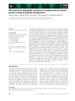
described in Methods. Representative histology and radial
alveolar counts at PN15 is shown in Figure 1. At PN15,
DEX-treated animals had larger, simpler distal air spaces
than saline controls, with a decreased RAC as compared to
control animals (*p < 0.05). These structural changes
were evident as early as PN5 (data not shown, see ref. 33).
RA-treated pups, on the other hand, had smaller, more
numerous alveoli and higher RAC (**p < 0.05) than CSO
controls as early as PN5 and up to PN15. Resolution of
corticosteroid-induced changes in architecture was seen
between PN10 and 15 in pups treated with concomitant
DEX and RA, such that, at PN15, the lungs displayed
architecture similar to that of controls and RAC were the
same as controls (# p < 0.05 vs. DEX).
Expression of Midkine and Effects of Hormonal Treatment
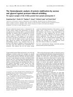
Northern blot analysis was carried out for each treatment
group at each time point studied (Figure 2). Data are
shown as percent PN1 control levels. Data from the three
control groups (no treatment, saline and CSO treatment)
were combined since the vehicle treatments had no effect
on MK mRNA expression. In control animals, MK mRNA
increased between PN5 and PN10. Dexamethasone treat-
ment had a biphasic effect, increasing MK mRNA preco-
ciously, between PN1 and PN5, and then decreasing
content at PN10 and PN15. RA alone had minimal effects
on the developmental pattern. However, with co-treat-
ment, the inhibition observed with dexamethasone was
delayed until PN15.
Respiratory Research 2009, 10:77 />Page 5 of 10
(page number not for citation purposes)
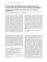
A representative Western blot for MK is shown in Figure
3a with a histogram demonstrating densitometric analysis
with normalization with β-actin for equal loading in Fig-
ure 3b; (β-actin blots not shown). In concordance with
the known temporal expression patterns of MK, protein
levels were highest in control animals at PN5, with a 10.5-
fold induction from PN1, and decreasing thereafter. Dex-
amethasone treatment delayed the increase in MK with a
3-fold reduction (p < 0.01, n = 3) compared to control
animals at PN5. Corresponding to the architecture in RA-
alone treated lungs, an increase in MK similar to control
animals was seen at PN5. This increase was sustained up
to PN10 in RA-treated animals being 1.5 fold higher than
the same day controls. Concomitant DEX+RA treatment
resulted in protein levels similar to those of controls.
These data confirmed that no relationship exists between
steady state mRNA and protein levels for MK [8].
Changes in Tie1 expression during hormonal treatment
partially correlated with the changes in MK expression and
lung morphology
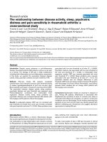
MK plays a significant role in angiogenesis. We therefore
wanted to test if Tie1, a marker of endothelial cells, would
change during hormonal treatment and correlate with the
changes in MK. Expression of Tie1 mRNA was determined
by real Time RT-PCR (n = 49 per group). As shown in Fig-
ure 4, Tie1 expression was significantly decreased in DEX-
treated animals at both PN10 and 15 compared to control
(*P = 0.0006 and 0.0022 respectively). At PN5, there was
a trend toward decreased Tie1 expression with DEX and
increased Tie1 expression when RA was added to DEX
treatment. However, this did not reach statistical signifi-
cance (p = 0.08). RA treatment alone did not change Tie 1
expression and also failed to restore DEX-induced
decrease in Tie 1 expression at PN10 and 15 (RA+DEX vs.
control: **p = 0.04 at PN10 and **p = 0.01 at PN15).
Hormonal Regulation of MK in ATII-like Cells
We next examined the expression and hormonal regula-
tions of MK in isolated human alveolar epithelial cells and
fibroblasts. We used a well-established method of alveolar
epithelial cell isolation and culture. DCI promotes the dif-
ferentiation of isolated undifferentiated epithelial cells
towards a type II epithelial cell phenotype. In the same
system, DEX or cAMP alone induces only partial differen-
tiation. We therefore examined the effect of different hor-
mone combinations on MK expression.
Western blot analysis of MK regulation in Type II-like cells
and lung fibroblasts are shown in Figure 5. The levels of
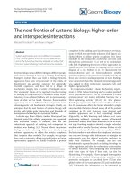
Morphologic changes in the lung at PN15 after hormonal treatmentsFigure 1
Morphologic changes in the lung at PN15 after hor-
monal treatments. (A). A simplified distal architecture was
seen in DEX-treated animals. RA-treated animals had smaller
and more numerous alveoli. Concomitant DEX and RA
administration resulted in septation similar to that of con-
trols. Vehicle (saline or CSO) treatment alone had no effects
on lung histology. Control: same handling, no injections.
DEX: Dexamethasone. RA: all-trans-retinoic acid. CSO: cot-
tonseed oil. (B). Radial Alveolar Counts confirm the
decreased septation seen with DEX treatment (*p < 0.001
DEX vs. control), the increased septation seen with RA (*p <
0.001 RA vs. control), and the resolution of DEX effects by
concomitant RA administration (**p < 0.001 DEX vs.
DEX+RA). Data are representative of at least 6 rats per
treatment group. All images 40× magnification.
A.
B.
Hormonal regulation of lung MK mRNA expressionFigure 2
Hormonal regulation of lung MK mRNA expression.
mRNA content expressed as percentage of PN1 control nor-
malized to 28 s. Data are shown as mean ± SEM. DEX treat-
ment inhibited and RA treatment had no effect on MK
mRNA expression on PN10 and 15. Concomitant RA treat-
ment was unable to restore DEX-induced decrease in MK
expression at PN15 (*p < 0.01 DEX vs. control at PN10, **p
< 0.001 DEX or DEX+RA vs. control at PN15).
Respiratory Research 2009, 10:77 />Page 6 of 10
(page number not for citation purposes)
MK protein expression with various treatments were sim-
ilar on PN3 and PN5. Therefore, combined densitometry
data are shown in figure 5B. MK expression increased 10-
fold during cell culture without hormones or serum. Cells
treated with hormones (DEX, cAMP, or DCI) had signifi-
cantly decreased MK protein levels, with an apparent
additive effect of GC and cAMP to repress the culture-
induced increase in MK and RA eliminated the repressive
effects of hormones (**p < 0.05 vs. no RA).
Fetal lung fibroblast had minimal MK expression with or
without hormone treatment (Figure 5c), whereas ATII-like
cells showed much more robust MK expression especially
in the presence of RA. These data suggest that alveolar epi-
thelial cells, and not fibroblasts, are the primary source of
MK.
Discussion
In the present study, we show that, in normal lungs, mid-
kine (MK) protein content is highest at PN5, and begins
to decline by PN10. This finding is in concordance with
Matsuura et al. who showed a transient increase in MK
expression in normal lungs between two to seven days
postnatally [8]. We extend these observations to demon-
strate that, in vivo, GC treatment is associated with lower
and RA treatment with higher lung MK protein expres-
sion. However, in our hands, changes in steady state MK
mRNA did not match MK protein expression after hormo-
nal treatments. Hormonally driven changes in protein
expression were also seen in cultured human type II-like
epithelial cells, but not fibroblasts, isolated from second
trimester human fetal lung tissue.
The regulation of the balance between the actions of GC
and RA on lung development is largely unknown. Studies
by Kaplan et al have suggested that MK might serve as a
potential bridge between these two systems [18]. MK is a
retinoic acid-responsive, heparin binding growth factor
that promotes angiogenesis, cell growth, and cell migra-
tion [19,20]. A bimodal temporal-spatial expression pat-
tern of MK is seen in the developing mouse lung. High
levels of MK expression are observed at embryonic day
(E)13-16.5 and then again at postnatal days 512, prima-
rily in respiratory epithelium early in lung development
and increasingly localized to lung stroma and pulmonary
Hormonal regulation of lung MK proteinFigure 3
Hormonal regulation of lung MK protein. A) Repre-
sentative Western blots of MK expression in neonatal rat
lungs after various treatments. B) Densitometry analysis con-
firmed that, in control animals, MK protein content was high-
est at PN5, with a 10.5-fold induction from day 1 (*p < 0.01,
n = 3), and decreased thereafter (**p = 0.02 PN5 control vs.
PN15 control, n = 3/group). In contrast, MK was significantly
decreased in DEX-treated lungs at PN5 with a 3-fold reduc-
tion compared to the same day control animals (**p < 0.01, n
= 3). An increase in MK similar to control animals was seen
at PN5, but this increase was sustained up to PN10 in RA-
treated animals being 1.5 fold higher than PN10 controls.
Concomitant DEX+RA treatment resulted in a return of
protein levels to that of control.
A.
B.
Tie 1 mRNA expression during hormonal treatmentFigure 4
Tie 1 mRNA expression during hormonal treatment.
mRNA content expressed as percentage of control normal-
ized to GAPDH. Data are shown as mean ± SEM (n = 49
group). DEX treatment significantly decreased Tie1 expres-
sion at both 10 and 15 days (*p = 0.0006 and **p = 0.0022
respectively) as compared to same days controls. RA treat-
ment alone did not change Tie 1 expression and also failed to
restore DEX-induced decrease in Tie 1 expression (* and **p
= 0.04).
Respiratory Research 2009, 10:77 />Page 7 of 10
(page number not for citation purposes)
blood vessels postnatally [21]. However, its expression is
completely absent from the adult mouse lung. These find-
ings suggest that MK may be involved in epithelial differ-
entiation, vascular growth and remodeling in the
developing lung and is not required for regular lung main-
tenance.
Although MK was initially identified as a retinoic acid-
responsive gene, mechanisms regulating its expression in
the lung have not been fully understood. Examples of
these MK regulators include thyroid transcription factor
(TTF)-1 [22], and hypoxia-inducible factor (HIF)-1 [23].
Through gene array analysis of GC receptor knockout
mice, Kaplan et al demonstrated that MK is dynamically
regulated by both GC and retinoic acid during normal
fetal lung development [10]. While these observations
provided a potential mechanism for the integration of GC
and retinoid effects in late gestation fetal lung develop-
ment, whether GC and RA also influence MK gene expres-
sion during postnatal lung development remained
unknown. In this study, we found that GC treatment
induced an early suppression of MK protein expression at
PN5, whereas RA treatment was associated with higher
and persistent MK expression to PN10 in neonatal rats.
This regulatory pattern of MK expression by GC and RA is
even clearer in the isolated human fetal lung epithelial
cells. Collectively, our data suggest that MK is likely differ-
entially regulated by GC and RA from the late saccular to
early alveolar stage of lung development.
Prolonged treatment with high doses of GC was widely
used in immature infants with evolving bronchopulmo-
nary dysplasia (BPD) during the 1990s. These treatments
were based on the belief that such treatment was associ-
ated with less early postnatal lung inflammation and a
reduction in the incidence of BPD among premature
infants [24]. However, subsequent clinical trials of DEX
treatment, beginning at 24 weeks after birth, failed to
demonstrate differences in ventilation requirements or
incidence of BPD, and showed toxic effects including
increased risk of infection, hyperglycemia and abnormal
neurodevelopmental outcome in exposed patients [25-
27]. These toxic effects of high-dose steroids have also
been documented in animal studies [28,29]. Further,
there is evidence from rodent studies that postnatal ster-
oid treatment also inhibits alveolarization and reduces
lung growth [30]. The serum concentration of GC reaches
a nadir during the period of maximum secondary septa-
tion, whether prenatal or postnatal, and increases as sep-
tation ends [4,31]. This suggests that endogenous
corticosteroids might be inhibitors of septation. Indeed,
our present study shows that treatment with DEX results
in simplified distal lung architecture with reduced second-
ary septation in neonatal rats. These results are in agree-
Hormonal regulation of MK in isolated human Type II-like cellsFigure 5
Hormonal regulation of MK in isolated human Type II-like cells. A) Representative western blot and B) Densitometry
analysis of MK expression in human fetal alveolar epithelial cells treated with different hormone combination: Alveolar epithe-
lial cells obtained from second trimester human fetal lung tissue treated with hormones (DEX, cAMP and IBMX, or DCI) to dif-
ferentiate them into alveolar type II (ATII) cells have significantly decreased MK protein content at day 3 and day 5 as
compared to controls with no treatment (*p < 0.01). However, RA treatment alone or concomitant RA treatment with hor-
mones was associated with significant increase in MK protein expression (**p < 0.05). C) Fetal lung fibroblasts isolated from
the same second trimester human fetal lung tissue were treated with DEX or DCI with/or without RA. Expression of MK was
very low irrespective of treatment groups. GAPDH expression was used as a loading control.
C.
A. B.
Respiratory Research 2009, 10:77 />Page 8 of 10
(page number not for citation purposes)
ment with the findings of Blanco et al [32] and our
previous studies [33].
The mechanism(s) by which DEX inhibits septation is not
well understood, but may be related to the inhibitory
effect of GC on DNA synthesis and cell proliferation [34].
Discontinuing corticosteroids after the "critical period" of
alveolarization is not followed by spontaneous septation.
The process of alveolar septation requires active replica-
tion of epithelial and other cells. GC therefore might pre-
vent septation via its ability to inhibit cell division [5,34].
In addition, this failed septation is accompanied by a
reduced number of pulmonary arteries and a restricted
alveolar capillary bed [35]. Our results demonstrating
decreased Tie1 expression with DEX treatment further
support these findings.
Several lines of evidence have indicated that retinoids
might be important regulators of alveolarization. Initial
evidence was provided by Brody et al. who reported that
fibroblasts rich in vitamin A storage granules form a large
fraction of the alveolar wall during septation [36,37].
These lipid-rich fibroblasts play a key role in producing
elastin at the sites of new secondary septa [38,39]. Retin-
oids signal through their receptors, RARs and RXRs.
Indeed, deletion or inhibition of RAR results in reduced
elastin and alveolar simplification [40,41]. Studies by
Massaro et al have shown that RA treatment results in
increased septation in newborn rats and also induces alve-
oli formation in adult rats with elastase-induced emphy-
sema [42,43]. In humans, low levels of vitamin A have
been found in premature babies at risk for BPD and vita-
min A supplementation reduces the incidence of BPD in
these babies [44,45]. Consistent with these studies, and
providing a potential mechanism by which retinoids
might decrease the incidence of BPD, we show that ani-
mals receiving retinoic acid (RA) treatment had smaller
and more numerous alveoli and that concomitant treat-
ment with DEX and RA prevented the DEX-induced
changes in septation.
Closely linked to the development of distal alveolar struc-
tures is the formation of a mature vascular plexus [46].
The transition from saccular to alveolar stages of lung
development correlates with microvascular development
and allows for close apposition of the vascular bed and
airspace for efficient gas exchange to occur [44]. The
molecular signals that link these two processes are not
clear. However, a complex interplay of epithelial-
endothelial cells is most likely required for normal lung
morphogenesis. Recently, the "vascular hypothesis" of
BPD [47] has proposed that inhibition of vascular growth
itself may directly impair alveolarization. Several observa-
tions support the importance of vascular formation as
vital for normal alveolar development. For example, treat-
ment of neonatal rat pups with anti-angiogenic drugs,
such as thalidomide, or VEGF receptor blocker is associ-
ated with a simplified distal lung architecture and
decreased vascularization [48]. In addition, FGF receptor
3 and 4 double knockout mice fail to develop a mature
distal lung architecture [49]. Further, decreased endothe-
lial cell migration by blocking anti-PECAM-1 antibody or
in PECAM-1 null mice is associated with disrupted alveo-
larization [50]. In humans, an abnormal alveolar capillary
network and decreased expression of endothelial cell
markers have been found in premature newborns dying
with BPD [51]. The fact that GC treatment decreased MK
expression both in vivo and in cultured type II lung epi-
thelial cells, (as demonstrated by the current study), and
also decreased Tie1 expression on PN10 and 15, suggests
that GC might inhibit alveolarization by interfering with
epithelial-endothelial communication via MK and alter-
ing normal alveolar septal vascular development. How-
ever, RA treatment had no effect on Tie1 expression and
also failed to rescue the decreased Tie1 expression caused
by DEX-treatment in our study. This suggests that the RA-
induced enhancement in septation and the rescue of GC-
induced inhibition of alveolarization may not be medi-
ated by affecting endothelial content.
Conclusion
In summary, we have demonstrated that MK is differen-
tially regulated by corticosteroid and retinoid treatment
during postnatal lung development, and that its expres-
sion matches the hormonal effects on alveolarization. MK
may, therefore, serve as a paracrine signal that originates
in the epithelium, targets pulmonary vascular cells and
influences lung vascularization during the alveolar and
microvascular maturation phase of lung development.
Competing interests
The authors declare that they have no competing interests.
Authors' contributions
HZ was responsible for part of the animal studies, per-
forming statistical analyses, performing real-rime PCR
analysis, and drafting the manuscript. SJG was responsible
for some animal studies and measuring radial alveolar
counts. ZC performed the Northern and Western blots for
MK from the animal samples. JPF, MJ and GSM assisted in
animal harvesting and injections, as well as some data
analysis. FK and NBS helped conceive the study and
design initial experiments. LWG was responsible for the
determination of MK expression in alveolar type II cells
and fibroblasts. RCS conceived the study, participated in
its design and coordination, and helped to write and
revise the manuscript. All authors read and approved the
final manuscript.
Respiratory Research 2009, 10:77 />Page 9 of 10
(page number not for citation purposes)
Acknowledgements
The experiments in this study were supported by NIH grants HL07930,
HL079090 and HL073896 to RCS. HZ was funded by the NIH Pediatric Sci-
entist Development Award (HD00850) and RCS holds the William Bucha-
nan Chair in Pediatrics at University of Texas Southwestern Medical
Center. We thank Dr. Philip L. Ballard for multiple discussions and critical
review of the manuscript.
References
1. Massaro GD, Massaro D: Formation of pulmonary alveoli and
gas-exchange surface area: quantitation and regulation. Annu
Rev Physiol 1996, 58:73-92.
2. Burri PH: The postnatal growth of the rat lung. 3. Morphol-
ogy. Anat Rec 1974, 180(1):77-98.
3. Burri PH, Dbaly J, Weibel ER: The postnatal growth of the rat
lung. I. Morphometry. Anat Rec 1974, 178(4):711-730.
4. Henning SJ: Plasma concentrations of total and free corticos-
terone during development in the rat. Am J Physiol 1978,
235(5):E451-456.
5. Massaro D, Teich N, Maxwell S, Massaro GD, Whitney P: Postnatal
development of alveoli: Regulation and evidence for a critical
period in rats. J Clin Invest 1985, 76:1297-1305.
6. Massaro GD, Massaro D: Postnatal treatment with retinoic acid
increases the number of pulmonary alveoli in rats. Am J Physiol
1996, 270:L305-L310.
7. Kadomatsu K, Huang RP, Suganuma T, Murata F, Muramatsu T: A
retinoic acid responsive gene MK found in the teratocarci-
noma system is expressed in spatially and temporally con-
trolled manner during mouse embryogenesis. J Cell Biol 1990,
110(3):607-616.
8. Matsuura O, Kadomatsu K, Takei Y, Uchimura K, Mimura S, Watan-
abe K, Muramatsu T: Midkine expression is associated with
postnatal development of the lungs. Cell Struct Funct 2002,
27(2):109-115.
9. Toriyama K, Muramatsu H, Hoshino T, Torii S, Muramatsu T: Evalu-
ation of heparin-binding growth factors in rescuing morpho-
genesis of heparitinase-treated mouse embryonic lung
explants. Differentiation 1997, 61(3):161-167.
10. Kaplan F, Comber J, Sladek R, Hudson TJ, Muglia LJ, Macrae T, Gag-
non S, Asada M, Brewer JA, Sweezey NB: The growth factor mid-
kine is modulated by both glucocorticoid and retinoid in fetal
lung development. Am J Respir Cell Mol Biol 2003, 28(1):33-41.
11. Gonzales LW, Angampalli S, Guttentag SH, Beers MF, Feinstein SI,
Matlapudi A, Ballard PL:
Maintenance of differentiated function
of the surfactant system in human fetal lung type II epithelial
cells cultured on plastic. Pediatr Pathol Mol Med 2001,
20(5):387-412.
12. Gonzales LW, Ballard PL, Ertsey R, Williams MC: Glucocorticoids
and thyroid hormones stimulate biochemical and morpho-
logical differentiation of human fetal lung in organ culture. J
Clin Endocrinol Metab 1986, 62(4):678-691.
13. Ballard PL, Ertsey R, Gonzales LK, Liley HG, Williams MC: Isolation
and characterization of differentiated alveolar type II cells
from fetal human lung. Biochim Biophys Acta 1986,
883(2):335-344.
14. Gonzales LW, Guttentag SH, Wade KC, Postle AD, Ballard PL: Dif-
ferentiation of human pulmonary type II cells in vitro by glu-
cocorticoid plus cAMP. Am J Physiol Lung Cell Mol Physiol 2002,
283(5):L940-951.
15. Savani RC, Godinez RI, Godinez MH, Wentz E, Zaman A, Cui Z,
Pooler PM, Guttentag SH, Beers MF, Gonzales LW, et al.: Respira-
tory distress after intratracheal bleomycin: selective defi-
ciency of surfactant proteins B and C. Am J Physiol Lung Cell Mol
Physiol 2001, 281:L685-L696.
16. Emery JL, Mithal A: The number of alveoli in the terminal res-
piratory unit of man during late intrauterine life and child-
hood. Arch Dis Child 1960, 35:544-547.
17. Cooney TP, Thurlbeck WM: The radial alveolar count method
of Emery and Mithal: a reappraisal 1 postnatal lung growth.
Thorax 1982, 37(8):572-579.
18. Morrisey EE, Savani RC: Midkine: a potential bridge between
glucocorticoid and retinoid effects on lung vascular develop-
ment. Am J Respir Cell Mol Biol 2003, 28(1):5-8.
19. Muramatsu H, Muramatsu T: Purification of recombinant midk-
ine and examination of its biological activities: functional
comparison of new heparin binding factors. Biochem Biophys
Res Commun 1991, 177(2):652-658.
20. Takada T, Toriyama K, Muramatsu H, Song XJ, Torii S, Muramatsu T:
Midkine, a retinoic acid-inducible heparin-binding cytokine
in inflammatory responses: chemotactic activity to neu-
trophils and association with inflammatory synovitis. J Bio-
chem 1997, 122(2):453-458.
21. Mitsiadis TA, Salmivirta M, Muramatsu T, Muramatsu H, Rauvala H,
Lehtonen E, Jalkanen M, Thesleff I: Expression of the heparin-
binding cytokines, midkine (MK) and HB-GAM (pleio-
trophin) is associated with epithelial-mesenchymal interac-
tions during fetal development and organogenesis.
Development 1995, 121(1):37-51.
22. Reynolds PR, Mucenski ML, Whitsett JA: Thyroid transcription
factor (TTF) -1 regulates the expression of midkine (MK)
during lung morphogenesis. Dev Dyn 2003, 227(2):227-237.
23. Reynolds PR, Mucenski ML, Le Cras TD, Nichols WC, Whitsett JA:
Midkine is regulated by hypoxia and causes pulmonary vas-
cular remodeling. J Biol Chem 2004, 279(35):37124-37132.
24. Yeh TF, Lin YJ, Hsieh WS, Lin HC, Lin CH, Chen JY, Kao HA, Chien
CH: Early postnatal dexamethasone therapy for the preven-
tion of chronic lung disease in preterm infants with respira-
tory distress syndrome: a multicenter clinical trial. Pediatrics
1997, 100(4):E3.
25. Papile LA, Tyson JE, Stoll BJ, Wright LL, Donovan EF, Bauer CR,
Krause-Steinrauf H, Verter J, Korones SB, Lemons JA, et al.: A mul-
ticenter trial of two dexamethasone regimens in ventilator-
dependent premature infants. N Engl J Med 1998,
338(16):1112-1118.
26. Yeh TF, Lin YJ, Huang CC, Chen YJ, Lin CH, Lin HC, Hsieh WS, Lien
YJ: Early dexamethasone therapy in preterm infants: a fol-
low-up study. Pediatrics 1998, 101(5):E7.
27. O'Shea TM, Kothadia JM, Klinepeter KL, Goldstein DJ, Jackson BG,
Weaver RG 3rd, Dillard RG: Randomized placebo-controlled
trial of a 42-day tapering course of dexamethasone to reduce
the duration of ventilator dependency in very low birth
weight infants: outcome of study participants at 1-year
adjusted age. Pediatrics 1999, 104:15-21.
28. Flagel SB, Vazquez DM, Watson SJ Jr, Neal CR Jr: Effects of taper-
ing neonatal dexamethasone on rat growth, neurodevelop-
ment, and stress response. Am J Physiol Regul Integr Comp Physiol
2002, 282(1):R55-63.
29. Edwards HE, Burnham WM: The impact of corticosteroids on
the developing animal. Pediatr Res 2001, 50(4):433-440.
30. Blanco LN, Frank L:
The formation of alveoli in rat lung during
the third and fourth postnatal weeks: effect of hyperoxia,
dexamethasone, and deferoxamine. Pediatr Res 1993,
34(3):334-340.
31. Jones CT: Corticosteroid concentrations in the plasma of fetal
and maternal guinea pigs during gestation. Endocrinology 1974,
95(4):1129-1133.
32. Blanco LN, Massaro GD, Massaro D: Alveolar dimensions and
number: developmental and hormonal regulation. Am J Physiol
1989, 257:L240-247.
33. Garber SJ, Zhang H, Foley JP, Zhao H, Butler SJ, Godinez RI, Godinez
MH, Gow AJ, Savani RC: Hormonal regulation of alveolariza-
tion: structure-function correlation. Respir Res 2006, 7:47.
34. Loeb JN: Corticosteroids and growth. N Engl J Med 1976,
295(10):547-552.
35. Le Cras TD, Kim DH, Gebb S, Markham NE, Shannon JM, Tuder RM,
Abman SH: Abnormal lung growth and the development of
pulmonary hypertension in the Fawn-Hooded rat. Am J Physiol
1999, 277(4 Pt 1):L709-718.
36. Maksvytis HJ, Vaccaro C, Brody JS: Isolation and characterization
of the lipid-containing interstitial cell from the developing
rat lung. Lab Invest 1981, 45(3):248-259.
37. Brody JS, Kaplan NB: Proliferation of alveolar interstitial cells
during postnatal lung growth. Evidence for two distinct pop-
ulations of pulmonary fibroblasts. Am Rev Respir Dis 1983,
127(6):763-770.
38. Okabe T, Yorifuji H, Yamada E, Takaku F: Isolation and character-
ization of vitamin-A-storing lung cells. Exp Cell Res 1984,
154(1):125-135.
Publish with BioMed Central and every
scientist can read your work free of charge
"BioMed Central will be the most significant development for
disseminating the results of biomedical research in our lifetime."
Sir Paul Nurse, Cancer Research UK
Your research papers will be:
available free of charge to the entire biomedical community
peer reviewed and published immediately upon acceptance
cited in PubMed and archived on PubMed Central
yours — you keep the copyright
Submit your manuscript here:
/>BioMedcentral
Respiratory Research 2009, 10:77 />Page 10 of 10
(page number not for citation purposes)
39. McGowan SE, Harvey CS, Jackson SK: Retinoids, retinoic acid
receptors, and cytoplasmic retinoid binding proteins in peri-
natal rat lung fibroblasts. Am J Physiol 1995, 269(4 Pt
1):L463-472.
40. Massaro GD, Massaro D, Chambon P: Retinoic acid receptor-
alpha regulates pulmonary alveolus formation in mice after,
but not during, perinatal period. Am J Physiol Lung Cell Mol Physiol
2003, 284(2):L431-433.
41. Yang L, Naltner A, Yan C: Overexpression of dominant negative
retinoic acid receptor alpha causes alveolar abnormality in
transgenic neonatal lungs. Endocrinology 2003,
144(7):3004-3011.
42. Massaro GD, Massaro D: Retinoic acid treatment partially res-
cues failed septation in rats and in mice. Am J Physiol Lung Cell
Mol Physiol 2000, 278(5):L955-960.
43. Massaro D, Massaro GD: Pulmonary alveolus formation: critical
period, retinoid regulation and plasticity. Novartis Found Symp
2001, 234:229-236. discussion 236241
44. Shenai JP, Chytil F, Jhaveri A, Stahlman MT: Plasma vitamin A and
retinol-binding protein in premature and term neonates. J
Pediatr 1981, 99(2):302-305.
45. Tyson JE, Wright LL, Oh W, Kennedy KA, Mele L, Ehrenkranz RA,
Stoll BJ, Lemons JA, Stevenson DK, Bauer CR, et al.: Vitamin A sup-
plementation for extremely-low-birth-weight infants.
National Institute of Child Health and Human Development
Neonatal Research Network. N Engl J Med 1999,
340(25):1962-1968.
46. Burri PH: Fetal and postnatal development of the lung. Annu
Rev Physiol 1984, 46:617-628.
47. Abman SH: Bronchopulmonary dysplasia: "a vascular hypoth-
esis". Am J Respir Crit Care Med 2001, 164(10 Pt 1):1755-1756.
48. Jakkula M, Le Cras TD, Gebb S, Hirth KP, Tuder RM, Voelkel NF,
Abman SH: Inhibition of angiogenesis decreases alveolariza-
tion in the developing rat lung. Am J Physiol Lung Cell Mol Physiol
2000, 279(3):L600-607.
49. Weinstein M, Xu X, Ohyama K, Deng CX: FGFR-3 and FGFR-4
function cooperatively to direct alveogenesis in the murine
lung. Development 1998, 125(18):3615-3623.
50. DeLisser HM, Helmke BP, Cao G, Egan PM, Taichman D, Fehrenbach
M, Zaman A, Cui Z, Mohan GS, Baldwin HS, et al.: Loss of PECAM-
1 function impairs alveolarization. J Biol Chem 2006,
281(13):8724-8731.
51. Bhatt AJ, Pryhuber GS, Huyck H, Watkins RH, Metlay LA, Maniscalco
WM: Disrupted pulmonary vasculature and decreased vascu-
lar endothelial growth factor, Flt-1, and TIE-2 in human
infants dying with bronchopulmonary dysplasia. Am J Respir
Crit Care Med 2001, 164(10 Pt 1):1971-1980.