Báo cáo y học: " Increased levels of (class switched) memory B cells in peripheral blood of current smokers" docx
Bạn đang xem bản rút gọn của tài liệu. Xem và tải ngay bản đầy đủ của tài liệu tại đây (465.29 KB, 11 trang )
BioMed Central
Page 1 of 11
(page number not for citation purposes)
Respiratory Research
Open Access
Research
Increased levels of (class switched) memory B cells in peripheral
blood of current smokers
Corry-Anke Brandsma*
1,2
, Machteld N Hylkema
2
, Marie Geerlings
1,2
,
Wouter H van Geffen
1
, Dirkje S Postma
1
, Wim Timens
2
and
Huib AM Kerstjens
1
Address:
1
Department of Pulmonary Diseases, University Medical Center Groningen, University of Groningen, P.O. Box 30.001, 9700 RB,
Groningen, The Netherlands and
2
Department of Pathology, University Medical Center Groningen, University of Groningen, P.O. Box 30.001,
9700 RB, Groningen, The Netherlands
Email: Corry-Anke Brandsma* - ; Machteld N Hylkema - ;
Marie Geerlings - ; Wouter H van Geffen - ; Dirkje S Postma - ;
Wim Timens - ; Huib AM Kerstjens -
* Corresponding author
Abstract
There is increasing evidence that a specific immune response contributes to the pathogenesis of
COPD. B-cell follicles are present in lung tissue and increased anti-elastin titers have been found in
plasma of COPD patients. Additionally, regulatory T cells (Tregs) have been implicated in its
pathogenesis as they control immunological reactions. We hypothesize that the specific immune
response in COPD is smoke induced, either by a direct effect of smoking or as a result of smoke-
induced lung tissue destruction (i.e. formation of neo-epitopes or auto antigens). Furthermore, we
propose that Tregs are involved in the suppression of this smoke-induced specific immune
response.
The presence of B cells, memory B cells and Tregs was assessed by flow cytometry in peripheral
blood of 20 COPD patients and 29 healthy individuals and related to their current smoking status.
COPD patients had lower (memory) B-cell percentages and higher Treg percentages in peripheral
blood than healthy individuals, with a significant negative correlation between these cells.
Interestingly, current smokers had higher percentages of (class-switched) memory B cells than ex-
smokers and never smokers, irrespective of COPD.
This increase in (class-switched) memory B cells in current smokers is intriguing and suggests that
smoke-induced neo-antigens may be constantly induced in the lung. The negative correlation
between B cells and Tregs in blood is in line with previously published observations that Tregs can
suppress B cells. Future studies focusing on the presence of these (class switched) memory B cells
in the lung, their antigen specificity and their interaction with Tregs are necessary to further
elucidate the specific B-cell response in COPD.
Published: 12 November 2009
Respiratory Research 2009, 10:108 doi:10.1186/1465-9921-10-108
Received: 13 May 2009
Accepted: 12 November 2009
This article is available from: />© 2009 Brandsma et al; licensee BioMed Central Ltd.
This is an Open Access article distributed under the terms of the Creative Commons Attribution License ( />),
which permits unrestricted use, distribution, and reproduction in any medium, provided the original work is properly cited.
Respiratory Research 2009, 10:108 />Page 2 of 11
(page number not for citation purposes)
Introduction
COPD is a leading cause of death worldwide and its mor-
bidity and mortality are still rising. Although the patho-
genesis of the disease is still not fully defined, tobacco
smoke is widely accepted as the most important cause for
the development of the disease certainly in the western
world. Until now, the only effective treatment to stop the
accelerated lung function decline is smoking cessation,
even though the inflammatory response may persist [1].
More information is needed about the origins and nature
of the chronic inflammatory response in COPD to find
better treatment targets for COPD patients.
The role of the innate immune response, i.e. neutrophils
and macrophages is well established in COPD, as is the
role of CD8 T cells [2,3]. Yet the role of other important
cells in specific immunity, in particular CD4 T cells and B
cells, have only recently attracted attention. We and others
have found both oligoclonal T- and B cells in the lungs of
COPD patients suggesting an antigen driven immune
response [4,5]. Furthermore, Lee et al recently demon-
strated a specific Th1 response against lung elastin in
patients with emphysema [6]. Additionally, an increased
number of small airways containing B cells and lymphoid
follicles has been shown in patients with GOLD stage III-
IV compared to stage 0-II [7], as well as an increase of B
cells in the mucosa of large airways in COPD patients
compared to controls [8]. At present it is largely unclear
against which antigen(s) this specific immune response in
the lungs of COPD patients is directed. In this respect, at
least three potential sources of antigens should be consid-
ered: 1) microbial, 2) cigarette smoke components or
derivatives, and 3) auto-antigens, encompassing (neo)
antigens derived from degradation products of extracellu-
lar matrix. The latter is supported by the recent findings
regarding an immune response against elastin [6] and the
presence of anti nuclear auto-antibodies in COPD [9].
An important modulator of the immune system is the reg-
ulatory T cell (Treg). Tregs express CD4, CD25 and fork-
head transcription factor 3 (Foxp3) and are important in
controlling immunological tolerance and preventing
auto-immune reactions by inhibiting T-cell responses
[10]. In addition, Tregs can directly inhibit B-cell
responses by suppressing class switch recombination and
Ig production [11,12]. Given this link between Tregs and
B cells, it is tempting to speculate about a diminished role
for Tregs in the suppression of the specific B-cell response
in COPD.
So far, only four studies have investigated the presence of
Tregs in COPD, but they reported different findings in
lung tissue and bronchoalveolar lavage (BAL). First,
decreased numbers of CD4
+
CD25
+
Tregs and Foxp3
mRNA levels were shown in lung tissue of emphysema
patients compared to control subjects [6]. Additionally,
increased numbers of CD4
+
CD25
bright
Tregs were shown
in BAL from COPD patients and healthy smokers com-
pared to healthy never smokers [13], while another group
showed decreased CD4
+
CD25
+
Tregs in BAL of COPD
patients and never smokers compared to healthy smokers
[14]. Finally, an immunohistochemical study demon-
strated increased numbers of Foxp3
+
cells in large airways
of asymptomatic smokers and COPD patients compared
to non-smokers, and decreased numbers of Foxp3
+
cells in
small airways of COPD patients compared to asympto-
matic smokers and non-smokers [15].
We hypothesize that the specific immune response in
COPD is smoke induced and is either a direct result of
smoking or a result of the smoke-induced lung tissue
destruction (i.e. formation of neo-epitopes or auto anti-
gens). We propose that Tregs are involved in the suppres-
sion of this smoke induced specific immune response and
that a diminished presence or function on these cells may
underlie the development of the specific humoral
immune response in COPD.
We investigated the presence of B cells, memory B cells,
and Tregs in peripheral blood obtained from smoking and
ex-smoking COPD patients and smoking, ex-smoking and
never-smoking healthy volunteers.
Methods
Subjects
COPD patients and healthy individuals were recruited to
participate in this study. Inclusion criteria for COPD
patients were; clinical diagnosis of COPD, post bron-
chodilator FEV
1
< 80% predicted, post bronchodilator
FEV
1
/FVC < 70%, and no exacerbation in the 6 weeks pre-
ceding the study. Inclusion criteria for healthy individuals
were; no signs and symptoms of pulmonary disease, FEV
1
> 90% predicted, and FEV
1
/FVC > 70%.
All participants met the following criteria: age > 40 years,
negative skin prick tests for the most common aeroaller-
gens, no use of (inhaled or systemic) corticosteroids in the
6 weeks preceding the study, and no major co morbidities.
To avoid the effect of gender only males were included in
the study. Smokers and ex-smokers had to have a smoking
history of at least 10 packyears and ex-smokers had to
have quit smoking for a least one year. The medical ethics
committee of the University Medical Center Groningen
approved the study and all participants gave their written
informed consent.
Cell isolation
All participants donated 20 ml of peripheral blood.
Peripheral blood mononuclear cells (PBMCs) were iso-
lated using ficoll-paque plus (GE Healthcare, UK) density
Respiratory Research 2009, 10:108 />Page 3 of 11
(page number not for citation purposes)
gradient centrifugation. Total isolated cells were counted
using a Sysmex pocH-100i cell counter (Sysmex, Roche,
Germany). Cells were used for flow cytometry and immu-
nocytochemical staining on cytospins.
Flow cytometry analysis
Two antibody cocktails were used to stain PBMCs for 1) B
cells and 2) Tregs.
1. CD20-PE-Cy5, CD27-FITC, and IgM-biotin followed by
Streptavidin-PE (all BD Biosciences).
2. CD4-AmCyan (BD Biosciences, San Jose, USA), CD25-
Pe-Cy7 (eBioscience, San Diego, USA) and Foxp3-Alexa
Fluor 700 (eBioscience).
Appropriate isotype controls were used for the CD25
(mouse IgG1-Pe-Cy7, eBioscience) and Foxp3 (rat IgG2a-
Alexa Fluor 700, eBioscience) staining.
Before staining the surface markers, 10
6
cells per 25 μl
were first incubated for 15 minutes on ice with cold 0.5%
human serum (Sigma-Aldrich, Zwijndrecht, the Nether-
lands) to block a-specific binding sites. Plates were centri-
fuged and cells were subsequently incubated with the
appropriate antibody cocktail for 30 minutes on ice, pro-
tected from light. After washing the cells of both cocktails
with phosphate buffered saline solution (PBS) supple-
mented with 2% bovine serum albumin (BSA, Serva, Hei-
delberg, Germany), the cells of cocktail 1 were incubated
for 15 minutes with Streptavidin-PE, washed three times
with PBS/2%BSA, resuspended in FACS lysing solution
(BD Biosciences), and kept in the dark on ice until flow
cytometry analysis. The cells of cocktail 2 were fixed and
permeabilized for 30 minutes using a fixation and perme-
abilization buffer kit (eBioscience), and then washed with
permeabilization buffer, blocked with 2% human serum
and then incubated with anti-Foxp3 for 1 hour. After-
wards the cells were washed with permeabilization buffer,
resuspended in FACS lysing solution, and kept in the dark
on ice until flow cytometric analysis. The fluorescent
staining of the cells was measured on a LSR-II flow cytom-
eter (BD Biosciences) and data were analyzed using
FlowJo Software (Tree Star, Ashland, USA).
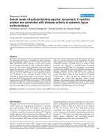
Based on the expression of CD20, CD27, and membrane
IgM, different B-cell subsets were distinguished. Within
the lymphocyte gate, total B cells were analyzed based on
CD20 expression, and total memory B cells were analyzed
based on co-expression of CD20 and CD27 (Figure 1).
Within the CD20 population, naive B cells (CD27
-
IgM
+
),
IgM
+
memory B cells (CD27
+
IgM
+
), and class-switched
memory B cells (CD27
+
IgM
-
) were distinguished.
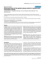
Tregs were defined as CD4
+
CD25
+
Foxp3
+
T cells. The pos-
itive gates for CD25 and Foxp3 expression were based on
the expression levels of the appropriate isotype controls,
and a separate CD25
high
gate was set on the high popula-
tion (Figure 2).
Immunocytochemistry
The presence of cells expressing the different Ig isotypes
IgE, IgG and IgA was assessed using immunocytochemical
staining of PBMC cytospins. IgE, IgG and IgA expression
was demonstrated by a rabbit-anti IgE antibody (Dako,
Heverlee, Belgium) followed by a biotin labeled goat-anti-
rabbit secondary antibody (SBA, Birmingham, USA) and
AB complex (Dako), a direct labeled anti-IgG-Fitc anti-
body (Protos Immunoresearch, Burlingame, USA), and an
anti-IgA (Dako) antibody followed by a biotin labeled
rabbit-anti-mouse secondary antibody (Dako) and AB
complex, respectively. Per cytospin, 600 cells were
counted and expressed as percentage positive cells.
Statistical analysis
A multiple linear regression model was used to determine
whether the levels of B cells, memory B cells and Tregs dif-
fered by current smoking status or by having COPD or
their combination. This method disentangles the separate
effects of COPD and current smoking and their interac-
tion. First, the effects of COPD and current smoking were
tested together with the interaction between COPD and
current smoking as independent variables. When the
interaction between COPD and current smoking was not
significant, the effects of COPD and current smoking were
tested again without the interaction term. The normal dis-
tribution of the residuals was analyzed with a Kol-
mogorov-Smirnov test and when needed the data were
log-transformed to normalize distributions. Additionally,
Mann Whitney U tests were used to establish differences
between all the subgroups according to the presence of
COPD and the current smoking status. The relation
between B cells and CD4
+
CD25
+
Foxp3
+
T cells, and (class-
switched) memory B cells and IgA expression was evalu-
ated with the Spearman correlation. A value of p < 0.05
was considered significant.
Results
Patient characteristics
The characteristics of the twenty COPD patients (current
and ex-smokers) and twenty-nine healthy volunteers (cur-
rent, ex- and never smokers), included in the study, are
shown in table 1. Healthy individuals were slightly
younger than the COPD patients, which was mainly
caused by the young age of the healthy smokers. Addition-
ally, COPD patients had more packyears of smoking
when compared to healthy current and ex-smokers. One
healthy person was included as "never smoker" who had
a smoking history of 2.5 packyears and had stopped
Respiratory Research 2009, 10:108 />Page 4 of 11
(page number not for citation purposes)
smoking for 40 years, the other never smokers had no
smoking history at all.
B cells, memory B cells, and Ig isotypes in peripheral blood
COPD versus healthy
COPD patients had lower percentages of total B cells (p =
0.006, Figure 3A) and memory B cells (p = 0.004, Figure
4) compared to healthy individuals. There was a similar
trend (p = 0.08, Figure 5A) for IgG positive cells. No dif-
ferences were found between COPD patients and healthy
controls with respect to numbers of IgA and IgE positive
cells (Figure 5).
When analyzing the groups based on their current smok-
ing status, COPD ex-smokers had lower B-cell percentages
than healthy smokers (p = 0.01), ex-smokers (p = 0.02)
and never smokers (p = 0.03) and a trend (p = 0.05) when
compared to COPD smokers (Figure 3B).
The lower percentages of B cells in COPD could not be
explained by the difference in age or packyears between
COPD patients and healthy individuals (p > 0.05, when
age or packyears was added to the multiple regression
analysis).
Flow cytometry plots of B cells and memory B cells in peripheral bloodFigure 1
Flow cytometry plots of B cells and memory B cells in peripheral blood. A representative example of the difference
in percentage of CD20
+
B cells between COPD (blue curve) and healthy (red curve) and the CD20
+
CD27
+
gate to analyze the
memory B cells is depicted in the upper panel. The CD27
+
IgM
-
gate for class switched memory B cells, the CD27
+
IgM
+
gate for
IgM
+
memory B cells, and the CD27
-
IgM
+
gate for naive B cells are shown for a current and a never smoker in the lower panel.
CD20 B cells;
COPD vs Healthy
CD20CD27 gate
CD27+IgM-
CD27+IgM+
CD27-IgM+
Current smoker
Never smoker
CD27+IgM-
CD27+IgM+
CD27-IgM+
CD20-Pe-Cy5
CD20-Pe-Cy5
CD27-FITC
CD27-FITC
CD27-FITC
IgM-Pe IgM-Pe
Respiratory Research 2009, 10:108 />Page 5 of 11
(page number not for citation purposes)
Effect of current smoking
Current smokers (COPD and healthy combined) had
higher percentages of memory B cells (p < 0.001, Figure
4A) and class-switched memory B cells (p < 0.001, Figure
4C) than ex-smokers and never smokers (combined).
There was a similar trend for total B cells (p = 0.05, Figure
3).
When analyzing the groups based on their current smok-
ing status, COPD smokers had higher percentages of
memory B cells than COPD ex-smokers (p = 0.03, Figure
4B). Also within healthy individuals, current smokers had
higher percentages of memory B cells than ex-smokers (p
= 0.03) and never smokers (p = 0.02). Similar results were
present for class switched memory B cells; healthy smok-
ers had higher percentages of class-switched memory B
cells than healthy ex-smokers (p = 0.002) and never
smokers (p = 0.003, Figure 4D).
The expression of the different Ig subtypes was analyzed
on PBMC cytospins to asses to which isotype the memory
B cells had switched. Current smokers (COPD and
healthy combined) had more IgA positive cells than ex-
and never smokers (p = 0.002, Figure 5C). This current
Flow cytometry plots of regulatory T cells in peripheral bloodFigure 2
Flow cytometry plots of regulatory T cells in peripheral blood. The CD25 expression (red curve) compared to the
isotype (blue curve), and the CD25 total and CD25high gates are depicted in the upper panel. The Foxp3 expression (red
curve) compared to the isotype (blue curve), and an example of the difference in Foxp3 expression between COPD (red
curve) and healthy (blue curve) are depicted in the lower panel.
CD25 expression
compared to
isotype control
CD25 total
CD25high
Foxp3 expression
compared to
isotype control
Foxp3 expression;
COPD vs Healthy
CD25-Pe-Cy7
Foxp3-Alexa Fluor 700
CD4-Amcyan
Foxp3-Alexa Fluor 700
CD25-Pe-Cy7
CD25
Foxp3
Respiratory Research 2009, 10:108 />Page 6 of 11
(page number not for citation purposes)
smoking effect was not present for IgE and IgG positive
cells.
When analyzing the groups based on their current smok-
ing status, COPD smokers had higher percentages of IgA
positive cells than COPD ex-smokers (p = 0.03, Figure
5D). Also within healthy individuals, current smokers had
higher percentages of IgA positive cells than ex-smokers (p
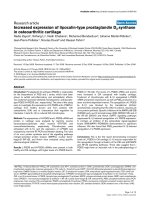
= 0.03). Furthermore, the percentages of IgA positive cells
were positively correlated with memory B cells (rho =
0.46, p = 0.001) and class switched memory B cells (rho =
0.56, p < 0.001, Figure 6).
There were no effects of COPD or current smoking on
IgM
+
memory B cells and naive B cells (data not shown).
Regulatory T cells in peripheral blood
COPD versus healthy
COPD patients had higher percentages of
CD4
+
CD25
+
Foxp3
+
T cells (p = 0.03, Figure 7A) and
CD4
+
CD25
high
Foxp3
+
T cells (p = 0.04, Figure 7C) than
healthy individuals.
When analyzing the groups based on their current smok-
ing status, COPD smokers had a higher percentage of
Table 1: Characteristics of COPD patients and healthy individuals
COPD patients Healthy individuals
Current smokers Ex-smokers Current smokers Ex-smokers Never smokers
Subjects (n) 10 10 9 10 10
Age (years) 65.9 (4.3) * 66.7 (7.4) * 52.8 (4.1)
#
61.1 (9.3) 58.1 (6.5)
Packyears 34 (13.5) * 36.7 (18.2) * 24.6 (11) 20.6 (5.9) 0.3 (0.8)
FEV
1
post BD
(% pred.)
44.9 (14.9)
$
60.7 (14.7) 105.6 (8.7) 115.7 (15.9) 111.1 (12.1)
FEV
1
/FVC post BD (%) 37.6 (10) 43.8 (10.7) 76.4 (4) 78.2 (5.9) 78.5 (4.1)
Mean (standard deviation) is depicted. Mann Whitney U tests were used to test differences between the groups. FEV
1
= Forced expiratory volume
in 1 second. FVC = Forced vital capacity. BD = Bronchodilator.
* COPD patients versus healthy individuals; packyears p = 0.006, age p = 0.000
#
Healthy current smokers versus healthy ex-smokers p = 0.01 and versus healthy never smokers p = 0.045
$
COPD smokers versus COPD ex-smokers p = 0.03
B cells in peripheral bloodFigure 3
B cells in peripheral blood. A) Percentages of total B cells in peripheral blood of COPD patients (closed symbols) and
healthy individuals (open symbols). The result of the multiple linear regression analysis (i.e. corrected for current smoking) is
depicted in the figure. B) The same results are depicted, but divided in subgroups based on the presence of COPD and the cur-
rent smoking status. In this figure the results of the Mann Whitney U tests are depicted. * indicates that p < 0.05
A
CD20
+
B cells
COPD Healthy
0
5
10
15
20
25
*
% CD20 of total lymphocytes
B
CD20
+
B cells
0
5
10
15
20
25
COPD Healthy
smoker ex-smoker smoker ex-smoker never smoker
p=0.05
*
*
*
% CD20 of total lymphocytes
Respiratory Research 2009, 10:108 />Page 7 of 11
(page number not for citation purposes)
CD4
+
CD25
high
Foxp3
+
T cells than healthy smokers (p =
0.049, Figure 7D), which was also true for the
CD4
+
CD25
+
Foxp3
+
T cells (trend (p = 0.065), Figure 7B).
No differences were found between COPD and healthy
individuals with respect to CD4 T cells, CD4
+
CD25
+
T
cells, and CD4
+
CD25
high
T cells (data not shown).
The differences in percentages of CD4
+
CD25
+
Foxp3
+
T
cells could not be explained by the difference in age or
packyears of smoking between COPD patients and
healthy individuals (p > 0.05, when age or packyears was
added to the multiple regression analysis).
Effect of current smoking
There were no effects of current smoking with respect to
CD4 T cells, CD4
+
CD25
+
T cells, CD4
+
CD25
high
T cells
and CD4
+
CD25
+
Foxp3
+
T cells in peripheral blood.
Correlation between regulatory T cells and B cells
The percentage of CD4
+
CD25
+
Foxp
+
T cells was negatively
correlated with the percentage of B cells (rho = -0.36, p =
0.01, Figure 8) and memory B cells (rho = -0.34, p = 0.02).
For COPD alone, the correlation between
CD4
+
CD25
+
Foxp
+
T cells and B cells was of the same mag-
nitude, but due to less power it did not reach statistical sig-
nificance (rho = -0.40, p = 0.08).
Discussion
In this study we had two main observations. First, patients
with COPD had lower percentages of (memory) B cells
and higher percentages of Tregs in peripheral blood com-
pared to healthy individuals. These higher Treg percent-
ages correlated significantly with both lower total B cell
and memory B cell percentages. Second, current smokers
had higher percentages of total memory B cells as well as
class-switched memory B cells in peripheral blood,
regardless of the disease state. Additional Ig subtype anal-
ysis suggested that this increased class switched memory B
cell population consists mainly of IgA expressing B cells.
In addition to our previous studies in which B cells were
studied in lung tissue of COPD patients [4,8], we now
have studied the presence of B cells and memory B cells in
peripheral blood of COPD patients and healthy individu-
Memory B cells in peripheral bloodFigure 4
Memory B cells in peripheral blood. A) Percentages of memory B cells and class switched memory B cells (D) in periph-
eral blood of current smokers (closed symbols) and non smokers (open symbols). In B), C) and E), F) the same results are
depicted, but divided into subgroups based on the current smoking status and COPD versus healthy controls. In A) and D) the
results of the multiple linear regression analysis corrected for having COPD are depicted. In C) and F) the results of the multi-
ple linear regression analysis corrected for smoking are depicted. In B) and E) the results of the Mann Whitney U tests are
depicted. * indicates that p < 0.05
Memory B cells
Current smokers Non smokers
0
2
4
6
8
*
% CD20CD27
of total lymphocytes
Memory B cells
0
2
4
6
8
*
*
*
COPD Healthy
smoker ex-sm oker sm oker ex-smoker never smoker
% CD20CD27
of total lymphocytes
Memory B cells
COPD Healthy
0
2
4
6
8
*
% CD20CD27
of total lymphocytes
Class switched memory B cells
Current smokers Non smokers
0
10
20
30
40
50
*
% CD27
+
IgM
-
of total B cells
Class switched memory B cells
0
10
20
30
40
50
*
*
COPD Healthy
smoker ex-sm oker sm oker ex-smoker never sm oker
*
% CD27
+
IgM
-
of total B cells
Class switched memory B cells
COPD Healthy
0
10
20
30
40
50
% CD27
+
IgM
-
of total B cells
AB
DE
C
F
Respiratory Research 2009, 10:108 />Page 8 of 11
(page number not for citation purposes)
als. Except for one earlier publication from our group that
showed decreased total B-cell percentages in COPD non-
smokers compared to COPD smokers [16], we could not
find any data assessing the presence of B-cells and mem-
ory B cells in peripheral blood of patients with COPD.
With respect to our first main observation, the lowest B-
cell percentages were detected in the COPD ex-smokers,
consistent with the earlier findings of de Jong et al. [16].
Although speculative, the decreased percentage of total B
cells in peripheral blood of COPD patients and the previ-
ously described increased presence of B cells in lung tissue
of COPD patients [7,8] could reflect an increased recruit-
ment of B cells from the periphery to the lung, perhaps
related to increased presence of antigens in the lungs.
Since B cells were expressed as the percentage of total lym-
phocytes, we can not exclude that the decreased percent-
age of B cells in COPD patients may be related to an
increased percentage of CD8 cells, which was already
demonstrated in COPD before [16,17].
Regarding our second main observation, current smokers
had significantly more memory B cells including class-
switched memory B cells than ex- and never smokers. This
is intriguing since class-switched memory B cells are
mature B cells that have replaced their primary encoded
membrane receptor (IgM) by IgG, IgA or IgE in response
to repeated antigen recognition [18]. This process of class-
switch recombination is mostly dependent on the pres-
ence of specific antigen-antibody complexes in germinal
centers (GC), and thus the extent of this GC mediated
level of class-switching is related to actual presence of
antigen and recognizing antibody. Therefore, the finding
of increased class-switched memory B cells in our current
smokers suggests the possibility of a chronic antigen-spe-
IgG, IgE and IgA positive cells in peripheral bloodFigure 5
IgG, IgE and IgA positive cells in peripheral blood. Percentages of A) IgG, B) IgE and D) IgA positive cells in peripheral
blood of COPD patients (closed symbols) and healthy individuals (open symbols) are depicted and divided in subgroups based
on the presence of COPD and the current smoking status. The results of the Mann Whitney U tests are depicted in these fig-
ures. In C) the same results for IgA are depicted, but divided in current smokers (closed symbols) and non-smokers (open
symbols). In this figure the result of the multiple linear regression analysis (i.e. corrected for having COPD) is depicted. * indi-
cates that p < 0.05
IgG positive cells
0
2
4
6
8
10
12
COPD Healthy
smoker ex-smoker smoker ex-smoker never smoker
% IgG positive cells
IgE positive cells
0
2
4
6
COPD Healthy
smoker ex-smoker smoker ex-smoker never smoker
% IgE positive cells
IgA positive cells
0
1
2
3
4
*
*
COPD Healthy
smoker ex-smoker smoker ex-smoker never smoker
% IgA positive cells
IgA positive cells
Current smokers Non smokers
0
1
2
3
4
*
% IgA positive cells
AB
CD
Respiratory Research 2009, 10:108 />Page 9 of 11
(page number not for citation purposes)
cific immune response that is particularly caused by ongo-
ing smoke-induced formation or release of (neo)-antigens
(e.g. matrix degradation products or smoke particles). The
primary immune response to these antigens may be weak,
but may still lead to the formation of memory B cells.
When the antigen stimulus (tobacco smoke) is present for
a prolonged period, secondary immune responses may
lead to increased numbers of memory B cells and plasma
cells, and a continued presence of memory B cells, as
shown in the current smokers in our study.
Because this increase in memory B cells is only present in
the current smokers and does not distinguish between
COPD patients and healthy controls, one might argue
whether it is important for COPD pathogenesis. We spec-
ulate that this specific immune response is to a certain
extent present in all smokers with a considerable smoking
history and is not the only factor leading to COPD patho-
genesis. Other important factors like the underlying
genetic predisposition, in combination with environmen-
tal factors may contribute to a large extent to the develop-
ment of the chronic inflammatory response and
emphysema development, which distinguishes COPD
patients from asymptomatic smokers. Genetic predisposi-
tion can have profound effects on many immunologic
processes, including the lack of immune suppression by
Tregs.
As mentioned in the introduction, the presence of
CD4
+
CD25
+
Tregs in COPD has been investigated previ-
ously [6,13-15]. These studies reported different, partially
contradictory, findings in lung tissue and bronchoalveolar
lavage, and reported no differences in CD4
+
CD25
+
Tregs
in peripheral blood between COPD patients and healthy
controls. However, in these studies, the presence of Tregs
was analyzed by measuring CD4
+
CD25
+
T cells. Foxp3
expression in these cells was assessed in separate analyses
to prove that a high percentage of these CD4
+
CD25
+
T
cells were positive for Foxp3 and thus Tregs. Instead, we
analyzed Tregs by measuring the percentage of Foxp3
expressing CD4
+
CD25
+
T cells and with this method
increased Treg percentages in peripheral blood of COPD
patients were found when compared to healthy individu-
als. In our view, the way of identifying Tregs explains the
discrepant findings between the previous studies and our
study. This is supported by the fact that with a similar
analysis compared to the previous studies we could also
not detect differences in CD4
+
CD25
+
or CD4
+
CD25
high
T
cells between COPD patients and healthy individuals.
Nevertheless, the observation that the percentage of Tregs
and the level of Foxp3mRNA is decreased in lung tissue of
COPD patients [6] together with the increased percentage
of Tregs in peripheral blood in our study could suggest a
decreased infiltration of Tregs to the lung in COPD.
Together with the increased B cell numbers in lung tissue
this might represent a local imbalance between B cells and
Tregs in the lung. Unfortunately there is no data yet ana-
lyzing the balance between Tregs and B cells in lung tissue.
The only data supporting a relation between Tregs and B
cells in the lung comes from our smoking mouse model,
in which we showed a relation between the levels of
Foxp3 positive cells and the number of B cell infiltrates in
lung tissue [19].
We assessed the presence of Tregs and B cells in peripheral
blood and it can be argued whether this gives a good
reflection of the inflammatory response in the lungs. Ani-
mal data showed that BAL lymphocytes can migrate to
regional lymph nodes and recirculate in the blood [20].
However, several studies investigating Tregs in different
compartments, i.e. BAL, lung tissue and blood, showed
discrepant findings in the different compartments com-
paring COPD and healthy controls [6,13,14]. Further-
more, it is known that the inflammatory environment,
particularly high levels of TNF-α, affects the Foxp3 expres-
sion and functionality of Tregs [21]. Thus, in order to
draw conclusions about a possible role for Tregs in COPD,
the crucial next step is to study the presence and particu-
larly the functionality of local Tregs in the lung.
With respect to the B cells, this study showed that smoking
can lead to increased levels of circulating memory B cells.
Correlation between class switched memory B cells and IgA positive cellsFigure 6
Correlation between class switched memory B cells
and IgA positive cells. Correlation between class switched
memory B cells and IgA positive cells for current smokers
(black circles) and ex-and never smokers (open circles). The
result of the Spearman correlation is depicted in the figure.
% of IgA positive cells
3.02.52.01.51.00.50.0
% of class switched memory B cells
50.0
40.0
30.0
20.0
10.0
0.0
Correlation between class switched memory B cells and IgA positive cells
current smokers
ex- and never smokers
rho=0.56, p<0.001
Respiratory Research 2009, 10:108 />Page 10 of 11
(page number not for citation purposes)
Given the fact that B cells traffic to the circulation after
antigen recognition in the lung, this smoke induced mem-
ory B-cell response in blood could very well be a reflection
of the specific B-cell response in the lung. This is sup-
ported by our observation that the increased class
switched memory B cell population consists mainly of IgA
expressing B cells, reflecting a mucosal immune response.
In conclusion, we showed that smoking may induce a spe-
cific immune response, which is reflected by increased
percentages of circulating (class switched) memory B cells.
We propose that a smoke-induced specific immune
response is involved in the chronic inflammatory
response in COPD. Future studies focusing on the pres-
ence of (class switched) memory B cells in the lung and
their antigen specificity are necessary to further elucidate
the specific B-cell response in COPD. Additionally, we
showed increased percentages of circulating Tregs in
COPD in association with decreased B cell percentages.
These findings provide support for a relation between
Tregs and B cells in COPD, which needs to be further
explored in lung tissue. Preferably, Treg functionality in
the lung should be related to parameters reflecting the
specific B cell response in the lung.
Competing interests
The authors declare that they have no competing interests.
Authors' contributions
CB recruited the patients, analyzed the data, performed
statistical analysis and drafted the manuscript. MH partic-
ipated in the study design and data analysis, and helped
Regulatory T cells in peripheral bloodFigure 7
Regulatory T cells in peripheral blood. A) Foxp3 percentages of CD4
+
CD25
+
T cells and C) CD4
+
CD25
high
T cells in
peripheral blood of COPD patients (closed symbols) and healthy individuals (open symbols). The results of the multiple linear
regression analysis (i.e. corrected for current smoking) are depicted in the figures. In B) and D) the same results are depicted,
but divided in subgroups based on the presence of COPD and the current smoking status. In these figures the results of the
Mann Whitney U tests are depicted. * indicates that p < 0.05
CD4
+
CD25
+
Foxp3 T cells
COPD Healthy
50
60
70
80
90
100
*
Foxp3 % of CD4CD25
T cells
A
CD4
+
CD25
+
Foxp3 T cells
50
60
70
80
90
100
COPD Healthy
smoker ex-smoker smoker ex-smoker never smoke
r
p=0.065
p=0.065
Foxp3 % of CD4CD25 T cells
B
CD4
+
CD25
high
Foxp3 T cells
COPD Healthy
60
70
80
90
100
110
Foxp3 % of CD4CD25
high
T cells
C
CD4
+
CD25
high
Foxp3 T cells
60
70
80
90
100
110
COPD Healthy
smoker ex-smoker smoker ex-smoker never smoker
*
p=0.07
Foxp3 % of CD4CD25
high
T cells
D
*
Publish with BioMed Central and every
scientist can read your work free of charge
"BioMed Central will be the most significant development for
disseminating the results of biomedical research in our lifetime."
Sir Paul Nurse, Cancer Research UK
Your research papers will be:
available free of charge to the entire biomedical community
peer reviewed and published immediately upon acceptance
cited in PubMed and archived on PubMed Central
yours — you keep the copyright
Submit your manuscript here:
/>BioMedcentral
Respiratory Research 2009, 10:108 />Page 11 of 11
(page number not for citation purposes)
to draft the manuscript. MG carried out the cell isolations,
the flow cytometry analyses and was involved in the
immunocytochemical analyses. WG performed the
immunocytochemical stainings and analyses. WT and DS
were involved in the study design and the patient recruit-
ment, and critically reviewed the manuscript. HK partici-
pated in the study design, was supervisor of the patient
recruitment, helped with the statistical analyses and criti-
cally reviewed the manuscript. All authors read and
approved the final manuscript.
Funding
This study was financially supported by the Graduate
School for Drug Exploration (GUIDE) of the University of
Groningen and the Dutch Asthma Foundation.
Acknowledgements
The authors would like to thank the dedicated people from the lung func-
tion department for performing all lung function and skin prick tests.
References
1. Willemse BW, ten Hacken NH, Rutgers B, Lesman-Leegte IG, Postma
DS, Timens W: Effect of 1-year smoking cessation on airway
inflammation in COPD and asymptomatic smokers. Eur
Respir J 2005, 26:835-845.
2. O'Donnell R, Breen D, Wilson S, Djukanovic R: Inflammatory cells
in the airways in COPD. Thorax 2006, 61:448-454.
3. Barnes PJ, Shapiro SD, Pauwels RA: Chronic obstructive pulmo-
nary disease: molecular and cellular mechanisms. Eur Respir J
2003, 22:672-688.
4. Strate BW van der, Postma DS, Brandsma CA, Melgert BN, Luinge
MA, Geerlings M, et al.: Cigarette smoke-induced emphysema:
a role for the B cell? Am J Respir Crit Care Med 2006, 173:751-758.
5. Sullivan AK, Simonian PL, Falta MT, Mitchell JD, Cosgrove GP, Brown
KK, et al.: Oligoclonal CD4+ T cells in the lungs of patients
with severe emphysema. Am J Respir Crit Care Med 2005,
172:590-596.
6. Lee SH, Goswami S, Grudo A, Song LZ, Bandi V, Goodnight-White S,
et al.: Antielastin autoimmunity in tobacco smoking-induced
emphysema. Nat Med 2007, 13:567-569.
7. Hogg JC, Chu F, Utokaparch S, Woods R, Elliott WM, Buzatu L, et al.:
The nature of small-airway obstruction in chronic obstruc-
tive pulmonary disease. N Engl J Med 2004, 350:2645-2653.
8. Gosman MM, Willemse BW, Jansen DF, Lapperre TS, van Schadewijk
A, Hiemstra PS, et al.: Increased number of B-cells in bronchial
biopsies in COPD. Eur Respir J 2006, 27:60-64.
9. Feghali-Bostwick CA, Gadgil AS, Otterbein LE, Pilewski JM, Stoner
MW, Csizmadia E, et al.: Autoantibodies in patients with chronic
obstructive pulmonary disease. Am J Respir Crit Care Med 2008,
177:
156-163.
10. Sakaguchi S: Naturally arising Foxp3-expressing CD25+CD4+
regulatory T cells in immunological tolerance to self and
non-self. Nat Immunol 2005, 6:345-352.
11. Lim HW, Hillsamer P, Banham AH, Kim CH: Cutting edge: direct
suppression of B cells by CD4+ CD25+ regulatory T cells. J
Immunol 2005, 175:4180-4183.
12. Lim HW, Hillsamer P, Kim CH: Regulatory T cells can migrate
to follicles upon T cell activation and suppress GC-Th cells
and GC-Th cell-driven B cell responses. J Clin Invest 2004,
114:1640-1649.
13. Smyth LJ, Starkey C, Vestbo J, Singh D: CD4-regulatory cells in
COPD patients. Chest 2007, 132:156-163.
14. Barcelo B, Pons J, Ferrer JM, Sauleda J, Fuster A, Agusti AGN: Phe-
notypic characterisation of T-lymphocytes in COPD: abnor-
mal CD4+CD25+ regulatory T-lymphocyte response to
tobacco smoking. Eur Respir J 2008, 31:555-562.
15. Isajevs S, Taivans I, Strazda G, Kopeika U, Bukovskis M, Gordjusina V,
et al.: Decreased FOXP3 expression in small airways of smok-
ers with COPD. Eur Respir J 2009, 33:61-67.
16. de Jong JW, Belt-Gritter B van der, Koeter GH, Postma DS: Periph-
eral blood lymphocyte cell subsets in subjects with chronic
obstructive pulmonary disease: association with smoking,
IgE and lung function. Respir Med 1997, 91:67-76.
17. Domagala-Kulawik J, Hoser G, Dabrowska M, Chazan R: Increased
proportion of Fas positive CD8+ cells in peripheral blood of
patients with COPD. Respir Med 2007, 101:1338-1343.
18. Edry E, Melamed D: Class switch recombination: a friend and a
foe. Clin Immunol 2007, 123:244-251.
19. Brandsma CA, Hylkema MN, Strate BW van der, Slebos DJ, Luinge
MA, Geerlings M, et al.: Heme oxygenase-1 prevents smoke
induced B-cell infiltrates: a role for regulatory T cells?
Respir
Res 2008, 9:17.
20. Lehmann C, Wilkening A, Leiber D, Markus A, Krug N, Pabst R, et al.:
Lymphocytes in the bronchoalveolar space reenter the lung
tissue by means of the alveolar epithelium, migrate to
regional lymph nodes, and subsequently rejoin the systemic
immune system. Anat Rec 2001, 264:229-236.
21. Valencia X, Stephens G, Goldbach-Mansky R, Wilson M, Shevach EM,
Lipsky PE: TNF downmodulates the function of human
CD4+CD25hi T-regulatory cells. Blood 2006, 108:253-261.
Correlation between regulatory T cells and B cellsFigure 8
Correlation between regulatory T cells and B cells.
Correlation between CD4
+
CD25
+
Foxp3
+
T cells and total B
cells for COPD patients (black circles) and healthy individuals
(open squares). The result of the Spearman correlation is
depicted in the figure.
% B cells
20.016.012.08.0
% regulatory T cells
90.0
80.0
70.0
60.0
Correlation between regulatory T cells and B cells
COPD
Healthy
rho= -0.36, p=0.01