Báo cáo y học: " Rapamycin attenuates hypoxia-induced pulmonary vascular remodeling and right ventricular hypertrophy in mice" pptx
Bạn đang xem bản rút gọn của tài liệu. Xem và tải ngay bản đầy đủ của tài liệu tại đây (443.43 KB, 12 trang )
BioMed Central
Page 1 of 12
(page number not for citation purposes)
Respiratory Research
Open Access
Research
Rapamycin attenuates hypoxia-induced pulmonary vascular
remodeling and right ventricular hypertrophy in mice
Renate Paddenberg
†1
, Philipp Stieger
†2
, Anna-Laura von Lilien
1
,
Petra Faulhammer
1
, Anna Goldenberg
1
, Harald H Tillmanns
2
,
Wolfgang Kummer
1
and Ruediger C Braun-Dullaeus*
3
Address:
1
Institute of Anatomy and Cell Biology, Giessen University, Giessen, Germany,
2
Department of Internal Medicine/Cardiology Giessen
University, Giessen, Germany and
3
Department of Internal Medicine/Cardiology, Dresden University of Technology, Dresden, Germany
Email: Renate Paddenberg - ; Philipp Stieger - ; Anna-Laura von
Lilien - ; Petra Faulhammer - ;
Anna Goldenberg - ; Harald H Tillmanns - ;
Wolfgang Kummer - ; Ruediger C Braun-Dullaeus* -
* Corresponding author †Equal contributors
Abstract
Background: Chronic hypoxia induces pulmonary arterial hypertension (PAH). Smooth muscle
cell (SMC) proliferation and hypertrophy are important contributors to the remodeling that occurs
in chronic hypoxic pulmonary vasculature. We hypothesized that rapamycin (RAPA), a potent cell
cycle inhibitor, prevents pulmonary hypertension in chronic hypoxic mice.
Methods: Mice were held either at normoxia (N; 21% O
2
) or at hypobaric hypoxia (H; 0.5 atm;
~10% O
2
). RAPA-treated animals (3 mg/kg*d, i.p.) were compared to animals injected with vehicle
alone. Proliferative activity within the pulmonary arteries was quantified by staining for Ki67
(positive nuclei/vessel) and media area was quantified by computer-aided planimetry after immune-
labeling for α-smooth muscle actin (pixel/vessel). The ratio of right ventricle to left ventricle plus
septum (RV/[LV+S]) was used to determine right ventricular hypertrophy.
Results: Proliferative activity increased by 34% at day 4 in mice held under H (median: 0.38)
compared to N (median: 0.28, p = 0.028) which was completely blocked by RAPA (median
HO+RAPA: 0.23, p = 0.003). H-induced proliferation had leveled off within 3 weeks. At this time
point media area had, however, increased by 53% from 91 (N) to 139 (H, p < 0.001) which was
prevented by RAPA (H+RAPA: 102; p < 0.001). RV/[LV+S] ratio which had risen from 0.17 (N) to
0.26 (H, p < 0.001) was attenuated in the H+RAPA group (0.22, p = 0.041). For a therapeutic
approach animals were exposed to H for 21 days followed by 21 days in H ± RAPA. Forty two days
of H resulted in a media area of 129 (N: 83) which was significantly attenuated in RAPA-treated
mice (H+RAPA: 92). RV/[LV+S] ratios supported prevention of PH (N 0.13; H 0.27; H+RAPA 0.17).
RAPA treatment of N mice did not influence any parameter examined.
Conclusion: Therapy with rapamycin may represent a new strategy for the treatment of
pulmonary hypertension.
Published: 24 February 2007
Respiratory Research 2007, 8:15 doi:10.1186/1465-9921-8-15
Received: 2 November 2006
Accepted: 24 February 2007
This article is available from: />© 2007 Paddenberg et al; licensee BioMed Central Ltd.
This is an Open Access article distributed under the terms of the Creative Commons Attribution License ( />),
which permits unrestricted use, distribution, and reproduction in any medium, provided the original work is properly cited.
Respiratory Research 2007, 8:15 />Page 2 of 12
(page number not for citation purposes)
Background
Pulmonary arterial hypertension (PAH), a disease of the
small pulmonary arteries, is characterized by vascular pro-
liferation and remodeling [1]. It results in a progressive
increase in pulmonary vascular resistance and, ultimately,
right ventricular failure and death. One trigger of PAH is
hypoxia which acutely causes a rise in pulmonary blood
pressure by vasoconstriction but chronically results in the
structural remodeling of the pulmonary vasculature [2].
Medial thickening of small pulmonary arteries has long
been recognized as one of the earliest pathologic features,
indicating proliferation of smooth muscle cells (SMC) [3].
Indeed, smooth muscle cell proliferation in small, periph-
eral, normally nonmuscular pulmonary arterioles is a
hallmark of PAH [4,5].
The current medical management of PAH is directed at
vasodilatation rather than towards inhibition of smooth
muscle cell proliferation [1]. However, recently an excit-
ing new therapeutic avenue has been taken using a plate-
let-derived growth factor (PDGF) receptor antagonist to
treat PAH in hypoxic rats [6]. This approach has even suc-
cessfully been used in a single patient with end stage pri-
mary pulmonary hypertension [7]. Anti-proliferative
therapy seems to offer a novel approach for treatment of
PAH.
Rapamycin (sirolimus) is another very potent anti-prolif-
erative drug. Through inhibition of its target, the m
amma-
lian T
arget of Rapamycin (mTOR), rapamycin blocks
mitogen-induced signaling via phosphoinositide 3-kinase
(PI3K) and protein kinase B (Akt) towards the cell cycle
machinery in SMC in vitro and in vivo [8]. In cardiovascu-
lar medicine, rapamycin is successfully used as stent-coat-
ing for prevention of in-stent restenosis [9-11]. However,
rapamycin also abrogates hypoxia-induced increase in
proliferation of cultured smooth muscle and endothelial
cells [12]. Furthermore, the requirement of PI3K, Akt, and
mTOR in hypoxia-induced pulmonary artery adventitial
fibroblast proliferation has been demonstrated recently
[13].
On this background we hypothesized that rapamycin pre-
vents and reverses hypoxia-induced vascular remodeling.
Mice were injected with rapamycin or with vehicle alone
(0.2% carboxymethylcellulose) and held either at nor-
moxia (21% O
2
) or at hypobaric hypoxia (0.5 atm; ~10%
O
2
). Frozen lung sections of mice kept for four days or
three weeks at normoxia or hypobaric hypoxia were
employed for double labeling for Ki67 (proliferating
cells) and α-smooth muscle actin to quantify the prolifer-
ative activity of the pulmonary vasculature and to deter-
mine the vessel media area by computer-aided
planimetry. In hematoxylin-eosin stained cross sections of
frozen hearts, calculation of the ratio of the areas of right
ventricular wall/[left ventricular wall + septum] and meas-
urement of the diameters of individual cardiomyocytes
served for the estimation of right ventricular hypertrophy.
Our results demonstrated that rapamycin is able to atten-
uate hypoxia-induced proliferation and thickening of the
pulmonary vasculature as well as right ventricular hyper-
trophy thereby supporting that anti-proliferative regimens
offer a novel approach for anti-remodeling therapy in
hypoxia-induced PAH.
Methods
Chemicals and antibodies
Rapamycin was a kind gift from Wyeth Pharmaceuticals
(Muenster, Germany). FITC-conjugated monoclonal anti-
α-smooth muscle actin antibody (clone 1A4) and 4',6-
diamidino-2-phenyl-inodole (DAPI) were obtained from
Sigma-Aldrich (Deisenhofen, Germany), rabbit polyclo-
nal anti-Ki67 antibody from Novocastra Laboratories Ltd.
(Dossenheim, Germany) and Cy3-conjugated donkey
anti-rabbit antibody from Dianova (Hamburg, Germany).
Animals and experimental protocol
FVB mice of both gender were obtained from Harlan Win-
kelmann (Paderborn, Germany) and used at 6–8 weeks of
age. The animals were fed standard mouse chow and were
allowed to take food and water ad libidum. All experi-
ments conformed to the NIH guidelines to the care and
use of experimental animals, and were approved by the
local authorities.
The kinetic of proliferation within the walls of intrapul-
monary vessels in response to reduced oxygen supply was
examined in mice kept for 2, 3, 4, 10, 16, or 21 days in a
hypobaric chamber. An air intake valve was adjusted to
maintain intrachamber pressure at 380 mmHg (0.5 atm)
while allowing adequate airflow through the chamber to
prevent accumulation of CO
2
and NH
3
. Control mice
were kept at normobaric pressure (760 mmHg) at room
air.
To examine the effect of rapamycin on hypoxia-induced
vascular remodeling and right ventricular hypertrophy,
age-matched mice were divided into 6 experimental
groups: 1. untreated normoxic mice, 2. vehicle-treated
normoxic mice, 3. rapamycin-treated normoxic mice, 4.
untreated hypobaric mice, 5. vehicle-treated hypobaric
mice, and 6. rapamycin-treated hypobaric mice. In some
experiments solely four groups (vehicle-/rapamycin-
treated mice at normoxia/hypoxia) were formed. For
application of rapamycin or vehicle, the chamber was
opened daily and the mice were weighed. An 1.75 mg/ml
stock solution of rapamycin was freshly prepared every
second day by homogenization of the drug in 0.2% car-
boxymethylcellulose as vehicle. Rapamycin was injected
i.p. at 3 mg/kg*d in a final volume of 100 µl. Control mice
Respiratory Research 2007, 8:15 />Page 3 of 12
(page number not for citation purposes)
received either the same volume of the vehicle or
remained untreated.
Tissue preparation
Mice were sacrificed by cervical dislocation and exsan-
guinated by cutting the vena cava inferior. The chest cavity
was opened, and the lungs were filled via the trachea with
Zamboni fixative (2% formaldehyde, 15% saturated pic-
ric acid in 0.1 mol/L phosphate buffer). Heart and lungs
were removed en block and transferred into Zamboni fix-
ative. After fixation for 6 h, the tissue was washed over-
night with 0.1 mol/L phosphate buffer and incubated for
3 days with increasing concentrations of sucrose solution
(9%, 18% and 40% sucrose in 0.1 mol/L phosphate
buffer). Finally, the specimens were embedded in optimal
cutting temperature (OCT) compound (Sakura; Zoeter-
woude, The Netherlands) and frozen in liquid nitrogen.
Immunohistochemistry
Immunohistochemical double-labeling of lung tissue for
Ki67 (proliferating cells) and α-smooth muscle actin (vas-
cular muscularization) was employed for a quantitative
analysis of the proliferative activity of the pulmonary vas-
culature. For that purpose, 10 µm thick frozen sections
were prepared and Ki67 antigen was unmasked by micro-
wave treatment (twice for 6 min at 800 W in 0.1 mol/L
sodium citrate buffer, pH 6.0). After blocking of unspe-
cific protein binding sites, the frozen sections were incu-
bated overnight simultaneously with FITC-conjugated
anti-α-smooth muscle actin antibody and anti-Ki67 anti-
body (1:500 and 1:1000, respectively, in 5% bovine
serum albumin, 5% normal goat serum in phosphate
buffered saline (PBS)) followed by Cy3-conjugated don-
key anti-rabbit antibody (1:2000 in 5% bovine serum
albumin, 5% normal goat serum in PBS, 1 h at room tem-
perature). After three washes with PBS the sections were
incubated with 1 µg/ml DAPI in PBS for 15 min followed
by three washes with PBS. Sections were evaluated with an
epifluorescence microscope (BX60; Olympus, Hamburg,
Germany) equipped with appropriate filter combinations.
The number of cells with Ki67 positive nuclei detectable
per cross section of a vessel was defined as "Ki67 positive
cells/vessel". Per condition, two lung sections were ana-
lyzed, and the mean was calculated. The obtained data
were statistically analyzed as described in "Statistical anal-
ysis".
The lung sections stained for α-smooth muscle actin were
also used to evaluate by computer-aided planimetry the
extent of muscularization of intrapulmonary vessels. For a
quantitative analysis, the ratio of the number of α-smooth
muscle actin positive pixels within a vessel wall and the
minimal vascular diameter [µm] was calculated. Per con-
dition about 100 vessels were analyzed and the median
was calculated. The obtained data were statistically ana-
lyzed as described in "Statistical analysis".
Assessment of right ventricular hypertrophy
Right ventricular hypertrophy was investigated employing
10 µm thick frozen sections. In detail, cross sections of the
heart embracing the walls of both ventricle and the sep-
tum were prepared and routinely stained with hematoxy-
lin-eosin, dehydrated, and embedded in Entellan (Merck,
Darmstadt, Germany). Heart sections were evaluated with
a BX60 microscope (Olympus, Hamburg, Germany)
employing Scion VisiCapture 1.0 software (Scion Coorpo-
ration, Frederick, Maryland, USA). The ratio of right ven-
tricular wall area to left ventricular wall area plus septum
area [RV/LV+S] was used as an index of right ventricular
hypertrophy. To analyze the size of individual cardiomy-
ocytes in cross sections of the right and left ventricle wall
the diameter of individual myocytes was measured using
an Axioplan 2 microscope (Zeiss, Jena, Germany) and
employing the AxioVision 3.0 software (Zeiss, Jena, Ger-
many).
Statistical analysis
Statistical analysis was performed by using SPSS Base 8.0
(SPSS Software, Munich, Germany). Percentiles 0, 25, 50,
75 and 100 are presented in box plots. Differences among
experimental groups were analyzed with the Kruskal-Wal-
lis and the Mann-Whitney tests, with p ≤ 0.05 being con-
sidered significant and p ≤ 0.01 highly significant.
Results
Rapamycin prevents hypoxia-induced increase of
proliferative activity within the pulmonary vasculature
To examine the effect of reduced oxygen supply on the
kinetic of the proliferative activity within the murine pul-
monary vasculature, frozen lung sections of mice housed
for 0, 2, 3, 4, 10, 16, or 21 days at hypobaric hypoxia were
stained for α-smooth muscle actin (smooth muscle cells)
and Ki67 (proliferating cells). Nuclei of individual cells
were labeled with DAPI (Fig. 1A). The quantitative analy-
sis revealed that within the first few days hypobaric
hypoxia resulted in an increased number of proliferating
cells/vessel which achieved a maximum at day 4 (Fig. 1B).
At that time the proliferative activity was 0.21 in normoxic
mice and 0.325 in mice kept at hypoxia (p = 0.001).
Thereafter, the number of proliferating cells/vessel
decreased and dropped even below that seen in the nor-
moxic control.
Based on these results we investigated the effect of
rapamycin on the proliferative activity within the pulmo-
nary vasculature on day four of hypobaric hypoxia at
which the highest proliferative activity was observed and
on day 21 at which a distinct thickening of the wall of the
pulmonary arteries has taken place (see below and [14]).
Respiratory Research 2007, 8:15 />Page 4 of 12
(page number not for citation purposes)
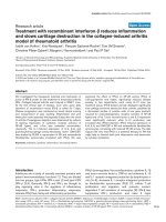
Proliferative activity in the murine pulmonary vasculature in response to hypobaric hypoxiaFigure 1
Proliferative activity in the murine pulmonary vasculature in response to hypobaric hypoxia. Frozen lung sections double immu-
nolabeled for Ki67 and α-smooth muscle actin were used for the detection of proliferating cell within the walls of intrapulmo-
nary vessels. Nuclei of individual cells were visualized by staining with DAPI. Exemplary immune histochemistries are
demonstrated in (A). The results of a quantitative analysis of the number of proliferating cells/vessel depending on time of
exposure to hypobaric hypoxia is given in (B). In the boxplots the middle horizontal line indicates the median, the top and bot-
tom of each box identifies the upper and lower quartiles of the distribution and the top and bottom horizontal line gives the
total distribution (n = number of animals. ** p ≤ 0.01).
anti α smooth muscle actin anti Ki67 DAPI
A
1866226361n =
21 d16 d10 d4 d3 d2 d0 d
Ki67-positive cells/vessel
0.6
0.5
0.4
0.3
0.2
0.1
0.0
**
**
**
days at hypobaric hypoxia
B
30 µm
Respiratory Research 2007, 8:15 />Page 5 of 12
(page number not for citation purposes)
Exposure to hypoxia for four days resulted in a significant
increase in proliferative activity by 34% in untreated ani-
mals and by 43% in vehicle-injected mice (Fig. 2A).
Administration of rapamycin completely abolished the
hypoxia-induced increase in proliferation. The anti-prolif-
erative effect of rapamycin was restricted to hypoxia-
induced proliferation: In mice housed at normoxia the
number of Ki67-positive cells/vessel was not significantly
changed by rapamycin compared to the untreated (p =
0.065) and the vehicle-injected control animals, implying
that rapamycin did not interfere with basal proliferative
activity.
Since proliferative activity had subsided after 3 weeks of
exposure to hypoxia (Fig. 1), no effect of rapamycin was
detectable after this time period (data not shown).
Rapamycin blocks the hypoxia-triggered media thickening
of intrapulmonary vessels
In lung sections of mice kept for 4 days at hypobaric
hypoxia a trend towards a thickened muscle layer com-
pared to normoxic controls was already detectable. How-
ever, this difference was not significant (data not shown).
Three weeks of hypoxia, however, induced a distinct
increase in muscularization of intrapulmonary vessels.
The extent of muscularization rose about 53% both in
untreated and vehicle-injected mice (in both cases p <
0.001) (Fig. 2B). Whereas under normoxic conditions the
degree of muscularization was unchanged by rapamycin
administration, in lungs of hypoxic mice a 26% reduction
of the muscularization was detectable. The rapamycin-
treated group did not differ significantly from animals
housed at normoxia.
Allocation of the distal arteries on one of five classes of
vessel caliber (inner diameter) ranging from 0 to 70 µm
revealed that hypoxia-induced a distinct shift towards ves-
sels with smaller calibers: The relative proportion of arter-
ies with diameters smaller than 20 µm was approximately
twice as high in mice kept for three weeks at hypobaric
hypoxia in comparison to normobaric control animals
(Fig. 3). The relative proportion of vessels with diameters
between 20 and 30 µm was comparable in the normoxic
and hypoxic groups. Accordingly, the relative proportion
of vessel calibers of 30.1 to 40 µm as well as 40.1 to 50 µm
was less in hypoxic mice. The relative proportion of ves-
sels with large diameters (50.1–70 µm) was not different
in mice housed at normoxia or hypoxia. Rapamycin treat-
ment of mice did not affect the distribution of the vessels
to the five caliber classes.
Proliferative activity and muscularization of intrapulmonary vessels of untreated mice and of animals injected with 0.2% carboxymethylcellulose as vehicle or with rapamycinFigure 2
Proliferative activity and muscularization of intrapulmonary
vessels of untreated mice and of animals injected with 0.2%
carboxymethylcellulose as vehicle or with rapamycin. Mice
were kept for four days (A) or three weeks (B) at normoxia
or at hypobaric hypoxia. In frozen lung sections stained with
anti-Ki67 and anti α-smooth muscle actin the number of pro-
liferating cells per cross section of a vessel was quantified
(A). The extent of muscularization of intrapulmonary arter-
ies was quantified by computer-aided planimetry (B). The
results are given as boxplots (N: normoxia; H: hypobaric
hypoxia; CMC: carboxymethylcellulose; Rapa: rapamycin; n =
number of animals).
888888n =
H
+Rapa
H
+CMC
HN
+Rapa
N
+CMC
N
Ki67-positive cells/vessel
0.6
0.5
0.4
0.3
0.2
0.1
0.0
**
*
A
NN
+CMC
N
+Rapa
HH
+CMC
H
+Rapa
0
50
100
150
pixel/min. diameter
n= 8 11 11 8 11 10
**
**
**
**
B
Respiratory Research 2007, 8:15 />Page 6 of 12
(page number not for citation purposes)
Hypoxia-induced right ventricular wall thickening is
attenuated by rapamycin
Hearts of mice housed for three weeks at hypobaric
hypoxia were characterized by a marked thickening of the
wall of the right ventricle (Fig. 4A). The index of right ven-
tricular hypertrophy increased about 53% and 65% in
untreated or vehicle-injected mice, respectively (in both
cases p < 0.001). In mice housed under conditions of
reduced oxygen supply rapamycin application partially
blocked the thickening of the right ventricular wall: The
median was reduced by 14% compared to the untreated
control group (p = 0.041) and no significant difference to
vehicle- or rapamycin-injected mice kept at normoxia was
detectable (p = 0.062 and p = 0.146, respectively).
Hypoxia-triggered hypertrophy of individual
cardiomyocytes is reduced by rapamycin
Untreated or vehicle-treated mice kept at hypobaric
hypoxia for 3 weeks exhibited a 20% increase in cardio-
myocyte diameter compared to the normoxic reference
groups (p < 0.001 in both cases). Whereas rapamycin had
no effect on cardiomyocyte size of mice housed at nor-
moxia, in hypoxic animals the diameter was significantly
reduced (Fig. 4B).
Cardiomyocytes of the left ventricular wall exhibited dis-
tinctly larger diameters than those of the right ventricular
wall. The size of the cells was affected neither by exposure
to hypobaric hypoxia nor by application of rapamycin.
Rapamycin reverses hypoxia-induced pulmonary vascular
remodeling
A therapeutic approach was probed: Mice were first
exposed to hypobaric hypoxia for 3 weeks followed by
another 3 weeks of hypoxia but daily rapamycin treat-
ment. Age-matched controls were held at normoxia and
treated for 3 weeks either with vehicle or with rapamycin.
Inner diameter-based classification of intrapulmonary vesselsFigure 3
Inner diameter-based classification of intrapulmonary vessels. Three weeks of hypoxia induced a distinct shift toward smaller
vessels which was not affected by CMC or rapamycin. Data are presented as means ± S.E.M (CMC: carboxymethylcellulose;
Rapa: rapamycin; n = number of animals).
0
10
20
30
40
50
60
0 µm-20 µm 20.1 µm-30 µm 30.1 µm-40 µm 40.1 µm-50 µm 50.1 µm-70 µm
rel. proportion of caliber class
normoxia (n=8) normoxia+CMC (n=11) normoxia+Rapa (n=11)
hypoxia (n=8) hypoxia+CMC (n=11) hypoxia+Rapa (n=10)
Respiratory Research 2007, 8:15 />Page 7 of 12
(page number not for citation purposes)
Rapamycin attenuates hypoxia-triggered thickening of the right ventricular wall and hypertrophy of individual cardiomyocytesFigure 4
Rapamycin attenuates hypoxia-triggered thickening of the right ventricular wall and hypertrophy of individual cardiomyocytes.
Hematoxylin-eosin stained frozen sections of cardiac ventricles were used to estimate the ratio of right ventricular wall area to
left ventricular wall area plus septum area [RV/LV+S] (A). The results of a quantitative analysis of the diameters of individual
cardiomyocytes of the right and left ventricular wall are given in (B). Data are presented as boxplots (N: normoxia; H: hypo-
baric hypoxia; CMC: carboxymethylcellulose; Rapa: rapamycin. n = number of animals; * p ≤ 0.05 and ** p ≤ 0.01).
8911101111n =
H
+Rapa
H
+CMC
HN
+Rapa
N
+CMC
N
RV/[LV+S]
0.4
0.3
0.2
0.1
0.0
**
**
*
*
A
B
77111089779999n =
H+
Rapa
H+
CMC
HN+
Rapa
N+
CMC
NH+
Rapa
H+
CMC
HN+
Rapa
N+
CMC
N
diameter [µm]
16
14
12
10
8
6
4
2
0
**
**
**
**
right ventricular wall left ventricular wall
18
Normoxia
Hypoxia+CMC
Hypoxia+Rapa
Respiratory Research 2007, 8:15 />Page 8 of 12
(page number not for citation purposes)
In hypoxic mice proliferative activity within the vascula-
ture was again determined even below the normoxic con-
trols which was not further attenuated by rapamycin
treatment (Fig. 5A). In contrast, 6 weeks of exposure to
hypoxia had resulted in a strong 55% increase of muscu-
larization of the pulmonary arteries (Fig. 5B). However,
this increase was similar to that observed in animals kept
under hypoxic conditions for only 3 weeks (see Fig. 2B)
indicating that remodeling processes had reached a home-
ostatic situation within 3 weeks. Despite the lack of appar-
ent proliferative activity, addition of rapamycin after 3
weeks was able to almost completely reverse vascular
muscularization despite ongoing hypoxia (Fig. 5B).
Accordingly, the index of right ventricular hypertrophy,
which had increased twofold (208%) during hypoxia, was
determined only 131% of normoxic controls when
hypoxic animals were treated with rapamycin. Similarly,
the increase in cardiomyocyte diameter had significantly
declined (Fig. 6A and 6B).
In comparison to normoxia, hypoxia had again induced a
shift of the relative proportion of arteries with diameters
smaller than 20 µm. This shift was not affected by rapamy-
cin treatment of the mice (data not shown).
Discussion
The current medical management of PAH is directed at
vasodilatation rather than towards inhibition of smooth
muscle cell proliferation, although progression of pulmo-
nary hypertension is known to be associated with
increased proliferation [1]. However, the data of this
experimental study imply that targeting vascular remode-
ling processes may represent a promising therapeutic
approach towards hypoxia-induced PAH, too.
This exciting avenue has very recently been gone by Scher-
muly et al. demonstrating a reversal of pulmonary remod-
eling processes in hypoxia-induced PAH by the platelet-
derived growth factor (PDGF) receptor antagonist imat-
inib mesylate [6]. In a case report of a patient in a desper-
ate situation of progressing pulmonary hypertension,
Seeger's group further substantiates this new concept [7].
PDGF represents a potent mitogen for pulmonary smooth
muscle cells [15] acting via PI3K/Akt/mTOR, a central sig-
naling pathway for cell cycle entry and progression. This
pathway is activated by other growth factors involved in
PAH as well [16] suggesting that it may represent a "final
common pathway" towards proliferation. We had, there-
fore, successfully aimed to inhibit this pathway through
usage of rapamycin which potently inhibits mTOR [8] to
not only prevent but also reverse vascular remodeling
processes and right ventricular signs of pulmonary hyper-
tension in mice held under hypoxic conditions.
Hypoxia is the main stimulus for the induction of pulmo-
nary hypertension accompanying chronic ventilatory dis-
orders such as chronic obstructive pulmonary disease and
interstitial lung disease. While acute hypoxia causes selec-
tive pulmonary arteriolar vasoconstriction, chronic expo-
sure to hypoxia results in morphological and functional
changes in the pulmonary vascular bed [17-20]. Indeed,
mTOR signaling seems to play a key role in hypoxia-trig-
gered smooth muscle and endothelial cell proliferation in
vitro [12]. The requirement of PI3K, Akt, and mTOR for
hypoxia-induced proliferation has also been demon-
strated for pulmonary artery adventitial fibroblasts [13].
Although it is generally accepted that proliferation is an
important contributor to hypoxia-induced vascular
remodeling, only few data regarding the kinetics of the
proliferative activity are available. Quinlan et al. [14]
reported that the number of 5-bromo-2'-deoxyuridine-
positive cells/vessel is about 50% higher in mice exposed
to hypoxia for 4 or 6 days. After three weeks no differences
in the proliferative index in the pulmonary vasculature of
animals housed at normoxia or hypoxia were detectable.
Our data confirm the finding of an only transient increase
of proliferative activity within the pulmonary vasculature
during hypoxia reaching a maximum within the first
week. In our study this increase was sensitive to rapamy-
cin treatment suggesting that inhibition of the early
hypoxia-triggered cell cycle activity results in reduced
chronic vascular remodeling. This way the drug may pre-
vent further hypoxia-triggered proliferation and disease
progression.
However, prevention of early proliferation does not
explain rapamycin's effectiveness when given therapeuti-
cally after 3 weeks of hypoxia when proliferative activity
within the pulmonary vasculature was determined even
below that of normoxic mice. Rapamycin may inhibit the
undetectable turnover the smooth muscle cells within the
vessel wall are subjected to and, by this means, revert vas-
cular muscularization when hypoxia had already resulted
in pulmonary arterial remodeling. However, mTOR holds
a critical role for activation of protein synthesis as well
and, this way, seems to be involved in smooth muscle
hypertrophy [21,22]. Our data, indeed, indicate that
rapamycin acts as a selective inhibitor of hypoxia-induced
thickening of the muscle layer: Histologically, pulmonary
vascular remodeling is characterized by de novo muscu-
larization of small precapillary vessels and by smooth
muscle cell hyperplasia and hypertrophy resulting in
media thickening [14,23]. With our assays we were able to
quantify both processes: A classification based on the ves-
sel caliber acted as an indicator for de novo musculariza-
tion of small arteries and the calculation of the ratio of
"number of α-smooth muscle actin positive pixels within
a vessel wall/minimal vascular diameter" was a criterion
Respiratory Research 2007, 8:15 />Page 9 of 12
(page number not for citation purposes)
Therapeutic effect of rapamycin after induction of pulmonary vascular remodelingFigure 5
Therapeutic effect of rapamycin after induction of pulmonary vascular remodeling. Mice were exposed for three week to nor-
moxia or hypoxia before treatment with rapamycin for three weeks. Rapamycin had no effect on proliferative activity but on
muscularization of intrapulmonary vessels. Quantitative analysis of the number of proliferating cells/vessel (A) and of the
extent of muscularization of intrapulmonary arteries as estimated by computer-aided planimetry (B) (N: normoxia; H: hypo-
baric hypoxia; CMC: carboxymethylcellulose; Rapa: rapamycin; n = number of animals; * p ≤ 0.05 and ** p ≤ 0.01)
6666n =
H
+Rapa
H
+CMC
N
+Rapa
N
+CMC
Ki67-positive cells/vessel
0.3
0.2
0.1
0.0
**
**
6666n =
H
+Rapa
H
+CMC
N
+Rapa
N
+CMC
pixel/min. diameter
160
140
120
100
80
60
40
20
0
A
B
**
*
**
Respiratory Research 2007, 8:15 />Page 10 of 12
(page number not for citation purposes)
Rapamycin reverses hypoxia-induced thickening of the right ventricular wall and hypertrophy of individual cardiomyocytesFigure 6
Rapamycin reverses hypoxia-induced thickening of the right ventricular wall and hypertrophy of individual cardiomyocytes.
Before treatment with rapamycin mice were housed for three weeks at normoxia or hypoxia. In (A) the results of the estima-
tion of the ratio of right ventricular wall/(left ventricular wall+septum) and in (B) a quantitative analysis of the diameters of
individual cardiomyoctes are given.
5566n =
H
+Rapa
H
+CMC
N
+Rapa
N
+CMC
RV/[LV+S]
0.4
0.3
0.2
0.1
0.0
**
6666n =
H
+Rapa
H
+CMC
N
+Rapa
N
+CMC
diameter [µm]
13
12
11
10
9
8
7
**
**
A
B
Respiratory Research 2007, 8:15 />Page 11 of 12
(page number not for citation purposes)
for the media thickening. Application of rapamycin pre-
vented the hypoxia-induced increase in media thickening
without affecting the degree of muscularization of lung
vessels obtained from mice kept at normoxia. The rela-
tively high proportion of vessels with calibers smaller
than 20 µm diameter observed in mice kept at hypoxia
was also detectable after rapamycin treatment. These
results indicate that rapamycin had no effect on de novo
muscularization of small arteries but acts as a selective
inhibitor of hypoxia-induced thickening of the muscle
layer. Presumably the de novo muscularization of precap-
illary vessels is caused by transdifferentiation of non-mus-
cle cells and/or migration of smooth muscle cells from
proximal to distal segments of the vascular system
[14,23]. Although rapamycin's effect on either processes
have been demonstrated in growth factor-stimulated cells
[24,25], its effect on hypoxia-induced migration and/or
transdifferentiation has not been investigated.
We propose that pulmonary vascular remodeling occurs
in two steps: An initial phase of increased proliferative
activity is followed by a second phase characterized by
enhanced hypertrophy of vascular smooth muscle precur-
sor cells leading to thickening of the media. Through its
anti-proliferative and anti-hypertrophic effects rapamycin
seems able to affect each phase.
An orally active derivative of rapamycin has previously
been shown to attenuate monocrotaline-induced pulmo-
nary hypertension in pneumonectomized rats [26]. In this
study, the drug was only effective when started simultane-
ously with the administration of monocrotaline. Late
therapy did not affect pulmonary hypertension. The dif-
ference to our study may be due to the different models
used. Monocrotaline has been proposed to cause perivas-
cular inflammation and platelet activation, subsequently
resulting in a proliferative response of the vascular media
which is augmented by increased shear stress in the pul-
monary bed after pneumoctomy [27]. The difference may
also lie in a transient inflammatory response after single
monocrotaline treatment of rats. In this study, mice were
exposed to hypoxia during the entire experimental period.
Yet, this is the first study to show the effectiveness of
rapamycin in preventing pulmonary hypertension
induced by a pathophysiologic stimulus.
In our study, the extent of hypoxia-induced myocardial
hypertrophy (RV/[LV+S]) was in good agreement with
data published by other groups [28]. Additionally, the
diameter of individual cardiomyocytes was determined.
Hypoxia induced a 20% increase in diameters of cardio-
myocytes of the right ventricular wall while cardiomyo-
cytes of the left ventricular wall were unaffected by
changes in the oxygen supply. Hypoxia-induced hypertro-
phy of the right ventricle and of individual right ventricu-
lar cardiomyocytes was attenuated by treatment with
rapamycin. In principle, hypertrophy of the right ventricu-
lar wall in response to hypoxia is caused by the ability to
respond to increased mechanical stretch caused by pul-
monary vasoconstriction and vascular remodeling. How-
ever, the capability of individual cardiomyocytes to sense
changes in the oxygen supply and respond with hypertro-
phy has been suggested, too [29]. In any case, recent data
suggest that, in response to certain hypertrophic stimuli,
signaling via mTOR is required for activation of protein
synthesis and cardiac hypertrophy [30]. Either mecha-
nisms may have resulted in attenuation of right ventricu-
lar hypertrophy and cardiomyocyte diameter. A direct
effect of the drug on cardiomyocyte size raises the possi-
bility of detrimental effects on right ventricular function.
However, when load-induced left ventricular hypertrophy
was induced in mice, rapamycin treatment resulted in a
decrease in chamber size and a normal systolic function of
the left ventricle [31]. However, disruption of coordinated
cardiac hypertrophy has recently also been shown to con-
tribute to the transition to heart failure as well [32].
Conclusion
In vascular medicine, local application of rapamycin
through drug-coated stent placement is currently the
favored invasive strategy to prevent restenosis [33]. Sys-
temic treatment with rapamycin is, furthermore, success-
fully used to prevent graft rejection and transplant
vasculopathy in de novo heart transplant recipients [34].
Rapamycin may represent a novel therapeutic strategy for
pulmonary arterial hypertension in humans as well to
delay further progression or even result in regression of
pulmonary vascular remodeling.
Competing interests
The author(s) declare that they have no competing inter-
ests.
Authors' contributions
RP contributed to the study design, performed histochem-
ical analyses and drafted the manuscript.
PS performed the animal studies, morphometrical analy-
ses, histochemical analyses and contributed to the evalua-
tion of the data.
AvL performed histochemical and morphometrical analy-
ses on the right heart architecture
PF and AG performed morphometrical analyses
HT contributed to the study design
WK contributed to the study design and revised the man-
uscript
Respiratory Research 2007, 8:15 />Page 12 of 12
(page number not for citation purposes)
RBD contributed to the study design, evaluated the data,
and revised the manuscript
Acknowledgements
The financial support of the Deutsche Forschungsgemeinschaft (Sonderfor-
schungsbereich (SFB) 547, project C1 and Sonderforschungsbereich (SFB)
655, project A11) is gratefully acknowledged.
References
1. Humbert M, Sitbon O, Simonneau G: Treatment of pulmonary
arterial hypertension. N Engl J Med 2004, 351:1425-1436.
2. Rubin LJ: Primary pulmonary hypertension. N Engl J Med 1997,
336:111-117.
3. Wagenvoort CA: Vasoconstriction and medial hypertrophy in
pulmonary hypertension. Circulation 1960, 22:535-546.
4. Kay JM, Kahana LM, Rihal C: Diffuse smooth muscle prolifera-
tion of the lungs with severe pulmonary hypertension. Hum
Pathol 1996, 27:969-974.
5. Wohrley JD, Frid MG, Moiseeva EP, Orton EC, Belknap JK, Stenmark
KR: Hypoxia selectively induces proliferation in a specific
subpopulation of smooth muscle cells in the bovine neonatal
pulmonary arterial media. J Clin Invest 1995, 96:273-281.
6. Schermuly RT, Dony E, Ghofrani HA, Pullamsetti S, Savai R, Roth M,
Sydykov A, Lai YJ, Weissmann N, Seeger W, Grimminger F: Reversal
of experimental pulmonary hypertension by PDGF inhibi-
tion. J Clin Invest 2005, 115:2811-2821.
7. Ghofrani HA, Seeger W, Grimminger F: Imatinib for the treat-
ment of pulmonary arterial hypertension. N Engl J Med 2005,
353:1412-1413.
8. Braun-Dullaeus RC, Mann MJ, Seay U, Zhang L, von Der Leyen HE,
Morris RE, Dzau VJ: Cell cycle protein expression in vascular
smooth muscle cells in vitro and in vivo is regulated through
phosphatidylinositol 3-kinase and mammalian target of
rapamycin. Arterioscler Thromb Vasc Biol 2001, 21:1152-1158.
9. Moses JW, Leon MB, Popma JJ, Fitzgerald PJ, Holmes DR, O'Shaugh-
nessy C, Caputo RP, Kereiakes DJ, Williams DO, Teirstein PS, Jaeger
JL, Kuntz RE: Sirolimus-eluting stents versus standard stents in
patients with stenosis in a native coronary artery. N Engl J Med
2003, 349:1315-1323.
10. Windecker S, Remondino A, Eberli FR, Juni P, Raber L, Wenaweser
P, Togni M, Billinger M, Tuller D, Seiler C, Roffi M, Corti R, Sutsch G,
Maier W, Luscher T, Hess OM, Egger M, Meier B: Sirolimus-eluting
and paclitaxel-eluting stents for coronary revascularization.
N Engl J Med 2005, 353:653-662.
11. Morice MC, Serruys PW, Sousa JE, Fajadet J, Ban Hayashi E, Perin M,
Colombo A, Schuler G, Barragan P, Guagliumi G, Molnar F, Falotico
R: A randomized comparison of a sirolimus-eluting stent
with a standard stent for coronary revascularization. N Engl J
Med 2002, 346:1773-1780.
12. Humar R, Kiefer FN, Berns H, Resink TJ, Battegay EJ: Hypoxia
enhances vascular cell proliferation and angiogenesis in vitro
via rapamycin (mTOR)-dependent signaling. Faseb J 2002,
16:771-780.
13. Gerasimovskaya EV, Tucker DA, Stenmark KR: Activation of phos-
phatidylinositol 3-kinase, Akt, and mammalian target of
rapamycin is necessary for hypoxia-induced pulmonary
artery adventitial fibroblast proliferation. J Appl Physiol 2005,
98:722-731.
14. Quinlan TR, Li D, Laubach VE, Shesely EG, Zhou N, Johns RA:
eNOS-deficient mice show reduced pulmonary vascular pro-
liferation and remodeling to chronic hypoxia. Am J Physiol Lung
Cell Mol Physiol 2000, 279:L641-L650.
15. Goncharova EA, Ammit AJ, Irani C, Carroll RG, Eszterhas AJ, Panet-
tieri RA, Krymskaya VP: PI3K is required for proliferation and
migration of human pulmonary vascular smooth muscle
cells. Am J Physiol Lung Cell Mol Physiol 2002, 283:L354-L363.
16. Barst RJ: PDGF signaling in pulmonary arterial hypertension.
J Clin Invest 2005, 115:2691-2694.
17. Ghofrani HA, Reichenberger F, Kohstall MG, Mrosek EH, Seeger T,
Olschewski H, Seeger W, Grimminger F: Sildenafil increased
exercise capacity during hypoxia at low altitudes and at
Mount Everest base camp: a randomized, double-blind, pla-
cebo-controlled crossover trial. Ann Intern Med 2004,
141:169-177.
18. Zhao L, Mason NA, Morrell NW, Kojonazarov B, Sadykov A, Maripov
A, Mirrakhimov MM, Aldashev A, Wilkins MR: Sildenafil inhibits
hypoxia-induced pulmonary hypertension. Circulation 2001,
104:424-428.
19. Frisdal E, Gest V, Vieillard-Baron A, Levame M, Lepetit H, Eddahibi S,
Lafuma C, Harf A, Adnot S, Dortho MP: Gelatinase expression in
pulmonary arteries during experimental pulmonary hyper-
tension. Eur Respir J 2001, 18:838-845.
20. Weissmann N, Nollen M, Gerigk B, Ardeschir Ghofrani H, Schermuly
RT, Gunther A, Quanz K, Fink L, Hanze J, Rose F, Seeger W, Grim-
minger F: Downregulation of hypoxic vasoconstriction by
chronic hypoxia in rabbits: effects of nitric oxide. Am J Physiol
Heart Circ Physiol 2003, 284:H931-H938.
21. Zhou L, Goldsmith AM, Bentley JK, Jia Y, Rodriguez ML, Abe MK, Fin-
gar DC, Hershenson MB: 4E-binding protein phosphorylation
and eukaryotic initiation factor-4E release are required for
airway smooth muscle hypertrophy. Am J Respir Cell Mol Biol
2005, 33:195-202.
22. Yamakawa T, Tanaka S, Kamei J, Kadonosono K, Okuda K: Phos-
phatidylinositol 3-kinase in angiotensin II-induced hypertro-
phy of vascular smooth muscle cells. Eur J Pharmacol 2003,
478:39-46.
23. Stenmark KR, Gerasimovskaya E, Nemenoff RA, Das M: Hypoxic
activation of adventitial fibroblasts: role in vascular remode-
ling. Chest 2002, 122:326S-334S.
24. Poon M, Marx SO, Gallo R, Badimon JJ, Taubman MB, Marks AR:
Rapamycin inhibits vascular smooth muscle cell migration. J
Clin Invest 1996, 98:2277-2283.
25. Fukuda D, Sata M, Tanaka K, Nagai R: Potent inhibitory effect of
sirolimus on circulating vascular progenitor cells. Circulation
2005, 111:926-931.
26. Nishimura T, Faul JL, Berry GJ, Veve I, Pearl RG, Kao PN: 40-O-(2-
hydroxyethyl)-rapamycin attenuates pulmonary arterial
hypertension and neointimal formation in rats. Am J Respir Crit
Care Med 2001, 163:498-502.
27. Wilson DW, Segall HJ, Pan LC, Lame MW, Estep JE, Morin D: Mech-
anisms and pathology of monocrotaline pulmonary toxicity.
Crit Rev Toxicol 1992, 22:307-325.
28. Minamino T, Christou H, Hsieh CM, Liu Y, Dhawan V, Abraham NG,
Perrella MA, Mitsialis SA, Kourembanas S: Targeted expression of
heme oxygenase-1 prevents the pulmonary inflammatory
and vascular responses to hypoxia. Proc Natl Acad Sci U S A 2001,
98:8798-8803.
29. Ito H, Adachi S, Tamamori M, Fujisaki H, Tanaka M, Lin M, Akimoto
H, Marumo F, Hiroe M: Mild hypoxia induces hypertrophy of
cultured neonatal rat cardiomyocytes: a possible endog-
enous endothelin-1-mediated mechanism. J Mol Cell Cardiol
1996, 28:1271-1277.
30. Proud CG: Ras, PI3-kinase and mTOR signaling in cardiac
hypertrophy. Cardiovasc Res 2004, 63:403-413.
31. Shioi T, McMullen JR, Tarnavski O, Converso K, Sherwood MC, Man-
ning WJ, Izumo S: Rapamycin attenuates load-induced cardiac
hypertrophy in mice. Circulation 2003, 107:1664-1670.
32. Siekierka JJ, Hung SH, Poe M, Lin CS, Sigal NH: A cytosolic binding
protein for the immunosuppressant FK506 has peptidyl-pro-
lyl isomerase activity but is distinct from cyclophilin. Nature
1989, 341:755-757.
33. Ong AT, Serruys PW: Technology Insight: an overview of
research in drug-eluting stents. Nat Clin Pract Cardiovasc Med
2005, 2:647-658.
34. Keogh A, Richardson M, Ruygrok P, Spratt P, Galbraith A, O'Driscoll
G, Macdonald P, Esmore D, Muller D, Faddy S: Sirolimus in de
novo heart transplant recipients reduces acute rejection and
prevents coronary artery disease at 2 years: a randomized
clinical trial. Circulation 2004, 110:2694-2700.