Báo cáo khoa học: "Evaluation of Clinical and Laboratory Variables as Prognostic Indicators in Hospitalised Gastrointestinal Colic Horse" pdf
Bạn đang xem bản rút gọn của tài liệu. Xem và tải ngay bản đầy đủ của tài liệu tại đây (161.18 KB, 10 trang )
Ihler CF, Larsen J and Skjerve E: Evaluation of clinical and laboratory variables
as prognostic indicators in hospitalised gastrointestinal colic horses. Acta vet.
scand. 2004, 45, 109-118. – The present prospective study included 106 horses referred
to the Department of Large Animal Sciences, The Norwegian School of Veterinary Sci-
ence, as non-responders to the initial colic treatment in general practise. In 14 of these
cases a required surgical treatment was not performed due to economical or other rea-
sons and were excluded from the study. Clinical and laboratory data were obtained at the
arrival in the hospital. The outcome for all analyses was survival/ non-survival. A mul-
tivariable logistic regression was performed. The analyses were used in medically (46
horses) and surgically treated cases (46 horses) separately. The same analyses were also
run for all 92 horses in a simulated "field" situation, where only clinical variables and
D-dimer values were included. The fraction of survivors was 78% in the medical and
48% in the surgical cases. In total 63% of the horses survived. In the final multivariable
logistic regression model packed cell volume (PCV) was the only important predictor
for medically treated cases, and heart rate and presence of hyperaemic or cyanotic mu-
cous membranes were the predictors in the surgically treated cases as well as in the sim-
ulated "field" situation. In conclusion, traditional variables as heart rate, mucous mem-
branes and PCV were the important predictors for the outcome in hospitalised colic
cases.
horse; colic; prognosis; clinical and laboratory variables; hospital; logistic regres-
sion; epidemiology.
Acta vet. scand. 2004, 45, 109-118.
Acta vet. scand. vol. 45 no. 1-2, 2004
Evaluation of Clinical and Laboratory Variables as
Prognostic Indicators in Hospitalised Gastrointesti-
nal Colic Horses
By Carl F. Ihler
1
, Jostein Larsen Venger
1
and Eystein Skjerve
2
1
Department of Large Animal Clinical Sciences, and
2
Department of Pharmacology, Microbiology and Food
Hygiene; The Norwegian School of Veterinary Science, Oslo, Norway.
Introduction
Equine colic caused by gastrointestinal disor-
ders is often encountered in general equine
practise and may lead to severe clinical condi-
tions and death. The incidence of colic is esti-
mated in several studies. From USA Kaneene et
al. (1997) and Tinker et al. (1997) reported an
incidence of 3.5 and 10.5 colic cases per 100
horse-years, respectively. From Norway Larsen
& Flåøyen (1997) estimated the incidence to be
4.8 colic cases per 100 horse-years. The case
fatality rate is by Kaneene et al. (1997) and Tin-
ker et al. (1997) reported to be 13 and 7%, re-
spectively.
Acute cases often need immediate surgical
treatment. Such treatment is expensive and the
mortality is relatively high, reported to be be-
tween 31 and 44% in different studies (Morris
1991, Sandholm et al. 1995, Kaneene et al.
1997).
In general, a correct diagnosis is necessary to
predict a reliable prognosis. In colic cases, how-
ever, a correct clinical diagnosis of the site and
type of the intestinal lesion is often difficult
(Blikslager & Roberts 1995). The identification
of significant clinical and laboratory variables
for the prognosis might therefore be useful
when taking the decision whether to let the pa-
tient undergo further treatment or not.
From an animal welfare point of view a reliable
prognosis is also important when veterinarians
in general practice consider transportation of
severe clinical colic cases long distances for
further treatment in hospitals.
From a multivariable model Parry et al. (1983)
concluded that variables assessing cardiovascu-
lar function were good prognostic guides. From
other studies clinical and laboratory variables
such as heart rate, packed cell volume (PCV),
colour of mucous membranes, capillary refill
time (CRT), acid- base variables and plasma
lactate are valuable prognostic predictors (Pas-
coe et al. 1990, Reeves et al. 1990, Sandholm et
al. 1995, Furr et al. 1995, Thoefner et al. 2000).
The fibrin degradation product, D-dimer, is
valuable in assessment of the cardiovascular
status. Increased levels indicate that excessive
amount of fibrin is formed within the vascular
system as a result of disseminated intra-vascu-
lar coagulation. D-dimer is of special interest
because the values can be measured as a horse
side test. Sandholm et al. (1995) concluded that
D-dimer values in plasma were valuable as a
predictor for outcome in equine gastrointestinal
colic cases.
The purpose of the present study was to evalu-
ate clinical and laboratory variables as prognos-
tic indicators in medically and surgically
treated colic cases in a hospital situation.
Materials and methods
Cases
The present study was a prospective study con-
sisting of 106 horses with colic symptoms
caused by gastrointestinal disorders referred to
The Norwegian School of Veterinary Science,
by veterinarians in general practise for further
examination and treatment from March out De-
cember 1997 (50 horses) and 1999 (56 horses).
In the 2 periods of the study all referred colic
cases were included. The horses were all non-
responders to the initial treatment in general
practise such as non-steroid anti-inflammatory
drugs (NSAIDs), fluid therapy and laxatives.
In 14 horses, for which the clinical examination
indicated that surgical treatment was required,
such treatment was not performed due to eco-
nomical or other reasons. These horses were eu-
thanised and excluded from the study.
Of the final 92 horses 30 horses were warm-
blooded riding horses, 15 Norwegian cold-
blooded trotters, 15 Standard-bred trotters, 16
Thoroughbreds and 16 horses of different pony
and pleasure horse breeds. The age ranged from
6 months to 25 years (median = 8.0 years).
Horses discharged from the hospital in good
general condition without any signs of colic
were classified as survivors. For survivors the
hospitalisation period ranged from 3 to 16 days.
Non-survivors were all euthanised when con-
sidered to be at the terminal stage, where no re-
sponse to treatment of the life threatening clin-
ical situation, was detected. This was done to
prevent unnecessary suffering.
Clinical examination
All horses were clinically examined immedi-
ately after arrival in the hospital according to a
standard protocol. Rectal temperature (°C), de-
gree of abdominal pain (1-3), heart and respira-
tory rate (per min), colour of mucous mem-
branes (normal, abnormal), CRT (sec), ab-
dominal auscultation (no sounds, decreased,
normal and increased activity), rectal examina-
tion findings and any reflux of gastro-intestinal
content through a naso-pharyngeal tube (0-1)
were recorded. The colour of the mucous mem-
branes was assessed using the gingival mem-
brane and classified as normal or abnormal (hy-
110 Carl F. Ihler et al.
Acta vet. scand. vol. 45 no. 1-2, 2004
peraemic or cyanotic as pallor and jaundice
were not recorded). Abdominal paracentesis
was not systematically performed, and the pro-
tein content and cytology of the abdominal fluid
were therefore not used in the statistical analy-
ses.
Decisions whether the patients should be
treated surgically or medically were primarily
based on the clinical variables. The criteria for
surgical treatment were: (i) Recurrent pain de-
spite heavy analgesic treatment and/or (ii) Rec-
tal findings of distended small intestines and/or
(iii) Rectal findings corresponding to displace-
ment of the large intestines. Non-responders to
the medical treatment were continuously evalu-
ated for surgical intervention.
The different clinical variables used in this
study are presented in Table 1.
Laboratory analyses
Packed cell volume (PCV) was analysed by a
capillary micro centrifuge using venous blood
from the jugular vein. Haematological variables
were analysed on Technicon H 1 Analyser
(Miles Inc., Tarrytown, NY, USA).
For blood gas analysis approximately 1 ml of
blood was collected from the transverse facial
artery into a Pico 70 heparinised arterial blood
sampler (Radiometer Medical A/S, Copenha-
gen, Denmark) for immediate acid-base and
blood gas evaluation on a Radiometer ABL 625
acid-base laboratory (Radiometer Medical A/S,
Copenhagen, Denmark). Values were adjusted
to body temperature.
Total serum protein was measured with a re-
fractometer (Atago SPR-NE, Atago Co LTD,
Japan). Serum electrophoresis to measure albu-
min, α-, ß- and γ-globulin fractions was per-
formed using Paragon Electrophoresis System
(Beckman Instruments Inc, Brea CA, USA).
Fibrinogen was measured indirectly by calcu-
lating the difference of total protein in EDTA-
plasma and serum. Serum sodium was mea-
sured with an ion-selective electrode,
1
serum
chloride was measured with a colorimetric
method,
2
and plasma lactate was measured en-
zymatically.
3
Prognostic indicators in colic horses 111
Acta vet. scand. vol. 45 no. 1-2, 2004
–––––––
1
AVL 982 Electrolyte analyzer, AVL List GmbH,
Graz, Austria.
2
Technicon RA-1000, Miles Inc., Tarrytown, NY.
3
Lactate fully enzymatic, Boehringer Mannheim,
Germany.
Table 1. Clinical and laboratory variables in the
study.
Clinical variables
Heart rate /min
Respiratory rate /min
Rectal temperature °C
Mucous membranes (normal/abnormal)
Capillary refill time (CRT) sec
Pain (no pain/moderate/severe)
Abdominal activity
(no sounds/decreased/normal/increased)
Distended small intestine by rectal examination
(yes/no)
Pain by rectal examination (yes/no)
Reflux through naso-pharyngeal tube (yes/no)
Laboratory variables
Packed cell volume (PCV) %
Haemoglobin g/l
Total protein g/l
α-, ß- and γ-globulin g/l
Albumin g/l
Fibrinogen g/l
Anion Gap (Na
+
+ K
+
-(Cl
-
+ HCO
3
)) mmol/l
Na
+
mmol/l
K
+
mmol/l
Cl
-
mmol/l
Total Ca mmol/l
Mg
++
mmol/l
Lactate mmol/l
HCO
3
-
mmol/l
Arterial pO
2
kP
a
Arterial pCO
2
kP
a
Standard Base Excess (SBE) mmol/l
PH
D-dimer mg/l
Plasma D-dimer values were assessed by the
Nycomed NycoCard D-Dimer test (Axis-
Shield PoC AS, Oslo, Norway) on citrated ve-
nous plasma. The test is based on an immuno-
filtration of citrated plasma through a mem-
brane attached with monoclonal antibody fol-
lowed by colloidal gold-labelled monoclonal
antibody staining (Gogstad et al. 1993). The in-
tensity of colour was measured semi quantita-
tively using a NycoCard Reader (Axis-Shield
PoC AS, Oslo, Norway). In addition, the D-
dimer test was used in 20 healthy horses as con-
trols. Two cut-off values, 0.5 and 1.5 mg/l were
tested in the statistical analysis.
The different laboratory variables used in this
study are presented in Table 1.
All clinical variables and blood samples were
obtained immediately after arrival at the hospi-
tal according to a standard protocol. Further,
clinical and laboratory variables obtained dur-
ing the stay in hospital were used as the basis of
further treatment of the patient and not used in
the statistical analysis.
Statistical methods
Initially, survivors and non-survivors were
compared with respect to year of inclusion
(1997 or 1999), breed, age, gender and clinical
and laboratory variables using Wilcoxon/
Kruskal Wallis Rank Sum Test for continuous
variables and Fischer Exact Test for nominal or
ordinal variables was performed using JMP for
Windows version 4.02 (SAS Institute Inc,
USA). Rectal findings were not used as a vari-
able in the analysis because the variable was es-
sential in the classification of surgically and
medically treated cases.
To handle collinearity, a correlation analysis of
the various explanatory variables was per-
formed for surgically and medically treated
cases separately. When variables showed strong
correlation (Spearman þ >0.5) the variable with
the lowest p-value in the univariable analyses
was selected. These selected variables and the
other variables showing p <0.15 in the univari-
able analysis were used in a multivariable logis-
tic model (Hosmer & Lemeshow 2000) to asso-
ciate survival/non-survival with clinical an
laboratory variables for surgically and medi-
cally treated horses. The same procedure was
also used for all cases to simulate a "field situa-
tion " where clinical variables and D-dimer, as
a horse-side test, were the only available vari-
ables.
112 Carl F. Ihler et al.
Acta vet. scand. vol. 45 no. 1-2, 2004
Table 2. Survivors and non-survivors of different diagnosis and treatment in 92 colic cases.
Survivors Non-survivors % Survivors
(n) (n)
Diagnosis
1
Non-specific colic 10 1 91
Large colon impaction 25 4 86
Displacement large colon 16 9 64
Strangulation small intestine 2 10 17
Enterocolitis 5 3 63
Peritonitis 0 7 0
Total 58 34 63
Treatment
Medically treated cases 36 10 78
Surgically treated cases 22 24 48
1
For survivors: Clinical diagnosis. For non-survivors: Diagnosis based on necropsy.
n = number of observations.
The final multivariable model was built using
logistic regression in a forward selection proce-
dure as described by Hosmer & Lemeshow
(2000) using Intercooled Stata for Windows 7.0
(Stata Corporation, College Station, TX, USA).
Model fit was assessed using the overall Pear-
son fit, the Hosmer-Lemeshow test and the Re-
ceiver Operating Characteristic (ROC) curve.
Possible outliers were identified using the Ƨ
and ∆ deviance test (Hosmer & Lemeshow
2000). The sensitivity and specificity for vari-
ous probability cut-offs were graphically as-
sessed.
Linearity for continuous variables was also as-
sessed comparing the continuous variable
grouped into quartiles and with a graphical ap-
Prognostic indicators in colic horses 113
Acta vet. scand. vol. 45 no. 1-2, 2004
Table 3. Descriptive analyses of clinical and laboratory variables in medically and surgically treated colic cases
demonstrating p-values < 0.15 in either Wilcoxon/Kruskall Wallis Rank Sum test for continuous variables or
Fisher Exact test for nominal variables.
Survivors Non-survivors
Variables n Median(range) or n Median (range) or
no/ (%) no (%)
Medically treated colic cases:
Heart rate /min 36 48 (32-96) 10 72 (32-120)
Respiratory rate /min 35 20 (8-50) 10 28 (15-34)
CRT sec 36 2 (2-4) 9 3 (2-6)
Rectal temperature °C 36 37.9 (36.6-38.8) 10 38.4 (37.9-39.6)
Abnormal mucous membrane 36 16 (44%) 10 7 (70%)
PCV % 36 34 (22-50) 10 48 (33-69)
HGB g/l
1
34 122 (82-181) 10 180 (123-248)
Fibronogen g/l 33 3 (2-7) 9 3 (1-5)
Alfa- globulin g/l 36 9 (6.9-14.3) 9 7.7 (2.9-12.4)
Gamma- globulin g/l 36 7.8 (4.2-14.7) 9 5.8 (3.5-13.7)
Lactate mmol/l 36 1.2 (0.5-11.1) 9 3.7 (1.1-13.1)
Total Ca mmol/l 34 3.0 (1.5-3.4) 9 2.7 (2.1-2.9)
Surgically treated colic cases:
Heart rate /min 22 44 (36-80) 24 79 (36-148)
Breath rate /min
2
22 20 (12-40) 24 30 (15-72)
Temperature difference from 38°C 20 0.3 (0.0-1.0) 24 0.6 (0.0-2.5)
Abnormal mucous membrane 22 5 (23 %) 24 19 (79%)
No abdominal sounds
2
22 2 ( 9%) 24 14 (58%)
CRT sec
2
22 3 (2-3) 24 4 (2-7)
PCV %
2
22 38 (29-43) 24 42 (28-56)
HGB g/l
2
22 138 (106-163) 24 153 (112-199)
Beta-globulin g/l 20 10.5 (7.7-20.0) 23 12.2 (7.4-19.8)
Gamma- globulin g/l 20 8.7 (3.2 -12.1) 23 7.6 (2.2-12.6)
Cl mmol/l 22 99 (89-108) 24 95 (87-108)
K mmol/l 22 3.5 (2.3-4.5) 24 3.8 (2.6-5.2))
Lactate mmol/l
2
20 1.6 (0.8-7.1) 24 3.6 (1.1-13.0)
D-dimer > 1.5 mg/l 22 2 (9%) 24 12 (50%)
D-dimer > 0.5 mg/l 22 7 (33%) 24 17 (71%)
1
Excluded from further analysis due to high correlation to PCV.
2
Excluded from further analysis due to high correlation to heart rate.
n = number of observations.
proach using the kernel smoothing graph pro-
cedure in Intercooled Stata for Windows 7.0
(Stata Corporation, College Station, TX, USA).
Interactions were not evaluated due to a limited
number of horses.
Results
Fifty-eight horses (63%) of the total 92 colic
cases survived. Forty-six horses where treated
medically (survival 78%) and 46 were treated
surgically (survival 48%). The numbers of sur-
114 Carl F. Ihler et al.
Acta vet. scand. vol. 45 no. 1-2, 2004
Table 4. Final multiple logistic regression models for surgically and medically treated colic cases and "simu-
lated field situation" based upon variables presented in Table 3. Results are given as estimates (95 % confidence
limits) and Odds Ratio (95 % confidence limits).
Treatment group Variable Estimate Odds Ratio
Surgically treated Heart rate 0.11 (0.04-0.18) 3.00 (1.49-6.05)*
cases Abnormal mucous membranes 2.90 (0.73-5.07) 18.23 (2.08-159)
Medically treated PCV 0.20 (0.07-0.32) 1.22 (1.07-1.37)**
cases
Simulated field Heart rate 0.07 (0.04-0.11) 2.05 (1.62-2.88)*
situation Abnormal mucous membranes 1.18 (0.03-2.34) 3.25 (1.03-10.3)
* Per increase of 10 units
**Per increase of 1 unit
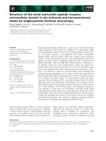
Figure 1. Estimated probability of non-survival for medically and surgically treated colic cases based upon the
final logistic models in Table 4. For surgically treated horses the left curve represents horses with abnormal mu-
cous membranes and the right curve represents horses with normal mucous membranes.
vivors and non-survivors according to diagno-
sis and treatment are presented in Table 2.
D-dimer values of the 20 controls of healthy
horses all showed D-dimer values < 0.5 mmol/l.
Univariable analysis of age, breed, gender and
year of inclusion (1997 or 1999) did not show
any statistical difference between survivors and
non- survivors for either medically or surgically
treated cases. Neither breed nor gender re-
vealed any difference between surgical and
medical cases. The median age was however
lower (p= 0.035) in the surgically treated horses
(6.5 and 8.0 years, respectively).
The results of the univariable analysis of clini-
cal and laboratory variables showing p-values
<0.15 between survivors and non-survivors are
given in Table 3.
The univariable analysis of other blood param-
eters as Mg, Na, total protein, albumin, arterial
pH, arterial pO
2
and pCO
2
, HCO
3
and Standard
Base Excess (SBE) all revealed p-values >0.15
for both medically and surgically treated cases.
The univariable analysis of the clinical dichoto-
mous variable intestinal reflux also showed a p-
value >0.15 for both treatment groups. The de-
gree of abdominal pain at the time of arrival to
the hospital did not differ statistically between
the survivors and non-survivors in the 2 treat-
ment groups.
As a result of a high correlation in the medically
treated cases the variable haemoglobin (þ=0.99
to PCV) were excluded in the following logistic
regression procedure. In the surgically treated
cases the variables abdominal sounds, breath
rate, CRT, PCV, haemoglobin and lactate were
excluded due to high correlation to heart rate
(heart rate/abdominal sounds þ=0,52, heart
rate/ respiratory rate þ=0.53, heart rate/CRT
þ=0.61, heart rate/PCV þ=0.67, heart rate/
HGB þ=0.63 and heart rate/lactate þ=0,59).
The final multiple logistic model included PCV
as the only significant predictor in medically
Prognostic indicators in colic horses 115
Acta vet. scand. vol. 45 no. 1-2, 2004
Figure 2. Sensitivity/specificity curves and Receiver Operating Characteristic (ROC) curves for the final lo-
gistic model for medically treated cases and surgically treated cases.
treated cases and heart rate and abnormal mu-
cous membranes as significant predictors in
surgically treated cases (Table 4). In the simu-
lated "field" situation the final model also in-
cluded heart rate and abnormal mucous mem-
branes (Table 4). The prediction curves for the
2 treatment groups are presented in Fig. 1,
while the ROC curves and sensitivity and speci-
ficity curves are illustrated in Fig. 2.The ROC
curve described an area under curve of 87% for
medical cases and 94% for surgical cases.
No influential outliers were found in the regres-
sion diagnostics. Grouping the continuous vari-
ables into quartiles gave no better model fit than
analysing the variables as continuous ones.
Supplementary tests also indicated (kernel
smoothing) linearity of the continuous vari-
ables.
Discussion
Material and methods
As all horses in the present study are referred
cases as non-responders to initial treatment(s)
in the general practice, they represent a selected
material compared to colic cases in general.
That means that neither colic cases with their
origin in simple spastic intestines and mild ob-
structions nor severe colic cases, in which the
pain was not possible to reduce to an acceptable
level before transportation to the hospital, are
represented in the study.
The exclusion of cases that required surgery but
were euthanised of economical or other reasons
was necessary to ensure that all horses were eu-
thanised at the terminal stage of the disease.
However, it may be difficult to classify a patient
to be at the terminal stage. To avoid unneces-
sary suffering when all possible further treat-
ment was performed was very important when
classifying non-survival.
The study was performed over 2 periods of time
(1997 and 1999). Year of inclusion might have
influenced the results. The same protocol for
clinical and laboratory examination throughout
the study was used to avoid such bias. In the sta-
tistical analysis no association between year of
inclusion and survival was detected.
Results
The survival percentage of 63 for all colic cases
in the present study is on the same level as re-
ported from other studies on hospitalised colic
cases (Ebert 1994, Sandholm et al. 1995). The
probabilities of survival in surgically and med-
ically colic cases (48 and 78%, respectively)
also correspond to previous studies (Sandholm
et al. 1995, Siebke 1995).
In the present study there was no association
between age and gender and outcome while
some studies have reported that older horses
have a higher risk of non-survival (Orsini et al.
1988, Reeves et al. 1989). In line with the pre-
sent study, Reeves et al. (1990) and Thoefner et
al. (2000) reported no association between age
and the outcome in colic cases.
Thoefner et al. (2000) reported an increased
risk of death in cold-blooded ponies compared
to a heterogeneous group of warm-blooded
horses. In the present study no association be-
tween breed and survival was found. In agree-
ment with Thoefner et al. (2000) and Reeves et
al. (1990) no association between gender and
outcome was found.
The degree of pain (pain score 1-3) at the initial
examination in the hospital did not show any
association with the outcome of the colic cases.
This is in disagreement with Thoefner et al.
(2000), Puotonen-Reinert (1986) and Reeves et
al. (1990). In the present study most horses
were given analgesics and/or NSAID before
and for some horses during the transportation.
When given NSAIDs and analgesics at differ-
ent times before the clinical evaluation the ini-
tial pain at arrival might not give the correct
picture of the real clinical situation and further
might explain why pain did not influence the
116 Carl F. Ihler et al.
Acta vet. scand. vol. 45 no. 1-2, 2004
outcome. Recurrent pain despite analgesics or
NSAIDs was, however, one of the criteria for
surgery, and since surgically treated cases in the
present study had a higher mortality, pain might
indirectly relate to the outcome.
Thoefner et al. (2000) found that a temperature
deviation from 38°C was a significant variable
in the multivariable logistic regression model
expressing the outcome of a colic case. In the
present study we did not find such a relationship
when temperature deviation from 38°C was in-
cluded in the model.
Heart rate and the presence of abnormal mu-
cous membranes were the only statistically sig-
nificant variables in the multivariate model for
surgical cases. Even if the initial laboratory
variables did not directly relate to the outcome,
they were of importance in the decision making
of supportive therapy such as fluid therapy and
correction of acid-base disturbances. In this
way they might indirectly influence the out-
come.
The classification of the mucous membranes
was based on presence of hyperaemia and
cyanosis. Since pallor or jaundice was not
recorded in any horse in this study the authors
chose a dichotomous classification (normal/ab-
normal) for mucous membranes.
In medical cases PCV was the only explanatory
variable in the final logistic model. Also PCV
represents a variable expressing the cardiovas-
cular status in the patient.
The final models gave an excellent fit by an area
under the ROC curve of 0.94 in surgically and
0.88 in medically treated cases, respectively.
The ROC curve plots the probability of deflect-
ing a true signal (sensitivity) and a false signal
(specificity) for the entire range of possible out-
points. While the ROC curve assess the overall
performance of the model, the prediction
curves in Fig. 1 give the magnitude of the prob-
ability of death given various levels of the pre-
dictor variables.
In the simulated "field" situation, where only
clinical variables and D-dimer were used, the
only reliable predictors for survival were heart
rate and the presence of abnormal mucous
membranes. The D-dimer values did not give
any additional information in explaining the
outcome. This is in agreement with Thoefner et
al. (2000). Sandholm et al. (1995), however,
found that D-dimer was included in the final lo-
gistic model together with the variables heart
rate and chloride. The reason for this might be
that Sandholm et al. (1995) did not use other
clinical variables than heart rate and respiratory
rate in their statistical procedure. D-dimer, as
the condition of the mucous membranes, is an
indicator of cardiovascular status. During our
logistic regressions D-dimer was excluded
from the final model. This supports that the
evaluation cardiovascular variables as the mu-
cous membrane in addition to heart rate tell
more about the outcome than the D-dimer
value.
In conclusion, traditional clinical variables as
heart rate and presence of abnormal mucous
membranes in surgical and PCV in medical
colic cases were the significant predictors for
the outcome. The other variables were, how-
ever, important in establishing supportive treat-
ment of the patients.
References
Blikslager AT, Roberts MC: Accuracy of clinicians in
predicting site and type of lesion as well as out-
come in horses with colic. J. Am. Vet. Med. As-
soc., 1995, 207, 1444-1447.
Ebert R: Lätalitätsaspekte der Kolikk des Pferdes.
(Mortobily in colic hourses). Pferdheilkunde,
1994, 10, 97-101.
Furr MO, Lessard P, White NA: Development of a
colic severity score for predicting the outcome of
equine colic. Vet. Surg., 1995, 24, 97-101.
Gogstad GO, Dale S, Brosstad F, Brandnes Ø,
Holtlund J, Gartner E, Borch SM: Assay of D-
dimer based on immunofiltration and staining
with gold colloids. Clin. Chem., 1993, 39, 2070-
2076.
Prognostic indicators in colic horses 117
Acta vet. scand. vol. 45 no. 1-2, 2004
Hosmer DW, Lemeshow S: In: Applied Logistic Re-
gression 2nd Edition, 2000p. 375 (Wiley, New
York).
Kaneene JB, Miller R, Ross WA, Gallagher K, Marte-
niuk J, Rook J: Risk factors for colic in the Michi-
gan (USA) equine population. Prev Vet Med
1997, 30, 23-36.
Larsen J, Flåøyen: Kolikk hos hest: En studie av 77
tilfeller i felt. (Colic in horses. A study of 77 field
cases). Nor.Vet.Tidsskr., 1997, 109, 655-661.
Morris DD: Endotoxemia in horses. A review of cel-
lular and humoral mediators involved in its
pathogenisis. J. Vet. Int. Med., 1991, 5, 167-181.
Orsini JA, Elser AH, Galligan DT, Donnawick WJ,
Kronfeld DS: Prognostic index for acute abdomi-
nal crises (colic) in horses. Am. J. Vet. Res.,
1988, 11, 1969-1971.
Parry BW, Anderson GA, Gay CC: Prognosis in
equine colic: A study of individual variables used
in case assessment. Equine Vet. J., 1983, 15, 337-
344.
Pascoe PJ, Ducharme NG, Ducharme GR, Lumsden
JH: A computer-derived protocol using recursive
partitioning to aid in estimating prognosis of
horses with abdominal pain in referral hospitals.
Can. J. Vet. Res., 1990, 54, 373-378.
Puotonen-Reinert A: Study of variables commonly
used in examination of equine colic cases to as-
sess prognostic value. Equine Vet. J., 1986, 18,
275-277.
Reeves MJ, Curtis CR, Salman MD, Hilbert BJ: Prog-
nosis in equine colic patients using multivariable
analysis. Can J. Vet. Res., 1989, 53, 87-94.
Reeves MJ, Curtis CR, Salman MD, Reif JS, Stashak
TS: A multivariable prognostic model for equine
colic patients. Prev. Vet. Med., 1990, 9, 241-257.
Sandholm M, Vidovic A, Poutunen-Reinert A,
Sankari S, Nyholm K, Rita H: D-Dimer improves
the prognostic value of combined clinical and
laboratory data in equine gastrointestinal colic.
Acta Vet. Scand., 1995, 36, 255-272.
Siebke AU, Keller H, Lauk HD, von Plocki KA: Statis-
tische Erhebung uber Kurz- und Langzeiter-
ergegnisse von 718 operativ behandelten Kolik-
patienten. Pferdheilkunde, 1995, 11, 299-312.
Thoefner MB, Ersbøll AK, Hesselholt M: Prognostic
indicators in a Danish hospital-based population
of colic horses. Equine Vet. J. Suppl., 2000, 32,
11-18.
Tinker MK, White NA, Lessard P, Thatcher CD,
Pelzer KD, Davis B, Carmel DK: Prospective
study of equine colic incidence and mortality.
Equine Vet. J., 1997, 29, 448-453.
Sammendrag
Kliniske parametere og laboratorieparametere som
prognostiske indikatorer hos hospitaliserte kolikk-
hester.
Kliniske parametere og laboratorieparametere fra
106 kolikkhester innsendt til Norges veterinær-
høgskole ble statistisk undersøkt som mulige prog-
nostiske indikatorer. Ingen hester hadde respondert
på den initiale behandlingen i felt. Fjorten kasus,
hvor kirurgisk behandling var påkrevd, men ikke ut-
ført på grunn av økonomiske eller andre årsaker, ble
utelukket i studien. Alle parametere ble registrert rett
etter ankomst og statistisk behandlet med hensyn på
overlevelse ved hjelp av en univariabel analyse med
etterfølgende korrelasjonsanalyse og multivariabel
logistisk regresjon. Overlevelsesprosenten var 78%
for de medisinsk be-
handlede kolikker og 48% for de kirurgisk be-
handlede. I den multivariable logistiske modellen var
hematokrit den eneste viktige variabel for overlevelse
for de medisinsk behandlede kolikkene, mens puls-
frekvens og abnormale slimhinner var de beste pre-
diktorene for de kirurgisk behandlede. Studien kon-
kluderer med at tradisjonelle kliniske parametere
som pulsfrekvens, slimhinneforandringer og hema-
tokrit var de eneste viktige prognostiske indikatorer
ved kolikk hos hospitaliserte hester.
118 Carl F. Ihler et al.
Acta vet. scand. vol. 45 no. 1-2, 2004
(Received March 22, 2002; accepted February 10, 2004).
Reprints may be obtained from: Carl Fredrik Ihler, Department of Large Animal Clinical Sciences, The
Norwegian School of Veterinary Science P.O. Box 8146 dep., N-003 Oslo, Norway. E-Mail:
, tel: +47 22 96 49 20, fax: +47 22 96 47 61.