Báo cáo y học: "Inverse association of plasma IL-13 and inflammatory chemokines with lung function impairment in stable COPD: a cross-sectional cohort study" doc
Bạn đang xem bản rút gọn của tài liệu. Xem và tải ngay bản đầy đủ của tài liệu tại đây (359.45 KB, 10 trang )
BioMed Central
Page 1 of 10
(page number not for citation purposes)
Respiratory Research
Open Access
Research
Inverse association of plasma IL-13 and inflammatory chemokines
with lung function impairment in stable COPD: a cross-sectional
cohort study
Janet S Lee*
1
, Matthew R Rosengart
2
, Venkateswarlu Kondragunta
3
,
Yingze Zhang
1
, Jessica McMurray
1
, Robert A Branch
2
, Augustine MK Choi
1
and Frank C Sciurba
1
Address:
1
Division of Pulmonary, Allergy, and Critical Care Medicine, Department of Medicine, University of Pittsburgh, Pittsburgh, PA, 15213,
USA,
2
Division of Trauma/General Surgery, Department of Surgery, University of Pittsburgh, Pittsburgh, PA, 15213, USA and
3
Division of Clinical
Pharmacology, Department of Medicine, University of Pittsburgh, Pittsburgh, PA, 15213, USA
Email: Janet S Lee* - ; Matthew R Rosengart - ; Venkateswarlu Kondragunta - ;
Yingze Zhang - ; Jessica McMurray - ; Robert A Branch - ;
Augustine MK Choi - ; Frank C Sciurba -
* Corresponding author
Abstract
Background: Chronic obstructive pulmonary disease (COPD) is a heterogeneous syndrome characterized by
varying degrees of airflow limitation and diffusion impairment. There is increasing evidence to suggest that COPD
is also characterized by systemic inflammation. The primary goal of this study was to identify soluble proteins in
plasma that associate with the severity of airflow limitation in a COPD cohort with stable disease. A secondary
goal was to assess whether unique markers associate with diffusion impairment, based on diffusion capacity of
carbon monoxide (DLCO), independent of the forced expiratory volume in 1 second (FEV
1
).
Methods: A cross sectional study of 73 COPD subjects was performed in order to examine the association of
25 different plasma proteins with the severity of lung function impairment, as defined by the baseline
measurements of the % predicted FEV
1
and the % predicted DLCO. Plasma protein concentrations were assayed
using multiplexed immunobead-based cytokine profiling. Associations between lung function and protein
concentrations were adjusted for age, gender, pack years smoking history, current smoking, inhaled
corticosteroid use, systemic corticosteroid use and statin use.
Results: Plasma concentrations of CCL2/monocyte chemoattractant protein-1 (CCL2/MCP-1), CCL4/
macrophage inflammatory protein-1β (CCL4/MIP -1β), CCL11/eotaxin, and interleukin-13 (IL-13) were inversely
associated with the % FEV
1
. Plasma concentrations of soluble Fas were associated with the % DLCO, whereas
CXCL9/monokine induced by interferon-γ (CXCL9/Mig), granulocyte- colony stimulating factor (G-CSF) and IL-
13 showed inverse relationships with the % DLCO.
Conclusion: Systemic inflammation in a COPD cohort is characterized by cytokines implicated in inflammatory
cell recruitment and airway remodeling. Plasma concentrations of IL-13 and chemoattractants for monocytes, T
lymphocytes, and eosinophils show associations with increasing severity of disease. Soluble Fas, G-CSF and
CXCL9/Mig may be unique markers that associate with disease characterized by disproportionate abnormalities
in DLCO independent of the FEV
1
.
Published: 14 September 2007
Respiratory Research 2007, 8:64 doi:10.1186/1465-9921-8-64
Received: 11 May 2007
Accepted: 14 September 2007
This article is available from: />© 2007 Lee et al; licensee BioMed Central Ltd.
This is an Open Access article distributed under the terms of the Creative Commons Attribution License ( />),
which permits unrestricted use, distribution, and reproduction in any medium, provided the original work is properly cited.
Respiratory Research 2007, 8:64 />Page 2 of 10
(page number not for citation purposes)
Background
Chronic obstructive pulmonary disease (COPD), while
defined by the presence of incompletely reversible airflow
obstruction, represents a syndrome of various physiologic
impairments [1,2]. COPD is also defined by "an abnor-
mal inflammatory response to noxious stimuli" [1,2], and
increasing evidence suggests that COPD is a disease char-
acterized by both local and systemic inflammation [3].
The best characterized systemic marker is C-reactive pro-
tein (CRP) [3,4], but its lack of specificity provides little
insight into potential mechanisms underlying the sys-
temic inflammation characterizing COPD. We hypothe-
size that this systemic inflammation may be further
characterized by examining associations between physio-
logic indices of lung function impairment and members
of various classes of soluble proteins. To date, studies
examining the association between a wide range of solu-
ble proteins in plasma and severity of lung function
impairment during stable COPD are lacking. This is due,
in part, to the limited amount of sample that can be
obtained from subjects at any given time.
We conducted an exploratory analysis to determine the
associations between increasing physiologic severity of
COPD, as defined by the % predicted FEV
1
or % DLCO,
during stable disease and plasma concentrations of 25 dif-
ferent cytokines and growth factors. We adjusted for cur-
rent cigarette smoking and corticosteroid use because
others have shown that these factors may be potential
modifiers of systemic inflammation in this cohort [5-7].
We also adjusted for variables such as gender, age, statin
use, and pack years smoking that may influence cytokine
levels. This analysis represents an important, initial stage
in identifying candidate plasma proteins for future pro-
spective, longitudinal studies and one that utilizes a new
technique to assay for multiple cytokines at a given time.
Methods
Patient selection
Seventy-three individuals enrolled in the Emphysema/
COPD Research Center (ECRC) of the University of Pitts-
burgh gave informed consent for the study. Inclusion cri-
teria included clinically stable COPD at the time of the
examination, tobacco exposure of at least 10 pack years,
and no clinical diagnosis of rheumatologic, infectious or
other systemic inflammatory disease. Exclusion criteria
included dominant restrictive spirometric impairment, a
significant allergic history, completely reversible airflow
obstruction or a history of clinical asthma. The study was
approved by the University of Pittsburgh Institutional
Review Board.
Pulmonary function measurements
Spirometry was performed on 73 subjects using standard
methodology at the time of entry into the study [8-10].
Fifty-three subjects also had single breath carbon monox-
ide diffusing capacity using standard methodology [11].
Standard reference equations for % FEV
1
and % DLCO
were used [12,13].
Plasma marker measurements
Plasma samples were obtained from subjects upon enroll-
ment into the ECRC registry. Blood was collected into acid
citrate dextrose (ACD) cell preparation tubes (CPT tubes).
Samples were processed immediately, and plasma was
isolated and stored immediately at -80°C until analyzed.
A detailed methods of the multiplex assay performed at
the University of Pittsburgh Cancer Institute Luminex
Core Facility has been previously described [14]. We have
previously used a multiplex immuno-bead assay system
(Luminex, Austin, TX, USA) to assay multiple systemic
cytokine concentrations using both mouse and human
plasma samples [15]. Reproducibility of cytokine signals
for inter-individual comparisons using stimulated plasma
samples has been previously demonstrated using the mul-
tiplex format [16]. Four sets of plates were used to assay a
total of 28 cytokines and inflammatory markers: Set 1)
Twenty-three cytokines in multiplex format (Biosource
Invitrogen, Camarillo, CA); Set 2) EGFR, Fas, and FasL
analytes in multiplex format (University of Pittsburgh
Luminex Core Facility, Pittsburgh, PA); Set 3) CRP con-
centrations (LINCO Research, St. Charles, Missouri); Set
4) MPO concentrations (LINCO Research, St. Charles,
Missouri). All samples were assayed simultaneously to
minimize day-to-day variability (Table 1).
Selection of specific cytokines in the study was based
upon two main criteria: (1) availability of reagent using
the Luminex platform, and (2) prior published data to
suggest biological plausibility of a cytokine or soluble
protein in either systemic or local inflammation observed
in COPD. We chose six broad classes of soluble proteins
and measured representative markers (Table 1). Apopto-
sis-related proteins included soluble Fas, FasL, soluble
TNFRI and TNFRII [17-19]. Acute phase reactants
included C-reactive protein (CRP) [4] and Myeloperoxi-
dase (MPO) [20]. Representative chemokines included
CCL2/MCP-1, CCL3/MIP-1α and CCL4/MIP-1β [21],
CCL5/RANTES [22], CCL11/eotaxin [23], CXCL8/IL-8
[24,25], and CXCL9/Mig [26]. T
H
related cytokines were
also of considerable interest, given recent findings regard-
ing the role of the T
H
phenotype in COPD [27-29]. Repre-
sentative T
H1
and T
H2
cytokines interferon-gamma (IFN-
γ), interleukin-2 (IL-2) and its soluble receptor IL-2R,
interleukin-4 (IL-4), and IL-13 were chosen on this basis.
Inflammation related proteins included TNF-α [30,31],
soluble TNFR1 and TNFRII [30,31], IL-1β [32], IL-6 [32],
Respiratory Research 2007, 8:64 />Page 3 of 10
(page number not for citation purposes)
and IL-10 [28]. Growth factors included epidermal
growth factor (EGF) and its soluble receptor epidermal
growth factor receptor (EGFR) [33-35], fibroblast growth
factor beta (FGFβ) [36,37], granulocyte-colony stimulat-
ing factor (G-CSF)[38], hepatocyte growth factor (HGF)
[39], and vascular endothelial growth factor (VEGF) [40].
Standard curves were generated according to the manufac-
turer's instructions. Goodness of fit for standard curves
was determined by the standards recovery method and
performed by calculating the following equation for the
concentration of each standard: (observed concentration/
expected concentration) × 100. Concentrations for the
unknown samples were calculated based upon a 5 para-
metric curve fitting program (Bio-Rad Laboratories, Her-
cules, CA). The 5 parametric curve fitting program yields
extrapolated values beyond the concentrations for a given
standard curve as determined by conventional linear
regression, and is the preferred mathematical modeling
for multiplex immunoassays [41,42]. This provided a
greater detectable range of observed concentrations, and
was particularly useful for analytes where plasma concen-
trations of samples were uniformly low.
We defined the lower limit of detection (LLD) for each
analyte as the lowest observed concentration in pg/mL.
This was, in some instances, an extrapolated value that
was lower than the lowest standard curve concentration.
Unknown sample concentrations, below the LLD for a
given analyte, were assigned a value set just below the LLD
using the following equation: undetectable value = LLD of
analyte/squared root 2. This method of assigning a value
for unknown sample concentrations with undetectable
levels has been previously used to examine the relation-
ship of impaired lung function to circulating levels of C-
reactive protein and fibrinogen [4]. This allowed for the
inclusion of all samples in our analysis, with data shown
in Table 1.
Statistical analysis
We performed univariate and multivariate linear regres-
sion analysis to test the association between the concen-
tration of each plasma cytokine and the physiologic
indices of interest: percent (%) predicted FEV
1
and the %
predicted DLCO. The dependent variable of interest,
plasma cytokine concentration, was not normally distrib-
uted; thus, values were log transformed to meet the
assumption of normality for linear regression. Standard
regression diagnostics were performed to ensure the
assumptions for linear regression were met. Covariates
previously published as associated with the outcomes of
interest (e.g. current smoking and corticosteroid use) were
identified a priori and also included [5-7]. We also
included variables presumed to alter cytokine values: age,
gender, statin use, and pack year smoking history. Statisti-
cal significance was determined at a p-value < 0.05. We
did not attempt to adjust for multiple comparisons as our
emphasis, being exploratory, was to minimize a Type I
error and any adjustment could potentially miss real dif-
ferences within the scope of this modest sample size [43].
SAS 8.2 (SAS Institute Inc., Cary, NC) and STATA 9.0
(Stata Corporation, College Station, Texas) softwares were
used for analysis.
Results
Subject demographics
Seventy-three individuals were recruited for analysis.
Table 2 shows the subject demographics for each Global
Table 1: Detectability of plasma marker concentrations
Classification Plasma
marker
Mean (pg/mL)
+ SE
LLD*
(pg/mL)
below
LLD (%)
Apoptosis Fas 74 ± 3 1.3 0
FasL 68 ± 4 6.5 0
Acute phase CRP 6268332 ±
1124813
78 0
MPO 4381 ± 589 13 3
Chemokines CCL2/
MCP -1
202 ± 6 10 0
CCL3/
MIP-1α
161 ± 33 6.8 5
CCL4/
MIP-1β
115 ± 20 1.3 1
CCL5/
RANTES
5249 ± 398 8.2 0
CCL11/
eotaxin
76 ± 3 2.3 0
CXCL8/
IL-8
11 ± 0.4 6.1 0
CXCL9/
Mig
1163 ± 87 35 0
T
H
Related
Cytokines
IFN-γ 55 ± 7 2.1 8
IL-2 103 ± 23 2.6 23
IL-2R
†
344 ± 28 39 0
IL-4 15 ± 3 0.6 25
IL-13 98 ± 7 8.2 11
Inflammation TNF-α 55 ± 7 5.3 0
TNFRI
†
1352 ± 95 36 0
TNFRII
†
3231 ± 138 39 0
IL-1β 72 ± 17 8.5 45
IL-6 19 ± 4 0.4 1
IL-10 0.3 ± 0.08 0.2 96
Growth
Factors
EGF 19 ± 1.5 2.5 0
EGFR
†
19769 ± 434 13.5 0
FGFβ NE
‡
NE
‡
NE
‡
G-CSF 2496 ± 180 379 0
HGF 196 ± 9 2.8 0
VEGF NE
‡
NE
‡
NE
‡
*LLD, Lower Limit of Detection
†
For clarity, the soluble receptors are grouped with their respective
ligand
‡
NE, Not Evaluable
Respiratory Research 2007, 8:64 />Page 4 of 10
(page number not for citation purposes)
initiative for Chronic Obstructive Lung Disease (GOLD)
classification. The prevalence of cigarette smoking
decreased and the use of inhaled or systemic corticoster-
oids increased with more severe airflow obstruction.
Fifty-three of the 73 individuals from the cohort received
DLCO measurements (Table 3). We addressed the poten-
tial for selection bias by comparing the patient character-
istics of those with and without DLCO measurements.
There was no significant difference between those with
and those without DLCO measurements for any of the
patient characteristics.
Detectability of plasma protein concentrations
Twenty-eight markers from 6 classes of soluble proteins
were originally measured. The mean plasma concentra-
tions in pg/mL are depicted in Table 1. Sixteen of 28 pro-
teins showed detectable concentrations for all samples
(Table 1). Ten of 28 proteins were below the detectable
range for some samples (MPO, CCL3/MIP-1α, CCL4/
MIP-1β, IFN-γ, IL-2, IL-4, IL-13, IL-1β, IL-6, IL-10). None
of the samples were above the detectable range for any of
the proteins measured. IL-10 concentrations were unde-
tectable in virtually all patients (70/73, 96%), and stand-
ard curves generated for FGFβ and VEGF were consistently
poor. Thus, IL-10, FGFβ, and VEGF were excluded from
further analysis, and a total of 25 cytokines were assessed
for an association with severity of lung function impair-
ment.
Association between systemic cytokines and FEV1
In univariate analyses, increasing concentrations of T
helper (T
H
) related cytokines interferon-γ (IFN-γ), inter-
leukin-2 (IL-2), interleukin-4 (IL-4) and IL-13 were asso-
ciated with increasing severity of airflow obstruction, as
characterized by decreasing % predicted FEV
1
(Table 4).
Increasing concentration of the monocyte and T lym-
phocyte chemokine CCL4/MIP-1β was also associated
with increasing severity of airflow obstruction (Table 4).
We did not observe significant associations between
plasma CRP concentrations and the % predicted FEV
1
. We
explored the effect of inhaled corticosteroids on the rela-
tionship between CRP and the % FEV
1
because of previous
findings that inhaled corticosteroids can suppress sys-
temic CRP levels [6]. In contrast to other cytokines exam-
ined, we noted interaction between corticosteroids with
CRP concentrations (p = 0.05). An overall association was
not observed between increasing plasma CRP with
increasing severity of airflow limitation because the mag-
nitude of the difference in CRP concentration across %
FEV
1
was diminished in those with corticosteroid use as
compared to those without (data not shown).
Multivariate model of the association between systemic
cytokines and FEV
1
After adjusting for age, gender, pack years smoking his-
tory, current smoking, inhaled corticosteroid use, sys-
Table 3: Demographics, comparison of subjects with and
without % DLCO measurements
Subjects with
DLCO
Subjects
without DLCO
p-value
Sample size 53 20 -
Age, years* 64 (1) 61 (2) 0.16
Sex, M/F 32/21 10/10 0.43
Pack years* 54 (3) 49 (5) 0.49
Current
smokers (%)
15 (28) 4 (20) 0.48
ICS use (%) 24 (45) 12 (60) 0.27
SCS use (%) 3 (6) 2 (10) 0.52
% FEV
1
*51 (4)51 (6)0.97
FEV
1
/FVC*45 (2)46 (4)0.89
*Data are presented as mean (SEM).
Table 2: Demographics, comparison of subjects by GOLD classification
GOLD 0 GOLD 1 GOLD 2 GOLD 3 GOLD 4 Total
Sample size5 8 21201973
Age, years* 61 (2) 59 (2) 64 (2) 67 (2) 60 (2) 63 (1)
Sex, M/F 3/2 5/3 12/9 13/7 9/10 42/31
Pack years*37 (4)57 (8)60 (7)53 (4)47 (4)53 (3)
Current smokers
(%)
2 (40) 4 (50) 7 (33) 5 (25) 1 (5) 19 (26)
ICS use (%) 0 (0) 1 (13) 7 (33) 12 (60) 16 (84) 36 (49)
SCS use (%) 0 (0) 0 (0) 0 (0) 2 (10) 3 (16) 5 (7)
% FEV
1
*87 (4)91 (3)66 (2)39 (1)21 (1)51 (3)
FEV
1
/FVC* 77(2) 63 (2) 55 (2) 37 (2) 28 (1) 45 (2)
% DLCO*
†
68 (4)58 (7)62 (4)37 (2)25 (1)46 (3)
ICS, inhaled corticosteroids; SCS, systemic corticosteroids
*Data are presented as mean (SEM)
†
DLCO % predicted measurements not available for 1 subject in GOLD 0, 2 subjects in GOLD 1, 6 subjects in GOLD 2, 6 subjects in GOLD 3, 5
subjects in GOLD 4.
Respiratory Research 2007, 8:64 />Page 5 of 10
(page number not for citation purposes)
temic corticosteroid use and statin use, three of the seven
chemokines examined were significantly associated with
% FEV
1
(Table 5). Increasing concentrations of chemok-
ines CCL4/MIP-1β, CCL2/MCP-1, and CCL11/eotaxin
were associated with increasing severity of airflow
obstruction. Of the 4 T
H
related cytokines that showed
associations with % FEV1 in univariate analysis (Table 4),
only IL-13 remained significant (Table 5). Thus, CCL4/
MIP-1β and IL-13 showed inverse associations with %
FEV1 both by univariate and multivariate analysis.
Association between systemic cytokines and DLCO
We examined the association between systemic cytokines
and the % predicted DLCO (Table 6). Increasing concen-
trations of chemokines CCL4/MIP -1β, CC chemokine lig-
and 5/Regulated on Activation Normal T cell Expressed
and Secreted (CCL5/RANTES), CXC chemokine ligand 8/
interleukin 8 (CXCL8/IL-8), and CXCL9/Mig were associ-
ated with increasing severity of diffusion impairment, as
characterized by decreasing % predicted DLCO. Similar to
FEV
1
, T
H
related cytokines IFN-γ, IL-2, IL-4 and IL-13
showed inverse associations with the % predicted DLCO.
We also observed that increasing concentrations of TNF-α,
epidermal growth factor (EGF) and G-CSF associated with
increasing severity of diffusion impairment. This is in con-
trast to soluble Fas where lower concentrations were asso-
ciated with increasing severity of diffusion impairment.
Systemic markers such as CRP, IL-6 and MPO did not
show significant associations with the % predicted DLCO.
Multivariate model of the association between systemic
cytokines and DLCO
We further examined the relationship between plasma
concentrations of inflammatory markers and the % pre-
dicted DLCO, adjusting for the % FEV
1
, age, gender, pack
years smoking history, current smoking, inhaled corticos-
teroid use, systemic corticosteroid use and statin use
(Table 7). The inverse associations between % DLCO and
CXCL9/Mig, G-CSF, and IL-13 remained significant. The
association between soluble Fas and % DLCO also
remained significant.
IL-13 and Bronchodilator Reversiblity
Of the 25 cytokines examined, increasing plasma concen-
trations of IL-13 showed inverse relationships with both
% FEV
1
and % DLCO (Figures 1 &2). We tested the possi-
bility that a subset of the population with bronchodilator
Table 5: Association between plasma markers and % FEV
1
,
adjusted*
Analyte β
†
95% CI p
‡
CCL2/MCP -1 -0.003 -0.005, -0.001 0.02
CCL4/MIP-1β -0.01 -0.02, -0.001 0.04
CCL11/eotaxin -0.005 -0.01, -0.002 0.004
CXCL9/Mig -0.01 -0.02, 0.0003 0.06
EGF -0.005 -0.01, 0.004 0.24
IFN-γ -0.01 -0.03, 0.002 0.08
IL-2 -0.02 -0.03, 0.004 0.12
IL-2R -0.005 -0.01, 0.002 0.15
IL-4 -0.02 -0.03, 0.001 0.07
IL-13 -0.01 -0.02, -0.001 0.04
*Adjusted for current smoking, pack years, ICS use, SCS use, statin
use, gender and age.
†
β = regression co-efficient
‡
p = p-value
Table 4: Association between plasma marker concentrations and
% FEV1, unadjusted
Classification Plasma
marker
β
†
95% CI p
‡
Apoptosis Fas 0.003 -0.001, 0.01 0.12
FasL 0.001 -0.004, 0.01 0.67
Acute phase CRP -0.01 -0.02, 0.003 0.19
MPO 0.0004 -0.01, 0.01 0.93
Chemokines CCL2/
MCP -1
-0.002 -0.004, 0.001 0.14
CCL3/
MIP-1α
-0.004 -0.02, 0.01 0.53
CCL4/
MIP-1β
-0.01 -0.02, -0.003 <
0.01
CCL5/
RANTES
-0.005 -0.01, 0.002 0.18
CCL11/
eotaxin
-0.003 -0.006, 0.0004 0.09
CXCL8/
IL-8
-0.002 -0.01, 0.001 0.18
CXCL9/
Mig
-0.01 -0.01, 0.001 0.09
T
H
Related
Cytokines
IFN-γ -0.01 -0.03, -0.001 0.04
IL-2 -0.02 -0.04, -0.002 0.03
IL-2R
§
-0.005 -0.01, 0.0003 0.06
IL-4 -0.02 -0.03, -0.003 0.02
IL-13 -0.01 -0.02, -0.0004 0.04
Inflammation TNF-α -0.01 -0.02, 0.002 0.11
TNFRI
§
-0.001 -0.01, 0.004 0.72
TNFRII
§
-0.001 -0.01, 0.004 0.83
IL-1β -0.01 -0.02, 0.01 0.47
IL-6 -0.003 -0.01, 0.01 0.61
IL-10 -0.001 -0.01, 0.004 NE*
Growth Factors EGF -0.01 -0.01, 0.001 0.09
EGFR
§
0.001 -0.001, 0.003 0.25
FGFβ NE* NE* NE*
G-CSF -0.003 -0.01, 0.002 0.28
HGF -0.002 -0.01, 0.001 0.20
VEGF NE* NE* NE*
†
β = regression co-efficient
‡
p = p-value
§
For clarity, the soluble receptors are grouped with their respective
ligand
*NE, Not Evaluable
Respiratory Research 2007, 8:64 />Page 6 of 10
(page number not for citation purposes)
reversibility may account for the inverse association
between IL-13 and % FEV
1
. Of those subjects with availa-
ble information, 12 out of the 73 subjects in the cohort
met ATS/ERS task force definition for bronchodilator
response [44]. Excluding these 12 individuals did not alter
the association between IL-13 and % FEV
1
(β = -0.01, p =
0.01). An additional 15 out of the 73 subjects did not have
bronchodilator reversibility testing at the time of study
entry, although 10 of these subjects had emphysema by
CT scan and/or abnormally low % predicted DLCO. Fur-
ther excluding these 15 individuals with unknown bron-
chodilator response from the cohort, the point estimates
for the association between IL-13 and % FEV
1
in the
remaining 46 subjects was essentially unchanged but did
not reach significance due to greater variation (β = -0.01,
p = 0.06).
Discussion
We examined the association between 25 different plasma
markers of inflammation and two physiologic parameters
of COPD in a well-defined clinical population. The main
observation was that increasing severity of airflow limita-
tion, as defined by the % FEV
1
, was associated with
increasing systemic concentrations of IL-13, and the
inflammatory chemokines CCL2/MCP-1, CCL4/MIP-1β,
and CCL11/eotaxin after adjusting for age, gender, pack
years smoking history, current smoking, inhaled corticos-
teroid use, systemic corticosteroid use and statin use. Fur-
thermore, increasing severity of diffusion impairment, as
defined by the % DLCO, was associated with increasing
IL-13, CXCL9/Mig, and G-CSF concentrations and
decreasing soluble Fas concentrations.
In both univariate and multivariate analysis, increasing
plasma concentration of the T helper 2 (T
H2
) type
cytokine IL-13 was associated with increasing severity of
airflow obstruction, suggesting that IL-13 may be an
important mediator in human COPD. The association
between increasing IL-13 concentrations and increasing
severity of airflow obstruction could not be accounted for
by a subset of the cohort with bronchodilator reversibility.
This finding suggests that the association is unlikely due
to misclassification of asthmatic patients in our COPD
cohort.
IL-13 is implicated in airway mucin production and air-
way inflammation [45,46]. IL-13 has been previously
shown to induce mucous metaplasia and chemokine
expression in animal models of allergic airway inflamma-
tion and emphysema [47,48]. Others have recently shown
that both CD4
+
and CD8
+
T cells in the bronchoalveolar
lavage fluid of COPD patients expressed significantly
higher percentages of IL-13 than smokers with normal
lung function and never smokers [28]. Similar to our find-
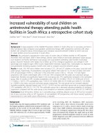
The relationship between natural log (LN) IL-13 concentra-tions in pg/mL and % predicted DLCOFigure 2
The relationship between natural log (LN) IL-13 concentra-
tions in pg/mL and % predicted DLCO. The line was calcu-
lated using conditional standardization of the regression
results for a patient with mean and modal values for the cov-
ariates in the model. The standardized line thus represents
the relationship between IL-13 and DLCO for a man, age 63,
with a FEV
1
of 51 % predicted, who does not currently
smoke, with mean pack year smoking history of 52.5 years,
who is not on statins or systemic steroids, but is on inhaled
steroids (β = -0.02).
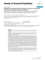
The relationship between natural log (LN) IL-13 concentra-tions in pg/mL and % predicted FEV1Figure 1
The relationship between natural log (LN) IL-13 concentra-
tions in pg/mL and % predicted FEV1. The line was calculated
using conditional standardization of the regression results for
a patient with mean and modal values for the covariates in
the model. The standardized line thus represents the rela-
tionship between IL-13 and FEV
1
for a man, age 63, who does
not currently smoke, with mean pack year smoking history of
52.5 years, who is not on statins or systemic steroids, but is
on inhaled steroids (β = -0.01).
Respiratory Research 2007, 8:64 />Page 7 of 10
(page number not for citation purposes)
ings, these authors showed a negative correlation between
intracellular IL-13 and % FEV
1
.
Three of seven chemokines tested were associated with
increasing severity of airflow obstruction: CCL2/MCP-1,
CCL4/MIP-1β, and CCL11/Eotaxin. In addition, CXCL9/
Mig was associated with increasing severity of diffusion
impairment. These chemokines recruit primarily mono-
cytes, T lymphocytes, and eosinophils, inviting the possi-
bility that soluble proteins that promote inflammatory
cell recruitment contribute to the low-grade systemic
inflammation observed in COPD. CCL2/MCP-1 recruits
monocytes and T lymphocytes expressing the receptor
CCR2 [49], and increased concentrations of this chemok-
ine have been reported in induced sputum, BAL and lung
tissue of COPD individuals [38,50]. CCL4/MIP-1β can
recruit CCR5 expressing monocytes and T lymphocytes
[49]. Our data corroborates findings showing a negative
correlation between CCL4/MIP-1β concentrations in the
BAL from patients with chronic bronchitis and the % FEV
1
[21]. CCL11/Eotaxin is involved in eosinophil recruit-
ment [51], and CCL11/eotaxin concentrations are
increased in the sputum of patients with exacerbations of
chronic bronchitis [23]. However, some COPD patients
with stable disease also show airway eosinophilic inflam-
mation [52].
A secondary goal of this study was to explore whether sys-
temic cytokines are associated with severity of diffusion
impairment, the physiologic parameter that corresponds
best to the loss of alveolar-capillary bed surface area in
emphysema. In the smaller cohort that received DLCO
measurements, it is interesting that CXCL9/Mig concen-
tration was inversely associated with % DLCO. CXCL9/
Mig recruits CXCR3 expressing T lymphocytes [49]. Saetta
and colleagues have previously shown increased numbers
of CXCR3 expressing T lymphocytes in peripheral airways
of COPD patients [53]. Upon stimulation with CXCL9/
Mig, CD14
+
CXCR3
+
macrophages of human emphysema-
tous lungs can increase metalloproteinase production in
vitro [26]. Thus, recent findings suggest a potential link
between this chemokine and the pro-elastolytic environ-
ment #of emphysema.
Increasing concentrations of plasma G-CSF are also asso-
ciated with increasing severity of diffusion impairment. G-
CSF is involved in neutrophil mobilization and survival
Table 7: Association between plasma markers and % DLCO,
adjusted*
Analyte β
†
95% CI p
‡
CCL2/MCP-1 -0.001 -0.01, 0.003 0.58
CCL4/MIP-1β 0.004 -0.02, 0.03 0.76
CCL5/RANTES -0.01 -0.03, 0.003 0.11
CCL11/eotaxin -0.005 -0.01, 0.002 0.15
CXCL8/IL-8 -0.004 -0.01, 0.002 0.18
CXCL9/Mig -0.02 -0.03, -0.002 0.02
EGF -0.01 -0.03, 0.01 0.29
Fas 0.01 0.003, 0.02 0.01
G-CSF -0.01 -0.02, -0.0001 0.05
HGF -0.0001 -0.01, 0.01 0.99
IFN-γ -0.02 -0.05, 0.01 0.11
IL-2 -0.03 -0.06, 0.01 0.11
IL-2R 0.01 -0.01, 0.02 0.46
IL-4 -0.02 -0.05, 0.02 0.30
IL-13 -0.02 -0.03, -0.002 0.03
TNF-α -0.01 -0.03, 0.003 0.11
*Adjusted for % FEV1, current smoking, pack years, ICS use, SCS use,
statin use, gender and age.
†
β = regression co-efficient
‡
p = p-value
Table 6: Association between plasma marker concentrations and
% DLCO, unadjusted
Classification Plasma
marker
β
†
95% CI p
‡
Apoptosis Fas 0.01 0.001, 0.01 0.01
FasL -0.0004 -0.007, 0.006 0.89
Acute phase CRP -0.01 -0.02, 0.01 0.33
MPO -0.005 -0.01, 0.004 0.30
Chemokines CCL2/MCP -1 -0.003 -0.01, 0.0004 0.09
CCL3/MIP-1α 0.001 -0.02, 0.02 0.92
CCL4/MIP-1β -0.02 -0.03, -0.001 0.04
CCL5/
RANTES
-0.01 -0.02, -0.002 0.02
CCL11/
eotaxin
-0.002 -0.01, 0.002 0.31
CXCL8/IL-8 -0.005 -0.01, -0.002 < 0.01
CXCL9/Mig -0.01 -0.02, -0.004 < 0.01
T
H
Related
Cytokines
IFN-γ -0.03 -0.04, -0.01 <
0.001
IL-2 -0.04 -0.06, -0.02 < 0.01
IL-2R
§
-0.005 -0.01, 0.002 0.18
IL-4 -0.03 -0.06, -0.01 < 0.01
IL-13 -0.02 -0.03, -0.01 <
0.001
Inflammation TNF-α -0.02 -0.03, -0.005 < 0.01
TNFRI
§
0.001 -0.01, 0.01 0.79
TNFRII
§
0.001 -0.004, 0.005 0.84
IL-1β -0.01 -0.03, 0.004 0.13
IL-6 -0.01 -0.02, 0.004 0.17
IL-10 -0.01 -0.01, 0.003 NE*
Growth
Factors
EGF -0.01 -0.02, 0.0001 0.05
EGFR
§
0.002 -0.001, 0.005 0.12
FGFβ NE* NE* NE*
G-CSF -0.01 -0.02, -0.002 0.02
HGF -0.005 -0.01, 0.001 0.08
VEGF NE* NE* NE*
†
β = regression co-efficient
‡
p = p-value
§
For clarity, the soluble receptors are grouped with their respective
ligand
*NE, Not Evaluable
Respiratory Research 2007, 8:64 />Page 8 of 10
(page number not for citation purposes)
[54], however its role in COPD is not yet known. There are
increased numbers of granulocytes in the sputum and BAL
[38] in addition to small airways [55] of COPD patients,
leading others to speculate that granulocyte survival in the
lungs may be enhanced in COPD by mediators such as G-
CSF [38].
Another molecule identified is soluble Fas. Decreasing
concentrations of soluble Fas are associated with increas-
ing severity of diffusion impairment. Soluble Fas, a result
of alternative mRNA splicing, inhibits apoptosis by com-
petitively binding FasL and preventing its interaction with
the membrane bound Fas receptor [56,57]. The relation-
ship between systemic levels of soluble Fas and COPD is
unclear, as other smaller studies have shown variable
findings of either elevation or no difference compared
with controls [17-19]. Our results suggests that a systemic
imbalance of the anti-apoptotic factor soluble Fas occurs
in the setting of a pro-apoptotic environment of the lungs
in COPD.
The limitations of this present study include the size of the
cohort and its cross-sectional nature. The modest size, par-
ticularly the number of subjects with milder lung function
impairment (GOLD 0–1 stages), may limit the ability to
detect significant associations between systemic markers
and lung function impairment. Furthermore, we included
age, gender, pack years smoking history, current smoking,
inhaled corticosteroid use, systemic corticosteroid use and
statin use in the multivariate model. It is uncertain
whether adjustment for these covariates is appropriate.
Thus, we present both univariate and multivariate analy-
sis. We also recognize that the observed associations
between plasma concentrations of a protein and lung
function severity do not necessarily invoke a cause-effect
relationship. However, the findings of this study can serve
as the basis for a larger prospective cohort study examin-
ing a narrower profile of cytokines on a longitudinal basis.
Conclusion
Systemic inflammation has been increasingly recognized
in patients with COPD. CRP has been shown to be
increased in COPD [3,4], yet many other disease states
characterized by inflammation are associated with
increased CRP concentrations. Our data suggests that sys-
temic inflammation in a COPD cohort is also character-
ized by cytokines implicated in inflammatory cell
recruitment and airway remodeling. We show associa-
tions between plasma concentrations of chemokines and
IL-13 with increasing severity of disease, as measured by
% FEV
1
or % DLCO. Increasing severity of diffusion
impairment is also associated with increasing G-CSF and
decreasing soluble Fas concentrations. We speculate that
disease characterized by disproportionate abnormalities
in DLCO may be associated with peripheral markers inde-
pendent of the FEV
1
. The biological plausibility of IL-13
and the discrete repertoire of inflammatory chemokines
identified in our model underscore the possibility to more
precisely characterize systemic inflammation of COPD.
Abbreviations
CCL2/MCP-1, CC chemokine ligand 2/monocyte chemo-
tattractant protein-1; CCL3/MIP-1α, CC chemokine lig-
and 3/macrophage inflammatory protein-1α; CCL4/MIP-
1β, CC chemokine ligand 4/macrophage inflammatory
protein-1β; CCL5/RANTES, CC chemokine ligand 5/regu-
lated on activation normal T cell expressed and secreted;
CCL11/eotaxin, CC chemokine ligand 11/eotaxin; CRP,
C-reactive protein; CXCL8/IL-8, CXC chemokine ligand 8/
interleukin-8; CXCL9/Mig, CXC chemokine ligand 9/
monkine induced by interferon-γ; EGF, epidermal growth
factor; EGFR, epidermal growth factor receptor; FasL, Fas
ligand; FGFβ, fibroblast growth factor β; G-CSF, granulo-
cyte-colony stimulating factor; HGF, hepatocyte growth
factor; IFN-γ, interferon-γ; IL-1β, interleukin-1β; IL-2,
interleukin-2; IL-2R, interleukin-2 receptor; IL-4, inter-
leukin-4, IL-6, interleukin-6; IL-10, interleukin-10; IL-13,
interleukin-13; MPO, myeloperoxidase; TNF-α, tumor
necrosis factor α, TNFRI, tumor necrosis factor receptor 1,
TNFRII, tumor necrosis factor receptor 2; VEGF, vascular
endothelial growth factor;
Competing interests
Frank C. Sciurba has received funding from GlaxoSmithK-
line and AstraZeneca in 2005 through 2006 for participa-
tion in multi-center clinical trials. He has served on
advisory boards for GlaxoSmithKline and AstraZeneca.
None of the other authors has any competing interests to
declare.
Authors' contributions
JSL, VK, YZ, JM, RAB, AMC and FCS participated in the
design of the study. JSL contributed to the statistical anal-
ysis, interpretation of the data, and wrote the manuscript.
MRR performed portions of the statistical analysis, con-
tributed to the interpretation of the data, and revised the
manuscript for important intellectual content. VK per-
formed the statistical analysis. YZ participated in the col-
lection of data. JM participated in the analysis of the data.
RAB contributed to the analysis and interpretation of data,
and revised the manuscript for important intellectual con-
tent. AMC and FCS conceived the study, contributed to
the acquisition of the data, and provided important intel-
lectual content to the manuscript. All authors read and
approved the final manuscript.
Acknowledgements
We gratefully acknowledge Naftali Kaminiski for his assistance in facilitating
the performance of luminex assays at the University of Pittsburgh Cancer
Institute Luminex Core Facility and for selection of some of the plasma
markers studied. We also thank Anna Loshkin, Director of the University
Respiratory Research 2007, 8:64 />Page 9 of 10
(page number not for citation purposes)
of Pittsburgh Cancer Insitute Luminex Core Facility, for her help in the per-
formance of the assays. We gratefully acknowledge Bill Slivka, Chad Karole-
ski, Denise Filippino, Mary Bryner for their assistance with the pulmonary
function testing, data entry, and clinical recruitment of patients. We are
deeply indebted to the participants of the ECRC registry.
This study was supported by the ATS Research Grant Innovative Research
in COPD (JSL), HL70178 (JSL), General Clinical Research Grant No. 5 MO1
RR 0056 (RAB), HL084948 (FCS).
References
1. Pauwels RA, Buist AS, Calverley PM, Jenkins CR, Hurd SS: Global
strategy for the diagnosis, management, and prevention of
chronic obstructive pulmonary disease. NHLBI/WHO Glo-
bal Initiative for Chronic Obstructive Lung Disease (GOLD)
Workshop summary. Am J Respir Crit Care Med 2001,
163(5):1256-1276.
2. Rabe K, Barnes P, Buist S, Calverley PM, Fabbri L, Fukuchi Y, MacNee
W, Rodriguez-Roisin R, Zielinski I: Global Strategy for the Diag-
nosis, Management, and Prevention of Chronic Obstructive
Pulmonary Disease. NHLBI/WHO Global Initiative for
Chronic Obstructive Lung Disease Workshop Executive
Summary, 2005 Update. GOLD website wwwgoldcopdorg 2005.
3. Gan WQ, Man SF, Senthilselvan A, Sin DD: Association between
chronic obstructive pulmonary disease and systemic inflam-
mation: a systematic review and a meta-analysis. Thorax 2004,
59(7):574-580.
4. Mannino DM, Ford ES, Redd SC: Obstructive and restrictive lung
disease and markers of inflammation: data from the Third
National Health and Nutrition Examination. Am J Med 2003,
114(9):758-762.
5. Gan WQ, Man SF, Sin DD: The interactions between cigarette
smoking and reduced lung function on systemic inflamma-
tion. Chest 2005, 127(2):558-564.
6. Man SF, Sin DD: Effects of corticosteroids on systemic inflam-
mation in chronic obstructive pulmonary disease. Proc Am
Thorac Soc 2005, 2(1):78-82.
7. Sin DD, Lacy P, York E, Man SF: Effects of fluticasone on systemic
markers of inflammation in chronic obstructive pulmonary
disease. Am J Respir Crit Care Med 2004, 170(7):760-765.
8. Lung function testing: selection of reference values and
interpretative strategies. American Thoracic Society. Am
Rev Respir Dis 1991, 144(5):1202-1218.
9. Standardization of Spirometry, 1994 Update. American
Thoracic Society. Am J Respir Crit Care Med 1995,
152(3):1107-1136.
10. Miller MR, Hankinson J, Brusasco V, Burgos F, Casaburi R, Coates A,
Crapo R, Enright P, van der Grinten CP, Gustafsson P, Jensen R, John-
son DC, MacIntyre N, McKay R, Navajas D, Pedersen OF, Pellegrino
R, Viegi G, Wanger J: Standardisation of spirometry. Eur Respir J
2005, 26(2):319-338.
11. Macintyre N, Crapo RO, Viegi G, Johnson DC, van der Grinten CP,
Brusasco V, Burgos F, Casaburi R, Coates A, Enright P, Gustafsson P,
Hankinson J, Jensen R, McKay R, Miller MR, Navajas D, Pedersen OF,
Pellegrino R, Wanger J: Standardisation of the single-breath
determination of carbon monoxide uptake in the lung. Eur
Respir J 2005, 26(4):720-735.
12. Crapo RO, Morris AH: Standardized single breath normal val-
ues for carbon monoxide diffusing capacity. Am Rev Respir Dis
1981, 123(2):185-189.
13. Crapo RO, Morris AH, Gardner RM: Reference spirometric val-
ues using techniques and equipment that meet ATS recom-
mendations. Am Rev Respir Dis 1981, 123(6):659-664.
14. Gorelik E, Landsittel DP, Marrangoni AM, Modugno F, Velikokhatnaya
L, Winans MT, Bigbee WL, Herberman RB, Lokshin AE: Multiplexed
immunobead-based cytokine profiling for early detection of
ovarian cancer. Cancer Epidemiol Biomarkers Prev 2005,
14(4):981-987.
15. Lee JS, Wurfel MM, Matute-Bello G, Frevert CW, Rosengart MR, Ran-
ganathan M, Wong VW, Holden T, Sutlief S, Richmond A, Peiper S,
Martin TR: The Duffy antigen modifies systemic and local tis-
sue chemokine responses following lipopolysaccharide stim-
ulation. J Immunol 2006, 177(11):8086-8094.
16. Wurfel MM, Park WY, Radella F, Ruzinski J, Sandstrom A, Strout J,
Bumgarner RE, Martin TR: Identification of high and low
responders to lipopolysaccharide in normal subjects: an
unbiased approach to identify modulators of innate immu-
nity. J Immunol 2005, 175(4):2570-2578.
17. Yasuda N, Gotoh K, Minatoguchi S, Asano K, Nishigaki K, Nomura M,
Ohno A, Watanabe M, Sano H, Kumada H, Sawa T, Fujiwara H: An
increase of soluble Fas, an inhibitor of apoptosis, associated
with progression of COPD. Respir Med 1998, 92(8):993-999.
18. Takabatake N, Arao T, Sata M, Inoue S, Abe S, Shibata Y, Kubota I:
Circulating levels of soluble Fas ligand in cachexic patients
with COPD are higher than those in non-cachexic patients
with COPD. Intern Med 2005, 44(11):1137-1143.
19. Takabatake N, Nakamura H, Inoue S, Terashita K, Yuki H, Kato S,
Yasumura S, Tomoike H: Circulating levels of soluble Fas ligand
and soluble Fas in patients with chronic obstructive pulmo-
nary disease. Respir Med 2000, 94(12):1215-1220.
20. Andelid K, Bake B, Rak S, Linden A, Rosengren A, Ekberg-Jansson A:
Myeloperoxidase as a marker of increasing systemic inflam-
mation in smokers without severe airway symptoms . Respir
Med 2007, 101:888-895 2007, 101:888-95.
21. Capelli A, Di Stefano A, Gnemmi I, Balbo P, Cerutti CG, Balbi B,
Lusuardi M, Donner CF: Increased MCP-1 and MIP-1beta in
bronchoalveolar lavage fluid of chronic bronchitics. Eur Respir
J 1999, 14(1):160-165.
22. Zhu J, Qiu YS, Majumdar S, Gamble E, Matin D, Turato G, Fabbri LM,
Barnes N, Saetta M, Jeffery PK: Exacerbations of Bronchitis:
bronchial eosinophilia and gene expression for interleukin-4,
interleukin-5, and eosinophil chemoattractants. Am J Respir
Crit Care Med 2001, 164(1):109-116.
23. Bocchino V, Bertorelli G, Bertrand CP, Ponath PD, Newman W,
Franco C, Marruchella A, Merlini S, Del Donno M, Zhuo X, Olivieri
D: Eotaxin and CCR3 are up-regulated in exacerbations of
chronic bronchitis. Allergy 2002, 57(1):17-22.
24. Nocker RE, Schoonbrood DF, van de Graaf EA, Hack CE, Lutter R,
Jansen HM, Out TA: Interleukin-8 in airway inflammation in
patients with asthma and chronic obstructive pulmonary dis-
ease. Int Arch Allergy Immunol 1996, 109(2):183-191.
25. Tanino M, Betsuyaku T, Takeyabu K, Tanino Y, Yamaguchi E, Miya-
moto K, Nishimura M: Increased levels of interleukin-8 in BAL
fluid from smokers susceptible to pulmonary emphysema.
Thorax 2002, 57(5):405-411.
26. Grumelli S, Corry DB, Song LZ, Song L, Green L, Huh J, Hacken J,
Espada R, Bag R, Lewis DE, Kheradmand F: An immune basis for
lung parenchymal destruction in chronic obstructive pulmo-
nary disease and emphysema. PLoS Med 2004, 1(1):e8.
27. Elias JA, Kang MJ, Crouthers K, Homer R, Lee CG: State of the art.
Mechanistic heterogeneity in chronic obstructive pulmonary
disease: insights from transgenic mice. Proc Am Thorac Soc 2006,
3(6):494-498.
28. Barcelo B, Pons J, Fuster A, Sauleda J, Noguera A, Ferrer JM, Agusti
AG: Intracellular cytokine profile of T lymphocytes in
patients with chronic obstructive pulmonary disease. Clin Exp
Immunol 2006, 145(3):474-479.
29. Barnes PJ, Cosio MG: Characterization of T lymphocytes in
chronic obstructive pulmonary disease. PLoS Med 2004,
1(1):e20.
30. Eid AA, Ionescu AA, Nixon LS, Lewis-Jenkins V, Matthews SB, Grif-
fiths TL, Shale DJ: Inflammatory response and body composi-
tion in chronic obstructive pulmonary disease. Am J Respir Crit
Care Med 2001, 164(8 Pt 1):1414-1418.
31. Vernooy JH, Kucukaycan M, Jacobs JA, Chavannes NH, Buurman WA,
Dentener MA, Wouters EF: Local and systemic inflammation in
patients with chronic obstructive pulmonary disease: soluble
tumor necrosis factor receptors are increased in sputum. Am
J Respir Crit Care Med 2002, 166(9):1218-1224.
32. Ferroni P, Basili S, Alessandri C, Vieri M, Martini F, Belogi A, Pulcinelli
FM, Cordova C, Gazzaniga PP: Proinflammatory cytokines and
hemostatic system in patients with chronic obstructive pul-
monary disease. Platelets 1997, 8(4):255-259.
33. Takeyama K, Jung B, Shim JJ, Burgel PR, Dao-Pick T, Ueki IF, Protin U,
Kroschel P, Nadel JA: Activation of epidermal growth factor
receptors is responsible for mucin synthesis induced by ciga-
rette smoke. Am J Physiol Lung Cell Mol Physiol 2001,
280(1):L165-72.
Publish with BioMed Central and every
scientist can read your work free of charge
"BioMed Central will be the most significant development for
disseminating the results of biomedical researc h in our lifetime."
Sir Paul Nurse, Cancer Research UK
Your research papers will be:
available free of charge to the entire biomedical community
peer reviewed and published immediately upon acceptance
cited in PubMed and archived on PubMed Central
yours — you keep the copyright
Submit your manuscript here:
/>BioMedcentral
Respiratory Research 2007, 8:64 />Page 10 of 10
(page number not for citation purposes)
34. Nadel JA: Role of epidermal growth factor receptor activation
in regulating mucin synthesis. Respir Res 2001, 2(2):85-89.
35. Nadel JA, Burgel PR: The role of epidermal growth factor in
mucus production. Curr Opin Pharmacol 2001, 1(3):254-258.
36. Kranenburg AR, De Boer WI, Van Krieken JH, Mooi WJ, Walters JE,
Saxena PR, Sterk PJ, Sharma HS: Enhanced expression of fibrob-
last growth factors and receptor FGFR-1 during vascular
remodeling in chronic obstructive pulmonary disease. Am J
Respir Cell Mol Biol 2002, 27(5):517-525.
37. Kranenburg AR, Willems-Widyastuti A, Mooi WJ, Saxena PR, Sterk
PJ, de Boer WI, Sharma HS: Chronic obstructive pulmonary dis-
ease is associated with enhanced bronchial expression of
FGF-1, FGF-2, and FGFR-1. J Pathol 2005, 206(1):28-38.
38. Barnes PJ: Mediators of chronic obstructive pulmonary dis-
ease. Pharmacol Rev 2004, 56(4):515-548.
39. Plantier L, Marchand-Adam S, Marchal-Somme J, Leseche G, Fournier
M, Dehoux M, Aubier M, Crestani B: Defect of hepatocyte
growth factor production by fibroblasts in human pulmonary
emphysema. Am J Physiol Lung Cell Mol Physiol 2005, 288(4):L641-7.
40. Kasahara Y, Tuder RM, Taraseviciene-Stewart L, Le Cras TD, Abman
S, Hirth PK, Waltenberger J, Voelkel NF: Inhibition of VEGF
receptors causes lung cell apoptosis and emphysema. J Clin
Invest 2000, 106(11):1311-1319.
41. Bio-Rad Laboratories I: Principles of Curve Fitting for Multiplex
Sandwich Immunoassays, Rev. B, Bulletin # 2861. wwwbio-rad-
com .
42. de Jager W, Rijkers GT: Solid-phase and bead-based cytokine
immunoassay: a comparison. Methods 2006, 38(4):294-303.
43. Hurst JR, Donaldson GC, Perera WR, Wilkinson TM, Bilello JA,
Hagan GW, Vessey RS, Wedzicha JA: Use of plasma biomarkers
at exacerbation of chronic obstructive pulmonary disease.
Am J Respir Crit Care Med 2006, 174(8):867-874.
44. Pellegrino R, Viegi G, Brusasco V, Crapo RO, Burgos F, Casaburi R,
Coates A, van der Grinten CP, Gustafsson P, Hankinson J, Jensen R,
Johnson DC, MacIntyre N, McKay R, Miller MR, Navajas D, Pedersen
OF, Wanger J: Interpretative strategies for lung function tests.
Eur Respir J 2005, 26(5):948-968.
45. Shim JJ, Dabbagh K, Ueki IF, Dao-Pick T, Burgel PR, Takeyama K, Tam
DC, Nadel JA: IL-13 induces mucin production by stimulating
epidermal growth factor receptors and by activating neu-
trophils. Am J Physiol Lung Cell Mol Physiol 2001, 280(1):L134-40.
46. Ma B, Liu W, Homer RJ, Lee PJ, Coyle AJ, Lora JM, Lee CG, Elias JA:
Role of CCR5 in the pathogenesis of IL-13-induced inflamma-
tion and remodeling. J Immunol 2006, 176(8):4968-4978.
47. Zhu Z, Homer RJ, Wang Z, Chen Q, Geba GP, Wang J, Zhang Y, Elias
JA: Pulmonary expression of interleukin-13 causes inflamma-
tion, mucus hypersecretion, subepithelial fibrosis, physio-
logic abnormalities, and eotaxin production. J Clin Invest 1999,
103(6):779-788.
48. Zheng T, Zhu Z, Wang Z, Homer RJ, Ma B, Riese RJ Jr., Chapman HA
Jr., Shapiro SD, Elias JA: Inducible targeting of IL-13 to the adult
lung causes matrix metalloproteinase- and cathepsin-
dependent emphysema. J Clin Invest 2000, 106(9):1081-1093.
49. Murphy PM, Baggiolini M, Charo IF, Hebert CA, Horuk R, Matsushima
K, Miller LH, Oppenheim JJ, Power CA: International union of
pharmacology. XXII. Nomenclature for chemokine recep-
tors. Pharmacol Rev 2000, 52(1):145-176.
50. Traves SL, Culpitt SV, Russell RE, Barnes PJ, Donnelly LE: Increased
levels of the chemokines GROalpha and MCP-1 in sputum
samples from patients with COPD. Thorax 2002,
57(7):590-595.
51. Fukakusa M, Bergeron C, Tulic MK, Fiset PO, Al Dewachi O, Lavio-
lette M, Hamid Q, Chakir J: Oral corticosteroids decrease eosi-
nophil and CC chemokine expression but increase
neutrophil, IL-8, and IFN-gamma-inducible protein 10
expression in asthmatic airway mucosa. J Allergy Clin Immunol
2005, 115(2):280-286.
52. Brightling CE, Monteiro W, Ward R, Parker D, Morgan MD, Wardlaw
AJ, Pavord ID: Sputum eosinophilia and short-term response
to prednisolone in chronic obstructive pulmonary disease: a
randomised controlled trial. Lancet 2000,
356(9240):1480-1485.
53. Saetta M, Mariani M, Panina-Bordignon P, Turato G, Buonsanti C, Bar-
aldo S, Bellettato CM, Papi A, Corbetta L, Zuin R, Sinigaglia F, Fabbri
LM: Increased expression of the chemokine receptor CXCR3
and its ligand CXCL10 in peripheral airways of smokers with
chronic obstructive pulmonary disease. Am J Respir Crit Care
Med 2002, 165(10):1404-1409.
54. Colotta F, Re F, Polentarutti N, Sozzani S, Mantovani A: Modulation
of granulocyte survival and programmed cell death by
cytokines and bacterial products. Blood 1992, 80(8):2012-2020.
55. Hogg JC, Chu F, Utokaparch S, Woods R, Elliott WM, Buzatu L, Cher-
niack RM, Rogers RM, Sciurba FC, Coxson HO, Pare PD: The
nature of small-airway obstruction in chronic obstructive
pulmonary disease. N Engl J Med 2004, 350(26):2645-2653.
56. Albertine KH, Soulier MF, Wang Z, Ishizaka A, Hashimoto S, Zimmer-
man GA, Matthay MA, Ware LB: Fas and fas ligand are up-regu-
lated in pulmonary edema fluid and lung tissue of patients
with acute lung injury and the acute respiratory distress syn-
drome. Am J Pathol 2002, 161(5):1783-1796.
57. Cheng J, Zhou T, Liu C, Shapiro JP, Brauer MJ, Kiefer MC, Barr PJ,
Mountz JD: Protection from Fas-mediated apoptosis by a sol-
uble form of the Fas molecule. Science 1994,
263(5154):1759-1762.