Báo cáo khoa học: " An Investigation of the Ability of the Glutaraldehyde Test to Distinguish between Acute and Chronic Inflammatory Disease in Horses" ppsx
Bạn đang xem bản rút gọn của tài liệu. Xem và tải ngay bản đầy đủ của tài liệu tại đây (203.25 KB, 10 trang )
Brink P, Wright JC, Schumacher J: An investigation of the ability of the glu-
taraldehyde test to distinguish between acute and chronic inflammatory disease in
horses. Acta vet. scand. 2005, 46, 69-78. – The glutaraldehyde test (GT), a rapid and
inexpensive test, has been utilized empirically for many years in bovine practice for di-
agnosing inflammatory diseases. GT is used primarily to demonstrate increased serum
concentrations of fibrinogen and globulin. Glutaraldehyde binds with free amino groups
in fibrinogen and immunoglobulin to create a clot in a first degree chemical reaction.
The clotting time of the GT estimates the content of proteins produced in response to in-
flammation. The applicability of GT for diagnosing inflammation in the horse has never
been investigated. The objective of this study was to determine the ability of GT to dis-
tinguish between acute and chronic inflammatory disease in horses. Thirty-seven horses
with suspected inflammatory diseases were evaluated using the GT, history, complete
clinical examination and routine blood analysis. GT-times, laboratory results and clini-
cal outcome were compared statistically. Horses that were determined to be acutely af-
fected (based on history, clinical examination and routine blood analysis) tended to have
a negative GT (75%). Results of the GT did not correlate with blood fibrinogen con-
centration. Positive GT also predicted a fatal outcome in 69% of the clinical cases. The
results of this trial indicate that GT can be a useful screening test to distinguish between
acute and chronic inflammatory disease in horses.
Glutaraldehyde test, inflammation, horse diseases, equine, diagnostic techniques,
prognosis, immunoglobulin, globulin, blood clot, infectious diseases, hypergamma-
globulinemia, serum biochemistries.
Acta vet. scand. 2005, 46, 69-78.
Acta vet. scand. vol. 46 no. 1-2, 2005
An Investigation of the Ability of the Glutaraldehyde
Test to Distinguish between Acute and Chronic
Inflammatory Disease in Horses
By P. Brink
1
, J.C. Wright
2
, and J. Schumacher
3
1
ATG Equine Clinic, Jägersro, 21237 Malmö, Sweden,
2
Department of Pathobiology and
3
Department of Clin-
ical Sciences, Auburn University, Auburn, AL 36849-5522, USA.
Introduction
The glutaraldehyde reagent in the glutaralde-
hyde test (GT) creates a clot with either fibrino-
gen or gammaglobulin in EDTA-stabilized
blood by chemical reaction between the alde-
hyde groups in glutaraldehyde and free amino
groups in fibrinogen and immunoglobulins
(Sandholm 1974a, Sandholm 1974b, Martin et
al. 1985). The process is believed to run as a
first degree chemical reaction, where the reac-
tion time is directly proportional to the concen-
tration of fibrinogen and immunoglobulins
(Sandholm 1974a, Sandholm 1974b, Eriksen
1984).
The rapid and inexpensive GT has been used
with success empirically in Europe for many
years for diagnosing inflammatory diseases in
cattle (Sandholm 1974a, Sandholm 1974b,
Liberg et al. 1975a, Liberg et al. 1975b, Nielsen
1975, Martens 1977, Liberg 1978, Tennant et
al. 1979, Liberg 1981, Liberg 1982, Eriksen
1984, Keulen et al. 1984, Doll et al. 1985, Lars-
son 1985, Mahlin et al. 1985, Chadli & Mahin
1986, Kovac 1988, Katholm & Jorgensen 1992,
Kantor et al. 1993, Tyler et al. 1996, Sen et al.
2000, Ramprabhu et al. 2002), pigs (Liberg
1979, Hansen 1985, Kovac et al. 1993), goats
(Satpathy et al. 1996, Vihan 1989), mink (Sand-
holm & Kangas 1973), dogs (Sandholm &
Kivisto 1975, Wolff 1986), and zoo animals
(O'-Rourke & Satterfield 1981, Carstairs-Grant
et al. 1988, Juyal & Uppal 1995). In these
species, the test was used to indicate whether an
inflammatory disease was acute or chronic
(Doll et al. 1985, Chadli & Mahin 1986).
The GT, because of its simplicity, is very useful
in bovine practice for rapidly diagnosing in-
flammation under circumstances where it is not
practical or economically possible to have
blood analyzed at a professional clinical labora-
tory (Sandholm 1974a, Sandholm 1974b, Li-
berg et al. 1975a, Liberg et al. 1975b, Nielsen
1975, Martens 1977, Liberg 1978, Tennant et
al. 1979, Liberg 1981, Liberg 1982, Eriksen
1984, Keulen et al. 1984, Doll et al. 1985, Lars-
son 1985,
Mahlin et al. 1985, Chadli & Mahin
1986, Kovac 1988, Katholm & Jorgensen 1992,
Kantor et al. 1993, Tyler et al. 1996, Sen et al.
2000, Ramprabhu et al. 2002).
A negative GT can be used as a semiquantita-
tive indicator of hypogammaglobulinemia
caused by failure of passive transfer of
colostrum in neonatal foals (Beetson et al.
1985, Clabough et al. 1989, Saikku et al. 1989,
Clabough et al. 1991, Kumaran & Bhuvanaku-
mar 1994, Kalinbacak & Or 1996, Bruijn et al.
2003), calves (Tennant et al. 1979, Keulen et al.
1984, Larsson 1985, Kovac 1988, Tyler et al.
1996, Sen et al. 2000), kids (Vihan 1989, Sat-
pathy et al. 1996), and zoo ruminants
(O'-Rourke & Satterfield 1981, Carstairs-
Grant et al. 1988, Juyal & Uppal 1995). The
GT also has been used to determine the content
of IgG in mare colostrum (Jones & Brook 1995,
Ezhilan & Bhuvanakumar 1998).
Clinical experience indicates that the GT may
not be as reliable in horses as it is in cattle
(Nielsen 1975). In horses, lack of reliability of
the GT has been proposed to be caused by gen-
erally lower or delayed peaks of concentrations
of fibrinogen and immunoglobulin or a differ-
ent distribution of immunoglobulins (IgG, IgM,
IgA) compared to cattle (Bendixen 1954,
Nansen & Nielsen 1966, Sandholm 1974a,
Nielsen 1975, Aasted et al. 1989).
The purpose of this clinical trial was to deter-
mine the ability of GT to distinguish between
acute and chronic inflammatory disease in
horses. During the trial we compared indicators
of inflammation (the concentration of blood fib-
rinogen and serum globulin) to the GT.
Materials and methods
Thirty seven horses admitted for investigation
of suspected inflammatory disease were evalu-
ated using the GT (Glutarvac
a
), a complete
clinical examination, CBC and routine serum
biochemistries that included total protein, albu-
min, globulin and fibrinogen. Blood for the GT
and laboratory analysis was collected at the
same time either upon arrival at the hospital or
the following day.
Horses having a history of clinical signs of in-
flammatory disease of total duration six days or
less were arbitrarily classified as acutely in-
flamed. Horses with a history of clinical signs
greater than six days were arbitrarily classified
as chronically inflamed. The clinical examina-
tion leading to the diagnosis and etiology was
also used to reinforce the distinction between
acute and chronic disease (Table 1).
The GT was performed by adding equal
amounts of fresh blood and glutaraldehyde in a
test tube, mixing by slowly turning the test tube
and visually observing and noting the time re-
70 P. Brink et al.
Acta vet. scand. vol. 46 no. 1-2, 2005
Glutaraldehyde test of inflammatory disease in horses 71
Acta vet. scand. vol. 46 no. 1-2, 2005
Table 1: Diagnosis and outcome.
Horse # Diagnosis Duration Outcome
1 Purulent, bilateral guttural pouch empyema Chronic Fatal (spontaneous)
2 Dorsal rectal abscess Chronic Discharged
3 Traumatic, infected joint capsular laceration Acute Discharged
4 Dorsal rectal abscesses Chronic Discharged
5 Purulent nephritis, lung abscesses, ulcerous dermatitis,
myocarditis, fatty liver Chronic Fatal (euthanasia)
6 Severe, idiopathic, systemic infection Acute Fatal (spontaneous)
7 Purulent (jugular) thrombophlebitis (abscess) Chronic Discharged
8 Transportation syndrome, bronchitis/pleuritis,
systemic infection Acute Discharged
9 Fibrinopurulent pleuropneumonia Acute Fatal (euthanasia)
10 Systemic, malign lymphoma, borrelia infection Chronic Fatal (euthanasia)
11 Infected tendovaginitis Acute Discharged
12 Intraabdominal abscess, squamous cell carcinoma
(ventricle) Chronic Fatal (euthanasia)
13 Septic, purulent arthritis Chronic Discharged
14 Fibrinopurulent pleuropneumonia Acute Fatal (euthanasia)
15 Septicemia, pneumonia, peritonitis Acute Fatal (euthanasia)
16 Severe, purulent, traumatic muscle laceration Chronic Discharged
17 Severe, iatrogenic, muscle abscesses Chronic Discharged
18 Purulent osteomyelitis Chronic Fatal (euthanasia)
19 Severe subcutaneous infection/abscess, funiculitis Chronic Discharged
20 Humerus fracture, subcutaneous infection/abscess Chronic Discharged
21 Bacterial diarrhea Acute Fatal (euthanasia)
22 Abscess, inguinal region Chronic Discharged
23 Scrotal abscesses, postoperative castration Chronic Discharged
24 Necrotizing myositis, multiple subcutaneous abscesses Chronic Discharged
25 Fibrinopurulent septic bicipital bursitis, muscular
septic cellulitis Chronic Fatal (euthanasia)
26 Pericarditis, mitral insufficiency, systemic infection Chronic Fatal (euthanasia)
27 Septic peritonitis Chronic Discharged
28 Septic meningitis Acute Discharged
29 Septicemia, premature foal Acute Discharged
30 M. Masseter, throat latch, parotid, jugular
abscesses/fistulae Chronic Discharged
31 Systemic infection, septic myositis Chronic Fatal (euthanasia)
32 Systemic infection, possible abdominal/kidney abscess,
emaciation Chronic Fatal (euthanasia)
33 Severe, multiple, purulent, septic arthritis Chronic Fatal (euthanasia)
34 Metritis, purulent peritonitis, abdominal abscesses,
adherences Chronic Fatal (euthanasia)
35 Purulent, pharyngeal inflammation, choke Acute Discharged
36 Thrombosis pulmonary vessels, Cushing disease,
laminitis Chronic Fatal (euthanasia)
37 Systemic intoxication, parasitic aneurysm, intestinal
volvulus, paralysis Acute Fatal (euthanasia)
72 P. Brink et al.
Acta vet. scand. vol. 46 no. 1-2, 2005
Table 2: Categorization of GT-time.
Group # GT-times Empiric categorization
1 0 < GT-time < 3 min. High increase in concentration of fibrinogen and/or immunoglobulin
2 3 < GT-time < 6 min. Moderate increase in concentration of fibrinogen and/or immunoglobulin
3 6 < GT-time < 15 min. Low increase in concentration of fibrinogen and/or immunoglobulin
4 GT >15 min. No increase in concentration of fibrinogen and/or immunoglobulin
Table 3: Blood values
Horse GT- Al- Glo- Alb/ Fibri- Total WBC
Differential cell count leukocytes
Hemo-
# time bumin bulin Glob nogen prot. (10.9/l) Bands Segm Eosin Mono Lymph Baso RBC globin PCV
(min) (g/l) (g/l) (ratio) (g/l) (g/l) (%) (%) (%) (%) (%) (%) (10.12/l) (g/l) (%)
1 2,0 30 58 0,5 7,1 88 9,4 1 63 0 4 32 0 8,8 113 31
2 NR 30 35 0,9 7,5 65 9,3 0 42 0 5 53 0 9,7 118 33
3 NR 38 30 1,3 3,6 68 9,4 0 67 1 1 31 0 8,5 123 34
4 NR 27 35 0,8 4,7 62 7,0 3 24 2 3 66 2 8,2 110 30
5 NR 41 33 1,2 2,3 74 16,7 0 85 0 5 9 1 11,6 178 48
6 3,5 31 40 0,8 8,3 71 10,2 2 79 0 4 15 0 7,3 117 33
7 NR 36 37 1,0 7,7 73 16,0 5 78 0 1 16 0 11,7 186 50
8 NR 34 42 0,8 4,4 76 9,3 2 68 1 3 25 1 7,3 125 35
9 NR 32 37 0,9 9,9 69 9,2 11 64 0 4 21 0 8,6 144 39
10 1,0 17 63 0,3 3,0 80 15,8 0 80 1 4 14 1 1,1 27 8
11 NR 31 48 0,6 8,2 79 6,2 0 61 1 4 34 0 5,9 102 28
12 6,0 32 60 0,5 5,2 92 7,0 0 74 1 5 20 0 7,0 130 34
13 NR 34 29 1,2 5,9 63 11,1 0 68 0 3 29 0 8,0 105 31
14 5,0 18 47 0,4 7,1 65 10,3 3 59 0 8 30 0 9,2 153 45
15 NR 23 27 0,9 6,4 50 2,8 7 11 0 5 77 0 12,8 176 50
16 NR 33 25 1,3 2,9 58 7,4 1 68 6 1 23 1 7,8 136 38
17 NR 31 29 1,1 5,4 60 7,3 0 56 2 6 36 0 10,1 168 48
18 NR 27 25 1,1 4,9 52 35,4 0 94 0 3 3 0 6,5 137 39
19 3,0 29 58 0,5 6,8 87 19,2 1 72 2 1 24 0 5,8 85 26
20 NR 33 20 1,7 12,2 53 15,0 0 75 0 6 19 0 9,1 121 35
21 NR 20 18 1,1 7,0 38 36,6 0 90 0 1 9 0 10,3 136 37
22 3,0 21 69 0,3 5,0 90 30,7 0 85 1 1 13 0 6,3 92 24
23 NR 21 36 0,6 6,0 57 7,5 0 54 2 2 41 1 6,3 107 28
24 NR 21 19 1,1 5,0 40 13,7 1 87 0 2 10 0 5,5 72 19
25 NR 36 31 1,2 6,6 67 8,6 0 77 0 8 14 0 5,6 101 27
26 NR 35 22 1,6 6,0 57 10,8 0 86 2 2 10 0 8,3 141 41
27 5,0 31 46 0,7 9,0 77 13,5 0 72 0 3 25 0 7,4 119 32
28 NR 25 41 0,6 10,0 66 33,4 0 93 0 6 1 0 10,5 126 32
29 NR 32 16 2,0 5,0 48 0,8 0 16 0 0 84 0 7,7 117 31
30 NR 28 21 1,3 14,7 49 26,4 0 72 0 9 18 1 8,1 98 25
31 15,0 40 27 1,5 7,3 67 11,5 1 78 1 2 17 1 6,8 118 31
32 NR 36 19 1,9 4,6 55 15,0 2 42 2 7 47 0 9,3 116 32
33 15,0 29 44 0,7 4,8 73 8,0 1 45 0 9 45 0 8,0 128 35
34 14,0 21 23 0,9 5,8 44 9,4 0 90 2 2 6 0 6,2 114 31
35 2,0 31 42 0,7 11,0 73 12,0 0 75 1 2 21 1 5,1 83 20
36 3,0 32 41 0,8 5,0 73 16,7 0 93 0 2 5 0 4,3 80 20
37 NR 33 39 0,8 6,3 72 11,9 0 76 0 7 17 0 11,7 185 53
* NR = no reaction
quired for full clot formation. The test result
was categorized respectively as high, moderate,
low or no increase in concentration of fibrino-
gen and/or immunoglobulin based on GT-time
(Table 2).
The results of the GT and fibrinogen, globulin
and albumin/globulin ratio were compared us-
ing regression and correlation. The association
of the GT results with fatality was analyzed us-
ing chi-square. All data from the blood analysis
were also tested for correlation with GT using
principal component analysis.
Results
In Table 1, diagnoses, estimated duration of the
diseases and outcome of the clinical cases are
summarized.
In Table 3, the GT-times and results of blood
analysis of the horses are summarized.
Table 4 shows the mean concentration of se-
lected blood values for horses whose blood had
positive reaction to the GT, compared to horses
whose blood had a negative reaction to the GT.
Table 5 shows the comparison of selected clini-
cal parameters and mean blood values of horses
with positive GT.
The GT-times were divided into groups as listed
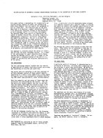
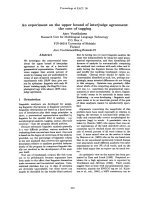
in Table 6. Table 7 shows the correlation of GT-
time and Group number versus globulin con-
centration and albumin/globulin ratio, respec-
tively, by linear regression. The regression
equations are also shown in Graphs 1-4. Group
number did not correlate with the mean fibrino-
gen concentration within groups.
Among the hospitalized horses, there was a
higher fatality rate in the GT positive horses
(69% = 9/13) when compared to the GT nega-
tive horses (38% = 9/24); however, this finding
was not statistically significant (p=0.06, Chi
square test).
Glutaraldehyde test of inflammatory disease in horses 73
Acta vet. scand. vol. 46 no. 1-2, 2005
Table 4: GT result versus mean blood values (+/- standard deviation).
Albumin (g/l) Globulin (g/l) Alb/Glo (ratio) Fibrinogen (g/l)
GT-positive 27,9 (+/- 6,6) 47,5 (+/- 13,7) 0,7 (+/- 0,3) 6,6 (+/- 2,1)
GT-negative 30,7 (+/- 5,7) 29,3 (+/- 8,7) 1,1 (+/- 0,4) 6,6 (+/- 2,9)
All horses 29,7 (+/- 6,1) 36,0 (+/- 13,6) 1,0 (+/- 0,4) 6,6 (+/- 2,6)
Table 5: Clinical parameters versus mean blood values.
GT-positive (%) Albumin (g/l) Globulin (g/l) Alb/Glo (ratio) Fibrinogen (g/l)
Duration Acute 23,1 26,7 43,0 0,6 8,8
Chronic 76,9 28,2 48,9 0,7 5,9
Outcome Fatal 69,2 27,8 44,8 0,7 6,0
Discharged 30,8 28,0 53,8 0,6 8,0
Table 6: GT-time groups versus mean blood values within groups (+/- standard deviation).
Group # GT-positive (No) Mean Globulin (g/l) Mean Alb/Glo (ratio) Mean Fibrinogen (g/l)
1: (0<GT-time<3 min.) 6 55,2 (+/- 11,3) 0,5 (+/- 0,2) 6,3 (+/- 2,7)
2: (3<GT-time<6 min.) 4 48,3 (+/- 8,4) 0,6 (+/- 0,2) 7,4 (+/- 1,7)
3: (6<GT-time<15 min.) 3 31,3 (+/- 11,2) 1,0 (+/- 0,4) 6,0 (+/- 2,0)
4: (GT-time>15 min.) 24 29,3 (+/- 8,7) 1,1 (+/- 0,4) 6,6 (+/- 2,9)
Among the 37 horses, the proportion of test
negatives of horses that were acutely inflamed
was 75% (9/12). The proportion of acutely in-
flamed test negatives was significantly greater
than the proportion of chronically inflamed test
positives (p=0.04, Chi square test). The propor-
tion of test positives of horses that were chron-
ically inflamed was 40% (10/25).
The GT did not show statistically significant
correlation with the concentration of blood fib-
rinogen in acute or chronic diseases.
All results from the blood analyses (Table 3)
were also compared to the GT using principal
component analysis without finding any statis-
tically significant correlation.
Discussion
The results of this study indicate that the GT
can be used to quickly differentiate chronic
from acute inflammatory disease in horses. The
high proportion of test negatives of horses hav-
ing acute inflammation indicates that horses
with inflammatory disease and negative GT are
likely to be acutely, rather than chronically, in-
flamed. Among GT positive horses, 77% were
chronically inflamed as shown in Table 5. The
GT was not reliable in predicting the blood con-
centration of fibrinogen in acute or chronic in-
flammatory diseases.
Useful clinical information could be obtained
by dividing GT-times into categories (groups)
as listed in Table 2 (Liberg et al. 1975a, Liberg
et al. 1975b). Comparison of category and re-
spectively globulin concentration and albu-
min/globulin ratio within a category seemed to
correlate, although this tendency was not statis-
tically significant. This could be due to the
small number of data points. A larger number
of horses included in a future study like ours
would probably eliminate this statistical uncer-
74 P. Brink et al.
Acta vet. scand. vol. 46 no. 1-2, 2005
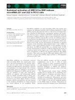
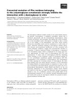
Table 7: GT-time and Group# correlation with globulin concentration and albumin/globulin. Linear regression
and regression coefficient.
Dependent variable Independent variable (equation) r
GT-time - 0,22 [globulin] + 16,33 0,61
GT-time 10,84 [albumin/globulin] - 1,21 0,67
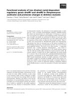
Group # - 0,10 [mean globulin within groups] + 6,50 0,96
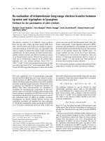
Group # 4,06 [mean albumin/globulin within groups] - 0,83 0,96
Graph 1.
GT-time/globulin
regression.
tainty. The correlation above has been observed
in cattle (Sandholm 1974a, Liberg et al. 1975a,
Liberg et al. 1975b, Nielsen 1975, Eriksen
1984). The difference between other studies of
other species and this study was that horses in
Group 1 had only moderately increased globu-
lin concentration and moderately decreased al-
bumin/globulin ratio, Group 2 horses had a
mildly increased globulin concentration and
mildly decreased albumin/globulin ratio, and
horses in Group 3 had a globulin concentration
and albumin/globulin ratio within normal
range.
If the clinical examination indicates systemic
infection (eg. increased rectal temperature) and
the GT is positive, the probability is high (77%
likelihood) for chronic inflammatory disease. A
positive GT acts then as an indicator for further
laboratory analysis of blood to determine
chronicity and etiology of the disease. If the test
is negative, the disease is most likely acute or
the systemic inflammatory response is either
insignificant or absent. The GT can also be
used as an additional diagnostic test to indicate
prognosis because a positive test predicted fatal
outcome in 69% of the clinical cases we stud-
ied. The test performance regarding the pre-
dictability of a fatal outcome might increase if
only severe inflammatory diseases are included
as compared to a study also including mild
cases (selection bias). Also, the lack of controls
will add bias to the percentages and will elimi-
nate false positives. Because the study did not
include a group of controls and a group of
Glutaraldehyde test of inflammatory disease in horses 75
Acta vet. scand. vol. 46 no. 1-2, 2005
Graph 2.
GT-time/A/G ratio
regression.
Graph 3. Regression of GT time Group vs Mean
Globulin.
Graph 4. Regression of GT-time Group vs Mean Al-
bumin/Globulin Ratio.
60
1,2
horses suffering from non-inflammatory dis-
eases, the data presented can only be consid-
ered valid for horses with inflammatory dis-
ease. For this reason, the conclusions are not
valid for the entire population of horses. The se-
lection of horses among patients submitted to a
large referral hospital also might introduce
spectrum bias as the hospitalized horses are
more likely to be severely affected than horses
treated in practice.
A positive GT in horses indicated the probabil-
ity of increased serum concentration of globu-
lin and a decreased albumin/globulin ratio, but
the GT was not correlated with the blood con-
centration of fibrinogen.
Taking into consideration the low cost and rapid
application of the GT and correlation of a posi-
tive test with increased concentration of globu-
lin, the GT is a useful screening test for horses
suspected to suffer from inflammatory disease.
a) Glutarvac Test tube; Jorgen Kruuse A/S, Marslev,
Denmark.
Acknowledgements
Dr. Joseph Spano is greatly appreciated for his help
and support! This investigation was supported by
grants from the Nortoft Thomsen Foundation, the
Goldschmidt Foundation and the Kruuse Company.
References
Aasted B, Leslie G, Agger R: Immunologi. (Im-
munology). DSR-forlag Landbohøjskolen, Co-
penhagen 1989, p. 29-36.
Beetson SA, Hilbert BJ, Mills JN: The use of the glu-
taraldehyde coagulation test for detection of hy-
pogammaglobulinaemia in neonatal foals. Aust.
Vet. J. 1985, 62, 279-281.
Bendixen HJ: Investigations on the relationship be-
tween the serum proteins and the formol-gel reac-
tion in cattle. Nord. Vet Med. 1954, 6, 187-194.
Bruijn CM, Wensing T, Nieuwstadt RA, Bruijn CM,
Nieuwstadt RA: Een onderzoek naar de betrouw-
baarheid van de glutaaraldehydetest voor de
bepaling van het gammaglobulinegehalte in het
serum van veulens en naar de bruikbaarheid van
deze test in de praktijk bij het controleren van de
colostrumopname bij veulens. (A study of the re-
liability of the glutaraldehyde test to determine
the level of gamma globulin in the serum of foals
and the suitability of this test in practice for the
control of colostrum intake in foals). Tijdschr.
Diergeneesk. 2003, 128, 240-246.
Carstairs-Grant SJ, Crawshaw GJ, Mehren KG: A
comparison of the glutaraldehyde coagulation test
and total serum protein estimation as indicator of
gamma globulin levels in neonatal ruminants. J.
Zoo. Anim. Med. 1988, 19, 14-17.
Chadli M, Mahin L: Test d'etable au glutaraldehyde,
indicateur preliminaire de la pathologie infec-
tieuse chronique au sein d'une exploitation
bovine. (The herd glutaraldehyde test, prelimi-
nary indicator of chronic infectious disease in cat-
tle). Act. Int. Agron. Vet. Hassan II. 1986, 6, 49-
57.
Clabough DL, Conboy HS, Roberts MC: Comparison
of four screening techniques for the diagnosis of
equine neonatal hypogammaglobulinemia. J. Am.
Vet. Med. Assoc. 1989, 194, 1717-1720.
Clabough DL, Levine JF, Grant GL, Conboy HS: Fac-
tors associated with failure of passive transfer of
colostral antibodies in standardbred foals. J. Vet.
Intern. Med. 1991, 5, 335-340.
Doll K, Schillinger D, Klee W: Der Glutaraldehyd-
Test beim Rind-seine Brauchbarkeit fur Diagnose
und Prognose innerer Entzundungen. (Suitability
of the glutaraldehyde test for diagnosis and prog-
nosis of internal inflammatory conditions in cat-
tle). Zentbl. Vet. Med. 1985, 32, 581-593.
Eriksen L: Klinisk undersøgelsesmetodik og journal-
skrivning. (Methods in clinical examination and
record writing). CF Mortensen A/S, Copenhagen
1984, p. 87-88+134.
Ezhilan V, Bhuvanakumar CK: Forecasting of IgG in
neonates from colostrum. Centaur Mylap. 1998,
15, 39-40.
Hansen K: Glutaraldehydprøvens anvendelighed på
svin, der ønskes nødslagtet. (The applicability of
the glutaraldehyde test in slaughter swine). Dansk
Vet. Tidsskr. 1985, 68, 151-156.
Jones D, Brook D: Investigation of the Gamma-
Check-C test as a mean of evaluating IgG levels in
equine colostrum. J. Equine Vet. Sci. 1995, 15,
269-271.
Juyal PD, Uppal SK: Determination of gamma glob-
ulins in young buffalo calves by glutaraldehyde
coagulation test. Indian J. Anim. Health 1995, 34,
161-162.
Kalinbacak A, Or ME: Yenidogan taylarda hipogam-
76 P. Brink et al.
Acta vet. scand. vol. 46 no. 1-2, 2005
maglobulinemi'nin saptanmasinda glutaraldehit
koaglasyon testi'nin kullanimi. (Use of the glu-
taraldehyde coagulation test to detect hypogam-
maglobulinaemia in newborn foals). Vet. Fakult.
Dergisi Ankara Univ. 1996, 43, 203-207.
Kantor IN, Lopez B, Torres P, Nader A, Garcia V, De-
Kantor IN: Preliminary evaluation of a simple
method for detection of bovine tuberculosis: the
glutaraldehyde test. J. Vet. Med. Series B. 1993,
40, 27-30.
Katholm J, Jorgensen RJ: Glutaraldehyd test. Til
"cow side" påvisning af colimastitis. (The glu-
taraldehyde test for "cow side" diagnosis of acute
coliform mastitis). Dansk Vet. Tidsskr. 1992, 75,
486-487.
Keulen KAS, Dobbelaar P, Noordhuizen JPTM,
Schwering C, Wensing T: Een onderzoek naar een
aantal aspecten van de biestverstrekking op
melkveebedrijven en naar de bruikbaarheid van
de glutaaraldehydetest bij de beoordeling van de
biestverstrekking. (Studies on a number of fea-
tures of the supply of colostrum on dairy farms
and the use of the glutaraldehyde test in evaluat-
ing the supply of colostrum). Tijdschr. Dierge-
neesk. 1984, 109, 605-611.
Kovac G: Diagnosis of hypogammaglobulinemia in
calves by means of the glutaraldehyde coagula-
tion test. Folia Vet. 1988, 32, 71-78.
Kovac G, Bartko P, Mudron P, Michna A, Bires J, Bal-
dovic J: Glutaraldehydovy koagulacny test u osi-
panych. (Glutaraldehyde coagulation test in pigs)
Sloven. Vet. Casop. 1993, 18, 66-68.
Kumaran D, Bhuvanakumar CK: Detection of im-
munoglobulin levels in neonatal foals. Centaur
Mylap. 1994, 10, 98-100.
Larsson B: The relationship between total protein in
serum, glutaraldehyde coagulation test and dis-
ease in feedlot calves. Nord. Vet Med. 1985, 37,
90-96.
Liberg P, Pehrson B, Sandholm M: Snabbtest för di-
agnosticering av inflammatoriska tilständ hos
nötkreatur. (Rapid test for diagnosis of inflamma-
tory disease in cattle). Svensk Vet. Tidn. 1975a,
27, 181-183.
Liberg P, Pehrson B, Sandholm M: The value of the
glutaraldehyde and formaldehyde tests in evalua-
tion of the globulin level in bovine blood. Acta
Vet. Scand. 1975b, 16, 236-243.
Liberg P: The fibrinogen concentration in blood of
dairy cows and its influence on the interpretation
of the glutaraldehyde and formol-gel test reac-
tions. Acta Vet. Scand. 1978, 19, 413-421.
Liberg P: Helblodstestning med glutaraldehyd vid
svinslakt - en preliminär undersökning. (Glu-
taraldehyde test on whole blood of slaughter
swine). Nord. Vet Med. 1979, 31, 360-366.
Liberg P: Glutaraldehyde and formol-gel tests in
bovine traumatic peritonitis. Acta Vet. Scand.
1981, 22, 78-84.
Liberg P: Blood protein screening in healthy and dis-
eased cattle. Agarose gel electrophoresis, the for-
mol-gel and glutaraldehyde tests. The Swedish
Veterinary University, Uppsala 1982.
Mahlin L, Chadli M, Marzou A, Maach L, Ychou M:
Differences in coagulability of three glutaralde-
hyde solutions in the glutaraldehyde test on
bovine whole blood. Zentbl. Vet. Med. 1985, 32,
151-154.
Martens HH: Untersuchungen mit der Glutaralde-
hydprobe nach Sandholm im Vollblut gesunder
und kranker Rinder. (Application of Sandholm's
glutaraldehyde test to whole blood from healthy
and diseased cattle). Thesis, Hannover 1977.
Martin DWJ, Mayes PA, Rodwell VW, Granner DK:
Harper's review of biochemistry. 20th ed. Lange
Medical Publications, California 1985, p. 637-
645.
Nansen P, Nielsen K: Metabolism of bovine im-
munoglobulin. 1. Metabolism of bovine IgG in
cattle with chronic pyogenic infections. Can. J.
Comp. Med. Vet. Sci. 1966, 30, 327-331.
Nielsen K: Glutaraldehydprøven, en metode til påvis-
ning af forhøjet immunoglobulin koncentration i
blod. (The glutaraldehyde test, a method for de-
termination of increased concentration of im-
munoglobulin in blood). Dansk Vet. Tidsskr.
1975, 58, 652-655.
O'-Rourke KI, Satterfield WC: Glutaraldehyde coag-
ulation for detection of hypogammaglobulinemia
in neonatal nondomestic ruminants. J. Am. Vet.
Med. Assoc. 1981, 179, 1144-1146.
Ramprabhu R, Dhanapalan P, Prathaban S: Efficacy
of Sulkowitch and glutaraldehyde tests in trau-
matic reticuloperitonitis and allied syndrome in
cattle. Indian J. Anim. Health 2002, 41, 74-76.
Saikku A, Koskinen E, Sandholm M: Detection of hy-
pogammaglobulinaemia in neonatal foals using
the glutaraldehyde coagulation test. J. Vet. Med.
Series B. 1989, 36, 168-174.
Sandholm M, Kangas J:
Coagulation of hyperglobu-
linaemic mink blood by glutaraldehyde. Zentbl.
Vet. Med. 1973, 20B, 206-211.
Sandholm M: A preliminary report of a rapid method
for the demonstration of abnormal gammaglobu-
Glutaraldehyde test of inflammatory disease in horses 77
Acta vet. scand. vol. 46 no. 1-2, 2005
lin levels in bovine whole blood. Res. Vet. Sci.
1974a, 17, 32-35.
Sandholm M: Die Feststellung der Hyper-ã-Globu-
linämie beim Rind unter Praxisbedingungen.
(The determination of hyperglobulinemia in cat-
tle under practice conditions). Tierärztl. Prax.
1974b, 2, 237-240.
Sandholm M, Kivisto AK: Determination of gamma
globulin in dog serum by glutaraldehyde. J. Small.
Anim. Pract. 1975, 16, 201-205.
Satpathy PK, Dutta NK, Mishra PR, Kar BC: Glu-
taraldehyde coagulation test: standard curve and
its applications to detect gammaglobulin level in
kids. Indian Vet. J. 1996, 73, 257-260.
Sen I, Basoglu A, Ok M, Birdane FM, Guzelbektas H,
Civelek T: Neonatal ishalli buzagilarda serum im-
munoglobulinlerin glutaraldehid koagulasyon
testi ile degerlendirilmesi. (Serum immunoglobu-
lins in neonatal diarrhoeic calves evaluation by
glutaraldehyde coagulation test). Vet. Bilim. Der-
gisi 2000, 16, 143-146.
Tennant B, Baldwin BH, Braun RK, Norcross NL,
Sandholm M: Use of the glutaraldehyde coagula-
tion test for detection of hypogammaglobuline-
mia in neonatal calves. J. Am. Vet. Med. Assoc.
1979, 174, 848-853.
Tyler JW, Besser TE, Wilson L, Hancock DD, Sanders
S, Rea DE: Evaluation of a whole blood glu-
taraldehyde coagulation test for the detection of
failure of passive transfer in calves. J. Vet. Intern.
Med. 1996, 10, 82-84.
Vihan VS: Glutaraldehyde coagulation test for detec-
tion of hypo-gammaglobulinaemia in neonatal
kids. Indian Vet. J. 1989, 66, 101-105.
Wolff B: Test a la glutaraldehyde: une method d'ap-
point dans le diagnostic du pyometre chez la chi-
enne. (Glutaraldehyde test: a supplementary diag-
nostic method for pyometra in the bitch). Point
Vet. 1986, 18, 69-71.
Sammendrag
Glutaraldehydprøvens evne til at skelne mellem akut
og kronisk inflammatorisk sygdom hos hest.
Glutaraldehydprøven (GP), en hurtig og billig test,
har været anvendt empirisk gennem mange år i
kvægpraksis for diagnosticering af inflammatoriske
sygdomme. GP bliver primært brugt til at påvise øget
serum koncentration af fibrinogen og globulin. Glu-
taraldehyd bindes til frie amino-grupper i fibrinogen
og globulin, som derpå danner et blodkoagel ved en
1. grads kemisk reaktion. Koaguleringstiden af GP
estimerer indholdet af de proteiner, som produceres i
et inflammatorisk respons. Anvendeligheden af GP
til diagnosticering af inflammatoriske tilstande i he-
stepraksis har aldrig været undersøgt før. Formålet
med dette studie er at bestemme GPs evne til at
skelne mellem akut og kronisk inflammatorisk syg-
dom hos hest. 37 heste, mistænkt for inflammatorisk
sygdom, blev evalueret på basis af GP, anamnese,
fuldstændig klinisk undersøgelse samt rutinemæssig
blodprøver. GP-tid, blodprøvesvar og klinisk udfald
blev sammenlignet statistisk. De heste, som var be-
stemt til at være akut afficeret på basis af anamnese,
klinisk undersøgelse og rutinemæssig blodprøve,
tenderede mod at have negativ GP (75%). Der kunne
ikke påvises sammenhæng mellem GP og fibrinogen
koncentration i blodet. Positiv GP forudsagde også et
fatalt udfald i 69% af de kliniske tilfælde. Resulta-
terne af dette studie indikerer, at GP kan være en
brugbar praktisk test til at skelne mellem akut og kro-
nisk inflammatorisk sygdom hos hest.
78 P. Brink et al.
Acta vet. scand. vol. 46 no. 1-2, 2005
(Received March 29, 2005; accepted March 30, 2005).
Reprints may be obtained from: Palle Brink, ATG Equine Clinic, Jägersro, 21237 Malmö, Sweden.