Báo cáo y học: " CpG oligonucleotide activates Toll-like receptor 9 and causes lung inflammation in vivo" ppsx
Bạn đang xem bản rút gọn của tài liệu. Xem và tải ngay bản đầy đủ của tài liệu tại đây (402.52 KB, 9 trang )
BioMed Central
Page 1 of 9
(page number not for citation purposes)
Respiratory Research
Open Access
Research
CpG oligonucleotide activates Toll-like receptor 9 and causes lung
inflammation in vivo
Pascal Knuefermann*
†1
, Georg Baumgarten
†1
, Alexander Koch
2
,
Markus Schwederski
1
, Markus Velten
1
, Heidi Ehrentraut
1
, Jan Mersmann
3
,
Rainer Meyer
4
, Andreas Hoeft
1
, Kai Zacharowski
2
and Christian Grohé
5
Address:
1
Department for Anesthesiology and Intensive Care Medicine, University Hospital Bonn, Sigmund-Freud-Strasse 25, 53125 Bonn,
Germany,
2
Molecular Cardioprotection & Inflammation Group, Department of Anesthesia, Bristol Royal Infirmary, Bristol BS2 8HW, UK,
3
Molecular Cardioprotection & Inflammation Group, Department of Anesthesia, University Hospital Düsseldorf, Moorenstrasse 5, 40225
Düsseldorf, Germany,
4
Institute of Physiology II, University Hospital Bonn, Wilhelmstrasse 31, 53111 Bonn, Germany and
5
Department of
Internal Medicine, University Hospital Bonn, Sigmund-Freud-Strasse 25, 53125 Bonn, Germany
Email: Pascal Knuefermann* - ; Georg Baumgarten - ;
Alexander Koch - ; Markus Schwederski - ; Markus Velten -
bonn.de; Heidi Ehrentraut - ; Jan Mersmann - ;
Rainer Meyer - ; Andreas Hoeft - ;
Kai Zacharowski - ; Christian Grohé -
* Corresponding author †Equal contributors
Abstract
Background: Bacterial DNA containing motifs of unmethylated CpG dinucleotides (CpG-ODN)
initiate an innate immune response mediated by the pattern recognition receptor Toll-like receptor
9 (TLR9). This leads in particular to the expression of proinflammatory mediators such as tumor
necrosis factor (TNF-α) and interleukin-1β (IL-1β). TLR9 is expressed in human and murine
pulmonary tissue and induction of proinflammatory mediators has been linked to the development
of acute lung injury. Therefore, the hypothesis was tested whether CpG-ODN administration
induces an inflammatory response in the lung via TLR9 in vivo.
Methods: Wild-type (WT) and TLR9-deficient (TLR9-D) mice received CpG-ODN
intraperitoneally (1668-Thioat, 1 nmol/g BW) and were observed for up to 6 hrs. Lung tissue and
plasma samples were taken and various inflammatory markers were measured.
Results: In WT mice, CpG-ODN induced a strong activation of pulmonary NFκB as well as a
significant increase in pulmonary TNF-α and IL-1β mRNA/protein. In addition, cytokine serum
levels were significantly elevated in WT mice. Increased pulmonary content of lung
myeloperoxidase (MPO) was documented in WT mice following application of CpG-ODN.
Bronchoalveolar lavage (BAL) revealed that CpG-ODN stimulation significantly increased total cell
number as well as neutrophil count in WT animals. In contrast, the CpG-ODN-induced
inflammatory response was abolished in TLR9-D mice.
Conclusion: This study suggests that bacterial CpG-ODN causes lung inflammation via TLR9.
Published: 9 October 2007
Respiratory Research 2007, 8:72 doi:10.1186/1465-9921-8-72
Received: 30 January 2007
Accepted: 9 October 2007
This article is available from: />© 2007 Knuefermann et al; licensee BioMed Central Ltd.
This is an Open Access article distributed under the terms of the Creative Commons Attribution License ( />),
which permits unrestricted use, distribution, and reproduction in any medium, provided the original work is properly cited.
Respiratory Research 2007, 8:72 />Page 2 of 9
(page number not for citation purposes)
Background
Acute lung injury (ALI) or its severe form, the acute respi-
ratory distress syndrome (ARDS) remains a major health
problem. Recent studies have estimated the incidence of
these conditions to be between 15 and 34 cases per
100,000 inhabitants per year showing an overall mortality
rate of 30–40% [1-3]. Depending on the underlying etiol-
ogies ARDS can be differentiated into a direct (pulmo-
nary) and an indirect (extrapulmonary) form (for details
see [4]).
ALI/ARDS are quite common in patients with sepsis [5]
and sepsis-associated ARDS carries the highest mortality
rates. Despite advances in the supportive care and
mechanical ventilation strategies of ALI/ARDS, mortality
rates remain unacceptably high [6-8]. As the pathophysi-
ology of the disease is not fully understood, the treatment
remains mainly supportive [9-13].
Experimental models of sepsis show that bacteria and bac-
terial cell components induce the expression of inflamma-
tory mediators in various tissues as well as in the blood
stream [14-17]. Among these mediators, proinflamma-
tory cytokines are regarded as a major cause for the devel-
opment of organ dysfunction during sepsis [18,19].
Bacterial DNA can initiate an innate immune response via
Toll-like receptor 9 (TLR9) potentially leading to septic
shock [20,21], septic arthritis [22], or meningitis [23]. The
bacterial genome, compared to vertebrate DNA, contains
a higher frequency of unmethylated cytosine-phosphate-
guanine (CpG) dinucleotides. Small oligodeoxynucle-
otides (ODN) with unmethylated CpG dinucleotides
(CpG-ODN) are able to perfectly mimic the immunostim-
ulatory activity of bacterial DNA since bacterial DNA and
synthetic oligodeoxynucleotides share similar base
sequences and bind to the same receptor system (TLR9)
[24-26].
The identification of TLRs has been a major advance in the
understanding of the pathogenesis of septic shock [27]. To
date, 13 TLRs (TLR1-13) have been described and TLR2
and TLR4 are the best-characterized receptors so far
[28,29]. TLR2 detects gram-positive bacterial cell wall
components, while TLR4 can recognize cell wall compo-
nents of gram-negative bacteria [30,31].
Little is known about the role of TLR9 in the lung, but
constitutive expression levels have been detected in
human and mouse lung endothelial cells and mouse
RAW264.7 cells. High TLR9 expression levels have been
found in lung tumors [15,32,33]. Others have shown that
CpG-ODN contributes to local inflammation of the lung
following intratracheal instillation [32,34]. However, to
our knowledge nothing is known regarding systemic
effects of CpG-ODN and pulmonary inflammation.
Therefore, we injected bacterial DNA intraperitoneally to
answer the question whether bacterial DNA induces lung
inflammation in a TLR9-dependent manner.
Methods
Animals
TLR9-deficient (TLR9-D) mice [25], back-crossed onto a
C57BL/6 background were handled according to the prin-
ciples of laboratory animal care (NIH publication No. 86-
23, revised 1985) and experimental procedures were
approved by the German government ethical and research
boards (50.203.2-BN 43, 28/01).
SIRS Model
The standard protocol for stimulation consisted of D-
galactosamine sensitization (D-GalN; Roth, Karlsruhe,
Germany) intraperitoneally (i.p.) with 1 mg/kg. 30 min
later, mice received i.p. either 1 mL/kg saline (sal) or 1
nmol/g CpG-ODN (Thioat 1668; containing a "CG-
motif": 5'-TCC-ATG-ACG-TTC-CTG-ATG-CT; TibMolBiol,
Berlin, Germany). The stimulatory dose of 1 nmol/g BW
was chosen according to earlier studies [20,21,25], which
was sufficient to induce clinical symptoms of sepsis.
Organs were harvested at 1, 2, 4 and 6 hours after stimu-
lation with CpG-ODN. Unless otherwise stated in the
manuscript groups consisted of 5 animals. In control
experiments, stimulation with D-GalN alone for up to 6
hrs did not influence the mRNA expression of TNF-α, IL-
1β and IL-6 detected by RNase Protection Assay.
Additional experiments were carried out injecting CpG-
ODN intratracheally to further understand its effect dur-
ing lung inflammation. Intratracheally, CpG-ODN was
administered at a dose of 1 nmol/g BW. After intratracheal
administration, lung myeloperoxidase, cytokine expres-
sion and leukocyte count were studied.
Real-Time PCR for TLR9
Total RNA from murine tissue was isolated with the gua-
nidinum thiocyanate method [35]. RNA concentration
was determined by absorbance at 260 nm. Until further
processing, RNA was dissolved in 100 μL of RNase-free
water and stored at -80°C. Reverse transcription was per-
formed using QIAGEN Omniscript Reverse Transcription
kit (Qiagen, Hilden, Germany) according to the manufac-
turer's protocol. 1 μg RNA was used in 20 μL reaction mix-
tures containing 2 μL 10× Reverse Transcription Buffer, 2
μL dNTP mixture (5 mM of each dNTP), 1 μL Omniscript
Reverse Transcriptase and 2 μL oligo-dT primers. The spe-
cific pre-made TaqMan
®
Gene Expression Assays (Applied
Biosystems, Foster City, CA, USA) for murine TLR9
(Mm00446193 m1, amplicon length: 60 bp) and murine
GAPDH (Mm999999915 q1) as housekeeping gene were
used in this study. Real-time PCR was performed accord-
Respiratory Research 2007, 8:72 />Page 3 of 9
(page number not for citation purposes)
ing to the manufacturer's protocol. 100 ng of single-
stranded cDNA was mixed with supplied 2 × TaqMan Uni-
versal Master Mix (PN 4304437, Applied Biosystems, Fos-
ter City, CA, USA) and 1 μL of TaqMan
®
Gene Expression
Assay to a final volume of 10 μl in a 384-well optical reac-
tion plate. Each sample underwent 40 cycles of amplifica-
tion in a 384-well optical reaction plate on an ABI PRISM
®
Sequence Detection Systems (Applied Biosystems, Foster
City, CA, USA). Relative quotients (RQ) of TLR9 gene
expression comparing control mice with stimulated mice
at different time-points were calculated with SDS Software
2.2 (Applied Systems, Foster City, CA, USA). RQ results
were analyzed with GraphPad Prism 4.05 (GraphPad Soft-
ware, San Diego, USA).
Western Blot Analysis for TLR9
Tissue cells were lysed in ice-cold buffer (150 mM NaCl,
50 mM Tris-HCl, pH 7.4, 1 mM EDTA, 5 μg/mL Leupep-
tin, 5 μg/mL aprotinin, 1 mM PMSF, 0.1% SDS, 1%
sodium deoxycholate, 1% Triton X-100) as previously
published [36]. After brief centrifugation (16.800 g),
supernatants were removed, total protein was determined
(bicinchoninic acid method), separated by SDS-PAGE
and blotted onto nitrocellulose membranes. The blots
were incubated with anti-TLR9-antibody (1:1,000, IMG-
431, Imgenex San Diego, CA, USA) at 4°C overnight.
Horseradish peroxidase (HRP)-conjugated anti-rabbit sec-
ondary antibody (1:3,000, GE Healthcare Europe, Braun-
schweig, Germany) was used. Signals were visualized by
enhanced chemiluminescence.
Pulmonary nuclear and cytoplasmic extraction
Pulmonary protein extracts were prepared with NE-PER™
Nuclear and Cytoplasmic Extraction Reagents (Perbio,
Bonn, Germany) according to the manufacturer's proto-
col [37].
Electrophoretic mobility shift assay (EMSA)
NFκB oligonucleotides were end-labeled with [γ-32P]
ATP. Binding reactions (25 μL total) were performed with
nuclear extracts and the specificity of the DNA-protein
binding was determined by cold chase analysis as well as
with supershift assays. Nuclear extracts were incubated
with 2 mg of polyclonal anti-p50 or anti-p65 antibody.
DNA-protein complexes were electrophoresed, gels were
dried, exposed overnight and scanned with a phosphoim-
ager (FLA3000, Fuji film Europe, Düsseldorf, Germany ).
Ribonuclease protection assay
Pulmonary RNA was extracted with the guanidinium thi-
ocyanate method [35]. The mRNA-expression was deter-
mined with an RNase protection assay system [16].
Pulmonary TNF-
α
and IL-1
β
protein expression
Pulmonary tissue was homogenized and incubated on ice
for 5 min in 1 mL of ELISA buffer containing PBS, Triton
X-100 (1 μL/mL), PMSF (250 mM in isopropanol, 1 μL/
mL) and protease inhibitors. Samples were incubated on
ice for 20 min, homogenized and centrifuged for 15 min
at 4°C. TNF-α and IL-1β were determined in the superna-
tant using ELISA (R&D systems, Minneapolis, MN, USA).
Plasma Cytokine Levels
Blood samples for plasma cytokine levels were obtained
by cardiac puncture. Plasma levels of TNF-α, IL-1β and IL-
6 (Mouse Cytokine multi-Plex for Luminex™ laser, Bio-
Source Europe, Nivelles, Belgium) were determined using
the microsphere array technique (Luminex 100 system,
Luminex Corp., Austin, TX, USA) as previously described
[36].
Lung Myeloperoxidase (MPO)-Assay
The MPO-Assay was performed as previously described
[38] with some minor modifications. Data are expressed
as % of controls.
Bronchoalveolar lavage (BAL) and cell counts
BAL was performed as described elsewhere [39]. Briefly, 4
h after CpG-ODN application, control- and TLR9-D mice
were anaesthetized with isoflurane (Forene
®
; Abbott
GmbH, Wiesbaden, Germany), and a midline incision
was made to expose the trachea. An 18-G catheter was
inserted into the trachea, and the lungs were lavaged two
times with 500 μL PBS. Approximately 50–70% of the
instilled volume was retrieved. All samples were kept on
ice until processed. Total and differential cell counts in
BAL fluid were determined. Subpopulations of leukocytes
were determined using as hemocytometer.
Leukocyte count
Lung tissue was fixed in 4% paraformaldehyde over night,
embedded in paraffin and cut into 5 μm sections. Hema-
toxylin and Eosin (H&E) staining was performed using
standard protocols and leukocyte accumulation was
quantified. A total of ten microscopic fields covering 1
mm
2
were photographed and leukocytes were counted by
a blinded investigator.
Statistical Evaluation
All values are expressed as mean ± SEM. One-way or two-
way ANOVA followed by Bonferroni-corrected post-hoc
analysis was used when appropriate. T-test was applied for
analysis of cell counts from bronchoalveolar lavage. Sig-
nificant differences were considered to exist at p ≤ 0.05.
Respiratory Research 2007, 8:72 />Page 4 of 9
(page number not for citation purposes)
Results
Clinical manifestation of inflammation
Clinical symptoms of inflammation were monitored after
CpG-ODN application in WT and TLR9-D mice. 2 hrs
after CpG-ODN challenge, WT mice developed shock-like
symptoms including ruffled hair, eye exudates, and leth-
argy, while TLR9-D mice were not affected.
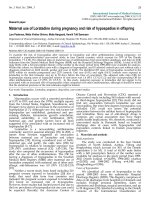
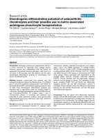
Pulmonary gene and protein expression of TLR9
The expression of TLR9 in whole native pulmonary tissue
was demonstrated using Real-time PCR and Western-blot
analysis. Both techniques showed a constitutive expres-
sion of TLR9 (Figure 1A–C). However, neither the mRNA
nor the protein expression pattern significantly changed
after agonist treatment with CpG-ODN (up to 6 hrs).
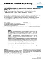
NF
κ
B activation in the lung after CpG-ODN stimulation
Systemic CpG-ODN treatment led to a time-dependent
(maximum at 2 hrs) substantial activation of pulmonary
NFκB in WT mice. In contrast, this effect was not detecta-
ble in TLR9-D mice (Figure 2).
Pulmonary cytokine mRNA expression after CpG-ODN
challenge
CpG-ODN induced a rapid and robust increase in TNF-α
and IL-1β mRNA transcripts in lungs of WT mice (Figure
3A). Densitometry (Figures 3B and 3C) revealed that peak
cytokine expression occurred 2 hrs after injection of CpG-
ODN and was not present in TLR9-D mice (p ≤ 0.05).
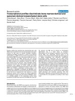
Pulmonary cytokine protein expression following CpG-
ODN challenge
To determine whether increased mRNA expression paral-
leled also increased cytokine protein levels in the lung, we
tested the in vivo induction of TNF-α and IL-1β protein
expression in WT and TLR9-D mice by ELISA. Figures 4A
and 4B illustrate that CpG-ODN administration led to a
significant increase in protein expression of TNF-α and IL-
1β in pulmonary tissue from control mice. A significant
increase in cytokine production can be observed 1 hr after
injection of CpG-ODN with a peak protein expression at
2 hrs. At 2 hrs, TNF-α and IL-1β protein levels were signif-
icantly higher in WT compared to TLR9-D mice. Figures
4A and 4B show that the kinetics of TNF-α and IL-1β pro-
tein production parallels the up-regulation of the corre-
sponding mRNA-transcripts.
To exclude solely extrapulmonary effects of CpG-ODN on
the lung, WT- and TLR9-D mice received CpG-ODN also
intratracheally. This route of administration again
resulted in lung inflammation, e.g. demonstrated by a sig-
nificant cytokine response in WT animals. 2 hrs after CpG-
ODN challenge, pulmonary TNF-α tissue levels were sig-
nificantly increased in WT mice (7.0 ± 0.6 pg/mg tissue)
when compared to TLR9-D animals (0.6 ± 0.2 pg/mg tis-
sue; p < 0.05). Also IL-1β levels were significantly raised in
WT mice (62 ± 12 pg/mg tissue) when compared to TLR9-
D animals (16 ± 1 pg/mg tissue; p < 0.05).
Plasma cytokine levels following CpG-ODN challenge
CpG-ODN-treated WT animals showed a significant
increase in the plasma levels of the cytokines TNF-α and
IL-6 after 2 hrs. Similarly, plasma levels of IL-1β increased
as well after 2 hrs without reaching statistical significance.
These effects were not detectable in CpG-ODN-treated
TLR9-D mice (Figure 5). After 6 hrs, cytokine levels in WT
mice return to baseline levels.
MPO activitiy
In WT mice, MPO increased significantly 6 hrs after i.p.
CpG-ODN stimulation. This effect was not detectable in
TLR9-D mice (Figure 6).
Pulmonary expression of TLR9Figure 1
Pulmonary expression of TLR9. TLR9 expression in the
lung was detected by Real-time PCR (A) and by Western
blot analysis (B, C). All data were normalized to control (0 h)
(C). TLR9 was present even under base line conditions; how-
ever, no significant increase in TLR9 was observed after
CpG-ODN stimulation (n = 3/group).
01246
0
1
2
3
4
RQ (mTLR9/mGAPDH)
time (h)
01246positive control
0.0
0.5
1.0
1.5
2.0
2.5
3.0
relative TLR9 protein expression
time (h)
A
time (h) 0
1
2
46
positive
control
TLR9
B
C
Respiratory Research 2007, 8:72 />Page 5 of 9
(page number not for citation purposes)
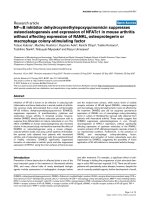
Bronchoalveolar lavage (BAL) after CpG-ODN stimulation
BALs demonstrated a significant increase in total cell
number as well as the number of recruited neutrophils
after CpG-stimulation in WT animals (Figure 7), which
was diminished in TLR9-D mice. BALs obtained from all
animal groups were not contaminated by peripheral
blood cells indicating cell migration into the lungs.
Leukocyte count
Under base line conditions, only a few leukocytes were
detectable in both genotypes (WT: 212 ± 25 leukocytes/
mm
2
; TLR9-D: 218 ± 34 leukocytes/mm
2
). 6 hrs after
intratracheal stimulation, leukocyte accumulation was
induced in both mouse strains. However, the detectable
levels in the lungs of WT mice were significantly higher
than those of TLR9-D animals (n = 5/group; 9465 ± 689
vs. 3509 ± 55 leukocytes/mm
2
, p < 0,05).
Discussion
Acute lung injury represents acute hypoxemic respiratory
failure and is associated with pulmonary and non-pulmo-
nary risk factors. Interestingly, direct lung injury caused by
bacteria and indirect lung injury associated with sepsis
share similar pathophysiological pathways.
The initial host's defense against bacterial infections is
essentially executed by pattern-recognition receptors. TLR
2, 4 and 5 have been implicated in bacterial signaling,
innate immunity and lung inflammation [40-44]. Little is
known about the role of TLR9 in the lung, but constitutive
expression levels have been detected in mouse lung
endothelial cells, mouse RAW264.7 cells, rat pulmonary
microvascular endothelial cells and rat pulmonary artery
endothelial cells [15]. High TLR9 expression levels have
been found in lung tumors [15,32,33,45]. Interestingly,
TLR9 is not expressed in all cells present in the lung. For
instance, TLR9 is absent in rat pulmonary arterial smooth
muscle cells [15], mouse pulmonary macrophages [46]
and in lung dendritic cells [47]. This is in conflict with
other reports demonstrating the existence of TLR9 in lung
dendritic cells [46-48].
It is thought that TLR9 is able to enhance the uptake of
long-chain double-stranded (ds) DNA, although single-
stranded (ss) CpG-ODNs appear to be sequence-inde-
pendently endocytosed. TLR9 is localized in the endoplas-
matic reticulum and following CpG stimulation recruited
to endosomal vesicles. Then, TLR9 and CpG-ODN co-
localize resulting in cell activation [49,50]. The exact
molecular structure of TLR9 is unknown, although some
evidence exists that leucine-rich repeats are responsible
for the recognition of distinct pathogen structures by
TLRs. Following CpG-ODN binding, TLR9 associates with
the adaptor molecule MyD88 resulting in activation of the
IL-1 receptor-associated kinase (IRAK) family, mitogen
activated kinases (MAPK), or IFN regulatory factors. The
latter events activate NFκB among other transcription fac-
tors (for detailed review please refer to [51]).
Our study demonstrates a TLR9-dependent mechanism of
lung inflammation. This is supported by the finding that
an intraperitoneal application of CpG-ODN (extrapulmo-
nary stimulus) leads to a systemic and local inflammatory
response in WT mice, which was abolished in TLR9-D ani-
mals. Our data are in accordance with others that TLR9 is
expressed in homogenisates of pulmonary tissue
[15,32,33]. In addition, we observed that CpG-ODN chal-
lenge did not significantly change TLR9 expression over
time. In gram-negative sepsis TLR4 expression in murine
lungs did also not change; however, the expression of
Activation of NFκB in the lungFigure 2
Activation of NFκB in the lung. A strong increase in pulmonary NFκB-DNA binding activity was observed in WT mice
within 2 hrs after stimulation with CpG-ODN, whereas there was only a reduced NFκB-DNA binding activity in TLR9-D mice
detectable by EMSA.
time (h)01 246 01246
WT + CpG-DNA TLR9-D + CpG-DNA
NFNB
Respiratory Research 2007, 8:72 />Page 6 of 9
(page number not for citation purposes)
CD14, a co-receptor of TLR4, was up-regulated [44]. This
may indicate that TLRs are differentially regulated. It is
known that TLR9 stimulation leads to the activation of
NFκB in various tissues [51]. To our knowledge, our study
shows for the first time that pulmonary NFκB activity is
up-regulated following CpG-ODN application. This is fur-
ther supported by the observation that NFκB is not acti-
vated in TLR9-D animals upon CpG-ODN stimulation. In
addition, CpG-ODN led to a significant increase of NFκB-
dependent, proinflammatory cytokine expression (TNF-
α, IL-1β) in pulmonary tissue. However, CpG-ODN did
not induce an inflammatory response in TLR9-D mice
indicating a TLR9-dependency. In correspondence with
the presented gene expression of proinflammatory
cytokines, the protein expression of TNF-α and IL-1β was
significantly higher in WT animals when compared to
TLR9-D mice. Furthermore, plasma levels of TNF-α and
IL-6 indicate systemic inflammation in WT animals. In
contrast, levels of these cytokines did not change in TLR9-
D mice after CpG-challenge. This further supports our
concept that CpG-ODN mediates its proinflammatory
effects via TLR9. In a small pilot study we could confirm
findings from others [34,52] that local (intratracheally)
CpG-ODN administration also caused an inflammatory
response in the lung (pulmonary stimulus), which was
absent in TLR9-D mice. These findings suggest that CpG-
ODN-induced lung inflammation can be initiated by
both, local and systemic TLR9 activation.
Increased content of lung myeloperoxidase activity, an
indicator of polymorphonuclear cells (PMNs) accumula-
tion, was documented in WT mice following application
Expression of pulmonary TNF-α and IL-1β proteinFigure 4
Expression of pulmonary TNF-α and IL-1β protein.
Expression of pulmonary TNF-α (A) and IL-1β (B) detected
by ELISA in WT and TLR9-D mice at different time points
following CpG-ODN stimulation. Results were normalized
to total protein content of lung tissue. A maximum in
cytokine production was observed 2 hrs after CpG-ODN
challenge. TNF-α (A) and IL-1β (B) protein expression were
significantly higher in WT compared to TLR9-D mice (mean
± SEM; * p < 0.05).
A
B
0 1 2 4 6
0
100
200
300
400
* *
time (h)
IL-1 (pg/mg protein)
0 1 2 4 6
0
5
10
15
20
*
*
TNF- (pg/mg protein)
WT
TLR9-D
Pulmonary proinflammatory cytokine gene expressionFigure 3
Pulmonary proinflammatory cytokine gene expres-
sion. Time course of pulmonary proinflammatory cytokine
gene expression of TNF-α and IL-1β and the house keeping
gene L32 following CpG-ODN stimulation in WT and TLR9-
D mice. Densitometric analysis of the RNase Protection
Assays revealed significant increases of TNF-α-mRNA/L32-
mRNA (B) and IL-1β-mRNA/L32-mRNA (C) in WT mice at
1 hr and 2 hrs compared to TLR9-D animals (mean ± SEM; *
p < 0.05; AU = arbitrary units).
Į
E
0 1 2 4 6
0
3
6
9
12
15
*
*
AU: TNF- -mRNA/L32-mRNA
0 1 2 4 6
0
25
50
75
100
**
time (h)
AU: IL-1 -mRNA/L32-mRNA
Respiratory Research 2007, 8:72 />Page 7 of 9
(page number not for citation purposes)
of CpG-ODN. In WT mice, MPO increased significantly 6
hrs after CpG-ODN stimulation, whereas TLR9-D mice
exhibited no increase in MPO activity. To further charac-
terize the cellular recruitment in the pulmonary system
after CpG-ODN induced inflammation a series of BALs
were carried out. Since PMNs are rarely found in BAL from
normal pathogen-free mice, we used this cell type as an
inflammatory marker. We found a significant induction of
total cell count in WT mice after CpG-ODN challenge. In
particular, neutrophil counts were induced in the BAL of
WT mice compared to TLR9-D animals. BALs obtained
from all animal groups were not contaminated by periph-
eral blood indicating migration as the underlying factor.
These data suggest that a significant recruitment of inflam-
matory cells into the alveolar space occurs after CpG-
ODN stimulation.
Our findings suggest that CpG-ODN induces an inflam-
matory response via TLR9. In an in vivo setting of inflam-
mation it is unlikely that bacterial DNA acts as the sole
virulence factor. Other pathogenic ligands such as
lipopolysaccharide and flagellin will contribute to the
induction of inflammation. Recent studies have demon-
strated that other TLRs and their respective ligands are also
responsible for pulmonary cytokine production and pul-
monary injury [42,43,53]. However, it remains unclear to
what extent single virulence factors contribute to an
inflammatory response. Further studies will be necessary
to solve this issue.
Conclusion
In summary, we demonstrate that CpG-ODN causes NFκB
activation, leading to the induction of various cytokines in
the lung and plasma and finally lung inflammation. These
effects were absent in TLR9-D mice. We propose the TLR9
signalling cascade as an additional pathway to induce pul-
monary inflammation.
Lung MPO contentFigure 6
Lung MPO content. Content of lung MPO was docu-
mented in WT mice following application of CpG-ODN. In
WT mice, MPO increased significantly 6 hrs after CpG-ODN
stimulation, whereas TLR9-D mice exhibited no increase in
MPO activity. Data are expressed as a % of controls (mean ±
SEM; * p < 0.05).
0 2 6
0
100
200
300
*
time (h)
MPO activity (% control)
WT
TLR9-D
Plasma cytokine levelsFigure 5
Plasma cytokine levels. Plasma levels of TNF-α, IL-1β and
IL-6 were determined using the microsphere array tech-
nique. CpG-ODN led to a significant increase in plasma
cytokine levels of TNF-α and IL-6 within 2 hrs (mean ± SEM;
* p < 0.05).
0 2 6
0
100
200
300
400
500
600
*
TNF- (pg/mL)
0 2 6
0
20
40
60
80
IL-1 (pg/mL)
0 2 6
0
250
500
*
2000
6000
10000
14000
time (h)
IL-6 (pg/mL)
WT
TLR9-D
TNF-
IL-6
IL-1
Respiratory Research 2007, 8:72 />Page 8 of 9
(page number not for citation purposes)
Competing interests
The author(s) declare that they have no competing inter-
ests.
Authors' contributions
PK and GB conceived the study and participated in its
design and coordination, both performed RNAse protec-
tion assay as well as ELISA. AK measured the MPO activ-
itiy. MS carried out the molecular genetic studies, the i.p.
injections, the sampling of the organs, Western blotting as
well as RT-PCR. MV was responsible for performing the
electromobility shift assay. HE performed RNAse protec-
tion assay and in particular the densitometric analysis. JM
performed the leukocyte count after intratracheal installa-
tion. RM participated in the design of the study and con-
tributed to the generation of the manuscript including the
statistical analysis. AH participated in its design and coor-
dination and helped to draft the manuscript. KZ carried
out the measurement of serum cytokine levels; CG was in
charge of the bronchoalveolar lavage (BAL) and cell
counts. All authors read and approved the final manu-
script.
Acknowledgements
This work was supported by BonFor (P.K.) and the Deutsche Forschungs-
gemeinschaft (P.K.; KN 521/2-1 and K.Z.; ZA 243/9-1). The authors thank
Shizuo Akira, Department of Host Defense, Research Institute for Micro-
bial Diseases, Osaka University, Japan for kindly providing the TLR9-defi-
cient mice. The authors thank Patrik Efferz and Dirk Böker for expert
technical assistance.
References
1. Wheeler AP, Bernard GR: Treating Patients with Severe Sepsis.
NEJM 1999, 340:207-215.
2. Frutos-Vivar F, Nin N, Esteban A: Epidemiology of acute lung
injury and acute respiratory distress syndrome. Curr Opin Crit
Care 2004, 10:1-6.
3. Ware LB, Matthay MA: The acute respiratory distress syn-
drome. N Engl J Med 2000, 342:1334-1349.
4. Pelosi P, D'Onofrio D, Chiumello D, Paolo S, Chiara G, Capelozzi VL,
Barbas CS, Chiaranda M, Gattinoni L: Pulmonary and extrapul-
monary acute respiratory distress syndrome are different.
Eur Respir J Suppl 2003, 42:48s-56s.
5. Suntharalingam G, Regan K, Keogh BF, Morgan CJ, Evans TW: Influ-
ence of direct and indirect etiology on acute outcome and 6-
month functional recovery in acute respiratory distress syn-
drome. Crit Care Med 2001, 29:562-566.
6. Kollef MH, Schuster DP: The acute respiratory distress syn-
drome. N Engl J Med 1995, 332:27-37.
7. Chow CW, Herrera Abreu MT, Suzuki T, Downey GP: Oxidative
stress and acute lung injury. Am J Respir Cell Mol Biol 2003,
29:427-431.
8. Vincent JL, Akca S, De Mendonca A, Haji-Michael P, Sprung C,
Moreno R, Antonelli M, Suter PM: The epidemiology of acute
respiratory failure in critically ill patients(*). Chest 2002,
121:1602-1609.
9. Ventilation with lower tidal volumes as compared with tra-
ditional tidal volumes for acute lung injury and the acute res-
piratory distress syndrome. The Acute Respiratory Distress
Syndrome Network [see comments]. N Engl J Med 2000,
342:1301-1308.
10. Poynter ME, Irvin CG, Janssen-Heininger YM: A prominent role for
airway epithelial NF-kappa B activation in lipopolysaccha-
ride-induced airway inflammation. J Immunol 2003,
170:6257-6265.
11. Held HD, Boettcher S, Hamann L, Uhlig S: Ventilation-Induced
Chemokine and Cytokine Release Is Associated with Activa-
tion of Nuclear Factor-kappaB and Is Blocked by Steroids.
Am J Respir Crit Care Med 2001, 163:711-716.
12. Stuber F, Wrigge H, Schroeder S, Wetegrove S, Zinserling J, Hoeft A,
Putensen C: Kinetic and reversibility of mechanical ventila-
tion-associated pulmonary and systemic inflammatory
response in patients with acute lung injury. Intensive Care Med
2002, 28:834-841.
13. Wrigge H, Zinserling J, Stuber F, von Spiegel T, Hering R, Wetegrove
S, Hoeft A, Putensen C: Effects of mechanical ventilation on
release of cytokines into systemic circulation in patients with
normal pulmonary function. Anesthesiology 2000, 93:1413-1417.
14. Knuefermann P, Sakata Y, Baker JS, Huang CH, Sekiguchi K, Hardar-
son HS, Takeuchi O, Akira S, Vallejo JG: Toll-like receptor 2 medi-
ates Staphylococcus aureus-induced myocardial dysfunction
and cytokine production in the heart. Circulation 2004,
110:3693-3698.
15. Li J, Ma Z, Tang ZL, Stevens T, Pitt B, Li S: CpG DNA-mediated
immune response in pulmonary endothelial cells. Am J Physiol
Lung Cell Mol Physiol 2004, 287:L552-L558.
16. Baumgarten G, Knuefermann P, Nozaki N, Sivasubramanian N, Mann
DL, Vallejo JG: In vivo expression of proinflammatory media-
tors in the adult heart after endotoxin administration: the
role of toll-like receptor-4. J Infect Dis 2001, 183:1617-1624.
17. Dofferhoff AS, Bom VJ, Vries-Hospers HG, van Ingen J, vd MJ, Hazen-
berg BP, Mulder PO, Weits J: Patterns of cytokines, plasma
Total and differential cell counts in BAL fluidsFigure 7
Total and differential cell counts in BAL fluids. WT
and TLR9-D mice were challenged i.p with. CpG-ODN or
saline control for 4 hrs. After CpG-ODN challenge total cell
counts were significantly higher in WT mice compared to
TLR9-D animals (A). In WT animals a significant increase of
neutrophils (PMNs) after CpG-ODN stimulation was
observed (B), which was absent in TLR9-D mice (mean ±
SEM; * p < 0.05).
lymphocytes PMNs
0
10
20
30
40
*
*
*
BAL cells (in %)
0
500
1000
1500
2000
2500
**
cell count (cells/µL)
WT
WT + CpG-ODN
TLR9-D
TLR9-D + CpG-ODN
A
B
Respiratory Research 2007, 8:72 />Page 9 of 9
(page number not for citation purposes)
endotoxin, plasminogen activator inhibitor, and acute-phase
proteins during the treatment of severe sepsis in humans.
Crit Care Med 1992, 20:185-192.
18. Knuefermann P, Nemoto S, Misra A, Nozaki N, Defreitas G, Goyert
SM, Carabello BA, Mann DL, Vallejo JG: CD14-deficient mice are
protected against lipopolysaccharide-induced cardiac
inflammation and left ventricular dysfunction. Circulation 2002,
106:2608-2615.
19. Hollingsworth JW, Chen BJ, Brass DM, Berman K, Gunn MD, Cook
DN, Schwartz DA: The critical role of hematopoietic cells in
lipopolysaccharide-induced airway inflammation. Am J Respir
Crit Care Med 2005, 171(8):806-813.
20. Sparwasser T, Miethke T, Lipford G, Borschert K, Hacker H, Heeg K,
Wagner H: Bacterial DNA causes septic shock [letter]. Nature
1997, 386:336-337.
21. Sparwasser T, Miethke T, Lipford G, Erdmann A, Hacker H, Heeg K,
Wagner H: Macrophages sense pathogens via DNA motifs:
induction of tumor necrosis factor-alpha-mediated shock.
Eur J Immunol 1997, 27:1671-1679.
22. Deng GM, Nilsson IM, Verdrengh M, Collins LV, Tarkowski A: Intra-
articularly localized bacterial DNA containing CpG motifs
induces arthritis. Nat Med 1999, 5:702-705.
23. Deng GM, Liu ZQ, Tarkowski A: Intracisternally localized bacte-
rial DNA containing CpG motifs induces meningitis. J Immu-
nol 2001, 167:4616-4626.
24. Krieg AM, Yi AK, Matson S, Waldschmidt TJ, Bishop GA, Teasdale R,
Koretzky GA, Klinman DM: CpG motifs in bacterial DNA trig-
ger direct B-cell activation. Nature 1995, 374:546-549.
25. Hemmi H, Takeuchi O, Kawai T, Kaisho T, Sato S, Sanjo H, Mat-
sumoto M, Hoshino K, Wagner H, Takeda K, Akira S: A Toll-like
receptor recognizes bacterial DNA. Nature 2000, 408:740-745.
26. Bauer S, Kirschning CJ, Hacker H, Redecke V, Hausmann S, Akira S,
Wagner H, Lipford GB: Human TLR9 confers responsiveness to
bacterial DNA via species-specific CpG motif recognition.
Proc Natl Acad Sci U S A 2001,
98:9237-9242.
27. Kopp EB, Medzhitov R: The Toll-receptor family and control of
innate immunity. Curr Opin Immunol 1999, 11:13-18.
28. Kawai T, Akira S: TLR signaling. Cell Death Differ 2006, 13:816-825.
29. Akira S, Uematsu S, Takeuchi O: Pathogen recognition and
innate immunity. Cell 2006, 124:783-801.
30. Yoshimura A, Lien E, Ingalls RR, Tuomanen E, Dziarski R, Golenbock
D: Cutting edge: recognition of Gram-positive bacterial cell
wall components by the innate immune system occurs via
Toll-like receptor 2. J Immunol 1999, 163:1-5.
31. Takeuchi O, Hoshino K, Kawai T, Sanjo H, Takada H, Ogawa T,
Takeda K, Akira S: Differential roles of TLR2 and TLR4 in rec-
ognition of gram-negative and gram-positive bacterial cell
wall components. Immunity 1999, 11:443-451.
32. Yamada H, Ishii KJ, Klinman DM: Suppressive oligodeoxynucle-
otides inhibit CpG-induced inflammation of the mouse lung.
Crit Care Med 2004, 32:2045-2049.
33. Droemann D, Albrecht D, Gerdes J, Ulmer AJ, Branscheid D, Vollmer
E, Dalhoff K, Zabel P, Goldmann T: Human lung cancer cells
express functionally active Toll-like receptor 9. Respir Res
2005, 6:1.
34. Schwartz DA, Quinn TJ, Thorne PS, Sayeed S, Yi AK, Krieg AM: CpG
motifs in bacterial DNA cause inflammation in the lower res-
piratory tract. J Clin Invest 1997, 100:68-73.
35. Chomczynski P, Sacchi N: Single-step method of RNA isolation
by acid guanidinium thiocyanate- phenol-chloroform extrac-
tion. Anal Biochem 1987, 162:156-9.
36. Zacharowski K, Zacharowski PA, Koch A, Baban A, Tran N, Berkels
R, Papewalis C, Schulze-Osthoff K, Knuefermann P, Zahringer U,
Schumann RR, Rettori V, McCann SM, Bornstein SR: Toll-like
receptor 4 plays a crucial role in the immune-adrenal
response to systemic inflammatory response syndrome. Proc
Natl Acad Sci U S A 2006, 103:6392-6397.
37. Knuefermann P, Chen P, Misra A, Shi SP, Abdellatif M, Sivasubrama-
nian N: Myotrophin/V-1, a protein up-regulated in the failing
human heart and in postnatal cerebellum, converts NFka-
ppa B p50-p65 heterodimers to p50-p50 and p65-p65
homodimers. J Biol Chem 2002, 277:23888-23897.
38. Uhlig S, Brasch F, Wollin L, Fehrenbach H, Richter J, Wendel A:
Functional and fine structural changes in isolated rat lungs
challenged with endotoxin ex vivo and in vitro. Am J Pathol
1995, 146:1235-1247.
39. Moffatt JD, Lever R, Page CP: Effects of inhaled thrombin recep-
tor agonists in mice. Br J Pharmacol 2004, 143:269-275.
40. Saito T, Yamamoto T, Kazawa T, Gejyo H, Naito M: Expression of
toll-like receptor 2 and 4 in lipopolysaccharide-induced lung
injury in mouse. Cell Tissue Res 2005, 321:75-88.
41. Knapp S, Wieland CW, van't veer C , Takeuchi O, Akira S, Florquin
S, van der Poll T: Toll-like receptor 2 plays a role in the early
inflammatory response to murine pneumococcal pneumo-
nia but does not contribute to antibacterial defense. J Immu-
nol 2004, 172:3132-3138.
42. Liaudet L, Szabo C, Evgenov OV, Murthy KG, Pacher P, Virag L, Mab-
ley JG, Marton A, Soriano FG, Kirov MY, Bjertnaes LJ, Salzman AL:
Flagellin from gram-negative bacteria is a potent mediator
of acute pulmonary inflammation in sepsis. Shock 2003,
19:131-137.
43. Jeyaseelan S, Chu HW, Young SK, Freeman MW, Worthen GS: Dis-
tinct roles of pattern recognition receptors CD14 and Toll-
like receptor 4 in acute lung injury. Infect Immun 2005,
73:1754-1763.
44. Baumgarten G, Knuefermann P, Wrigge H, Putensen C, Stapel H, Fink
K, Meyer R, Hoeft A, Grohé C: Role of Toll-like receptor 4 for
the pathogenesis of acute lung injury in Gram-negative sep-
sis. Eur J Anaesthesiol 2006, 23:1041-1048.
45. Zarember KA, Godowski PJ: Tissue expression of human Toll-
like receptors and differential regulation of Toll-like recep-
tor mRNAs in leukocytes in response to microbes, their
products, and cytokines. J Immunol 2002, 168:554-561.
46. Suzuki K, Suda T, Naito T, Ide K, Chida K, Nakamura H: Impaired
toll-like receptor 9 expression in alveolar macrophages with
no sensitivity to CpG DNA. Am J Respir Crit Care Med 2005,
171:707-713.
47. Chen L, Arora M, Yarlagadda M, Oriss TB, Krishnamoorthy N, Ray A,
Ray P: Distinct responses of lung and spleen dendritic cells to
the TLR9 agonist CpG oligodeoxynucleotide. J Immunol 2006,
177:2373-2383.
48. Demedts IK, Bracke KR, Maes T, Joos GF, Brusselle GG: Different
roles for human lung dendritic cell subsets in pulmonary
immune defense mechanisms. Am J Respir Cell Mol Biol 2006,
35:387-393.
49. Ahmad-Nejad P, Hacker H, Rutz M, Bauer S, Vabulas RM, Wagner H:
Bacterial CpG-DNA and lipopolysaccharides activate Toll-
like receptors at distinct cellular compartments. Eur J Immu-
nol 2002, 32:1958-1968.
50. Latz E, Schoenemeyer A, Visintin A, Fitzgerald KA, Monks BG, Knet-
ter CF, Lien E, Nilsen NJ, Espevik T, Golenbock DT: TLR9 signals
after translocating from the ER to CpG DNA in the lyso-
some. Nat Immunol 2004, 5:190-198.
51. Vollmer J: TLR9 in health and disease. Int Rev Immunol 2006,
25:155-181.
52. Schwartz DA, Wohlford-Lenane CL, Quinn TJ, Krieg AM: Bacterial
DNA or oligonucleotides containing unmethylated CpG
motifs can minimize lipopolysaccharide-induced inflamma-
tion in the lower respiratory tract through an IL-12-depend-
ent pathway. J Immunol 1999, 163:224-231.
53. Jeyaseelan S, Manzer R, Young SK, Yamamoto M, Akira S, Mason RJ,
Worthen GS: Toll-IL-1 receptor domain-containing adaptor
protein is critical for early lung immune responses against
Escherichia coli lipopolysaccharide and viable Escherichia
coli. J Immunol 2005, 175:7484-7495.