Báo cáo y học: " Patterns of airway inflammation and MMP-12 expression in smokers and ex-smokers with COPD" potx
Bạn đang xem bản rút gọn của tài liệu. Xem và tải ngay bản đầy đủ của tài liệu tại đây (349.71 KB, 9 trang )
BioMed Central
Page 1 of 9
(page number not for citation purposes)
Respiratory Research
Open Access
Research
Patterns of airway inflammation and MMP-12 expression in
smokers and ex-smokers with COPD
Agne Babusyte
1
, Kristina Stravinskaite
2
, Jolanta Jeroch
1
, Jan Lötvall
3
,
Raimundas Sakalauskas
2
and Brigita Sitkauskiene*
1,2
Address:
1
Laboratory of Pulmonology, Institute for Biomedical Research, Kaunas University of Medicine, Eiveniu 4, LT-50009, Kaunas, Lithuania,
2
Department of Pulmonology and Immunology, Kaunas University of Medicine, Eiveniu 2, LT-50009, Kaunas, Lithuania and
3
The Lung
Pharmacology Group, Department of Respiratory Medicine and Allergology, Institute of Internal Medicine, Göteborg University, Guldhedsgatan
10A, 413 46 Gothenburg, Sweden
Email: Agne Babusyte - ; Kristina Stravinskaite - ;
Jolanta Jeroch - ; Jan Lötvall - ; Raimundas Sakalauskas - ;
Brigita Sitkauskiene* -
* Corresponding author
Abstract
Background: Smoking activates and recruits inflammatory cells and proteases to the airways.
Matrix metalloproteinase (MMP)-12 may be a key mediator in smoke induced emphysema.
However, the influence of smoking and its cessation on airway inflammation and MMP-12
expression during COPD is still unknown. We aimed to analyse airway inflammatory cell patterns
in induced sputum (IS) and bronchoalveolar lavage (BAL) from COPD patients who are active
smokers and who have ceased smoking >2 years ago.
Methods: 39 COPD outpatients – smokers (n = 22) and ex-smokers (n = 17) were studied. 8
'healthy' smokers and 11 healthy never-smokers were tested as the control groups. IS and BAL
samples were obtained for differential and MMP-12
+
-macrophages count analysis.
Results: The number of IS neutrophils was higher in both COPD groups compared to both
controls. The amount of BAL neutrophils was higher in COPD smokers compared to healthy
never-smokers. The number of BAL MMP-12
+
-macrophages was higher in COPD smokers (1.6 ±
0.3 × 10
6
/ml) compared to COPD ex-smokers, 'healthy' smokers and healthy never-smokers (0.9
± 0.4, 0.4 ± 0.2, 0.2 ± 0.1 × 10
6
/ml respectively, p < 0.05).
Conclusion: The lower amount of BAL neutrophils in COPD ex-smokers, compared to COPD
smokers, suggests positive alterations in alveolar compartment after smoking cessation. Smoking
and disease itself may stimulate MMP-12 expression in airway compartments (IS and BAL) from
COPD patients.
Background
Smoking is the major known risk factor for the develop-
ment of chronic obstructive pulmonary disease (COPD),
which is characterized by progressive and not fully revers-
ible airflow limitation [1]. The pathogenesis of COPD is
multifactor, involving airway inflammation, associated
with an infiltration of inflammatory cells and protease-
antiprotease imbalance [2,3].
Published: 14 November 2007
Respiratory Research 2007, 8:81 doi:10.1186/1465-9921-8-81
Received: 21 June 2007
Accepted: 14 November 2007
This article is available from: />© 2007 Babusyte et al; licensee BioMed Central Ltd.
This is an Open Access article distributed under the terms of the Creative Commons Attribution License ( />),
which permits unrestricted use, distribution, and reproduction in any medium, provided the original work is properly cited.
Respiratory Research 2007, 8:81 />Page 2 of 9
(page number not for citation purposes)
Over 85% COPD patients have been regular smokers
[4,5]. It is well known, that inflammation initiated by
smoking leads to a changes in both – airways and lung
parenchyma. The main known contribution of smoking is
activation and recruitment of inflammatory cells to the
lungs [6-8]. We have previously observed a tendency of
neutrophils to be increased in the airways of stable COPD
patients [9]. Other studies have also shown that cigarette
smoke produces an increase of neutrophils in bronchoal-
veolar lavage (BAL) and lung tissue [10-12]. Although, the
major environmental risk factor – smoking, for COPD
development is well known, the changes of COPD
induced by inflammation after smoking cessation are less
evaluated.
It was also suggested, that various metalloproteinases
(MMPs), especially MMP-2 and MMP-9, mediate airway
inflammation and remodelling [13-15]. Since, it is nearly
impossible to investigate which individual MMP is the
most important in COPD pathogenesis. MMP-12 was first
detected as an elastolytic proteinase in alveolar macro-
phages of cigarette smokers [16]. Whilst, animal studies
have shown that MMP-12 is important in cigarette smoke
induced emphysema [17-19], the relevance of MMP-12 in
human disease is controversial.
Thus, we aimed to analyse airway inflammatory cell pat-
terns in smokers and ex-smokers with COPD and to com-
pare whether it differs from 'healthy' smokers and never-
smokers. Also, according to a previous study, showing an
increase in MMP-12 in the induced sputum (IS) of COPD
patients [20], we have assessed an expression of MMP-12
in IS and BAL cells from these COPD and healthy subjects
groups. Furthermore, we analysed if the decline of pulmo-
nary function in COPD patients is related to the smoking
history and MMP-12 expression in airway cells.
Methods
Study population
We studied 39 outpatients with stable COPD, according
to GOLD (stage II-III) [1]. All patients met following cri-
teria: has not used inhaled and systemic steroids at least 1
month before the study and had more than 20 pack-years
smoking history. None of the subjects showed signs of
acute respiratory infection at least one month before the
investigation. All patients were screened for deficiency of
alfa-1 antitrypsin (AAT) by quantitative ELISA test (Euro-
diagnosta, Sweden) and was established, that none of the
patients had the Z allele, which may cause the deficiency
of AAT. The patients were divided into 2 groups: COPD
smokers (n = 22), who are currently smokers and COPD
ex-smokers (n = 17), who ceased smoking at least 2 years
before investigation (however, we did not test a cotinine
level to ensure, if they have really ceased smoking).
8 smokers without airways obstruction ('healthy' smok-
ers) and 11 healthy never-smokers with normal lung func-
tion were tested as control groups.
Smoking history was calculated in pack-years as the prod-
uct of tobacco use (in years) and the average number of
cigarettes smoked per day/20 (years × cig. per day/20).
The study was approved by the Regional Bioethics Com-
mittee in Kaunas University of Medicine and written
informed consent was received from all participants.
Lung function testing
Pulmonary function was tested using a pneumotachomet-
ric spirometer "CustovitM" (Custo Med, Germany) with
subjects in the sitting position, and the highest value of
forced expiratory volume in 1 sec (FEV
1
) and forced vital
capacity (FVC) from at least two technically satisfactory
maneuvers differing by less than 5% was recorded. Nor-
mal values were characterized according to Quanjer and
colleagues [21]. Subjects had to avoid the use of short-act-
ing β
2
-agonists at least 8 h prior the test.
Sputum induction and processing
After lung function test, subjects inhaled 10 mL of sterile
hypertonic saline solution (3%, 4% or 5% NaCl (Ivex
Pharmaceuticals, USA)) at room temperature (RT) from
an ultrasonic nebulizer (DeVilbiss Health Care, USA). The
duration of each inhalation was 5 min and was stopped
after expectoration an adequate amount of sputum.
Spirometry was performed after each inhalation, in order
to detect a possible decrease of FEV
1
. Sputum was poured
into a Petri dish and separated from saliva. A fourfold vol-
ume of freshly prepared 0.1% dithiothreitol (DTT; Sigma-
Aldrich, Germany) was added. The mixture was vortexed
and placed on a bench rocker for 15 min. at RT. Next, an
equal volume of phosphate-buffered saline (PBS; Sigma-
Aldrich, Germany), solution was added to the DTT. The
cell pellet was separated using 40 µm cell stainer (Becton
Dickinson, USA). The mixture was centrifuged for 10 min
at 4°C, the supernatant was aspirated and stored at -70°C
for later assay.
The total cell counts, percentage of epithelial cells and cell
viability were investigated using a Neubauer hemocytom-
eter (Heinz-Herenz; Germany) by microscope (B5 Profes-
sional, Motic, China), using Trypan blue exclusion
method. Cytospin samples of induced sputum were pre-
pared using a cytofuge instrument (Shandon Southern
Instruments, USA). The cytospin preparations for immu-
nocytochemistry were air dried for 2 h and stored at -70°C
until further investigation.
Respiratory Research 2007, 8:81 />Page 3 of 9
(page number not for citation purposes)
Bronchoscopy and BAL processing
Bronchoscopy was performed in a week after sputum
induction procedure. Subjects were not allowed to drink
or eat at least 4 h, to smoke at least 10 h before the proce-
dure. To perform BAL, the local upper airways anesthesia
with 5 mL of 2% lidocaine (Grindex, Latvia) was used. All
bronchoscopic examinations were performed in the
morning. The bronchoscope (Olympus, USA) was
wedged into the segmental bronchus of the middle lobe
and 20 mL × 7, a total 140 mL of sterile saline solution
(0.9% NaCl) was infused. Fluid was gently aspirated
immediately after the infusion has been completed and
was collected into a sterile container. The fluid was imme-
diately filtered using 40 µm cell stainer (Becton Dickin-
son, USA) and centrifuged at 4°C for 10 min.
Supernatants were removed and frozen at -70°C for fur-
ther investigation. Preparation of BAL cytospins was the
same as the preparation of IS samples described above.
Cell analysis
Prepared IS and BAL cytospins were stained by the May-
Grünwald-Giemsa method for differential cell counts.
Cell differentiation was determined by counting approxi-
mately 400 cells in random fields of view under light
microscope, excluding squamous epithelial cells. The cells
were identified using standard morphological criteria, by
nuclear morphology and cytoplasmic granulation. Cell
counts were expressed as percentages of total cells and
absolute values (10
6
/ml).
MMP-12 immunocytochemistry (ICC)
MMP-12 expression in IS and BAL cytospin preparations
was detected immunocytochemically. Cytospin prepara-
tions were fixed in 4% paraphormaldehyde (Merck, USA)
in PBS for 20 min. and subsequently washed in PBS. All
incubations were performed at RT. Non-specific binding
sites were blocked with 5% normal blocking serum (Goat
ABC Staining System, Santa Cruz, USA) for 35 min. The
slides were incubated with optimum concentration of
goat anti-human MMP-12 antibody (Santa Cruz, USA),
which is raised against a peptide mapping near the C-ter-
minus of MMP-12, and negative control (rabbit IgG,
Santa Cruz, USA) for 30 min. After washings in PBS, the
slides were incubated with biotinylated secondary anti-
body (Santa Cruz, USA) for 30 min. Followed by wash-
ings in PBS, slides were incubated with avidin-
biotinylated peroxydase (Santa Cruz, USA) complex for
35 min. After washings, the staining with chromogenic
substrate 3,3'diaminobenzidine system (Santa Cruz,
USA) was developed for 10–15 min monitoring under
light microscope. The slides were counterstained with
Mayer's haematoxylin (Sigma-Aldrich, Germany) for 1–2
min and mounted in Crystal Mounting Medium (Santa
Cruz, USA). All slides were evaluated under light micro-
scope in random fields of view counting up to 300–400
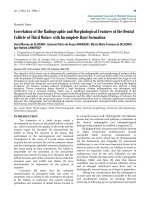
cells. Morphologically, all MMP-12 expressing cells were
macrophages. Macrophages with brown staining in cyto-
plasm were counted as MMP-12 positive macrophages
(MMP-12
+
-macrophages) (Fig. 1A). Figure 1B represents
the negative staining with rabbit IgG. The absolute
amount of MMP-12
+
-macrophages (10
6
/ml) was calcu-
lated according to the number of MMP-12
+
-macrophages
and total inflammatory cell count. The intensity of stain-
ing was evaluated as: 0 – negative; +++ – very strong
expression. The variations MMP-12
+
-macrophages were
counted by two "blinded" researchers and the mean of
their results was calculated. In most cases, the variation of
cell count between examinators was less than 5%.
It is important to note, that we used DTT for preparation
of IS samples, which may interfere with expression of
MMP-12. Therefore, we have compared a preparation of
few IS samples for MMP-12 and inflammatory cell count
with DTT and without it, and we did not notice any signif-
icant differences.
Statistical analysis
Statistical analysis was performed using Statistical Package
for the Social Sciences, version 12.0 for Windows (SPSS
12.0). Data was expressed as the mean of percentage or
absolute value (10
6
/ml) ± standard error of mean (SEM).
Differences between all groups were explored using one-
way ANOVA followed by Kruskal-Wallis test. Mann-Whit-
ney U-test was used to assess the statistical significance of
MMP-12 expression in BALFigure 1
MMP-12 expression in BAL. Representative photomicro-
graph (original magnification: ×1000) of BAL cells immunocy-
tochemical staining for MMP-12 (brown cytoplasm). 1 –
MMP-12
+
-macrophage, 2 – MMP-12
-
-macrophage. A – posi-
tive control, B – negative control (rabbit IgG).
Respiratory Research 2007, 8:81 />Page 4 of 9
(page number not for citation purposes)
differences between the groups. A P-value < 0.05 was con-
sidered significant. Correlations between analysed param-
eters were assessed using Spearman's rank coefficient.
Results
Characteristics of subjects
The average age did not differ between investigated groups
(Table 1). The number of pack-years did not significantly
differ between COPD smokers, COPD ex-smokers and
'healthy' smokers. Lung function parameters did not differ
between COPD groups, but were lower compared to con-
trols.
Cellular composition of IS
The total cell count of IS did not differ between all groups
(Fig. 2A). The composition of inflammatory cells did not
differ between COPD smokers and COPD ex-smokers.
COPD groups showed a predominance of neutrophils,
compared to both healthy subjects groups in percentages
(Table 2). An absolute amount of these cells was higher in
COPD smokers and COPD ex-smokers compared to
healthy never-smokers, but not 'healthy' smokers (Fig.
2A).
Macrophages in IS were more obvious in 'healthy' smok-
ers and healthy never-smokers, due to higher percentage
of neutrophils in both COPD groups. The percentage of
macrophages was significantly lower in COPD groups
compared to both healthy subjects groups, and did not
significantly differ between both COPD and between
both controls groups. An absolute amount of macro-
phages in COPD smokers was lower compared to healthy
never-smokers and did not significantly differ from
'healthy' smokers, however a tendency was seen (p = 0.06)
(Fig. 2A).
Cellular composition of BAL
The total BAL cell number was higher in COPD groups,
compared to healthy subjects groups, while it did not dif-
fer between COPD smokers and COPD ex-smokers and
between 'healthy' smokers and healthy never-smokers
(Fig. 2B). Also, the recovery of BAL was significantly
higher in COPD ex-smokers, compared to COPD smokers
(Table 2). While, this volume was significantly higher in
both healthy subjects groups, than in COPD smokers and
COPD ex-smokers. The recovery of BAL did not differ
between both 'healthy' smokers and healthy never-smok-
ers.
The percentage of neutrophils was increased in COPD
smokers, compared to COPD ex-smokers and healthy
subjects groups. Whereas, the percentage of these inflam-
matory cells in COPD ex-smokers was higher compared to
healthy never-smokers, but did not differ from 'healthy'
smokers. The percentage of BAL neutrophils in 'healthy'
smokers was also higher than in healthy never-smokers.
The absolute amount of neutrophils in COPD smokers
was higher compared to all other groups (Fig. 2B).
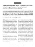
Expression of MMP-12 in IS and BAL cells
An immunocytochemical staining of IS cells for MMP-12
did not show significant differences between COPD
smokers and COPD ex-smokers neither in percentages
(Fig. 3), nor in absolute values. The percentage of IS MMP-
12
+
-macrophages was higher in COPD groups compared
to healthy subjects groups. 'Healthy' smokers had higher
percentage of these cells than healthy never-smokers (Fig.
3), but the absolute amount of MMP-12
+
-macrophages
did not differ.
The amount of BAL MMP-12
+
-macrophages was also sig-
nificantly higher in COPD groups than in controls in per-
centages and absolute values. Furthermore, the number of
BAL MMP-12
+
-macrophages was higher in COPD smok-
ers compared to COPD ex-smokers, and in 'healthy'
smokers compared to healthy never-smokers (Fig. 3).
Analysing the BAL samples we have observed macro-
phages differentiating in size and granularity of cyto-
plasm, while we did not evaluate the relations of MMP-12
expression with their morphology.
Table 1: Characteristics of subjects
Variables COPD smokers COPD ex-smokers 'Healthy' smokers Healthy never-smokers
Subjects (n) 22 17 8 11
Male/Female 22/0 14/3 7/1 4/7
Age (years) 64.2 ± 4.9 62.7 ± 6.3 61.7 ± 6.2 59.8 ± 8.2
Smoking (pack-years) 33.4 ± 5.7 27.9 ± 5.1 28.8 ± 12.1 -
FEV
1
(L) 1.5 ± 0.4*
#
1.7 ± 0.2*
#
3.2 ± 0.6 3.7 ± 0,2
FEV
1
(% pred.) 53.3 ± 4.2*
#
57.1 ± 4.7*
#
109.6 ± 5.3 117.5 ± 4.1
FVC (L) 2.7 ± 0.5*
#
2.9 ± 0.4*
#
3.0 ± 0.2 3.3 ± 0.2
FVC (% pred.) 69.8 ± 9.1*
#
71.7 ± 7.3*
#
108.1 ± 8.2 110.0 ± 6.4
FEV
1
/FVC ratio 50.2 ± 5.9*
#
52.5 ± 6.8*
#
91.0 ± 4.6 93.5 ± 1.0
Values are mean of percentage ± SEM. *: p < 0.05 compared to healthy never-smokers;
#
: p < 0.05 compared to 'healthy' smokers
Respiratory Research 2007, 8:81 />Page 5 of 9
(page number not for citation purposes)
Smoking history relation with cellular patterns, MMP-12
expression and lung function parameters
The number of pack-years correlated with FEV
1
(%) in
COPD smokers (R = -0.70, p < 0.05) and 'healthy' smok-
ers (R = -0.61, p < 0.05). Also, the pack-years correlated
with IS neutrophils in COPD ex-smokers (R = 0.66, p <
0.05). A correlation between pack-years and BAL neu-
trophils in COPD smokers, COPD ex-smokers and
'healthy' smokers groups (Fig. 4) was also obtained. More-
over, the pack-years correlated with BAL macrophages in
COPD smokers (R = 0.87, p < 0.05) and 'healthy' smokers
(R = 0.68, p < 0.05). These parameters did not correlate
with IS inflammatory cells.
The number of IS macrophages negatively correlated with
FEV
1
(%) in COPD smokers (R = -0.53, p < 0.05) and
COPD ex-smokers (R = -0.58, p < 0.05). The correlation
between BAL macrophages and FEV
1
(%) in all studied
groups was also obtained (R = -0.88; -0.62; -0.67; -0.78, p
< 0.05 respectively).
The number of pack-years correlated with IS MMP-12
+
-
macrophages in COPD smokers (R = 0.54, p < 0.05),
Differential cell counts in IS and BAL (10
6
/ml)Figure 2
Differential cell counts in IS and BAL (10
6
/ml). Differential cell composition in IS (A) and BAL (B) from COPD smokers,
COPD ex-smokers, 'healthy' smokers and healthy never-smokers. Data are shown as mean ± SEM. *p < 0.05 compared to
healthy never-smokers, #p < 0.05 compared to 'healthy' smokers.
Cell count of IS (x 10
6
/ml)
Cell count of BAL (x 10
6
/ml)
Total cell number Neutrophils Eosinophils Lymphocytes Macrophages
0
1.0
2.0
3.0
5.0
4.0
Healthy never-smokers
COPD smokers
COPD ex-smokers
‘Healthy’ smokers
*
*
*
A
0
0.5
1.0
1.5
2.0
2.5
3.0
Total cell number
Neutrophils Eosinophils Lymphocytes Macrophages
*
*
*
p<0.05
#
#
#
*
B
Healthy never-smokers
COPD smokers
COPD ex-smokers
‘Healthy’ smokers
Respiratory Research 2007, 8:81 />Page 6 of 9
(page number not for citation purposes)
COPD ex-smokers (R = 0.64, p < 0.05) and 'healthy'
smokers (R = 0.78, p < 0.05). Much stronger correlation
between pack-years and BAL MMP-12
+
-macrophages was
obtained (Fig. 5).
Discussion
We aimed to analyse the patterns of airway inflammation
in COPD patients depending on their smoking status, and
compare it to smokers without airways obstruction
('healthy' smokers) and healthy never-smokers. We have
evaluated different tissue compartments (IS and BAL), as
IS is thought to be a combination of resident mucus [22]
and the composition of its cells may be influenced by
inflammation in proximal airways [22]. While BAL cellu-
lar composition represents mainly the alveolar compart-
ment [23-25], however this method usually is limited due
invasiveness. We have analysed IS sputum and BAL,
because differences in these patterns are still unclear. Also,
we have analysed whether the possible differences in
MMP-12 expression are influenced by smoking history
and its cessation, as previous animal [17-19] and human
[26,27] studies showed, that smoking exposure may
increase an expression of MMP-12.
The number and composition of IS inflammatory cells did
not significantly differ between smokers and ex-smokers
with COPD, while the number of neutrophils was
Smoking history and neutrophilsFigure 4
Smoking history and neutrophils. Correlation between
smoking history (pack-years) and neutrophils (%) in BAL
samples from COPD smokers, COPD ex-smokers and
'healthy' smokers.
0
5
10
15
0102030405060
Pack-years
Neutrophils in BAL (%)
COPD smokers (Rs=0.75, p<0.05)
COPD ex-smokers (Rs=0.82, p<0.05)
‘Healthy’ smokers (Rs=0.79, p<0.05)
Table 2: Differential cell counts in IS and BAL samples
Variable COPD
smokers
COPD
ex-smokers
'Healthy'
smokers
Healthy
never-smokers
CS/CE CS/HS CS/HN CE/HS CE/HN HS/HN
Induced sputum
Neutrophils 67.7 ± 7.7 75.9 ± 9.5 22.6 ± 3.3 16.1 ± 7.0 >0.05 <0.05 <0.01 0.05 0.01 >0.05
Eosinophils 4.5 ± 2.2 3.4 ± 1.6 1.8 ± 0.4 2.3 ± 0.5 >0.05 >0.05 >0.05 >0.05 >0.05 >0.05
Lymphocytes 4.7 ± 1.6 2.7 ± 0.9 6.3 ± 1.1 4.9 ± 0.8 >0.05 >0.05 >0.05 >0.05 >0.05 >0.05
Macrophages 23.1 ± 7.5 18.0 ± 3.5 69.3 ± 9.5 64.8 ± 10.2 >0.05 <0.05 <0.05 0.05 <0.05 >0.05
BAL
Recovery of BAL fluid 43.1 ± 8.3 59.3 ± 6.7 83.3 ± 6.9 81.0 ± 3.8 <0.05 <0.05 <0.05 <0.05 <0.05 >0.05
Neutrophils 17.4 ± 4.8 3.2 ± 1.5 2.7 ± 0.4 1.2 ± 0.5 <0.01 0.05 <0.01 >0.05 0.01 <0.01
Eosinophils 0.8 ± 0.3 0.8 ± 0.3 0.2 ± 0.1 0.2 ± 0.1 >0.05 >0.05 >0.05 >0.05 <0.05 >0.05
Lymphocytes 22.6 ± 6.5 19.8 ± 3.9 24.2 ± 4.5 21.0 ± 4.4 >0.05 >0.05 >0.05 >0.05 >0.05 >0.05
Macrophages 59.2 ± 6.9 76.2 ± 9.3 72.9 ± 10.6 77.6 ± 10.6 <0.01 >0.05 <0.05 >0.05 >0.05 >0.05
Values are mean percentage of total cells ± SEM. CS: COPD smokers; CE: COPD ex-smokers; HS: 'healthy' smokers; HN: healthy never-smokers.
MMP-12
+
-macrophages in IS and BALFigure 3
MMP-12
+
-macrophages in IS and BAL. The relative
number of MMP-12
+
-macrophages in IS and BAL samples
from COPD smokers, COPD ex-smokers, 'healthy' smokers
and healthy never-smokers. Data are shown as mean ± SEM.
*p < 0.05 compared to healthy never-smokers, #p < 0.05
compared to 'healthy' smokers.
MMP-12
+
-macr ophages(% of total macrophages)
COPD smokers
COPD ex-smokers
'Healthy' smokers
Healthy never-
smokers
0
10
20
30
40
50
60
70
IS BAL
80
*
#
*
#
*
*
#
*
#
p<0.05
*
Respiratory Research 2007, 8:81 />Page 7 of 9
(page number not for citation purposes)
increased compared to healthy subjects. These results are
in agreement with major previous studies [23,28], which
have shown that cellular inflammatory response in COPD
is characterized by an increase of total inflammatory cells,
especially neutrophils, macrophages and lymphocytes in
small and large airways [2,3]. Thus, our results may indi-
cate the similar inflammatory response in smokers and ex-
smokers with COPD, which is associated not only with
smoking, but also with systemic inflammation. Influence
of smoking may explain an increased number of BAL neu-
trophils in COPD smokers, compared to COPD ex-smok-
ers, 'healthy' smokers and healthy never-smokers.
Interestingly, the number of BAL neutrophils did not dif-
fer between COPD ex-smokers and 'healthy' smokers,
while the amount of these cells was increased compared
to healthy never-smokers. This finding supports the
hypothesis, that cigarette smoking may cause cellular
alterations [3,22], which may intensify an inflammation
process, induced by disease itself.
Macrophage is predominant cell in IS from healthy never-
smokers and 'healthy' smokers as well. The lower relative
number of these cells obtained in COPD groups may indi-
cate an ongoing inflammatory process. Also, the similar
amount of BAL macrophages in COPD ex-smokers,
'healthy' smokers and never-smokers, suggests the possi-
bility of positive alterations in the alveolar compartment
after smoking cessation.
Also, we have obtained a higher recovery of BAL fluid in
healthy subjects, compared to both COPD groups.
According to Lofdahl et al. [29] suggestions, the extent of
emphysema (measured as an emphysema index and the
carbon monoxide diffusing capacity of the lung) may pre-
dict a low BAL recovery in patients with moderate-to-
severe COPD. Moreover, the lower recovery of BAL fluid
in COPD smokers than in COPD ex-smokers may indicate
an increased inflammatory process in alveolar compart-
ment strengthened by smoking. Furthermore, differences
in BAL cell composition between COPD smokers and ex-
smokers encouraged us to evaluate a correlation between
smoking history, pulmonary function and inflammatory
cells. We obtained, that smoking history (pack-years) pos-
itively correlates with number of BAL neutrophils in both
COPD groups and 'healthy' smokers. Such relation once
more supports the role of neutrophils recruitment in
response to cigarette smoke and suggests that longer
smoking history leads to more serious lung function dam-
age. Smoking may have accumulative effect of inflamma-
tory cells and may increase an inflammatory response in
COPD and 'healthy' smokers as well. Also, we observed
the positive correlation between BAL macrophages and
smoking history in COPD smokers and 'healthy' smokers.
It is known that cigarette smoke increases protease-anti-
protease imbalance and alveolar macrophages, which are
significant source of some MMPs [16,18]. According to
animal studies, MMP-12 deficiency protects against ciga-
rette smoke induced emphysema [18,19]. Though, most
studies investigating MMP-12 were performed using ani-
mal models and exact role of MMP-12 in human COPD
inflammation is not fully understood.
We analysed an expression of MMP-12 active form using
immunocytochemistry.
The number of IS MMP-12
+
-macrophages did not differ
between COPD groups, but it was higher compared to
healthy subjects. Absence of significant differences in
MMP-12 expression in IS may be explained by predomi-
nance of neutrophils, in COPD smokers and ex-smokers,
which obviously do not express MMP-12. An expression
of MMP-12 in IS from 'healthy' smokers was increased,
compared to never-smokers, supporting the suggestion
that smoking may increase an expression of this enzyme.
Our results are in agreement to Demedts et al. [20], who
found an increased sputum MMP-12 level in COPD
patients, compared to healthy smokers, former smokers
(>1 year) and never smokers, while they have not divided
COPD patients into smokers and ex-smokers. Also, Molet
et al., have reported an increase of MMP-12 in BAL and
bronchial biopsies of COPD patients compared to con-
trols [30], while they have not investigated an expression
of MMP-12 according to smoking status.
One of the most interesting our findings was an increased
number of MMP-12
+
-macrophages in BAL from COPD
smokers compared to COPD ex-smokers. Also, the
number of MMP-12
+
-macrophages was increased in both
Smoking history and MMP-12
+
-macrophagesFigure 5
Smoking history and MMP-12
+
-macrophages. Correla-
tion between smoking history (pack-years) and MMP-12
+
-
macrophages (%) in BAL samples from COPD smokers,
COPD ex-smokers and 'healthy' smokers (p < 0.05).
Pack-years
0
10
20
30
40
50
60
70
80
0 102030405060
MMP-12
+
-macrophages in BAL (%)
100
COPD smokers (Rs=0.86, p<0.05)
COPD ex-smokers (Rs=0.68, p<0.05)
‘Healthy’ smokers (Rs=0.63, p<0.05)
Respiratory Research 2007, 8:81 />Page 8 of 9
(page number not for citation purposes)
COPD groups, compared to controls. Nevertheless we
observed a lower amount of BAL macrophages in COPD
smokers, compared to COPD ex-smokers, the absolute
and relative number of BAL MMP-12
+
-macrophages in
COPD smokers was higher than in COPD ex-smokers.
'Healthy' smokers had higher number of BAL MMP-12
+
-
macrophages, than never-smokers supporting the fact of
smoking impact in MMP-12 expression. Actually, we did
not evaluate the activity of macrophages in this study,
thus we were not able to investigate the ratio of MMP-12
release and activated macrophages in this study.
Also, an increased number of BAL MMP-12
+
-macrophages
in COPD ex-smokers, compared to 'healthy' smoking sub-
jects, let us hypothesize that MMP-12 expression is
induced not only by cigarette smoking, but may be an
obligatory to the development of COPD.
Previous studies have shown that contribution of MMP-
12 to smoke induced emphysema is probably enhanced
by indirect effects, such as inactivation of AAT [31] and
MMP-12 mediated recruitment of neutrophils to the lung
[18]. Otherwise, our data suggests that MMP-12 may accu-
mulate and do not rapidly decreases or inactivates after
smoking cessation, exaggerating a persistent inflamma-
tion. An increased expression of MMP-12 in 'healthy'
smokers, also may be a reason for COPD development in
the future.
Conclusion
Smokers and ex-smokers with COPD had close to similar
number and type of IS inflammatory cells, indicating an
ongoing inflammation in proximal airways after smoking
cessation. Although, the lower amount of BAL neutrophils
in COPD ex-smokers, compared to COPD smokers sug-
gests, that smoking cessation may cause positive altera-
tions in alveolar compartment.
Also, a higher number of MMP-12
+
-macrophages in IS
and BAL from COPD smokers and COPD ex-smokers,
indicates that smoking, which is an initial step contribut-
ing to the development of COPD, may stimulate MMP-12
expression in airway cells. Moreover, it let as argue that
MMP-12 expression may be induced not only by smok-
ing, but by the disease itself. A lower amount of BAL
MMP-12
+
-macrophages and other mentioned inflamma-
tory cells, compared to COPD smokers, may indicate a
decrease of alveolar inflammation after smoking cessa-
tion.
Abbreviations
BAL bronchoalveolar lavage
COPD chronic obstructive pulmonary disease
DTT dithiothreitol
FEV
1
forced expiratory volume in 1 sec.
FVC forced vital capacity
ICC immunocytochemistry
IS induced sputum
MMP-12 matrix metalloproteinase
PBS phosphate-buffered saline
RT room temperature
Competing interests
The author(s) declare that they have no competing inter-
ests.
Authors' contributions
AB carried out the major part of cytological analysis and
immunocytochemistry, participated in the writing of
manuscript;
KS carried out screening and clinical evaluation of study
subjects;
JJ participated in the study design, carried out the part of
immunocytochemistry and performed some statistical
analysis;
JL participated in the study design and in the sequence
alignment
RS participated in the study design and in the sequence
alignment
BS conceived and supervised the study and participated in
its design, participated in the writing of the manuscript.
All authors read and approved the final manuscript.
Acknowledgements
We are grateful to Elvyra Draugeliene, MD and Vytis Dudzevicius, PhD for
their invaluable help performing bronchoscopies; Kestutis Malakauskas,
PhD for helpful discussions; Algirda Krisiukeniene, MDSandra Ragaisiene,
MD, Irena Jakubanis, BSc and Inesa Jermalaviciene for their technical sup-
port. This study was in part supported by a Scientific Foundation of Kaunas
University of Medicine (Project Grant PAR8), Lithuania.
References
1. Global Initiative for Chronic Obstructive Lung Disease. Glo-
bal strategy for the diagnosis, management and prevention
of chronic obstructive pulmonary disease NHLBI/WHO Work-
shop Report 2006 [
].
Publish with Bio Med Central and every
scientist can read your work free of charge
"BioMed Central will be the most significant development for
disseminating the results of biomedical research in our lifetime."
Sir Paul Nurse, Cancer Research UK
Your research papers will be:
available free of charge to the entire biomedical community
peer reviewed and published immediately upon acceptance
cited in PubMed and archived on PubMed Central
yours — you keep the copyright
Submit your manuscript here:
/>BioMedcentral
Respiratory Research 2007, 8:81 />Page 9 of 9
(page number not for citation purposes)
2. Barnes PJ, Shapiro SD, Pauwels RA: Chronic obstructive pulmo-
nary disease: molecular and cellular mechanisms. Eur Respir J
2003, 22:672-88.
3. Domagala-Kulawik J, Maskey-Warzechowska M, Kraszewska I,
Chazan R: The cellular composition and macrophage pheno-
type in induced sputum in smokers and ex-smokers with
COPD. Chest 2003, 123:1054-1059.
4. Snider GL: Chronic obstructive pulmonary disease: a defini-
tion and implications of structural determinants of airflow
obstruction for epidemiology. Am Rev Respir Dis 1989, 140:S3-8.
5. Fletcher C, Peto R: The natural history of chronic airflow
obstruction. Br Med J 1977, 1(6077):1645-1648.
6. Hogg JC, Chu F, Utokaparch S, Woods R, Elliott WM, Buzatu L, Cher-
niack RM, Rogers RM, Sciurba FC, Coxson HO, Paré PD: The
nature of small airway obstruction in chronic obstructive
pulmonary disease. N Engl J Med 2004, 350:2645-2653.
7. Saetta M, Turato G, Maestrelli P, Mapp CE, Fabbri LM: Cellular and
structural bases of chronic obstructive pulmonary disease.
Am J Respir Crit Care Med 2001, 163:1304-9.
8. Di Stefano A, Capelli A, Lusuardi M, Balbo P, Vecchio C, Maestrelli P,
Mapp CE, Fabbri LM, Donner CF, Saetta M: Severity of airflow lim-
itation is associated with severity of airway inflammation in
smokers. Am J Respir Crit Care Med 1998, 158:1277-1285.
9. Sitkauskiene B, Sakalauskas R, Malakauskas K, Lotvall J: Reversibility
to a β
2
-agonist in COPD: relationship to atopy and neu-
trophil activation. Respir Med 2003, 97:591-598.
10. Ekberg-Jansson A, Bake B, Andersson B, Skoogh BE, Lofdahl CG: Res-
piratory symptoms relate to physiological changes and
inflammatory markers reflecting central but not peripheral
airways. A study in 60-year-old 'healthy' smokers and never-
smokers. Respir Med 2001, 95:40-7.
11. Finkelstein R, Fraser RS, Ghezzo H, Cosio MG: Alveolar inflamma-
tion and its relation to emphysema in smokers. Am J Respir Crit
Care Med 1995, 152:1666-72.
12. Eidelman D, Saetta MP, Ghezzo H, Wang NS, Hoidal JR, King M,
Cosio MG: Cellularity of the alveolar walls in smokers and its
relation to alveolar destruction. Functional implications. Am
Rev Respir Dis 1990, 141:1547-52.
13. Russell RE, Thorley A, Culpitt SV, Dodd S, Donnelly LE, Demattos C,
Fitzgerald M, Barnes PJ: Alveolar macrophage-mediated elasto-
lysis: roles of matrix metalloproteinases, cysteine, and serine
proteases. Am J Physiol Lung Cell Mol Physiol 2002, 283:L867-L873.
14. Maus UA, Koay MA, Delbeck T, Mack M, Ermert M, Ermert L, Black-
well TS, Christman JW, Schlondorff D, Seeger W, Lohmeyer J: Role
of resident alveolar macrophages in leukocyte traffic into the
alveolar air space of intact mice. Am J Physiol Lung Cell Mol Physiol
2002, 282:L1245-52.
15. Parks WC, Shapiro SD: Matrix metalloproteinases in lung biol-
ogy. Respir Res 2001, 2:10-9.
16. Shapiro SD, Kobayashi DK, Ley TJ: Cloning and characterization
of a unique elastolytic metalloproteinase produced by
human alveolar macrophages. J Biol Chem 1993, 268:23824-9.
17. Houghton AM, Grisolano JL, Baumann ML, Kobayashi DK, Hautamaki
RD, Nehring LC, Cornelius LA, Shapiro SD: Macrophage elastase
(matrix metalloproteinase-12) suppresses growth of lung
metastases. Cancer Res 2006, 66:6149-55.
18. Churg A, Wang RD, Tai H, Wang X, Xie C, Dai J, Shapiro SD, Wright
JL: Macrophage metalloelastase mediates acute cigarette
smoke-induced inflammation via tumour necrosis factor-
release. Am J Respir Crit Care Med 2003, 167:1083-9.
19. Hautamaki RD, Kobayashi DK, Senior RM, Shapiro SD: Require-
ment for macrophage elastase for cigarette smoke-induced
emphysema in mice. Science 1997, 277:2002-4.
20. Demedts IK, Morel-Montero A, Lebecque S, Pacheco Y, Cataldo D,
Joos GF, Pauwels RA, Brusselle GG: Elevated MMP-12 protein
levels in induced sputum from patients with COPD. Thorax
2006, 61:196-201.
21. Quanjer PH, Tammeling GJ, Cotes JE, Pedersen OF, Peslin R, Yernault
JC: Lung volumes and forced ventilatory flows. Report work-
ing party. Standartization of lung function tests. European
Community for Steel and Coal. Official statement of the
European Respiratory Society. Eur Respir J 1993, 6:5-40.
22. Thompson PB, Daughton D, Robbins GA, Ghafouri MA, Oehlerking
M, Rennard SI: Intraluminal airway inflammation in chronic
bronchitis. Characterization and correlation with clinical
parameters. Am Rev Respir Dis 1989, 140:1527-37.
23. Gaga M, Zervas E, Loukides S: Inflammatory markers in moni-
toring response to treatment for asthma and chronic
obstructive pulmonary disease. Pneumon 2004, 17:242-250.
24. Alexis NE, Hu SC, Zeman K, Alter T, Bennett WD: Induced spu-
tum derives from the central airways: confirmation using a
radiolabeled aerosol bolus delivery technique. Am J Respir Crit
Care Med 2001, 164(10 Pt 1):1964-1970.
25. Pizzichini E, Pizzichini MM, Efthimiadis A, Hargreave FE, Dolovich J:
Measurement of inflammatory indices in induced sputum:
effects of selection of sputum to minimize salivary contami-
nation. Eur Respir J 1996, 9:1174-1180.
26. Segura-Valdez L, Pardo A, Gaxiola M, Uhal BD, Becerril C, Selman M:
Upregulation of gelatinases A and B, collagenases 1 and 2,
and increased parenchymal cell death in COPD. Chest 2000,
117:684-94.
27. Betsuyaku T, Nishimura M, Takeyabu K, Tanino M, Venge P, Xu S,
Kawakami Y: Neutrophil granule proteins in bronchoalveolar
lavage fluid from subjects with subclinical emphysema. Am J
Respir Crit Care Med 1999, 159(6):1985-1991.
28. Willemse BWM, Hacken NHT, Rutgers B, Lesman-Leegte IGAT,
Postma DS, Timens W: Effect of 1-year smoking cessation on
airway inflammation in COPD and asymptomatic smokers.
Eur Respir J 2005, 26:835-845.
29. Lofdahl JM, Cederlund K, Nathell L, Eklund A, Skold CM: Broncho-
alveolar lavage in COPD: fluid recovery correlates with the
degree of emphysema. Eur Respir J 2005, 25:275-281.
30. Molet S, Belleguic C, Lena H, Germain N, Bertrand CP, Shapiro SD,
Planquois JM, Delaval P, Lagente V: Increase in macrophage
elastase (MMP-12) in lungs from patients with chronic
obstructive pulmonary disease. Inflamm Res 2005, 54:31-36.
31. Gronski TJ Jr, Martin RL, Kobayashi DK, Walsh BC, Holman MC,
Huber M, Van Wart HE, Shapiro SD: Hydrolysis of a broad spec-
trum of extracellular matrix proteins by human macrophage
elastase. J Biol Chem 1997, 272:12189-94.