Báo cáo khoa học: "Effect of ACTH and CRH on Plasma Levels of Cortisol and Prostaglandin F2α Metabolite in Cycling Gilts and Castrated Boars" pdf
Bạn đang xem bản rút gọn của tài liệu. Xem và tải ngay bản đầy đủ của tài liệu tại đây (115.15 KB, 8 trang )
Madej A, Mwanza AM, Kindahl H, Einarsson S: Effect of ACTH and CRH on
plasma levels of cortisol and prostaglandin F
2
αα
metabolite in cycling gilts and cas-
trated boars. Acta vet. scand. 2005, 46, 249-256. – The present study was designed to
evaluate the effects of synthetic ACTH (1-24, tetracosactid) and porcine CRH on the
plasma levels of cortisol and PGF
2α
metabolite in cycling gilts (n = 3) and castrated
boars (n = 3). The experiments were designed as crossover studies for each gender sep-
arately. Each animal received, during three consecutive days; 1) ACTH (Synacthen
®
De-
pot) at a dose of 10 µg/kg body weight in 5 ml physiological saline, 2) porcine CRH at
a dose 0.6 µg/kg body weight in 5 ml physiological saline or 3) physiological saline (5
ml). The test substances were administered via an indwelling jugular cannula in ran-
domized order according to a Latin square. The administration of ACTH to cycling gilts
resulted in concomitant elevations of cortisol and PGF
2α
metabolite with peak levels
reached at 70.0 ± 10.0 and 33.3 ± 6.7 min, respectively. Similarly, the administration of
ACTH to castrated boars resulted in concomitant elevation of cortisol and PGF
2α
metabolite with peak levels reached at 60.0 ± 0.0 and 20.0 ± 0.0 min, respectively. Cor-
tisol peaked at 20 min after administration of CRH in both cycling gilts and castrated
boars with maximum levels of 149.3 ± 16.5 nmol/l and 138.3 ± 10.1 nmol/l, respec-
tively. It can be concluded that administration of synthetic ACTH (tetracosactid) to pigs
caused a concomitant elevation of cortisol and PGF
2α
metabolite levels in both cycling
gilts as well as castrated boars. The administration of CRH to pigs resulted in an eleva-
tion of cortisol levels in both cycling gilts and castrated boars. Conversely, PGF
2α
metabolite levels were not influenced by the administration of CRH either in cycling
gilts or in castrated boars.
ACTH; CRH; cortisol; PGF
2α
metabolite; gilts; castrated boars
Acta vet. scand. 2005, 46, 249-256.
Acta vet. scand. vol. 46 no. 4, 2005
Effect of ACTH and CRH on Plasma Levels of
Cortisol and Prostaglandin F
2α
Metabolite in Cycling
Gilts and Castrated Boars
By A. Madej
1
, A.M. Mwanza
2,3
, H. Kindahl
2
and S. Einarsson
2
1
Department of Anatomy and Physiology,
2
Department of Clinical Sciences, Centre for Reproductive Biology
in Uppsala, Swedish University of Agricultural Sciences (SLU), P.O. Box 7011, SE- 750 07 Uppsala, Sweden
and
3
Department of Clinical Studies, School of Veterinary Medicine, University of Zambia, Box 32379, Lusaka,
Zambia.
Introduction
Corticotropin releasing hormone (CRH) plays a
central role in regulating the release of adreno-
corticotropic hormone (ACTH) during a stress
response. ACTH acts on the adrenal glands, in-
ducing the secretion of cortisol. In our previous
study, we reported that ACTH administration to
ovariectomized gilts results in the plasma ele-
vation of cortisol, progesterone and prosta-
glandin F
2α
metabolite (Mwanza et al. 2000b).
How ACTH is capable of stimulating the secre-
tion of PGF
2α
metabolite remains unanswered.
However, it was previously suggested by Lay-
chok & Rubin (1975) that ACTH enhances the
conversion in vitro of
3
H-arachidonic acid to
prostaglandins in feline adrenocortical cells.
The findings of Anthonisen et al. (1997) indi-
cate that prostaglandins in the brain interact in
their stimulatory regulation of ACTH secretion.
Such an interaction may also be involved in
prostaglandins mediation of the ACTH re-
sponse to immunochallenges. Abraham et al.
(1998) reported that stimulation of porcine pi-
tuitary cells by relatively low concentrations of
prostaglandin E
2
support increased secretion of
ACTH but exposure to greater concentrations
of this prostaglandin in fact suppresses ACTH
secretion. Food deprivation, which is a form of
stress, has been shown to result in the plasma
elevation of both cortisol and PGF
2α
metabolite
(Tsuma et al. 1996; Mburu et al. 1998; Mwanza
et al. 2000a; Razdan et al. 2001).
Intracerebroventricular as well as intravenous
injections of CRH resulted in an increased
plasma cortisol concentration in pigs (Saku-
mato et al. 2004; Lang et al. 2004). Previously,
it was suggested that CRH may also act directly
or indirectly to enhance cortisol secretion be-
yond the level achieved through adrenal stimu-
lation by ACTH (Minton & Parsons 1993).
The objectives of the present study were to
evaluate the effects of synthetic ACTH (tetra-
cosactid) and porcine CRH on the plasma lev-
els of cortisol and PGF
2α
metabolite in cycling
gilts and castrated boars.
Materials and Methods
Animals
Six crossbred pigs (Landrace x Yorkshire; three
gilts and three castrated boars) aged approxi-
mately 6 months weighing between 110 and
125 kg were used for this experiment. The pigs
were brought to the Division of Comparative
Reproduction, Obstetrics and Udder Health,
and were housed in individual pens. The stable
had a light period of 12 h starting from 06:30 h
and the room temperature varied between 20
and 23°C. The pigs were fed according to the
Swedish breeding stock standard (Simonsson
1994). The gilts were stimulated by boars in the
vicinity and were expected to come in oestrus
within the first week after arrival. After the sec-
ond oestrus, the experiments were carried out in
the early luteal phase (days 5-10). The gilts
were checked twice daily at 06:00 h and 18:00
h for signs of oestrus in the presence of a fertile
boar by back- pressure test. All the six animals
were vein-cannulated (Rodriguez & Kunavong-
krit 1983) at about one week before the experi-
ments. The experiments were designed as
crossover studies for each gender separately.
Each animal received, during three consecutive
days; 1) ACTH (1-24) (Synacthen
®
Depot, No-
vartis Pharma AG, Basel, Schweiz) at a dose of
10 µg/kg body weight in 5 ml physiological
saline, 2) porcine CRH (American Peptide
Company, Inc., Sunnyvale, CA, USA) at a dose
0.6 µg/kg body weight in 5 ml physiological
saline or 3) physiological saline (5 ml). The test
substances were administered via an indwelling
jugular cannula in randomized order according
to a Latin square. On the day of the experiment,
blood samples were taken at -40, -20 min and
immediately before injection. Treatment was
performed at 10:00 h (time = 0) and blood sam-
ples were taken 20, 40, 60, 80, 100, 120, 140,
160, 180, 210, 240, 270, 300, 330 and 360 min
after injection. Blood was collected in ten ml
heparinised tubes, centrifuged immediately and
plasma stored at -20°C until analysed. The care
of the animals and the experimental design of
this study were approved by the Local Animal
Ethics Committee in Uppsala, Sweden.
Hormone assays
Cortisol. Plasma cortisol was determined by ra-
dioimmunoassay (Coat-A-Count Cortisol, Di-
agnostic Products Corporation, Los Angeles,
CA, USA) according to the manufacturer's in-
structions. Serial dilutions of porcine plasma
with high concentrations of cortisol produced
displacement curves parallel to the standard
curve. The intra-assay coefficients of variation
250 A. Madej et al.
Acta vet. scand. vol. 46 no. 4, 2005
calculated from 5 assays were 22% at 14
nmol/l, 14% at 28 nmol/l and decreased below
8% for concentrations between 138 and 552
nmol/l. The inter-assay coefficients of variation
for three control samples were 13% (33 nmol/l),
9% (74 nmol/l) and 9% (541 nmol/l). The aver-
age detection limit of the assay was 7 nmol/l.
Prostaglandin F
2α
metabolite. The main initial
blood plasma metabolite of prostaglandin F
2α
,
15-keto-13,14-dihydro-PGF
2α
(15-ketodihydro-
PGF
2α
), was analysed by radioimmunoassay
according to Kunavongkrit et al. (1983). The
relative cross-reactions of the antibody were
16% with 15-keto-PGF
2α
, and 4% with 13,14-
dihydro-PGF
2α
. The intra-assay coefficients of
variation ranged between 3.4 and 7.6% for dif-
ferent ranges of the standard curve and the in-
ter-assay coefficient of variation was around
14%. The practical limit of sensitivity for the
assay analyzing 0.2 ml of plasma was 60
pmol/l.
Statistical analyses
Data were examined by analysis of variance us-
ing MIXED procedure according to SAS pack-
age (Statistical Analysis Systems 1989). Addi-
tionally, the area under the curve, peak value,
time and duration of the peak were calculated
for each animal according to the GraphPad
Prism version 3.02 for Windows (GraphPad
Software, San Diego, CA, USA). Data are ex-
pressed as means ± S.E.M. Probabilities less
then 0.05 were considered significant.
Results
No significant (P>0.05) differences were seen
in the pretreatment plasma levels of cortisol or
PGF
2α
metabolite between the saline, ACTH
and CRH treated cycling gilts (Figures 1 and 2).
The administration of ACTH to cycling gilts re-
sulted in concomitant elevations of cortisol
(Figure 1) and PGF
2α
metabolite (Figure 2)
with peak levels reached at 70.0 ± 10.0 and 33.3
± 6.7 min, respectively. The durations of the
peaks were 153.3 ± 28.2 and 103.2 ± 11.4 min,
respectively and their maximum concentrations
were 270.7 ± 16.5 nmol/l and 1517.7 ± 137.2
pmol/l, respectively.
No significant (P>0.05) differences were seen
in the pretreatment plasma levels of cortisol or
PGF
2α
metabolite between the saline, ACTH
and CRH treated castrated boars (Figures 3 and
4). The administration of ACTH to castrated
boars resulted in concomitant elevation of cor-
tisol (Figure 3) and PGF
2α
metabolite (Figure
4) with peak levels reached at 60.0 ± 0.0 and
20.0 ± 0.0 min, respectively. The durations of
these peaks were 199.1 ± 30.0 and 86.3 ± 13.8
min, respectively and their maximum concen-
trations were 289.0 ± 10.1 nmol/l and 1262.3 ±
53.2 pmol/l, respectively.
The administration of CRH to both cycling gilts
and castrated boars resulted in the cortisol peak
20 min later with maximum levels of 149.3 ±
16.5 nmol/l (Figure 1) and 138.3 ± 10.1 nmol/l
(Figure 3), respectively. The durations of these
peaks were 57.3 ± 18.5 min and 255.2 ± 43.6
min, respectively.
Prostaglandin F
2α
metabolite levels were not in-
fluenced by the injection of CRH either in cy-
cling gilts or castrated boars (Figures 2 and 4).
Physiological saline did not alter significantly
either cortisol or PGF
2α
metabolite levels in any
animal (Figures 1-4).
No significant (P>0.05) differences were seen
in the measured responses between females and
males.
Discussion
The present study clearly demonstrates that
cortisol reach peak levels much lower and ear-
lier in CRH (approximately after 20 min) than
in ACTH (approximately after 70 min) treated
cycling gilts or castrated boars. This confirmed
earlier results by Beerda et al. (2004) who re-
ported that the cortisol concentration peaked
Effect of ACTH and CRH on cortisol and PGF
2
α
on pigs 251
Acta vet. scand. vol. 46 no. 4, 2005
252 A. Madej et al.
Acta vet. scand. vol. 46 no. 4, 2005
-60 0 60 120 180 240 300 360
0
50
100
150
200
250
300
TIME (min)
Cortisol nmol/l
D
-60 0 60 120 180 240 300 360
0
200
400
600
800
1000
1200
1400
1600
TIME (min)
PGF
2
D
metabolite pmol/l
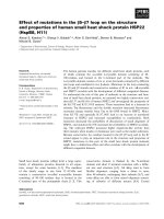
Figure 2. Plasma PGF
2α
metabolite concentrations (LSmeans ± SEM) in cycling gilts given the injection (time
= 0) of saline (ᮀ, n = 3), ACTH (, n = 3) and CRH (᭺, n = 3).
Figure 1. Plasma cortisol concentrations (LSmeans ± SEM) in cycling gilts given the injection (time = 0) of
saline (ᮀ, n = 3), ACTH (, n = 3) and CRH (᭺, n = 3).
Effect of ACTH and CRH on cortisol and PGF
2
α
on pigs 253
Acta vet. scand. vol. 46 no. 4, 2005
-60 0 60 120 180 240 300 360
0
50
100
150
200
250
300
TIME (min)
Cortisol nmol/l
D
-60 0 60 120 180 240 300 360
0
200
400
600
800
1000
1200
1400
1600
TIME (min)
PGF
2
D
metabolite pmol/l
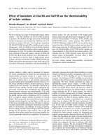
Figure 4. Plasma PGF
2a
metabolite concentrations (LSmeans ± SEM) in castrated boars given the injection
(time = 0) of saline (ᮀ, n = 3), ACTH (, n = 3) and CRH (᭺, n = 3).
Figure 3. Plasma cortisol concentrations (LSmeans ± SEM) in castrated boars given the injection (time = 0)
of saline (ᮀ, n = 3), ACTH (, n = 3) and CRH (᭺, n = 3).
approximately 30 min after administration of
CRH and approximately 60-90 min after ad-
ministration of synthetic ACTH to dairy cows.
The present study also demonstrates that the
administration of ACTH stimulates a concomi-
tant elevation of both cortisol and PGF
2α
metabolite levels in both cycling gilts and cas-
trated boars. In addition, peak PGF
2α
metabo-
lite levels occur earlier than peak cortisol lev-
els. Apparently, it takes approximately twice
the time for cortisol than for PGF
2α
metabolite
to reach peak levels following ACTH adminis-
tration. This is consistent with our previous
findings in ovariectomized gilts (Mwanza et al.
2000b) and suggests that ACTH stimulates the
secretion of PGF
2α
earlier than cortisol. The
frequency of blood collection may have impact
on the occurrence of PGF
2α
metabolite peak in
relation to cortisol peak. When blood samples
were taken only at 1-h intervals, both PGF
2α
metabolite and cortisol peak were seen one
hour after ACTH administration in recently
ovulated sows (Razdan et al. 2002).
Cooke & Ahmad (1994) have demonstrated that
daily administration of ACTH from day 13 to
day 16 of the oestrous cycle in multiparous
Welsh Mountain ewes suppressed the levels of
PGF
2α
metabolite. They further showed that in
ovariectomized multiparous Welsh Mountain
ewes, primed first with progesterone and then
with oestradiol-17ß, ACTH reduced the ability
of oxytocin to cause the release of PGF
2α
into
the peripheral circulation. However, there is ev-
idence that feline and rat adrenocortical cells
synthesise prostaglandins F
2α
and E
2
and that
the total prostaglandins synthesis is stimulated
by ACTH (Laychock & Rubin 1976; Chander-
bhan et al. 1979). Winter et al. (1990) demon-
strated that in vitro, the cytokine interleukin-1
enhances the conversion of
3
H-arachidonic acid
to prostaglandins by cultured bovine adrenal
cells. The secreted prostaglandins i.e. PGD
2
,
PGF
2α
and PGE
2
were in turn found to stimu-
late cortisol secretion. Furthermore, Nashuhita
et al. (1997) reported that intravenously admin-
istered PGE
1
, PGE
2
or PGF
2α
had significant
ACTH-releasing activity in the rat and sug-
gested that prostaglandins are playing a role in
regulating the hypothalamo-pituitary-adrenal
axis. In sows, injection of PGF
2α
after ovulation
resulted in a dramatic cortisol elevation, which
lasted approximately 1.5 h (Mwanza et al.
2002).
In contrast to ACTH-treated pigs, no peak
PGF
2α
metabolite levels were seen in any CRH
treatment. We can speculate that a combination
of CRH and lysine vasopressin (LVP) could
have been a better option since LVP + CRH was
seen to have a better ACTH response than CRF
or LVP alone in pigs (Minton & Parsons 1993).
It might also simply indicate that CRH does not
stimulate the secretion of PGF
2α
.
Interestingly, food deprivation which is a form
of stress has been shown to result in the plasma
elevation of both cortisol and PGF
2α
metabolite
(Mburu et al. 1998; Mwanza et al. 2000a; Raz-
dan et al. 2001; Tsuma et al. 1996). It is postu-
lated (Silver & Fowden 1982) that in food de-
prived animals, PGF
2α
metabolite levels are
elevated owing to increased levels of free fatty
acids that includes arachidonic acid, the precur-
sor of prostaglandin synthesis. In addition,
Madej et al. (2005) reported that during artifi-
cial insemination of sows housed in crates, a
dramatic elevation of cortisol levels was seen
before PGF
2α
metabolite reached its maximum.
It is still unclear what role if any ACTH plays
either directly or indirectly in the stimulation of
PGF
2α
production.
It can be concluded from the present study that
the administration of synthetic ACTH to pigs at
a dose of 10 µg/kg body weight caused a con-
comitant increase of cortisol and PGF
2α
metabolite levels in both cycling gilt as well as
castrated boars. The administration of CRH to
pigs resulted in an elevation of cortisol levels in
254 A. Madej et al.
Acta vet. scand. vol. 46 no. 4, 2005
both cycling gilts and castrated boars. Con-
versely, PGF
2α
metabolite levels were not influ-
enced by the administration of CRH either in
cycling gilts or in castrated boars.
Acknowledgements
This work was supported financially by the Swedish
Council for Forestry and Agricultural Research,
Swedish Farmers Foundation for Agricultural Re-
search and SLU´s Research Programme "Animal
Welfare for Quality in Food Production"
References
Abraham EJ, Morris-Hardeman JN, Sswenson LM,
Knoppel EL, Ramanathan B, Wright KJ, Grieger
DM, Minton JE: Pituitary function in the acute
phase response in domestic farm animals: Cy-
tokines, prostaglandins, and secretion of ACTH.
Domest. Anim. Endocrinol. 1998, 15, 389-396.
Anthonisen M, Knigge U, Kjoer A, Warberg J: His-
tamine and prostaglandin interaction in the cen-
tral regulation of ACTH secretion. Neuroen-
docrinology 1997, 66, 68-74.
Beerda B, Kornalijnslijper JE, van der Werf JTN, No-
ordhuizen-Stassen EN, Hopster H: Effect of milk
production capacity and metabolic status on HPA
function in early postpartum dairy cows. J. Dairy
Sci. 2004, 87, 2094-2102.
Chanderbhan R, Hodges VA, Treadwell CR, Vahouny
GV: Prostaglandins synthesis in rat adrenocortical
cells. J. Lipid Res. 1979, 20, 116-124.
Cooke RG, Ahmad N: The effect of ACTH on oxy-
tocin-induced release of prostaglandin F
2α
and on
uterine oxytocin receptors in the ewe. Anim. Re-
prod. Sci. 1994, 35, 201-208.
Kunavongkrit A, Kindahl H, Madej A: Clinical and
endocrinological studies in primiparous zero-
weaned sows. 2. Hormonal patterns of normal cy-
cling sows after zero-weaning. Zbl. Vet. Med. A
1983, 30, 616-624.
Lang, A, Kaeoket K, Kindahl H, Madej, A, Einarsson
S: Influence of CRH and ACTH administration on
endocrine profile and ovulation in sows. Reprod.
Domest. Anim. 2004, 39, 181-189.
Laychock SG, Rubin RP: ACTH-induced prosta-
glandin biosynthesis from 3H-arachidonic acid
by adrenocorticla cells. Prostaglandins 1975, 10,
529-540.
Laychock SG, Rubin RP: Radioimmunoassay mea-
surements of ACTH-facilitated PGE
2
and PGF
2α
release from isolated cat adrenocortical cells.
Prostaglandins 1976, 11, 753-765.
Madej A, Lang A, Brandt Y, Kindahl H, Madsen MT,
Einarsson S: Factors regulating ovarian function
in pigs. Domest. Anim. Endocrinol. 2005, 29,
347-361.
Mburu JN, Einarsson S, Kindahl H, Madej A, Ro-
driguez-Martinez H: Effects of food post-ovula-
tory deprivation on oviductal sperm concentra-
tion, embryo development and hormonal profiles
in the pig. Anim. Reprod. Sci. 1998, 52, 221-234.
Minton JE, Parsons KM: Adrenocorticotropic hor-
mone and cortisol response to corticotropin-re-
leasing factor and lysine vasopressin in pigs. J.
Anim. Sci. 1993, 71, 724-729.
Mwanza AM, Einarsson S, Madej A, Lundeheim N,
Rodriguez-Martinez H, Kindahl H: Postovulatory
effect of repeated administration of prostaglandin
F
2α
on the endocrine status, ova transport, binding
of accessory spermatozoa to the zona pellucida
and embryo development of recently ovulated
sows. Theriogenology 2002, 58, 1111-1124.
Mwanza AM, Englund P, Kindahl H, Lundeheim N,
Einarsson S: Effects of postovulatory food depri-
vation on the hormonal profiles, activity of the
oviduct and ova transport in sows. Anim. Reprod.
Sci. 2000a, 59, 185-199.
Mwanza AM, Madej, A, Kindahl H, Lundeheim N,
Einarsson S: Plasma levels of cortisol, proges-
terone, oestradiol-17ß and prostaglandin F
2α
metabolite after ACTH (Synacthen Depot
®
) ad-
ministration in ovariectomized gilts. J. Vet. Med.
A 2000b, 47, 193-200.
Nasushita R., Watanobe H, Takebe K: A comparative
study of adrenocorticotropic-releasing activity of
prostaglandin E1, E2, F2 alpha and D2 in the rat.
Prostaglandins Leukot. Essent. Fatty Acids 1997,
56, 165-168.
Razdan P, Mwanza AM, Kindahl H, Hultén F, Einars-
son S: Impact of postovulatory food deprivation
on the ova transport, hormonal profiles and
metabolic changes in sows. Acta vet. scand. 2001,
42, 45-55.
Razdan P, Mwanza AM, Kindahl H, Rodriguez-Mar-
tinez H, Hultén F, Einarsson S: Effect of repeated
ACTH-stimulation on early embryonic develop-
ment and hormonal profiles in sows. Anim. Re-
prod. Sci. 2002, 70, 127-137.
Rodriguez-Martinez H, Kunavongkrit A: Chronic ve-
nous catheterisation for frequent blood sampling
in unrestrained pigs. Acta. vet. scand. 1983, 42,
45-55.
Effect of ACTH and CRH on cortisol and PGF
2
α
on pigs 255
Acta vet. scand. vol. 46 no. 4, 2005
Sakumoto R, Kasuya E, Saito T, Akita T: A technique
for long-term implantation of a microcatheter into
the third ventricle of post-pubertal Chinese Meis-
han pigs based on ventriculography. J. Neurosci.
Methods 2004, 137, 97-101.
Silver M, Fowden AL: Uterine prostaglandin F
metabolite production in relation to glucose avail-
ability in late pregnancy and a possible influence
of diet on the time of delivery in the mare. J. Re-
prod. Fertil. Suppl. 1982, 32, 511-519.
Simonsson A: Näringsrekommendationer och foder-
medelstabeller till svin (Nutrient and metaboliz-
able energy recommendations for swine).
Swedish University of Agricultural Sciences,
SLU Info rapporter, Husdjur 75, 1994. (In
Swedish).
Statistical Analysis Systems Institute Inc.:
SAS/STAT User's Guide, Version 6. fourth ed,
vol. 2. Cary. NC, SAS Institute Inc., 1989.
Tsuma VT, Einarson S, Madej A, Kindahl H, Lunde-
heim H: Effect of food deprivation during early
pregnancy on endocrine changes in primiparous
sows. Anim. Reprod. Sci. 1996, 41, 71-76.
Winter JS, Gow KW, Perry YS, Greenberg AH: A
stimulatory effect of interleukin-1 on adrenocor-
tical cortisol secretion mediated by prosta-
glandins. Endocrinology 1990, 127, 1904-1909.
Sammanfattning
Behandlingseffekt av syntetiskt ACTH (tetracosactid)
och CRH på blodplasmakoncentrationerna av korti-
sol och PGF
2
α
metaboliten hos gyltor med normal
brunstcykel och hos kastrerade galtar.
Målsättningen med denna studie var att utvärdera be-
handlingseffekten av syntetiskt ACTH (1-24, tetra-
cosactid) och CRH från gris på blodplasmakoncen-
trationerna av kortisol och PGF
2α
metaboliten hos
gyltor med normal brunstcykel och hos kastrerade
galtar. Experimenten utfördes enligt crossover mod-
ellen separat för varje kön. Varje djur behandlades
under tre dagar med 1) ACTH (Synacthen
®
Depot),
10 µg/kg kroppsvikt i 5 ml fysiologisk koksaltlös-
ning, 2) CRH från gris 0,6 µg/kg kroppsvikt i 5 ml fy-
siologisk koksaltlösning eller 3) 5 ml fysiologisk
koksaltlösning. Testsubstanserna injicierades via en
permanent jugularkateter slumpartat enligt Latinkva-
drat principen. Behandlingen av gyltor med ACTH
resulterade i samtidig stegring av kortisol och PGF
2α
metaboliten, med högsta koncentrationerna efter
70,0 ± 10,0 respektive 33,3 ± 6,7 minuter. På samma
sätt resulterade behandling av kastrerade galtar med
ACTH i samtidig stegring av kortisol och PGF
2α
metaboliten och med högsta koncentrationerna efter
60,0 ± 0,0 respektive 20,0 ± 0,0 minuter. Kortisol
nådde sitt högsta värde 20 minuter efter behandling
med CRH både hos gyltor (149,3 ± 16,5 nmol/l) och
kastrerade galtar (138,3 ± 10,1 nmol/l).
Sammanfattningsvis resulterade behandling med
syntetiskt ACTH (tetracosactid) i samtidig stegring
av kortisol och PGF
2α
metaboliten hos både gyltor
och kastrerade galtar. Behandling med CRH resulter-
ade i stegring av kortisol hos både gyltor och kastr-
erade galtar. Blodplasmakoncentrationerna av PGF
2α
metaboliten var oförändrade hos både gyltor och kas-
trerade galtar efter CRH behandlingen.
256 A. Madej et al.
Acta vet. scand. vol. 46 no. 4, 2005
(Received May 2, 2005; accepted August 8, 2005).
Reprints may be obtained from: Andrzej Madej, Swedish University of Agricultural Sciences (SLU), Depart-
ment of Anatomy and Physiology, P.O. Box 7011, SE-750 07, Uppsala, Sweden. E-mail:
, tel: +46 18 672114, fax: +46 18 672111.