Báo cáo khoa học: " Neither Hippurate-negative Brachyspira pilosicoli nor Brachyspira pilosicoli Type Strain Caused Diarrhoea in Early-weaned Pigs by Experimental Infection" pdf
Bạn đang xem bản rút gọn của tài liệu. Xem và tải ngay bản đầy đủ của tài liệu tại đây (1.7 MB, 11 trang )
Fossi M, Ahlsten K, Pohjanvirta T, Anttila M, Kokkonen T, Jensen TK, Boye M,
Sukura A, Pelkola K, Pelkonen S: Neither hippurate-negative Brachyspira pilosi-
coli nor Brachyspira pilosicoli type strain caused diarrhoea in early-weaned pigs by
experimental infection. Acta vet. scand. 2005, 46, 257-267. – A hippurate-negative
biovariant of Brachyspira pilosicoli (B. pilosicoli
hipp-
) is occasionally isolated in diar-
rhoeic pigs in Finland, often concomitantly with hippurate-positive B. pilosicoli or Law-
sonia intracellularis. We studied pathogenicity of B. pilosicoli
hipp-
with special attention
paid to avoiding co-infection with other enteric pathogens. Pigs were weaned and moved
to barrier facilities at the age of 11 days. At 46 days, 8 pigs were inoculated with B. pi-
losicoli
hipp-
strain Br1622, 8 pigs were inoculated with B. pilosicoli type strain P43/6/78
and 7 pigs were sham-inoculated. No signs of spirochaetal diarrhoea were detected; only
one pig, inoculated with P43/6/78, had soft faeces from day 9 to 10 post inoculation. The
pigs were necropsied between days 7 and 23 after inoculation. Live pigs were culture-
negative for Brachyspira spp., but B. pilosicoli
hipp-
was reisolated from necropsy sam-
ples of two pigs. The lesions on large colons were minor and did not significantly differ
between the three trial groups. In silver-stained sections, invasive spirochaetes were de-
tected in colonic mucosae of several pigs in all groups. Fluorescent in situ hybridisation
for genus Brachyspira, B. pilosicoli and strain Br1622 was negative. However, in situ de-
tection for members of the genus Leptospira was positive for spirochaete-like bacteria
in the colonic epithelium of several pigs in both infected groups as well as in the con-
trol group. L. intracellularis, Salmonella spp., Yersinia spp. and intestinal parasites were
not detected. The failure of B. pilosicoli strains to cause diarrhoea is discussed with re-
spect to infectivity of the challenge strains, absence of certain intestinal pathogens and
feed and management factors.
Brachyspira pilosicoli, intestinal spirochaetosis, spirochaetal colitis, pig, diarrhoea,
experimental infection, early weaning, Lawsonia intracellularis.
Acta vet. scand. 2005, 46, 257-267.
Acta vet. scand. vol. 46 no. 4, 2005
Neither Hippurate-negative Brachyspira pilosicoli nor
Brachyspira pilosicoli Type Strain Caused Diarrhoea
in Early-weaned Pigs by Experimental Infection
By M. Fossi
1
, K. Ahlsten
2
, T. Pohjanvirta
3
, M. Anttila
4
, T. Kokkonen
4
, T. K. Jensen
5
, M. Boye
5
,
A. Sukura
6
, K. Pelkola
7
and S. Pelkonen
3
1
National Veterinary and Food Research Institute, Seinäjoki Unit, PB 198, 60101 Seinäjoki, Finland,
2
National
Veterinary and Food Research Institute, Department of Virology and Epidemiology, PB 45, 00581 Helsinki, Fin-
land,
3
National Veterinary and Food Research Institute, Kuopio Department, PB 92, 70701 Kuopio, Finland,
4
National Veterinary and Food Research Institute, Department of Pathology, PB 45, 00581 Helsinki, Finland,
5
Danish Institute for Food and Veterinary Research, Bülowsvej 27, 1790 Copenhagen V, Denmark,
6
Department
of Basic Veterinary Sciences, University of Helsinki, PB 57, 00014 Helsinki, Finland.
7
National Veterinary and
Food Research Institute, Department of Bacteriology, PB 45, 00581 Helsinki, Finland.
Introduction
Porcine intestinal spirochaetosis (PIS) in
weaned pigs is caused by an anaerobic, weakly
ß-haemolytic spirochaete, Brachyspira pilosi-
coli (Taylor et al. 1980, Trott et al. 1996), and it
occurs worldwide (Duhamel 1998, Møller et al.
1998, Barcellos et al. 2000, Heinonen et al.
2000, de Arriba et al. 2002, Choi et al. 2002).
The pathogenicity of different B. pilosicoli
strains has been shown to vary (Thomson et al.
1997). B. pilosicoli usually hydrolyses hippu-
rate, which is a major criterion for the bio-
chemical differentiation of B. pilosicoli from
the other porcine Brachyspira species (Fell-
ström & Gunnarsson 1995). However, some
porcine B. pilosicoli strains in Finland are hip-
purate-negative and their pathogenicity is un-
clear since other potentially enteropathogenic
bacteria, such as Lawsonia intracellularis and
B. pilosicoli with a positive hippurate reaction,
have simultaneously been diagnosed in the
herds (Fossi et al. 2004).
Co-infections with L. intracellularis and B. pi-
losicoli are highly prevalent among weaned
pigs with diarrhoea in Sweden (Jacobson et al.
2003), and co-infections with L. intracellularis
and various species of Brachyspira have also
been reported in Denmark and the UK (Møller
et al. 1998, Thomson et al. 1998). The con-
comitant ileitis caused by L. intracellularis or
colitis due to the pathogenic species of
Brachyspira aggravates the clinical outcome. L.
intracellularis and pathogenic species of
Brachyspira characteristically cause diarrhoea
in pigs after weaning. The time-point of the in-
testinal colonisation by these organisms during
the nursing period is unknown.
In this work, we evaluated the pathogenicity of
a single strain of hippurate-negative B. pilosi-
coli (B. pilosicoli
hipp-
), paying particular atten-
tion to the simultaneous presence or absence of
other enteric pathogens. The pigs were weaned
and moved to barrier facilities at the age of 11
days to enable the effect of early separation on
transmission of opportunistic pathogens from
sows to piglets to be assessed.
Materials and methods
The experimental infection was approved by
the Ethics Committee for Animal Experiments
of the National Veterinary and Food Research
Institute and carried out under license number
2-2002. Piglets were purchased from a herd of
120 sows with a long history of good health sta-
tus. Freedom from swine enzootic pneumonia,
atrophic rhinitis and Salmonella was verified by
regular laboratory investigations. Several stud-
ies for Brachyspira spp. performed within the
last two years had occasionally yielded weakly
ß-haemolytic spirochaetes other than B. pilosi-
coli.
On the farm, nine sows were moved to a far-
rowing department two weeks before the ex-
pected farrowing date. Colostrum samples were
collected on the day of farrowing, and faecal
samples were taken two days after farrowing.
Twelve Landrace piglets and 12 mixed-breed
piglets were chosen from four litters which had
been born within 24 hours of each other. Equal
numbers of male and female piglets for the both
races were chosen. No creep feed was served
before weaning. At the age of 11 days, the
piglets were transported to barrier facilities of
the Finnish National Veterinary and Food Re-
search Institute.
The piglets were raised together in a pen of 6.20
m
2
until the age of 45 days. The pen had a solid
floor which was scraped clean daily. The peat
bedding was changed to wood shavings 4-7
days post-inoculation (p.i.). The piglets were
fed a commercial sow milk substitute until the
age of 24 days. Commercial creep feed was
served from the age of 13 to 31 days. Commer-
cial grower feed was based on wheat and barley
until the age of 57 days, and thereafter, on bar-
ley. The crude protein content of the initial feed
was 17.2%, and from the age of 57 days on-
wards, 17.4%. No growth-promoting feed addi-
tives were used, and the feed was granulated
without heating.
Most piglets had diarrhoea at the age of 15-18
days. Abundant growth of haemolytic Es-
cherichia coli was observed in faecal cultures
during this period. One piglet died. All piglets
were medicated intramuscularly with trimeto-
258 M. Fossi et al.
Acta vet. scand. vol. 46 no. 4, 2005
prime-sulphadiazine at the age of 16 and 17
days. At 45 days of age, the pigs were divided
into three trial groups in which littermates, sex
and race were evenly distributed. Two groups of
eight pigs were placed in two identical isolated
rooms, where pen space per pig was 0.60 m
2
.
The third group of seven pigs was divided into
two subgroups of four and three pigs and placed
in two smaller rooms with pen spaces per pig of
0.68 m
2
and 0.51 m
2
.
The health of the pigs was monitored daily.
Body temperature and weight gain were
recorded, and faecal samples were initially
taken weekly, and after the challenge, three
times a week. Blood samples were collected
weekly.
Challenge
B. pilosicoli
hipp-
strain Br1622 had originally
been isolated in the year 2000 from a pig herd
in which weaners and young fatteners had diar-
rhoea. B. pilosicoli type strain P43/6/78 (ATCC
51139
T
) originated from a diarrhoeic pig in the
UK (Taylor et al. 1980). The strains were stored
at –70°C in beef broth supplemented with 12%
horse serum and 15% glycerol.
Br1622 and P43/6/78 were cultured on fastidi-
ous anaerobe agar and incubated anaerobically
for three days at 39°C. The next passages were
propagated anaerobically at 39°C in brain heart
infusion broth supplemented with 10% foetal
calf serum and 0.5% glucose. Broth cultures
were propagated through three passages for a fi-
nal volume of 300 ml. The final broth was incu-
bated for 36±3 hours to achieve a density of
1.6-2.0 × 10
8
cells ml
-1
. The second cascade of
broth cultures was started one day after the first
one.
At the ages of 46 and 47 days, the eight pigs in
one room were inoculated intragastrically with
3.2 × 10
9
cells of Br1622 on both days, and the
eight pigs in the second room received 4.0 × 10
9
and 3.2 × 10
9
cells of P43/6/78 on two consec-
utive days. The seven pigs in the two small
rooms were sham-inoculated with a sterile
broth on both days. Bacterial viability in the
broth containers was controlled after inocula-
tion by microscopy and cultivation.
Necropsy
One pig from both challenge groups and the
control group was euthanised and necropsied as
blind on days 7, 9, 11, 15, 16, 17, 21 and 23 p.i
Blood samples were taken and the intestines
were dissected immediately after the bolt stun-
ning. The routine gross examination was per-
formed paying special attention to intestinal
mucosae. The caecum, the ascending, mid-spi-
ral and descending colon, the rectum and the in-
testinal lymph nodes were sampled for histo-
pathology.
The tissue samples were fixed in 10% buffered
formalin, processed routinely, sectioned at 5
µm and stained with haematoxylin and eosin,
and Warthin-Starry silver stain. The sections
were blind-examined. For transmission electron
microscopy (TEM), 1.0 mm
2
samples from the
mucosa of the large intestine were fixed in 2.5%
glutaraldehyde in phosphate buffer. The sam-
ples were embedded in Epon
TM
, and thin sec-
tions were stained with uranyl acetate and lead
citrate. Samples were then visualised using a
Jeol Jem 100-s electron microscope.
Mucosal scrapings for Brachyspira cultivation
were taken from the caecum, ascending, mid-
spiral and descending colon and rectum. Con-
tent from the large intestine and ileum was col-
lected for further microbiological examination.
Fluorescent in situ hybridisation
The sections from the large colon were studied
by fluorescent in situ hybridisation (FISH). The
oligonucleotide probes used are presented in
Table 2. The general bacterial probe EUB338,
the genus-specific probe for Brachyspira and
the species-specific probe for B. pilosicoli have
Diarhoea in early-weaned pigs by experimetal infection 259
Acta vet. scand. vol. 46 no. 4, 2005
been described elsewhere (Boye et al. 1998).
The strain-specific probe for Br1622, the spe-
cific probe for Treponema (pallidum group) and
the genus-specific probes for Leptospira and
Borrelia were selected using ARB software
(Strunk et al. 2000). The probes were 5'-la-
belled with fluorescein isothiocyanate (MWG-
Biotech AG, Ebersberg, Germany). The probe
for strain Br1622 had at least two mismatches
to all other Brachyspira species in the ARB
database. Processing of the sections and hy-
bridisation were carried out as described previ-
ously (Jensen et al. 2000). An Axioplan2 epi-
fluorescence microscope was used for simul-
taneous detection of red and green fluores-
cence.
260 M. Fossi et al.
Acta vet. scand. vol. 46 no. 4, 2005
Table 1. Pigs inoculated with B. pilosicoli
hipp-
strain Br1622, B. pilosicoli type strain P43/6/78 or sterile broth
(control). Pathology and results from fluorescent in situ hybridisation are shown.
Pig Necropsy Gross
Histology FISH
4
No. Inoculum day post- pathology
1
Histo- Spiral-shaped 1 2
inoculation pathology
2
bacteria
3
1 Br1622 7 - + - + -
2 Br1622 9 - (+) + - -
3 Br1622 11 + + ++ + -
4 Br1622 15 ++ + ++ , s + -
5 Br1622 16 ++ (+) - , s + -
6 Br1622 17 + + ++ + -
7 Br1622 21 + (+) ++ - -
8 Br1622 23 ++ + - - -
9 P43/6/78 7 + (+) + - -
10 P43/6/78 9 - (+) - + -
11 P43/6/78 11 + (+) + - -
12 P43/6/78 15 ++ (+) ++ - -
13 P43/6/78 16 ++ (+) ++ + -
14 P43/6/78 17 + + + - -
15 P43/6/78 21 + (+) ++ + -
16 P43/6/78 23 ++ (+) - - -
17 control 7 - (+) - - -
18 control 15 + + - + -
19 control 17 - (+) ++ + -
20 control 23 + + - - -
21 control 9 + (+) - - -
22 control 11 - (+) - - -
23 control 21 + (+) ++ + -
1
Gross findings in caecum and/or colon. - = normal; + = mucosa slightly hyperaemic; ++ = mucosa hyperaemic and slightly
oedematous.
2
(+) = diffuse lymphocytic and plasmacytic infiltration in lamina propria, and multifocal accumulation of phagocytic
macrophages beneath the epithelial cells of the caecum and/or colon; + = in addition, occasional microabscesses in crypts
and/or mild exocytosis of neutrophils.
3
Spiral-shaped bacteria in caecum and/or colon in silver-stained sections. - = none; + = very rare or irregularly observed in
crypts and/or near tips of villi; ++ = invading through epithelial lining into lamina propria and/or abundantly in crypts; s =
Br1622 reisolated from necropsy samples.
4
Fluorescent in situ hybridisation. Positive (+) and negative (-) results with probes designed for 1 = domain bacteria and genus
Leptospira, and 2 = genus Brachyspira, species B. pilosicoli and strain Br1622.
Microbiology, parasitology and haematology
The selective culture and the biochemical dif-
ferentiation for Brachyspira spp. were per-
formed as previously described (Jenkinson &
Wingar 1981, Fellström & Gunnarsson 1995,
Olson 1996). The primary cultures from intesti-
nal scrapings were studied also by B. pilosicoli-
specific polymerase chain reaction (PCR) as-
says targeting 16S rDNA and 23S rDNA
(Fellström et al. 1997, Leser et al. 1997). The
PCR studies were repeated with frozen gut
samples when invasive spirochaetes were ob-
served by histopathology. Pulsed-field gel elec-
trophoresis (PFGE) with MluI restriction en-
zyme was used to compare DNA macro-
restriction profiles between isolated spiro-
chaetes and the challenge strains, as described
earlier (Fossi et al. 2003). The banding patterns
of DNA were compared visually.
Faecal and intestinal samples were cultured for
facultative anaerobic bacteria and anaerobic
bacteria, selectively for Campylobacter spp.
and by enrichment methods for Salmonella spp.
and Yersinia spp., using standard laboratory
methods. Cultivation for Campylobacter spp.
was done only from colon and caecum scrap-
ings at necropsy.
Faecal samples of the sows and 26-day-old
piglets were tested for L. intracellularis by
nested PCR. Sample preparation was per-
formed according to McOrist et al. (1994), and
primer pairs used for DNA amplification were
as described by Jones et al. (1993). Antibodies
for L. intracellularis were analysed by using an
indirect fluorescent antibody test (IFAT)
(McOrist et al. 1987, Lawson et al. 1988, Knit-
tel et al. 1998) from the sera of pigs at the age
of 42 days and at necropsy. L. intracellularis
antibodies were analysed also from the
colostrum of the pigs' dams, as well as from the
colostrum of the other five sows farrowing in
the same department during the same time.
Faecal samples from the challenge groups were
pooled and investigated for rotavirus by Ani
™
Rotatest (Ani Biotech Ltd., Vantaa, Finland)
according to the manufacturer's instructions,
and for parasite eggs by the flotation method.
Blood samples were studied for total and differ-
ential leucocyte count, haematocrit value,
haemoglobin concentration and platelet count.
Results
After the challenge, the pigs remained clini-
cally healthy. Only one pig (pig no. 13, Table 1)
inoculated with B. pilosicoli P43/6/78, had soft
faeces on days 9 and 10 p.i Intestinal spiro-
chaetes were not isolated from this pig. During
the first week p.i. the mean daily weight gain
Diarhoea in early-weaned pigs by experimetal infection 261
Acta vet. scand. vol. 46 no. 4, 2005
Table 2. Names, sequences and specificities of rRNA-targeted oligonucleotide probes used.
Probe Systematic name
1
Sequence (5'-3') Specificity
Eub338
2
S-D-Bact-0338-a-A-18 GCTGCCTCCCGTAGGAGT Domain bacteria
NON-Eub338
2
S-*-non-0338-a-S-18 CGACGGAGGGCATCCTCA Unspecific control probe
SER1410
3
L-G-Brachy-1410-a-A-19 GTCATTCCATCGAAACATA Genus Brachyspira
Pilosi209
3
S-S-B.pilo-0209-a-A-18 GCTCATCGTGAAGCGAAA B. pilosicoli
Br1622 S-S-Br1622-0637-a-A-18 CCAAGATCTACAGTATCC Br1622
Treponema S-G-Trepon-0728-a-A-18 TCGGCCAGAAACCCGCCT Treponema spp.
Borrelia S-G-Borre-0688-a-A-18 TATCAACAGATTCCACCC Borrelia spp.
Leptospira S-G-Leptospi-1414-a-A-18 CGGGTGCTCCCCACTCAG Leptospira spp.
1
According to the Oligonucleotide Probe Database (OPD) nomenclature (Alm et al. 1996).
2
Amann et al. (1990).
3
Boye et al. (1998).
262 M. Fossi et al.
Acta vet. scand. vol. 46 no. 4, 2005
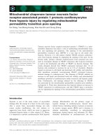
Figure 1. Invasion of spirochaetes in and below the colonic epithelium is shown in pig no. 13, which was in-
oculated with B. pilosicoli P43/6/78 and necropsied 16 days post-inoculation. Warthin-Starry silver staining.
1000 ×.
Figure 2. Invasive spirochaetes in a colonic crypt. Same pig as in Fig. 1. Warthin-Starry silver staining. 1000 ×.
(DWG) was 0.48, 0.39 and 0.49 kg in the
Br1622-, P43/6/78- and sham-inoculated
groups, respectively. During the second week,
the corresponding mean DWG was 0.58, 0.44
and 0.55 kg. The difference in mean DWG be-
tween the groups was statistically not signifi-
cant. The body temperatures of the pigs re-
mained within the normal range throughout the
trial period.
Pathology
At necropsy, a moderate hyperaemia was noted
on colonic and/or caecal mucosae of six, seven
and four pigs infected with Br1622, P43/6/78
and sterile broth, respectively. The colonic mu-
cosae were also slightly oedematous in three
pigs infected with Br1622 as well as in three
pigs infected with P43/6/78 (Table 1). In light
microscopy, minor inflammatory lesions were
seen in sections of the large colon of all pigs
(Table 1), but the severity of the microscopic le-
sions did not differ notably between the groups.
In silver-stained sections from the large intes-
tine, spiral-shaped bacteria were seen in the ep-
ithelium of five, six and two pigs in the Br1622,
P43/6/78 and control groups, respectively. Spi-
ral-shaped bacteria were more common in sec-
tions from the colon than from the caecum. The
spiral-shaped bacteria crossed the epithelial
cell layer into the lamina propria of the caecum
and/or colon in samples from four, three and
two pigs in these groups, respectively (Figs 1
and 2). Invasion of spiral-shaped bacteria below
the lamina muscularis mucosae of the colon
was seen in pig no.13, which was inoculated
with P43/6/78. In TEM, trans-sections of end-
oflagellated bacteria were seen between and be-
low the colonic epithelial cells of one pig inoc-
ulated with Br1622 (no. 6) and one control pig
(no. 23). The bacteria were 0.28-0.32 µm in di-
ameter and had 10-14 endoflagella.
Fluorescent in situ hybridisation
FISH with probes specific for Br1622, B. pi-
losicoli and the genus Brachyspira did not give
a signal in any section of the large intestines.
Diarhoea in early-weaned pigs by experimetal infection 263
Acta vet. scand. vol. 46 no. 4, 2005
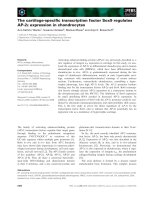
Figure 3. Fluorescent in situ hybridisation with a genus-specific probe Leptospira. Demonstration of
spirochaetes infiltrating the colonic epithelium. Pig no. 3 inoculated with B. pilosicoli
hipp-
strain Br1622 and
necropsied 11 days post-inoculation. 1000 ×.
However, hybridisation with the probes for
genus Leptospira and for domain bacteria re-
vealed multiple spiral-shaped bacteria in and
below the epithelial lining of the colon and/or
caecum in five, three and three pigs in the
groups inoculated with Br1622, P43/6/78 and
sterile broth, respectively (Fig. 3). These spiral-
shaped bacteria seemed uniform and were 6-11
µm in length.
Microbiology, parasitology and haematology
Brachyspira spp. were not isolated from the fae-
cal samples of live pigs, but B. pilosicoli
hipp-
was recovered from colon scrapings of two pigs
challenged with Br1622 (pig nos. 4 and 5, Table
1) and necropsied on day 15 or 16 p.i Accord-
ing to the PFGE macrorestriction profiles, the
B. pilosicoli
hipp-
isolates were the same as those
of the challenge strain Br1622. By B. pilosicoli-
specific PCR, all primary cultures for Bra-
chyspira, excluding these two Brachyspira iso-
lations, were negative.
Haemolytic E. coli was isolated from the faeces
of most pigs until one week before challenge.
Neither Salmonella spp. nor Yersinia spp. were
isolated. Campylobacter coli or Campylobacter
spp. were isolated from colon scrapings of four,
five and four pigs inoculated with Br1622,
P43/6/78 and sterile broth, respectively. Intesti-
nal parasites were not detected. The latex test
for rotavirus was positive in the group infected
with Br1622 from the age of 46 days onwards,
and in all other groups from the age of 48 days
onwards. Beyond the age of 55 days, the test re-
sults became ambiguous.
Pigs' sera were negative for L. intracellularis
antibodies. Four of the nine sows in the same
farrowing group had colostral antibodies to L.
intracellularis. However, these sows were not
the dams of the trial pigs. L. intracellularis was
not detected by PCR in faeces of the sows or the
pigs.
The leucocyte count of one pig in both inocu-
lated groups and two pigs in the control group
exceeded the upper limit of reference (20 000
cells µl
-1
) once in the first week p.i The values
of the other haematological parameters re-
mained within the normal range throughout the
trial period.
Discussion
Neither B. pilosicoli type strain nor hippurate-
negative field strain Br1622 caused diarrhoea in
pigs. Reisolation of Br1622 succeeded for only
two pigs at necropsy, whereas type strain
P43/6/78 was not detected in any of the sam-
ples. The inoculated pigs possibly may have
shed spirochaetes below the detection limits.
The sensitivity of the cultivation for B. pilosi-
coli is approximated as 5.4 × 10
5
CFU g
-1
fae-
ces (Stege et al. 2000) or 1.5 × 10
2
CFU g
-1
fae-
ces (Fellström et al. 1997). The sensitivity of
our cultivation method lies between these fig-
ures (unpublished data). In our experience,
PCR from a primary culture rarely yields a pos-
itive result in cases where the culture has been
negative for Brachyspira.
One interpretation is that true colonisation did
not occur in most of the pigs. An oral inocula-
tion of 10
9
cells ml
-1
of B. pilosicoli given once
(Thomson et al. 1997, 2001), twice (Neef et al.
1994) or thrice (Jensen et al. 2004) has been
satisfactory for induction of spirochaetal diar-
rhoea. Thus, the bacteria doses here should
have been sufficient. However, undefined fac-
tors may exist that affect the actively growing
bacteria inoculated as a pure suspension in an
empty stomach.
An experimental infection by B. pilosicoli type
strain P43/6/78 has been described to cause di-
arrhoea in gnotobiotic pigs (Neef et al. 1994).
This strain isolated and reported by Taylor et al.
in 1980, was later deposited in the American
Type Culture Collection as a type strain of
species B. pilosicoli (Trott et al. 1996). An at-
tenuation of this strain before its deposition can
264 M. Fossi et al.
Acta vet. scand. vol. 46 no. 4, 2005
not be excluded. Strain Br1622 has undergone
from four to six passages before its deposition,
and a further four passages for the final volume
of inocula. We assume that strain Br1622 is no
more attenuated than the type strain at the stage
of inoculation in this experiment. Some loss of
pathogenicity of Br1622 through the passages
can not, however, be excluded.
No infection of pigs by L. intracellularis was
detected. Their early separation from the sows
and the apparently low prevalence of infection
in the herd of origin could explain the freedom
from L. intracellularis. Co-infections, espe-
cially with L. intracellularis and B. pilosicoli,
have been suggested to aggravate clinical diar-
rhoea among growers in field conditions
(Thomson et al. 1998, Jacobson et al. 2003).
The absence of L. intracellularis could also
promote the health of the pigs during the trial.
Several environmental and management-re-
lated risk factors which enhance the outcome of
diarrhoea among weaners were missing in this
trial. The feed was not heat-treated and its crude
protein content was relatively low. Heat treat-
ment of feed during pelleting increases the
prevalence of weakly ß-haemolytic intestinal
spirochaetes (Stege et al. 2001) and non-spe-
cific colitis in herds (Smith & Nelson 1987).
Moreover, high protein content in feed has been
shown to cause loose stools in pigs (Reese
1999). The feed used in this trial might have
supported the clinical health of the pigs to some
extent.
Peat bedding is popular due to its high capacity
for fluid absorption and low pH. In this trial, the
peat was changed to wood shavings at 4-7 days
p.i However, we can speculate that sour peat
may have controlled the number of circulating
spirochaetes at the early stage of infection. The
other environmental conditions in the barrier
facilities were undoubtedly more favourable
than those on conventional farms, possibly pro-
moting the health of the trial pigs. The ventila-
tion in the barrier facilities was filtrated, and di-
urnal variation in room temperature and
draught was minimal. The pen area per pig in-
creased gradually after seven days p.i. due to
the necropsy schedule, which might have de-
creased the number of circulating bacteria. Fur-
ther experiments for pathogenicity of B. pilosi-
coli
hipp-
strains should be done for several B.
pilosicoli
hipp-
strains of various genotypes in a
conventional-like environment using an unat-
tenuated porcine B. pilosicoli strain as a posi-
tive control.
The bacteria observed in TEM sections of two
pigs were deemed not to belong to the species
B. pilosicoli because of their inconsistent end-
oflagella count (Sellwood & Bland 1997). In
FISH, the positive signal merely by the genus-
specific probe Leptospira was somewhat unex-
pected. Further investigations are needed to
clarify the taxonomic position of these intesti-
nal bacteria.
Acknowledgements
We thank senior researcher Kirsi Partanen of MTT
Agrifood Research Finland for the animals and feed.
Päivi Olonen, Anssi Isoniemi, Minttu Mikkonen,
Raili Heinonen and Pirkko Niemelä are thanked for
technical assistance.
This study was supported by the Finnish Ministry for
Agriculture and Forestry and the Finnish Association
for Food Animal Practitioners.
References
Alm EW, Oerther DW, Larsen N, Stahl DA, Raskin L:
The oligonucleotide probe database. Appl. Envi-
ron. Microbiol. 1996, 62, 3557-3559.
Amann RI, Binder BJ, Olson RJ, Chrisholm SW, Dev-
ereux R, Stahl DA: Combination of 16S rRNA-
targeted oligonucleotide probes with flow cytom-
etry for analyzing mixed microbial populations.
Appl. Environ. Microbiol. 1990, 56, 1919-1925.
Barcellos DESN, Mathiesen MR, de Uzeda M, Kader
IITA, Duhamel GE: Prevalence of Brachyspira
species isolated from diarrhoeic pigs in Brazil.
Vet. Rec. 2000, 146, 398-403.
Diarhoea in early-weaned pigs by experimetal infection 265
Acta vet. scand. vol. 46 no. 4, 2005
Boye M, Jensen TK, Møller K, Leser TD, Jorsal SE:
Specific detection of the genus Serpulina, S. hyo-
dysenteriae and S. pilosicoli in porcine intestines
by fluorescent rDNA in situ hybridization. Mol.
Cell. Probe. 1998, 12, 323-330.
Choi C, Han DU, Kim J, Cho WS, Chung H-K, Jung
T, Yoon BS, Chae C: Prevalence of Brachyspira
pilosicoli in Korean pigs determined using a
nested PCR. Vet. Rec. 2002, 150, 217-218.
de Arriba ML, Duhamel GE, Vidal AB, Carvajal A,
Pozo J, Rubio P: Porcine Colonic Spirochetosis in
Spanish pig herds. Proceedings of the 17th IPVS
Congress, Ames, Iowa, USA, June 2-5, 2002, Vol.
2, p. 373.
Duhamel GE: Colonic spirochetosis caused by Ser-
pulina pilosicoli. Large Animal Practice 1998,
19, 14-22.
Fellström C, Gunnarsson A: Phenotypical character-
ization of intestinal spirochaetes isolated from
swine. Res. Vet. Sci. 1995, 59, 1-4.
Fellström C, Pettersson B, Thomson J, Gunnarsson
A, Persson M, Johanssen KE: Identification of
Serpulina species associated with porcine colitis
by biochemical analysis and PCR. J. Clin. Micro-
biol. 1997, 35, 462-467.
Fossi M, Pohjanvirta T, Sukura A, Heinikainen S, Lin-
decrona R, Pelkonen S: Molecular and ultrastruc-
tural characterization of porcine hippurate-nega-
tive Brachyspira pilosicoli. J. Clin. Microbiol.
2004, 42, 3153-3158.
Fossi M, Pohjanvirta T, Pelkonen S: Molecular epi-
demiological study of Brachyspira pilosicoli in
Finnish sow herds. Epidemiol. Infect. 2003, 131,
967-973.
Heinonen M, Fossi M, Jalli J-P, Saloniemi H, Tuovi-
nen V: Detectability and prevalence of Brachy-
spira species in herds rearing health class feeder
pigs in Finland. Vet. Rec. 2000, 146, 343-347.
Jacobson M, Hård af Segerstadt C, Gunnarsson A,
Fellström C, de Verdier Klingenberg, K, Wallgren
P, Jensen-Waern M: Diarrhoea in the growing pig
– a comparison of clinical, morphological and mi-
crobial findings between animals from good and
poor performing herds. Res. Vet. Sci. 2003, 74,
163-169.
Jenkinson SR, Wingar CR: Selective medium for the
isolation of Treponema hyodysenteriae. Vet. Rec.
1981, 109, 384-385.
Jensen TK, Boye M, Møller K: Extensive intestinal
spirochaetosis in pigs challenged with Brachy-
spira pilosicoli. J. Med. Microbiol. 2004, 53, 309-
312.
Jensen TK, Møller K, Boye M, Leser TD, Jorsal SE:
Scanning electron microscopy and fluorescent in
situ hybridization of experimental Brachyspira
(Serpulina) pilosicoli infection in growing pigs.
Vet. Pathol. 2000, 37, 22-32.
Jones GF, Ward GE, Murtaugh M P, Lin G, Gebhart
C J: Enhanced detection of the intracellular or-
ganism of swine proliferative enteritis, Ileal sym-
biont intracellularis, in feces by polymerase chain
reaction. J. Clin. Microbiol. 1993, 31, 2611-2615.
Knittel JP, Jordan DM, Schwartz KJ, Janke BH, Roof,
MB, McOrist, S, Harris DL: Evaluation of ante-
mortem polymerase chain reaction and serologic
methods for detection of Lawsonia intracellu-
laris-exposed pigs. Am. J. Vet. Res. 1998, 59,
722-726.
Lawson GHK, McOrist S, Rowland AC, McCartney
E, Roberts L: Serological diagnosis of the porcine
proliferative enteropathies: implications for aeti-
ology and epidemiology. Vet. Rec. 1988, 122,
554-557.
Leser TD, Møller K, Jensen TK, Jorsal SE: Specific
detection of Serpulina hyodysenteriae and poten-
tially pathogenic weakly ß-haemolytic porcine in-
testinal spirochetes by polymerase chain reaction
targeting 23S rDNA. Mol. Cell. Probe. 1997, 11,
363-372.
McOrist S, Boid R, Lawson GH, McConnell I: Mon-
oclonal antibodies to intracellular campylobacter-
like organisms of the porcine proliferative en-
teropathies. Vet. Rec. 1987, 121, 421-422.
McOrist S, Gebhart CJ, Lawson GHK: Polymerase
chain reaction for diagnosis of porcine prolifera-
tive enteropathy. Vet. Microbiol. 1994, 41, 205-
212.
Møller K, Jensen TK, Jorsal SE, Leser TD, Car-
stensen B: Detection of Lawsonia intracellularis,
Serpulina hyodysenteriae, weakly beta-haemo-
lytic spirochaetes, Salmonella enterica, and hae-
molytic Escherichia coli from swine herds with
and without diarrhoea among growing pigs. Vet.
Microbiol. 1998, 62, 59-72.
Neef NA, Lysons RJ, Trott DJ, Hampson DJ, Jones
PW, Morgan JH: Pathogenity of porcine intestinal
spirochetes in gnotobiotic pigs. Infect. Immun.
1994, 62, 2395-2403.
Reese, DE: Nutrient deficiencies and excesses. In:
B.E. Straw, S. D'Allaire, W.L. Mengeling, D.J.
Taylor, editors. Diseases of Swine. 8th edition.
Iowa State University Press, Ames, Iowa 50014,
USA. 1999. p. 743-755.
Olson LD: Enhanced isolation of Serpulina hyo-
266 M. Fossi et al.
Acta vet. scand. vol. 46 no. 4, 2005
Diarhoea in early-weaned pigs by experimetal infection 267
Acta vet. scand. vol. 46 no. 4, 2005
dysenteriae by using sliced agar media. J. Clin.
Microbiol. 1996, 34, 2937-2941.
Sellwood R, Bland AP: Ultrastructure of Intestinal
Spirochaetes. In: DJ Hampson, TB Stanton, edi-
tors. Intestinal Spirochaetes in Domestic Animals
and Humans. CAB International, Wallingford,
OXON OX10 8DE, UK.1997, p. 109-121.
Smith WJ, Nelson EP: Grower scour/non-specific
colitis. Vet. Rec. 1987, 121,334.
Stege H, Jensen TK, Møller, K, Bækbo P, Jorsal S:
Risk factors for intestinal pathogens in Danish
finishing pig herds. Prev. Vet. Med. 2001, 50, 153-
164.
Strunk O, Gross O, Reichel B, May M, Hermann S,
Stuckmann N., et al. [Online] ARB: a software en-
vironment for sequence data. Department of Mi-
crobiology, Technische Universität München,
Munich, Germany. 2000. Available from:
.
Taylor DJ, Simmons JR, Laird HM: Production of di-
arrhoea and dysentery in pigs by feeding pure cul-
tures of a spirochaete differing from Treponema
hyodysenteriae. Vet. Rec. 1980, 106, 326-332.
Thomson JR, Smith WJ, Murray BP, McOrist S:
Pathogenity of three strains of Serpulina pilosi-
coli in pigs with a naturally acquired intestinal
flora. Infect. Immun. 1997, 65, 3693-3700.
Thomson JR, Smith WJ, Murray BP: Investigations
into field cases of porcine colitis with particular
reference to infection with Serpulina pilosicoli.
Vet. Rec. 1998, 142, 235-239.
Thomson JR, Smith WJ, Murray BP, Dick JE, Sump-
tion KJ: Porcine enteric spirochete infections in
the UK: surveillance data and preliminary inves-
tigation of atypical isolates. Anim. Health Res.
Review 2001, 2, 31-36.
Trott DJ, Stanton TB, Jensen NS, Duhamel GE, John-
son JL, Hampson DJ: Serpulina pilosicoli sp.
nov., the agent of porcine intestinal spirochetosis.
Int. J. Syst. Bacteriol. 1996, 46, 206-215.
Sammanfattning
Varken hippurat negativa Brachyspira pilosicoli eller
Brachyspira pilosicoli typ stammar framkallade di-
arré hos tidigt avvanda grisar vid experimentell in-
fektion.
Den hippurat negativa biovarianten av Brachyspira
pilosicoli (B. pilosicoli
hipp-
) kan ofta isoleras ur diar-
résjuka grisar i Finland, vanligen tillsammans med
hippurat positiva B. pilosicoli eller Lawsonia intra-
cellularis. Vi undersökte hur patogen B. pilosicoli
hipp-
är, och tog notis om andra enteropatogener. Grisar av-
vandes och avskiljdes vid 11 dygns ålder. Vid 46
dygns ålder inokulerades 8 grisar med B. pilosicoli-
hipp-
, 8 grisar med B. pilosicoli stam P43/6/78 och på
7 grisar utfördes inokuleringsproceduren utan bak-
terier (skeninokulation). Mellan 7 och 23 dygn efter
inokuleringen avlivades och obducerades grisarna.
Endast en (1) gris som blivit inokulerad med stam-
men P43/6/78 hade lindrig diarré från och med dygn
9 till och med dygn 10 efter inokulering. De levande
grisarna var odlingsnegativa för B. pilosicoli, medan
B. pilosicoli
hipp-
kunde återisoleras i obduktionsma-
terialet på två grisar. Endast lindriga tarmförän-
dringar observerades och dessa skiljde sig inte sig-
nifikant från varandra mellan de tre prövnings-
grupperna. I tjocktarmen från grisar i alla grupper
sågs under mikroskop invasiva spiroketer. Tre olika
oligonukleotidprober användes för att identifiera
Brachyspira med fluorescens in situ hybridisering,
men dessa prov utföll negativt. I kolonepiteliet hos
hälften av grisarna gav emellertid användningen av
en prob för Leptospira positivt utslag. Avsaknaden av
L. intracellularis påvisades ytterligare med serolo-
giska test och med artspecifik innesluten PCR
(nested PCR). B. pilosicoli-stammar kunde således
inte framkalla diarré och detta diskuteras i förhål-
lande till saminfektioner med vissa tarmpatogener
och i förhållande till en eventuell enteropatogenlin-
drande effekt hos B. pilosicoli.
(Received January 10, 2005; accepted September 1, 2005).
Reprints may be obtained from: M. Fossi, National Veterinary and Food Research Institute, Senäjoki Unit, PB
198, FIN-60101 Seinäjoki, Finland. E-mail: marja.fossi@eela.fi, tel: +358-6-4218100, fax: +358-6-4218180.