Báo cáo y học: " Characterization of the innate immune response to chronic aspiration in a novel rodent model" ppsx
Bạn đang xem bản rút gọn của tài liệu. Xem và tải ngay bản đầy đủ của tài liệu tại đây (1.34 MB, 12 trang )
BioMed Central
Page 1 of 12
(page number not for citation purposes)
Respiratory Research
Open Access
Research
Characterization of the innate immune response to chronic
aspiration in a novel rodent model
James Z Appel III
1
, SeanMLee*
1
, Matthew G Hartwig
1
, Bin Li
1
, Chong-
Chao Hsieh
1
, Edward Cantu III
1
, Yonghan Yoon
1
, Shu S Lin
1
,
William Parker
1
and R Duane Davis
†1,2
Address:
1
Transplant Immunobiology Laboratory, Department of Surgery, Duke University Medical Center, Durham, NC 27710, USA and
2
Box
3864, Department of Thoracic Surgery, Duke University Medical Center, Durham, NC 27710, USA
Email: James Z Appel - ; Sean M Lee* - ; Matthew G Hartwig - ;
Bin Li - ; Chong-Chao Hsieh - ; Edward Cantu - ;
Yonghan Yoon - ; Shu S Lin - ; William Parker - ; R
Duane Davis -
* Corresponding author †Equal contributors
Abstract
Background: Although chronic aspiration has been associated with several pulmonary diseases,
the inflammatory response has not been characterized. A novel rodent model of chronic aspiration
was therefore developed in order to investigate the resulting innate immune response in the lung.
Methods: Gastric fluid or normal saline was instilled into the left lung of rats (n = 48) weekly for
4, 8, 12, or 16 weeks (n = 6 each group). Thereafter, bronchoalveolar lavage specimens were
collected and cellular phenotypes and cytokine concentrations of IL-1alpha, IL-1beta, IL-2, IL-4, IL-
6, IL-10, GM-CSF, IFN-gamma, TNF-alpha, and TGF-beta were determined.
Results: Following the administration of gastric fluid but not normal saline, histologic specimens
exhibited prominent evidence of giant cells, fibrosis, lymphocytic bronchiolitis, and obliterative
bronchiolitis. Bronchoalveolar lavage specimens from the left (treated) lungs exhibited consistently
higher macrophages and T cells with an increased CD4:CD8 T cell ratio after treatment with
gastric fluid compared to normal saline. The concentrations of IL-1alpha, IL-1beta, IL-2, TNF-alpha
and TGF-beta were increased in bronchoalveolar lavage specimens following gastric fluid aspiration
compared to normal saline.
Conclusion: This represents the first description of the pulmonary inflammatory response that
results from chronic aspiration. Repetitive aspiration events can initiate an inflammatory response
consisting of macrophages and T cells that is associated with increased TGF-beta, TNF-alpha, IL-
1alpha, IL-1beta, IL-2 and fibrosis in the lung. Combined with the observation of gastric fluid-
induced lymphocyitic bronchiolitis and obliterative bronchiolitis, these findings further support an
association between chronic aspiration and pulmonary diseases, such as obliterative bronchiolitis,
pulmonary fibrosis, and asthma.
Published: 27 November 2007
Respiratory Research 2007, 8:87 doi:10.1186/1465-9921-8-87
Received: 6 August 2007
Accepted: 27 November 2007
This article is available from: />© 2007 Appel et al; licensee BioMed Central Ltd.
This is an Open Access article distributed under the terms of the Creative Commons Attribution License ( />),
which permits unrestricted use, distribution, and reproduction in any medium, provided the original work is properly cited.
Respiratory Research 2007, 8:87 />Page 2 of 12
(page number not for citation purposes)
Background
Gastroesophageal reflux disease (GERD) has been associ-
ated with a number of pulmonary diseases, including idi-
opathic pulmonary fibrosis, asthma, chronic bronchitis,
cystic fibrosis, and chronic obstructive pulmonary disease
[1-5]. It is generally believed that GERD-associated pul-
monary pathology is mediated by repetitive aspiration
events. Indeed, GERD is said to be the most common
cause of chronic intermittent aspiration [6,7]. DeMeester
et al. found that 70% of patients with respiratory symp-
toms of persistent cough, wheezing, or recurrent pneumo-
nia had GERD based on 24-hour pH monitoring of the
distal esophagus [8].
It is likely that many of these pulmonary responses to
repetitive aspiration are related to immune-mediated
events. Lung transplant recipients with GERD represent
one group for whom a chronic aspiration-induced
immune reaction likely results in a particularly adverse
clinical effect. Data from a number of retrospective clini-
cal studies performed at our institution implicates chronic
aspiration in the context of GERD as a reversible cause of
pulmonary dysfunction and bronchiolitis obliterans syn-
drome (BOS) in lung transplant recipients [9-13]. The
prevalence of pH-confirmed GERD is particularly high
among patients with end-stage lung disease, approaching
50%, but increases to over 70% following lung transplan-
tation [9,10,14]. However, lung transplant recipients with
GERD that undergo antireflux surgery early in the post-
transplant period exhibit decreased rates of BOS, mortal-
ity, and acute rejection [9,14].
Based on these data, our group has proposed that chronic
aspiration associated with GERD may represent a repeti-
tive non-allogeneic stimulus for immune-mediated injury
in lung transplant recipients. Chronic aspiration may
facilitate an innate immune response, predisposing lung
transplant recipients to acute and chronic rejection. Fur-
thermore, since these rejection processes are thought to
involve primarily cell-mediated responses, it is possible
that chronic aspiration additionally initiates or recruits an
acquired immune response, facilitating or exacerbating
pulmonary allograft dysfunction.
Although mounting evidence supports the clinical associ-
ation between GERD and pulmonary dysfunction in lung
transplant recipients and in patients with pulmonary
fibrosis and end-stage lung disease, the physiologic and
immunologic reaction to chronic aspiration has not been
investigated using a suitable model system. In order to
better characterize the changes in the lung that result from
chronic aspiration, our laboratory has developed a rodent
model of repetitive gastric fluid aspiration. Herein, we
describe the inflammatory response to chronic aspiration
in this novel rodent model. The results shed light on the
mechanisms by which chronic aspiration may lead to pul-
monary fibrosis, exacerbate end-stage lung disease, or
stimulate an allogeneic response in lung transplant recip-
ients.
Methods
Animals
Male, pathogen-free F344 rats were obtained from Charles
River Laboratories (Wilmington, MA). All experiments
were performed in accordance with the Guide for the Care
and Use of Laboratory Animals prepared by the National
Academy of Sciences and published by the National Insti-
tutes of Health. Protocols were approved by the Duke
University Medical Center Institutional Animal Care and
Use Committee.
Collection of gastric fluid
Rats were anesthetized using inhaled isoflurane. A surgical
gastrotomy was performed through which a silastic cathe-
ter was inserted. Gastric fluid was collected by gravity over
a 12-hour period, the fluid from several animals was
pooled together, and the mixture was then filtered
through a 70-micron strainer before being stored at -80°C
until immediately prior to use. The pH of all specimens
utilized for experimentation was 1.0 – 2.5. Animals were
not fasted prior to gastric fluid collection.
Instillation of gastric fluid or normal saline
Male pathogen-free F344 rats (250–300 g) were sedated
with ketamine (40 mg/kg IM), intubated orotracheally
using the sheath from a 14-gauge IV catheter, and main-
tained on a mechanical ventilator (Inspira, Harvard Appa-
ratus, Holliston, MA). Rats were subsequently
disconnected from the ventilator and placed in the left lat-
eral decubitus position with their head elevated at a 30°
angle. A silastic catheter was inserted through the endotra-
cheal tube to a distance 5 mm past the tip, where 150
microliters of either gastric fluid or normal saline (0.9%
NaCl) was slowly injected. Rats were maintained in this
position for 15 minutes and then extubated after recover-
ing from sedation.
Methods of the instillation procedure were developed
based on studies in our laboratory demonstrating that
gastrograffin contrast could be consistently instilled into
the left lung while sparing the right lung with 100% repro-
ducibility (Figure 1). The volume of fluid administered
was equal to one half the lethal dose used in acute aspira-
tion studies [15,16]. With the exception of control rats (n
= 6), instillation of gastric fluid or normal saline into the
left lung was repeated on a weekly basis for a predeter-
mined period (i.e. 4, 8, 12, or 16 weeks, n = 6 in each
group). A weekly regimen was selected after preliminary
studies indicated that more frequent aspiration resulted in
an unacceptably high mortality rate.
Respiratory Research 2007, 8:87 />Page 3 of 12
(page number not for citation purposes)
Sample collection
One week after the predetermined number of left lung
instillations, rats were sedated and a tracheotomy was per-
formed through which rats were intubated. The trachea,
lungs, and heart were then explanted en bloc. The right
and left mainstem bronchi were sequentially lavaged with
a total of 6 ml PBS buffer (37°C). 1 ml of bronchoalveolar
lavage (BAL) specimens was centrifuged and the superna-
tant stored at -80°C for cytokine analysis. The remainder
of the BAL specimens was utilized for FACS analysis.
Histology
Lung tissue was fixed using 2% paraformaldehyde and
stained using hematoxylin and eosin as well as Masson tri-
chrome stain for collagen. The extent of fibrosis in tri-
chrome-stained specimens was graded by a pathologist in
a blinded fashion using a numerical scale described else-
where [17].
Flow cytometry
The phenotypes of cells in BAL specimens were deter-
mined using FITC-conjugated anti-rat CD172a (Serotec,
Oxford, UK), TCR, CD4, and CD8 (Becton Dickinson,
Franklin Lakes, NJ) monoclonal antibodies and quanti-
fied using a FACSCalibur flow cytometer (Becton Dickin-
son, Franklin Lakes, NJ).
Cytokine assays
BAL specimens were thawed and concentrations of IL-
1alpha, IL-1beta, IL-2, IL-4, IL-6, IL-10, GM-CSF, IFN-
gamma, and TNF-alpha were measured using Bio-Plex
multiplex bead-based immunoassays (BioRad Laborato-
ries, Hercules, CA). Specimens were analyzed using a
Luminex 100 flow-based, dual-laser array reader
(Luminex, Austin, TX) and concentrations quantified
using Bio-Plex Manager Software (BioRad Laboratories,
Hercules, CA). TGF-beta concentrations were determined
using ELISA-based sandwich immunoassays (R&D Sys-
tems, Minneapolis, MN).
Statistical analysis
Unless otherwise noted, reported values represent mean ±
standard error of the mean. Chi-square, ANOVA, and
unpaired Student's t-tests were performed, where appro-
priate. For all statistical calculations, a p-value < 0.05 was
considered significant.
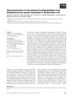
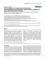
Instillation of fluid into the left lungFigure 1
Instillation of fluid into the left lung. The instillation of gastrograffin into the distal trachea of sedated rats in the left lateral
decubitus position resulted in consistent localization in the left lung, sparing the right.
Respiratory Research 2007, 8:87 />Page 4 of 12
(page number not for citation purposes)
Results
Histology
Histology specimens from left lungs after 4, 8, 12, and 16
weeks of gastric fluid aspiration were compared to those
from right lungs and to specimens from untreated rats and
from rats receiving normal saline (Figure 2). Four weeks of
gastric fluid aspiration was associated with an increase in
peribronchiolar and interstitial fibrosis compared to con-
trols (Figures 2a and 2b). These differences were greatest
after 8 weeks of gastric fluid aspiration. Fibrosis was also
noted after 12 or 16 weeks of gastric fluid aspiration,
although it was less prominent. Based on a blinded assess-
ment of the pathology, the fibrosis grade [17] was signifi-
cantly more severe in left lung specimens compared to
right lung specimens after 4, 8, 12, and 16 weeks of gastric
fluid aspiration (Figure 3).
As early as 4 weeks after the initiation of gastric fluid aspi-
ration, cellular infiltrates were observed in the left lung
(Figure 2). These infiltrates tended to be most prominent
after 8 weeks, diminishing but still persisting to a degree
after 12 and 16 weeks of gastric fluid aspiration. Lesions
consisted primarily of scattered giant cells (Figure 2c),
increased peribronchiolar and perivascular lymphocytes
(Figures 2a and 2d). In some instances, complete luminal
obstruction of the small airways was observed (Figure 2e).
Giant cell or lymphocytic infiltrates were not observed in
specimens from untreated rats or from rats receiving nor-
mal saline (Figure 2b) or in the right lung of rats receiving
gastric fluid (Figure 2f).
Cellular analysis of BAL specimens
Based on FACS analysis, the numbers of macrophages
were increased in BAL specimens from left (treated) lungs
of rats receiving gastric fluid, but not in BAL specimens
from right (untreated) lungs of rats receiving gastric fluid,
left lungs from rats receiving normal saline, or left lungs
from untreated rats. Furthermore, the ratio of macro-
phages in the left (treated) lung to macrophages in the
right (untreated) lung was significantly higher among rats
receiving gastric fluid for 4 weeks compared to rats receiv-
ing normal saline for the same time period (p = 0.04).
This trend was still observed after 8, 12, and 16 weeks,
although the difference between experimental animals
and controls was not significant (Figure 4). Among all rats
receiving gastric fluid, the ratio of macrophages in the left
(treated) lung to macrophages in the right (untreated)
lung was significantly higher among rats receiving gastric
fluid compared to rats receiving normal saline (Figure 4
inset, p < 0.01).
BAL specimens from rats receiving gastric fluid exhibited
a greater number of T cells compared to specimens from
rats receiving normal saline after 4, 8, 12, and 16 weeks of
treatment. Accordingly, the ratio of T cells in the left
(treated) lung to T cells in the right (untreated) lung was
consistently higher in BAL specimens from rats receiving
gastric fluid compared to those from rats receiving normal
saline after 4, 8, 12, or 16 weeks, although this difference
was not statistically significant (Figure 5). On the other
hand, when all rats receiving gastric fluid were compared
to all rats receiving normal saline, regardless of the dura-
tion of treatment, the ratio of T cells in the left (treated)
lung to T cells in the right (untreated) lung was signifi-
cantly higher in BAL specimens from rats receiving gastric
fluid compared to those from rats receiving normal saline
(Figure 5 inset, p < 0.001).
Further analysis of BAL T cell subpopulations revealed
that the CD4:CD8 T cell ratio was consistently higher in
left (treated) lung specimens compared to right
(untreated) lung specimens in rats receiving gastric fluid
aspiration. In contrast, this increase in the CD4:CD8 T cell
ratio in the left (treated) lung compared to the right
(untreated) lung was not observed after normal saline
aspiration (Figure 6). Using this measure, the difference
between the gastric fluid and normal saline groups was
not statistically significant after 4, 8, 12, or 16 weeks of
treatment. However, when all rats were evaluated collec-
tively, regardless of the duration of treatment, the relative
increase in CD4:CD8 T cell ratios in the left (treated) lung
compared to the right (untreated) lung was significantly
greater among rats receiving gastric fluid compared to
those receiving normal saline. (Figure 6 inset, p < 0.01).
Cytokine analysis of BAL specimens
Of the cytokines tested, only IL-1alpha, IL-1beta, IL-2,
TNF-alpha, and TGF-beta were consistently detected in
BAL specimens. Compared to untreated rats or rats receiv-
ing normal saline, IL-1alpha, IL-1beta, IL-2, and TNF-
alpha were higher in BAL specimens from rats receiving
gastric fluid (Figure 7). IL-1alpha was significantly higher
among left (treated) lung BAL specimens from rats receiv-
ing gastric fluid compared to specimens from the right
(untreated) lungs of the same animals (p < 0.05), speci-
mens from rats receiving normal saline (p < 0.01), or spec-
imens from untreated rats (p < 0.05) (Figure 7a). In
repetitive aspiration rats, left lung specimens exhibited IL-
1beta concentrations substantially higher than right lung
specimens from the same rats (p = 0.12) and significantly
higher than specimens from untreated rats (p < 0.01) or
rats receiving normal saline (p < 0.001) (Figure 7b).
Interestingly, IL-2 and TNF-alpha were elevated to compa-
rable levels in both left and right lung BAL specimens
from rats receiving gastric fluid (Figure 7c and 7d). How-
ever, IL-2 concentrations in left lung specimens from rats
receiving gastric fluid were markedly higher than those
from untreated rats (p = 0.18) and rats receiving normal
saline (p < 0.05). Similarly, TNF-alpha levels were also
Respiratory Research 2007, 8:87 />Page 5 of 12
(page number not for citation purposes)
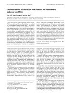
Histology following chronic gastric fluid aspirationFigure 2
Histology following chronic gastric fluid aspiration. Evaluation of Masson trichrome-stained tissue demonstrated an increase in
peribronchiolar and interstitial fibrosis in (a) left lung specimens after 8 weeks of gastric fluid aspiration compared to (b) lung
specimens from untreated rats or rats receiving normal saline. Scattered cellular infiltrates were most apparent in left lungs fol-
lowing chronic aspiration of gastric fluid after 8 weeks and consisted primarily of (c) giant cells (GC), apparent in specimens
stained with trichrome and (d) perivascular lymphocytes (arrows) as noted in specimens stained with H&E. In many trichrome-
stained left lung specimens from rats receiving gastric fluid, (e) complete airway occlusion was observed reminiscent of lesions
observed in lung transplant recipients exhibiting obliterative bronchiolitis (OB). Neither fibrosis nor cellular infiltrates were
apparent in right lung specimens from rats undergoing gastric fluid aspiration for (f) 8 weeks or at other time points.
Respiratory Research 2007, 8:87 />Page 6 of 12
(page number not for citation purposes)
markedly higher among specimens from rats receiving
gastric fluid compared to untreated rats (p < 0.05) and rats
receiving normal saline (p = 0.08).
In all instances, cytokine levels were highest in BAL speci-
mens from rats after 8 weeks of gastric fluid aspiration
(Figures 8a–d and 9). BAL levels of IL-1alpha, IL-1beta, IL-
2, and TNF-alpha peaked after 8 weeks of gastric fluid
aspiration and tapered off by 12 weeks and 16 weeks of
gastric fluid aspiration. At most time points, BAL IL-
1alpha, IL1-beta, and TNF-alpha levels were significantly
higher among rats receiving gastric fluid compared to
untreated rats or rats receiving normal saline (Figures 8a,
b, and 8d). Furthermore, TGF-beta, which was not detect-
able in the majority of BAL specimens, was detected in 5
of 6 specimens from rats undergoing 8 weeks of gastric
fluid aspiration (Figure 9, p < 0.0001 based on chi-square
analysis).
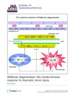
Changes in CD4:CD8 T cell ratios as a result of chronic aspi-ration of gastric fluidFigure 6
Changes in CD4:CD8 T cell ratios as a result of chronic aspi-
ration of gastric fluid. Relative CD4:CD8 T cell ratios in
bronchoalveolar lavage (BAL) specimens following aspiration
of either gastric fluid or normal saline are shown.
†
p < 0.10,
**p < 0.01 based on two-tailed Student's t test.
Macrophage infiltration following chronic aspiration of gastric fluidFigure 4
Macrophage infiltration following chronic aspiration of gastric
fluid. The relative macrophage quantities in bronchoalveolar
lavage (BAL) specimens following chronic aspiration of either
gastric fluid or normal saline are shown. The ratio of macro-
phages in the left to the macrophage in the right lung is
shown as a function of time. In the inset, the ratio of macro-
phages in the left to the macrophages in the right lung is
shown for all rats, regardless of the duration of treatment.
†
p
< 0.10, *p < 0.05, **p < 0.01 based on two-tailed Student's t
test.
Fibrosis following gastric fluid aspirationFigure 3
Fibrosis following gastric fluid aspiration. The fibrosis grade
was evaluated as described in the Methods. The mean peri-
bronchiolar fibrosis grade was significantly higher in left
(treated) lung specimens compared to right (untreated) lung
specimens after 4, 8, 12, and 16 weeks of gastric fluid aspira-
tion. **p < 0.01, ***p < 0.001 based on two-tailed Student's t
test.
T-cell infiltrates as a result of chronic aspiration of gastric fluidFigure 5
T-cell infiltrates as a result of chronic aspiration of gastric
fluid. Relative T cell quantities in bronchoalveolar lavage
(BAL) specimens following aspiration of either gastric fluid or
normal saline are shown. Left:right lung T cell quantities were
substantially higher in BAL specimens from rats receiving gas-
tric fluid compared to rats receiving normal saline after 4, 8,
12, and 16 weeks of aspiration.
†
p < 0.10, ***p < 0.001 based
on two-tailed Student's t test.
Respiratory Research 2007, 8:87 />Page 7 of 12
(page number not for citation purposes)
Discussion
The inflammatory response to massive acute aspiration
events has been described previously based on animal
models dating back 35 to 40 years [15,16,18]. The initial
1–2 hours are characterized by an immediate chemical
burn, primarily attributed to the acidity of gastric con-
tents, which is associated with endothelial cell damage,
increased capillary permeability, and scattered intraalveo-
lar hemorrhage. Several hours later, an acute inflamma-
tory response follows, comprised primarily of alveolar
neutrophils and macrophages. After approximately 15
hours, however, the inflammatory response resolves and
pulmonary capillary permeability returns to baseline [16].
In contrast, the pathophysiologic effects of chronic aspira-
tion are much less clear. Clinically, lung injury due to
repetitive aspiration in patients with GERD has been asso-
ciated with a number of pulmonary disorders including
idiopathic pulmonary fibrosis, asthma, chronic bronchi-
tis, cystic fibrosis, and chronic obstructive pulmonary dis-
ease [1-5]. Data from a number of clinical studies suggest
that chronic aspiration in the context of GERD is associ-
ated with increased rates of BOS and mortality in lung
transplant recipients [9-14]. However, it is unclear
whether chronic aspiration in the context of GERD causes
and/or exacerbates pulmonary disease or vice-versa. Fur-
thermore, the cellular processes that contribute to such
injury have not yet been characterized.
This work provides the first experimental animal model
aimed at evaluating the pathogenesis of chronic aspira-
tion-associated disease. In the current study, histology
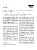
Cytokine response to chronic aspiration of gastric fluidFigure 7
Cytokine response to chronic aspiration of gastric fluid. Cytokine levels were evaluated in the bronchoalveolar lavage (BAL)
fluid from the left lung of rats receiving gastric fluid in their left lung, the left lung of rats receiving normal saline in their left
lung, the right lung of rats receiving gastric fluid in their left lung, and the left lung of rats receiving no treatment. Levels of (a)
IL-1alpha (b) IL-1beta, (c) IL-2 and (d) TNF-alpha are shown.
††
p < 0.05 vs. right (untreated) lung after gastric fluid; *p < 0.10,
**p < 0.05, ***p < 0.01, ****p < 0.001 vs. left (treated) lung after normal saline;
‡‡
p < 0.05,
‡‡‡
p < 0.01 vs. BAL from untreated
rats based on two-tailed Student's t test.
Respiratory Research 2007, 8:87 />Page 8 of 12
(page number not for citation purposes)
specimens from rats undergoing experimentally induced
gastric fluid aspiration demonstrated increased fibrosis
and a considerable giant cell infiltrate. Although the
extent of fibrosis in rats undergoing aspiration of gastric
fluid remained persistently greater than controls, the mag-
nitude decreased slightly with prolonged aspiration, the
relevance of which is unclear from these studies. Addi-
tionally, perivascular and peribronchial lymphocytic infil-
trates were seen, as well as fibroproliferative lesions
obstructing terminal airways, histologic findings compa-
rable to those of acute and chronic rejection, respectively,
in lung transplant recipients. Although these lesions were
most prominent in rats following 8 weeks of gastric fluid
aspiration, they were evident at all time points. Interest-
ingly, these lesions were similar to the scattered granulo-
matous response and obstructive bronchiolitis pattern
described by Teabeaut that occurred in some instances fol-
lowing an acute aspiration event in rabbits [18].
Analysis of BAL specimens revealed a substantial increase
in macrophages and T cells (particularly after 4 weeks of
gastric fluid aspiration) that persisted throughout the
study period. Notably, the increase in T cells was charac-
terized by a prominent shift toward a higher CD4:CD8 T
cell ratio, which some authors have previously correlated
with OB (obliterans bronchiolitis) in lung transplant
recipients [19,20]. Furthermore, repetitive gastric fluid
aspiration also resulted in increased TGF-beta, TNF-alpha,
IL-1alpha, IL-1beta, and IL-2 concentrations in BAL speci-
mens compared to controls, a TH1 cytokine-dominated
profile, whereas no increase in TH2 cytokines, such as IL-
4, IL-6, and IL-10, was detected.
One explanation for these observations is that repetitive
aspiration events may result in an early macrophage
response that generates TGF-beta, TNF-alpha, IL-1alpha,
and IL-1beta. Chemotactic for fibroblasts, TGF-beta
Cytokine response as a function of duration of gastric fluid aspirationFigure 8
Cytokine response as a function of duration of gastric fluid aspiration. Cytokine levels in bronchoalveolar lavage (BAL) fluid are
shown after 4, 8, 12 and 16 weeks of aspiration. Levels of (a) IL-1alpha, (b) IL-1beta, (c) IL-2, and (d) TNF-alpha are shown.
†
p <
0.10,
††
p < 0.05 vs. right (untreated) lung after gastric fluid; *p < 0.10, **p < 0.05 vs. left (treated) lung after normal saline based
on two-tailed Student's t test.
Respiratory Research 2007, 8:87 />Page 9 of 12
(page number not for citation purposes)
induces fibrosis and remodeling of the extracellular
matrix [21]. Production of TNF-alpha stimulates a gener-
alized inflammatory response, not only potentiating
fibrosis, but also inducing the upregulation of adhesion
molecules and the production of additional cytokines,
including IL-1alpha and IL-1beta. TNF-alpha also plays a
critical role in leukocyte trafficking and homing of T and
B cells [22,23]. The synthesis of IL-1alpha and IL-1beta by
endothelial cells, fibroblasts, and macrophages can be
profibrotic and not only exacerbates the inflammatory
response, but also activates and stimulates proliferation of
T and B cells [21]. Activated T cells produce additional IL-
1alpha and IL-1beta as well as IL-2, which further propa-
gate the inflammatory response by activating macro-
phages, natural killer cells, and lymphokine-activated
killer cells. Furthermore, these cytokines stimulate differ-
entiation and proliferation of T and B lymphocytes
thereby directing or upregulating the cell-mediated and
humoral immune responses. Such an upregulated
immune response may have substantial effects on pulmo-
nary pathology beyond mediation of lung transplant
rejection. Since the lung is exposed constantly to numer-
ous environmental antigens, altered immunoreactivity
against these antigens may have a substantial effect on
normal lung function.
By exacerbating the pulmonary immune response, it is
possible that such an inflammatory milieu could initiate
the development of various pulmonary diseases. An indi-
vidual's response may influence the phenotypic response
in the lung – the majority of patients may exhibit a nor-
mal reparative response whereas certain individuals may
be more prone to fibrosis or other pathophysiology. For
instance, such altered function may play a role in the asth-
matic response, which is thought to depend primarily
upon the interaction of mast cells, eosinophils, macro-
phages, CD4+ T cells, and IgE-producing B cells [24-26]. It
is possible that the presence of macrophages and CD4+ T
cells in patients with chronic aspiration may lower the
threshold for an immune-mediated asthmatic response by
inducing isotype-switching to IgE production in B cells
[24].
It is also quite possible that, in the setting of a pulmonary
allograft, the development of these inflammatory media-
tors could recruit and/or exacerbate immune responses
that predispose recipients to acute and/or chronic rejec-
tion. The activation of TH1 immune pathways and the
generation of a cytotoxic response has been associated
with rejection in a number of lung allograft models
[27,28]. A principal promoter of T cell activation and cyto-
toxic function, IL-2 is commonly detected in recipients of
lung and other allografts during OB and/or rejection [29-
32]. Additionally, TGF-beta has been associated with the
tissue remodeling response that occurs during the devel-
opment of OB and has been used as an early marker for
the process [33-35]. TNF-alpha increases class I MHC
expression and has been associated with acute and
chronic rejection in recipients of lung and other allografts
[21,36-38]. Furthermore, blocking TGF-beta, TNF-alpha,
or IL-1 prevents airway matrix deposition and OB in ani-
mal models [38-40].
In our study, it appears that the native lung eventually
develops tolerance to the injury induction by chronic
aspiration. Peribronchiolar and interstitial fibrosis, as well
as cellular infiltrates began at 4 weeks, peaked at 8 weeks,
and then appeared to regress after that time. BAL speci-
mens showed that macrophage infiltrates appeared to
peak by 8 weeks, and that cytokine levels peaked at 8
weeks. The immune responses initially induced by repeti-
tive aspiration events thus build over the first 8 weeks with
corresponding worsening of the histopathologic appear-
ance of the involved lung. As cytokine concentrations and
inflammatory cell populations then diminish, presuma-
bly via mechanisms of immunologic tolerance and/or
protective structural changes in the lung, the degree of
fibrosis and airway pathology begin to normalize. It is
possible that this improvement would eventually plateau
at some level of permanent fibrosis and inflammatory
activation above the initial baseline, or that improvement
would continue to resolution given enough time.
These preliminary studies have several inherent limita-
tions. First, weekly administration of gastric fluid to rats
TGF-beta production in response to chronic aspiration of gastric fluidFigure 9
TGF-beta production in response to chronic aspiration of
gastric fluid. TGF-beta was detected in BAL fluid from 5 of 6
rats after 8 weeks of gastric fluid aspiration. In contrast, TGF-
beta was undetectable in almost all BAL specimens from
untreated rats or from rats receiving normal saline (p <
0.0001 based on chi-square analysis).
Respiratory Research 2007, 8:87 />Page 10 of 12
(page number not for citation purposes)
may not be representative of clinical GERD, which often
affects patients at more frequent intervals. For this reason,
more physiologic studies involving GERD induction in
rats are underway in our laboratory. This study does not
address what component of gastric fluid is primarily
responsible for the observed pathologic changes. Normal
saline controls also reveal that gastric fluid is not neces-
sary to induce elevation of TNF-alpha levels (see figure 7).
This could be due to mechanical effects of even physiolog-
ically inert fluid in air spaces, or to repetitive anesthesia,
intubation, and mechanical ventilation. Vaneker et. al.
recently showed in a rodent model that mechanical venti-
lation with clinically relevant ventilator settings caused
reversible increases in immune cytokine concentrations
(including TNF-alpha) and leukocyte influx in lung tissue
[41]. The absence of significant histologic changes seen in
their study also agrees with our findings.
Nevertheless, this work is expected to provide a basis for
future studies. Foremost among those studies will be the
evaluation of the components of gastric fluid that are pri-
marily responsible for the inflammatory response result-
ing from chronic aspiration. Major components of gastric
fluid include hydrogen, potassium, sodium, and chloride
ions, pepsin, bile salts, and food particles. Although the
concentrations of these different components are highly
variable depending on the animal's time since feeding,
diet, age, state of stress, and numerous other variables,
approximate values have been reported in the literature:
pH 1.0 – 3.5, Sodium 50 mEq/l, Potassium 9.0 mEq/L,
Chloride 135 mEq/l, Pepsin 0.5 mmol/L (or 2300 – 3100
U/mL), Bile Salts 0.05 – 0.15 mmol/L [42-45]. Given its
known role in acute aspiration, the acidity of gastric fluid
might be expected to be a major etiologic factor in the
injury seen in this model in which gastric fluid pH ranged
between1.0 – 2.5. Based on data from our institution,
however, it seems unlikely that the acidic component of
the gastric contents is solely responsible for poor out-
comes, since the administration of H2 blockers or proton
pump inhibitors to lung transplant recipients with GERD
does not prevent their clinical deterioration [9,11,13,14].
Other components may thus play an even more impor-
tant role. For instance, lipopolysaccharide, commonly
present in gastric secretions, can induce a neutrophilic
alveolitis [46], and a high concentration of bile acids in
post lung transplant BAL samples were associated with
earlier onset of BOS [47]. Determination of the compo-
nents of gastric fluid that are primarily responsible for the
observed pathology may facilitate the development of
pharmacologic interventions aimed at the pathologic
processes associated with chronic GERD.
Conclusion
Clinical data suggests that chronic aspiration contributes
to pulmonary injury, resulting in a variety of pulmonary
pathologies. Based on the rodent model of chronic aspira-
tion described herein, chronic aspiration can initiate an
inflammatory response consisting of macrophages and T
cells and characterized by increased TGF-beta, TNF-alpha,
IL-1alpha, IL-1beta, IL-2 and fibrosis in the lung. The
increased production of TGF-beta and TH1 cytokines fol-
lowing repetitive aspiration events further suggests that
chronic aspiration augments pulmonary injury. These
observations provide further support for the role for
chronic aspiration in the development of pulmonary
fibrosis, OB, and asthma. This work also provides a
springboard for future studies aimed at better characteri-
zation of the pathways and effector molecules involved in
chronic aspiration-associated pulmonary dysfunction.
Competing interests
The author(s) declare that they have no competing inter-
ests.
Authors' contributions
JA carried out gastric fluid aspirations, assisted with anal-
ysis of cytokine levels, and histology data, and helped to
prepare the manuscript. SML helped with analysis of
cytokine data and assisted with manuscript preparation.
MH helped perform the gastric fluid aspirations and with
data analysis. BL carried out the cytokine analysis, and
helped prepare figures for the manuscript. CH assisted
with animal care and organ preparation for histologic
examination, assisted with histology data analysis. EC
helped to design the project and carried out preliminary
experiments validating aspiration technique. YY assisted
in developing the methodology of gastric fluid collection
and aspiration. SSL obtained funding for this project,
assisted with experimental design, and performed histo-
logic analysis. WP assisted with the experimental design,
and with manuscript preparation. RDD obtained funding
for this project and assisted with project conception and
design.
Acknowledgements
We thank our colleagues at the Transplantation Immunobiology Labora-
tory, Duke University Medical Center for their support. In particular, we
thank Mary Lou Everett for her technical support and Leonie M. Appel for
her helpful comments in the preparation of this manuscript.
1
Supported in
part by the American College of Surgeons Faculty Research Grant.
2
Sup-
ported in part by NIH R01 HL60232-03.
References
1. Tobin RW, Pope CE 2nd, Pellegrini CA, Emond MJ, Sillery J, Raghu G:
Increased prevalence of gastroesophageal reflux in patients
with idiopathic pulmonary fibrosis. Am J Respir Crit Care Med
1998, 158(6):1804-1808.
2. Vaezi MF: Extraesophageal manifestations of gastroesopha-
geal reflux disease. Clin Cornerstone 2003, 5(4):32-38. discussion
39–40
3. Harding SM: Recent clinical investigations examining the asso-
ciation of asthma and gastroesophageal reflux. Am J Med 2003,
115(Suppl 3A):39S-44S.
Respiratory Research 2007, 8:87 />Page 11 of 12
(page number not for citation purposes)
4. Ducolone A, Vandevenne A, Jouin H, Grob JC, Coumaros D, Meyer
C, Burghard G, Methlin G, Hollender L: Gastroesophageal reflux
in patients with asthma and chronic bronchitis. Am Rev Respir
Dis 1987, 135(2):327-332.
5. Ledson MJ, Tran J, Walshaw MJ: Prevalence and mechanisms of
gastro-oesophageal reflux in adult cystic fibrosis patients. J R
Soc Med 1998, 91(1):7-9.
6. Henderson RD, Woolfe CR: Aspiration and gastroesophageal
reflux. Can J Surg 1978, 21(4):352-354.
7. Crausaz FM, Favez G: Aspiration of solid food particles into
lungs of patients with gastroesophageal reflux and chronic
bronchial disease. Chest 1988, 93(2):376-378.
8. DeMeester TR, Bonavina L, Iascone C, Courtney JV, Skinner DB:
Chronic respiratory symptoms and occult gastroesophageal
reflux. A prospective clinical study and results of surgical
therapy. Ann Surg 1990, 211(3):337-345.
9. Cantu E 3rd, Appel JZ 3rd, Hartwig MG, Woreta H, Green C, Messier
R, Palmer SM, Davis RD Jr: Early fundoplication prevents
chronic allograft dysfunction in patients with gastroesopha-
geal reflux disease. Ann Thorac Surg 2004, 78(4):1142-1151. dis-
cussion 1142–1151
10. Hadjiliadis D, Duane Davis R, Steele MP, Messier RH, Lau CL, Eubanks
SS, Palmer SM: Gastroesophageal reflux disease in lung trans-
plant recipients. Clin Transplant 2003, 17(4):363-368.
11. Davis RD Jr, Lau CL, Eubanks S, Messier RH, Hadjiliadis D, Steele MP,
Palmer SM: Improved lung allograft function after fundoplica-
tion in patients with gastroesophageal reflux disease under-
going lung transplantation. J Thorac Cardiovasc Surg 2003,
125(3):533-542.
12. Lau CL, Palmer SM, Howell DN, McMahon R, Hadjiliadis D, Gaca J,
Pappas TN, Davis RD, Eubanks S: Laparoscopic antireflux sur-
gery in the lung transplant population. Surg Endosc 2002,
16(12):1674-1678.
13. Palmer SM, Miralles AP, Howell DN, Brazer SR, Tapson VF, Davis RD:
Gastroesophageal reflux as a reversible cause of allograft
dysfunction after lung transplantation. Chest 2000,
118(4):1214-1217.
14. Hartwig M, Appel JZ III, Cantu E III, Woreta H, Palmer SM, Davis RD:
Non-alloimmune Injury Mediated by Gastroesophageal
Reflux Precipitates Alloimmune Injury in Lung Transplant
Patients. J Heart Lung Transplant 23(2):s43.
15. Knight PR, Rutter T, Tait AR, Coleman E, Johnson K: Pathogenesis
of gastric particulate lung injury: a comparison and interac-
tion with acidic pneumonitis. Anesth Analg 1993, 77(4):754-760.
16. Kennedy TP, Johnson KJ, Kunkel RG, Ward PA, Knight PR, Finch JS:
Acute acid aspiration lung injury in the rat: biphasic patho-
genesis. Anesth Analg 1989, 69(1):87-92.
17. Ashcroft T, Simpson JM, Timbrell V: Simple method of estimating
severity of pulmonary fibrosis on a numerical scale. J Clin
Pathol 1988, 41(4):467-470.
18. Teabeaut JR 2nd: Aspiration of gastric contents; an experimen-
tal study. Am J Pathol 1952, 28(1):51-67.
19. Duncan SR, Leonard C, Theodore J, Lega M, Girgis RE, Rosen GD,
Theofilopoulos AN: Oligoclonal CD4(+) T cell expansions in
lung transplant recipients with obliterative bronchiolitis. Am
J Respir Crit Care Med 2002, 165(10):1439-1444.
20. Reynaud-Gaubert M, Thomas P, Gregoire R, Badier M, Cau P, Sampol
J, Giudicelli R, Fuentes P: Clinical utility of bronchoalveolar lav-
age cell phenotype analyses in the postoperative monitoring
of lung transplant recipients. Eur J Cardiothorac Surg 2002,
21(1):60-66.
21. Kirk A: Immunology of Transplantation. In Surgery: Basic Science
and Clinical Evidence Edited by: Norton J, Bollinger RR, Chang AE,
Lowry SE, Mulvihill SJ, Pass HI, Tohmpson RW. New York: Springer-
Verlag; 2001.
22. Sedgwick JD, Riminton DS, Cyster JG, Korner H: Tumor necrosis
factor: a master-regulator of leukocyte movement. Immunol
Today 2000, 21(3):110-113.
23. Gunn MD, Ngo VN, Ansel KM, Ekland EH, Cyster JG, Williams LT: A
B-cell-homing chemokine made in lymphoid follicles acti-
vates Burkitt's lymphoma receptor-1. Nature 1998,
391(6669):799-803.
24. Kapsenberg ML, Hilkens CM, Wierenga EA, Kalinski P:
The role of
antigen-presenting cells in the regulation of allergen-specific
T cell responses. Curr Opin Immunol 1998, 10(6):607-613.
25. Wills-Karp M, Luyimbazi J, Xu X, Schofield B, Neben TY, Karp CL,
Donaldson DD: Interleukin-13: central mediator of allergic
asthma. Science 1998, 282(5397):2258-2261.
26. Robinson DS, Hamid Q, Ying S, Tsicopoulos A, Barkans J, Bentley AM,
Corrigan C, Durham SR, Kay AB: Predominant TH2-like bron-
choalveolar T-lymphocyte population in atopic asthma. N
Engl J Med 1992, 326(5):298-304.
27. Boehler A, Bai XH, Liu M, Cassivi S, Chamberlain D, Slutsky AS, Kes-
havjee S: Upregulation of T-helper 1 cytokines and chemokine
expression in post-transplant airway obliteration. Am J Respir
Crit Care Med 1999, 159(6):1910-1917.
28. Zuo XJ, Matsumura Y, Prehn J, Saito R, Marchevesky A, Matloff J, Jor-
dan SC: Cytokine gene expression in rejecting and tolerant
rat lung allograft models: analysis by RT-PCR. Transpl Immunol
1995, 3(2):151-161.
29. Gu Y, Takao M, Kai M, Lu L, Shimamoto A, Onoda K, Shimono T, Tan-
aka K, Shimpo H, Shiraishi T, et al.: The role of cyclosporine A and
interleukin-2 in obliterative airway disease in a rat tracheal
transplant model. Ann Thorac Cardiovasc Surg 2000, 6(4):224-231.
30. Sundaresan S, Alevy YG, Steward N, Tucker J, Trulock EP, Cooper JD,
Patterson GA, Mohanakumar T: Cytokine gene transcripts for
tumor necrosis factor-alpha, interleukin-2, and interferon-
gamma in human pulmonary allografts. J Heart Lung Transplant
1995, 14(3):512-518.
31. Jordan SC, Marchevski A, Ross D, Toyoda M, Waters PF: Serum
interleukin-2 levels in lung transplant recipients: correlation
with findings on transbronchial biopsy. J Heart Lung Transplant
1992, 11(5):1001-1004.
32. Nickerson P, Steiger J, Zheng XX, Steele AW, Steurer W, Roy-
Chaudhury P, Strom TB: Manipulation of cytokine networks in
transplantation: false hope or realistic opportunity for toler-
ance? Transplantation 1997, 63(4):489-494.
33. Blobe GC, Schiemann WP, Lodish HF: Role of transforming
growth factor beta in human disease. N Engl J Med 2000,
342(18):1350-1358.
34. El-Gamel A, Sim E, Hasleton P, Hutchinson J, Yonan N, Egan J, Camp-
bell C, Rahman A, Sheldon S, Deiraniya A, et al.: Transforming
growth factor beta (TGF-beta) and obliterative bronchiolitis
following pulmonary transplantation. J Heart Lung Transplant
1999, 18(9):828-837.
35. Charpin JM, Valcke J, Kettaneh L, Epardeau B, Stern M, Israel-Biet D:
Peaks of transforming growth factor-beta mRNA in alveolar
cells of lung transplant recipients as an early marker of
chronic rejection. Transplantation 1998, 65(5):752-755.
36. Magnan A, Mege JL, Reynaud M, Thomas P, Capo C, Garbe L, Meric
B, Badier M, Bongrand P, Viard L, et al.: Monitoring of alveolar
macrophage production of tumor necrosis factor-alpha and
interleukin-6 in lung transplant recipients. Marseille and
Montreal Lung Transplantation Group. Am J Respir Crit Care
Med 1994, 150(3):684-689.
37. Steinmuller C, Steinhoff G, Bauer D, You XM, Denzin H, Franke-Ull-
mann G, Hausen B, Bruggemann C, Wagner TO, Lohmann-Matthes
ML, et al.: Analysis of leukocyte activation during acute rejec-
tion of pulmonary allografts in noninfected and cytomegalo-
virus-infected rats. J Leukoc Biol 1997, 61(1):40-49.
38. Smith CR, Jaramillo A, Lu KC, Higuchi T, Kaleem Z, Mohanakumar T:
Prevention of obliterative airway disease in HLA-A2-trans-
genic tracheal allografts by neutralization of tumor necrosis
factor. Transplantation 2001, 72(9):1512-1518.
39. Liu M, Suga M, Maclean AA, St George JA, Souza DW, Keshavjee S:
Soluble transforming growth factor-beta type III receptor
gene transfection inhibits fibrous airway obliteration in a rat
model of Bronchiolitis obliterans. Am J Respir Crit Care Med 2002,
165(3):419-423.
40. Farivar AS, Mackinnon-Patterson B, Woolley S, Namkung J, Shi-
mamoto A, Verrier ED, Mulligan MS: FR167653 reduces oblitera-
tive airway disease in rats. J Heart Lung Transplant 2004,
23(8):985-992.
41. Vaneker M, Halbertsma FJ, van Egmond J, Netea MG, Dijkman HB,
Snijdelaar DG, Joosten LA, van der Hoeven JG, Scheffer GJ:
Mechan-
ical ventilation in healthy mice induces reversible pulmonary
and systemic cytokine elevation with preserved alveolar
integrity: an in vivo model using clinical relevant ventilation
settings. Anesthesiology 2007, 107(3):419-426.
Publish with BioMed Central and every
scientist can read your work free of charge
"BioMed Central will be the most significant development for
disseminating the results of biomedical research in our lifetime."
Sir Paul Nurse, Cancer Research UK
Your research papers will be:
available free of charge to the entire biomedical community
peer reviewed and published immediately upon acceptance
cited in PubMed and archived on PubMed Central
yours — you keep the copyright
Submit your manuscript here:
/>BioMedcentral
Respiratory Research 2007, 8:87 />Page 12 of 12
(page number not for citation purposes)
42. Carone FA, Cooke RE: Effect of potassium deficiency on gastric
secretion in the rat. The American journal of physiology 1953,
172(3):684-688.
43. Lozzio BB, Biempica L, Royer M, Gorodisch S: Action of aminogua-
nidine on the stomach of pylorus-ligated rats. The American
journal of digestive diseases 1961, 6:534-545.
44. Kotani T, Murashima Y, Kobata A, Amagase K, Takeuchi K: Patho-
genic importance of pepsin in ischemia/reperfusion-induced
gastric injury. Life sciences 2007, 80(21):1984-1992.
45. Campbell NB, Ruaux CG, Shifflett DE, Steiner JM, Williams DA,
Blikslager AT: Physiological concentrations of bile salts inhibit
recovery of ischemic-injured porcine ileum. American journal of
physiology 2004, 287(2):G399-407.
46. Nikula KJ, Green FH: Animal models of chronic bronchitis and
their relevance to studies of particle-induced disease. Inhal
Toxicol 2000, 12(Suppl 4):123-153.
47. D'Ovidio F, Mura M, Ridsdale R, Takahashi H, Waddell TK, Hutcheon
M, Hadjiliadis D, Singer LG, Pierre A, Chaparro C, et al.: The effect
of reflux and bile acid aspiration on the lung allograft and its
surfactant and innate immunity molecules SP-A and SP-D.
Am J Transplant 2006, 6(8):1930-1938.