Báo cáo y học: " Bronchiolar chemokine expression is different after single versus repeated cigarette smoke exposure" docx
Bạn đang xem bản rút gọn của tài liệu. Xem và tải ngay bản đầy đủ của tài liệu tại đây (1.17 MB, 12 trang )
BioMed Central
Page 1 of 12
(page number not for citation purposes)
Respiratory Research
Open Access
Research
Bronchiolar chemokine expression is different after single versus
repeated cigarette smoke exposure
Tomoko Betsuyaku*
1
, Ichiro Hamamura
2
, Junko Hata
2
, Hiroshi Takahashi
2
,
Hiroaki Mitsuhashi
2
, Tracy L Adair-Kirk
3
, Robert M Senior
3
and
Masaharu Nishimura
1
Address:
1
First Department of Medicine, Hokkaido University School of Medicine, Kita-15, Nishi-7, Kita-ku, Sapporo, 060-8683, Japan,
2
Teijin
Institute for Bio-medical Research, Teijin Pharma Ltd., 4-3-2 Asahigaoka, Hino, Tokyo 191-8512, Japan and
3
Division of Pulmonary and Critical
Care Medicine, Department of Medicine, Washington University School of Medicine and Barnes-Jewish Hospital, 660 So. Euclid Avenue St. Louis,
MO 63110, USA
Email: Tomoko Betsuyaku* - ; Ichiro Hamamura - ;
Junko Hata - ; Hiroshi Takahashi - ; Hiroaki Mitsuhashi - ;
Tracy L Adair-Kirk - ; Robert M Senior - ; Masaharu Nishimura -
* Corresponding author
Abstract
Background: Bronchioles are critical zones in cigarette smoke (CS)-induced lung inflammation.
However, there have been few studies on the in vivo dynamics of cytokine gene expression in
bronchiolar epithelial cells in response to CS.
Methods: We subjected C57BL/6J mice to CS (whole body exposure, 90 min/day) for various
periods, and used laser capture microdissection to isolate bronchiolar epithelial cells for analysis of
mRNA by quantitative reverse transcription-polymerase chain reaction.
Results: We detected enhanced expression of keratinocyte-derived chemokine (KC), macrophage
inflammatory protein-2 (MIP-2), tumor necrosis factor-α (TNF-α), and interleukin-1β (IL-1β) by
bronchial epithelial cells after 10 consecutive days of CS exposure. This was mirrored by increases
in neutrophils and KC, MIP-2, TNF-α, and IL-1β proteins in the bronchoalveolar lavage (BAL) fluid.
The initial inhalation of CS resulted in rapid and robust upregulation of KC and MIP-2 with
concomitant DNA oxidation within 1 hr, followed by a return to control values within 3 hrs. In
contrast, after CS exposure for 10 days, this initial surge was not observed. As the CS exposure
was extended to 4, 12, 18 and 24 weeks, the bronchiolar KC and MIP-2 expression and their levels
in BAL fluid were relatively dampened compared to those at 10 days. However, neutrophils in BAL
fluid continuously increased up to 24 weeks, suggesting that neutrophil accumulation as a result of
long-term CS exposure became independent of KC and MIP-2.
Conclusion: These findings indicate variable patterns of bronchiolar epithelial cytokine expression
depending on the duration of CS exposure, and that complex mechanisms govern bronchiolar
molecular dynamics in vivo.
Published: 21 January 2008
Respiratory Research 2008, 9:7 doi:10.1186/1465-9921-9-7
Received: 3 September 2007
Accepted: 21 January 2008
This article is available from: />© 2008 Betsuyaku et al; licensee BioMed Central Ltd.
This is an Open Access article distributed under the terms of the Creative Commons Attribution License ( />),
which permits unrestricted use, distribution, and reproduction in any medium, provided the original work is properly cited.
Respiratory Research 2008, 9:7 />Page 2 of 12
(page number not for citation purposes)
Background
Chronic obstructive pulmonary disease (COPD) is charac-
terized by irreversible airflow limitation due to structural
alterations of the small airways, chronic inflammation in
the airways and alveolar spaces, and loss of elastic recoil
caused by destruction of lung parenchyma. Since the
pathology of COPD is that of a chronic inflammatory
process, many studies have focused on identifying the
inflammatory cell types and/or cytokines that play a role
in this condition. Increased numbers of neutrophils, mac-
rophages, and lymphocytes in the airways are found asso-
ciated with COPD [1-3], and various mediators derived
from these cells, such as interleukin (IL)-1β, IL-6, IL-8,
tumor necrosis factor (TNF)-α, monocyte chemoattract-
ant protein (MCP-1), and matrix metalloproteinase
(MMP)-2, MMP-8, and MMP-9, are suggested to contrib-
ute to the development of COPD [4,5].
Cigarette smoke (CS) is the main risk factor for the devel-
opment of COPD. Oxidative stress caused by CS can
injure lung cells directly and can trigger cytokine produc-
tion, leading to the recruitment of inflammatory cells into
the lungs [6-8]. The induction of these cytokines is regu-
lated by the activation of redox-sensitive transcription fac-
tors, such as nuclear factor-kappa B (NF-κB) [9,10].
Increased expression of NF-κB has been detected in the
airway epithelium of smokers compared to non-smokers
[11].
Airway epithelium is an important site of cytokine expres-
sion in COPD and in response to CS [12,13]. For example,
cultured airway epithelial cells produce IL-6 and IL-8 in
response to CS exposure [14-16], and TNF-α, IL-8, MCP-
1, and macrophage inflammatory protein (MIP)-1α are
upregulated in the bronchiolar epithelium of subjects
with COPD [17-19]. However, there is scant data on the
time course of cytokine responses to CS by airway epithe-
lium. Therefore, we decided to examine the temporal rela-
tionship of airway epithelial cytokine production after CS
exposure in vivo utilizing a mouse model of mainstream
CS exposure.
We hypothesized that CS would induce changes in gene
expression of pro-inflammatory cytokines, and that the
kinetics of the response would differ depending on dura-
tion of exposure and the cytokine. Accordingly, we exam-
ined the expression of keratinocyte-derived chemokine
(KC)/CXCL1 and MIP-2/CXCL2, the combined functional
homologues to human IL-8, as well as TNF-α and IL-1β by
bronchiolar epithelial cells following either a single CS
exposure, repeated exposures for 10 days, or repeated
exposure for 24 weeks. We have identified previously
unrecognized dynamics in gene expression in bronchiolar
epithelium in vivo following CS exposure.
Methods
CS Exposure
Male C57BL/6J mice, 9–10 weeks of age (Charles River,
Atsugi, Japan), were exposed to whole body mainstream
CS generated from commercially available filtered ciga-
rettes (12 mg tar/1.0 mg nicotine, Philip Morris, Rich-
mond, VA) by the INH06-CIGR0A smoking system (MIPS
Co., Osaka, Japan) using the following parameters: 15.5
puff/min/cigarette; air flow, 0.07 L/min; and volume, 280
mL/second, as described elsewhere [20]. The CS was
diluted with filtered air at 1:7 ratio and directed into the
exposure chamber (50(L) × 50(W) × 25(H) cm) at a
smoke to air ratio of 1:2. The box was fitted with an
exhaust vent of the same size as a blower vent in order to
avoid the accumulation of mainstream smoke. In initial
experiments, mice were exposed to CS for 90 min per day
for 1, 3, 7 or 10 days, and were sacrificed 24 hrs after the
last CS exposure. For assessment of kinetic patterns in
gene expression following CS exposure, mice received
either a single 90-min CS exposure or daily exposure for
10 days, and then were sacrificed at 1, 3, 6 or 24 hrs after
the last CS exposure. In long-tem CS exposure experi-
ments, mice were exposed to CS for 90 min per day, 6 days
per week, for 4, 12, 18 or 24 weeks, and were sacrificed 24
hrs after the last CS exposure. Age-matched, air-exposed
mice served as controls. All animal procedures were per-
formed in accordance with the regulations of the Animal
Care and Use Committee of Teijin Institute for Bio-medi-
cal Research.
Analysis of plasma cotinine levels
Blood samples were collected at 1 and 3 hrs after the last
CS exposure and the levels of cotinine in the plasma were
measured using a quantitative enzyme immunoassay kit
(Salimetrics, State College, PA), as described previously
[21]. Data represent average concentration from 3 mice
per condition performed in duplicate.
Collection of Broncholalveolar Lavage (BAL) fluid
At various times after CS exposure, mice were anesthetized
with urethane and α-chloralose and then exsanguinated
by severing the abdominal aorta, and BAL fluid was
retrieved by injecting 1.0 ml saline through the trachea as
described previously [22]. An aliquot of each BAL fluid
was mixed with an equal volume of Turk's solution
(Wako, Osaka, Japan) and the total cell number was deter-
mined using a hemocytometer. Differential cell counts
were performed on Diff-Quik™ (International Reagents,
Kobe, Japan)-stained cytospin preparations. Data repre-
sent the average numbers of cells per ml of BAL fluid from
8 mice per condition. The BAL fluid was centrifuged, and
the cell-free supernatants were stored at -80°C until use.
Respiratory Research 2008, 9:7 />Page 3 of 12
(page number not for citation purposes)
Detection of albumin, MIP-2, KC, TNF-
α
, and IL-1
β
in BAL
fluid
The concentration of albumin in BAL fluid was deter-
mined using an albumin B test-Wako kit (Wako) accord-
ing to manufacturer's protocol. The quantity of KC, MIP-
2, TNF-α, and IL-1β in the BAL fluid was determined by
ELISA kits (R&D Systems, Minneapolis, MN) according to
manufacturer's protocols. The detection limit was 7 pg/
mL for KC, MIP-2 and IL-1β, and 15 pg/mL for TNF-α.
Data represent the average concentration of 8 mice per
condition performed in duplicate.
Immunohistochemical evaluation of DNA oxidation in the
lung
Lungs were inflated with diluted Tissue-Tek OCT (Sakura
Finetek U.S.A., Torrance, CA) (50% vol/vol in ribonucle-
ase (RNase)-free PBS containing 10% sucrose) and imme-
diately frozen on dry ice as previously described [23].
Antigen retrieval was done on 5 µm sections by incubat-
ing in L.A.B. solution (Polysciences, Warrington, PA) at
room temperature for 10 min. Sections were incubated
with 3% bovine serum albumin (Sigma, St. Louis, MO)
and the mouse immunoglobulin blocking reagent from
the M.O.M. immunodetection kit (Vector Laboratories,
Burlingame, CA) in the TNB solution included in the TSA
Biotin System Immunohistochemistry kit (PerkinElmer
Life and Analytical Sciences, Wellesley, MA) for 30 min in
order to block non-specific binding. Sections were then
incubated with the mouse monoclonal anti-8-hydroxy-2'-
deoxyguanosine (8-OHdG) antibody (10 µg/mL) (Japan
Institute for the Control of Aging, Shizuoka, Japan) for 1
hr at room temperature, followed by 3% hydrogen perox-
ide for 10 min at room temperature [24]. Immunostain-
ing was developed using the M.O.M. detection kit (Vector
Laboratories) with DAB substrate and counterstained with
Mayer's hematoxylin.
Collection of bronchiolar epithelial cells by Laser Capture
Microdissection (LCM)
LCM was performed on 7 µm frozen sections to retrieve
cells within 100 µm of the bronchoalveolar junction using
the PixCell II System (Arcturus Engineering, Mountain
View, CA) with the following parameters: laser diameter,
30 µm; pulse duration, 5 ms; and amplitude, 50 mW, as
described previously [23]. Approximately 10,000 laser
bursts were used to collect cells for RNA isolation from
each mouse.
RNA isolation and real-time RT-PCR
Total RNA was extracted from LCM-retrieved bronchiolar
epithelial cells using an RNeasy Mini kit (Qiagen, Hilden,
Germany), or from whole lung homogenates using the
ISOGEN RNA isolation kit (Nippon Gene Co. Ltd.
Toyama, Japan). The quantity and quality of RNA were
determined using an RNA LabChip kit (Agilent Technolo-
gies, Palo Alto, CA) or a NanoDrop spectrophotometer
(NanoDrop Inc., Wilmington, DE). RNA was reverse tran-
scribed using TaqMan Reverse Transcription Reagents kit
(Applied Biosystems, Foster City, CA) as described previ-
ously [25]. The resulting first-strand cDNAs were used as
templates for quantitative real-time RT-PCR using the ABI
Prism 7700 Sequence Detector (Applied Biosystems) and
gene-specific TaqMan Gene Expression Assays probes
(Applied Biosystems) as described previously [18]. Probes
for mouse KC (Assay ID: Mm00433859_m1) were
derived from the boundary of exons 3 and 4 of the murine
KC gene [26]. Probes for mouse MIP-2
(Mm00436450_m1) were derived from the boundary of
exons 3 and 4 of the murine MIP-2 gene [27]. Probes for
mouse TNF-α (Mm00443258_m1) were derived from the
boundary of exons 1 and 2 of the murine TNF-α gene [28].
Probes for mouse IL-1β (Mm00434228_m1) were derived
from the boundary of exons 3 and 4 of the murine IL-1β
gene [29]. Probes for mouse β2-macroglobulin (β2-MG;
Mm00437764_m1) were used as an endogenous control
as described previously [25]. The relative amounts of each
mRNA in the samples were assessed by interpolation of
their cycle thresholds from a standard curve, and were
then normalized against β2-MG mRNA. RT-PCR data rep-
resent 6–12 mice per condition performed in triplicate.
Statistical analysis
All results are reported as means ± standard error of the
mean (SEM). Statistical significance of the values at each
time point after CS exposure was evaluated by Dunnett's
type multiple comparative analyses against the values in
pretreatment groups. Differences were considered signifi-
cant at p < 0.05. Statistical analyses were performed using
SAS version 8.2 for Windows XP (SAS Institute, Tokyo,
Japan).
Results
CS Exposure
To confirm adequate CS exposure, the levels of plasma
cotinine were measured. Cotinine was essentially unde-
tectable in mice unexposed to CS (<5 ng/mL) (Figure 1A).
However, following a single 90-min CS exposure, a dra-
matic increase in plasma cotinine levels was detected
within 1 hr of CS exposure, which was reduced but still
elevated 3 hr after CS exposure. Exposure of mice to CS for
10 consecutive days did induce a slight progressive
increase of cotinine in the plasma. The levels of plasma
cotinine in our studies are similar to that detected in
blood samples of ICR mice following CS exposure [30]
and in blood samples of humans who smoke >5 cigarettes
a day [31].
BAL fluid albumin, a biomarker of tissue injury, was also
measured. A significant increase in albumin in the BAL
fluid was detected after 3 days of CS exposure, compared
Respiratory Research 2008, 9:7 />Page 4 of 12
(page number not for citation purposes)
to levels in unexposed controls (Figure 1B). The levels of
albumin in the BAL fluid continued to increase following
10 consecutive days of CS exposures. These data indicate
that the conditions for CS exposure utilized for these stud-
ies were sufficient to induce known effects caused by
mainstream CS exposure [30,32,33].
CS-induced DNA oxidative stress in bronchiolar and
alveolar epithelium
To determine whether CS exposure induces oxidative
stress in lung cells, sections were immunostained for 8-
OHdG, a marker of oxidative DNA stress. Oxidative stress
was not detected in the lungs of mice unexposed to CS
(Figure 2A). Within 1 hr after a single 90-min CS expo-
sure, nuclear staining of 8-OHdG was markedly increased
in the bronchiolar and alveolar type II epithelial cells (Fig-
ure 2B), confirming that both cell types are major targets
of CS oxidants. However, 24 hr after a single CS exposure,
the staining was back almost to baseline (Figure 2C).
These data are consistent with the findings of Aoshiba et
al. [34] who examined the kinetics of oxidative stress in
mice following a single CS exposure.
Surprisingly, after repeated CS exposure for 10 days,
nuclear staining of 8-OHdG was not detected in the bron-
chiolar or alveolar epithelium either before (Figure 2D) or
at 1 hr (Figure 2E) following the final CS exposure. In
long-tem CS exposure experiments (4 or 24 weeks), 8-
OHdG staining was not observed at 4 or 24 weeks, either
(data not shown). Normal mouse IgG1 negative control
(DakoCytomation, Glostrup, Denmark) in place of the 8-
OHdG antibody resulted in no tissue staining (Figure 2F).
These data suggest that repeated CS exposure elicits a
mechanism in airway and alveolar epithelial cells to pro-
tect against DNA oxidative stress.
Inflammatory cells in BAL fluid during 10 days of CS
exposure
To determine whether short-term CS exposure elicits an
inflammatory response, mice were exposed to CS for up to
10 days and the BAL fluids collected 24 hr after the last CS
exposure were examined for the presence of inflammatory
cells. After 10 days of CS exposure, the total number of
cells in the BAL fluid was significantly increased compared
to the BAL fluid of unexposed mice (Figure 3A). Although
slightly elevated after 3 days of CS exposure, there was no
significant change in the number of macrophages in the
BAL fluid irrespective of duration of CS exposure (Figure
3B). In contrast, a significant increase in the number of
neutrophils in the BAL fluid was observed after 3 days of
CS exposure, which continued to increase following con-
secutive CS exposures (Figure 3C). A significant increase
in the number of lymphocytes was also detected after 10
days of CS exposure (Figure 3D). However, based on the
total number of cells relative to the number of each cell
type in the BAL fluid, the predominant infiltrating cells in
response to CS exposure were neutrophils.
Neutrophilic chemokines in BAL fluid during 10 days of CS
exposure
Since the primary infiltrating cells in response to CS expo-
sure were neutrophils, we examined the BAL fluid for
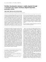
Plasma cotinine and BAL albumin levels are elevated follow-ing CS exposureFigure 1
Plasma cotinine and BAL albumin levels are elevated
following CS exposure. (A) Blood samples were collected
at 1 and 3 hrs after the last CS exposure and the levels of
cotinine in the plasma was measured using a quantitative
enzyme immunoassay kit. Data represent average concentra-
tion of three mice per condition ± SEM. (B) BAL fluids were
collected at 24 hr after the last CS exposure and assayed for
the presence of albumin using the albumin B test-Wako kit.
Data represent the average concentration of eight mice per
condition ± SEM. Statistical significance: ** = p < 0.01; *** = p
< 0.001.
Respiratory Research 2008, 9:7 />Page 5 of 12
(page number not for citation purposes)
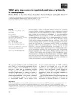
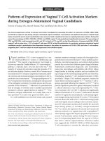
Initial CS exposure induces oxidative stress in airway epithelial cellsFigure 2
Initial CS exposure induces oxidative stress in airway epithelial cells. Mice were unexposed (A), exposed to a single
CS exposure (B and C), or repeatedly exposed to CS for 10 days (D and E). Lung sections were stained for oxidative DNA
stress using an anti-8-OHdG antibody at 1 hr (B and E) or 24 hrs (C and D) following the last CS exposure. Normal mouse IgG1
in place of the 8-OHdG antibody served as a negative control (F). Images are representative of five mice per condition.
Respiratory Research 2008, 9:7 />Page 6 of 12
(page number not for citation purposes)
cytokines that attract neutrophils. After 3 days of CS expo-
sure, a significant increase in the level of KC in the BAL
fluid was observed compared to the BAL fluid from unex-
posed mice (Figure 4A). The levels of KC in the BAL fluid
continued to increase following consecutive CS expo-
sures, paralleling the accumulation of neutrophils in the
BAL fluid. A significant increase in the levels of MIP-2
(Figure 4B), TNF-α (Figure 4C) and IL-1β (Figure 4D) was
also detected after 10 days of CS exposure.
Whole lung and bronchiolar cytokine expression during 10
days of CS exposure
Since CS produced oxidative stress in the airways (Figure
2), we examined whether bronchiolar epithelial cells
express cytokines in response to CS by real-time RT-PCR
analyses of RNA isolated from LCM-retrieved terminal
bronchiolar epithelial cells. Furthermore, we compared
the expression levels of KC, MIP-2, TNF-α, and IL-β in
LCM-retrieved bronchiolar epithelial cells to the levels in
whole lung homogenates. We found that KC was signifi-
cantly upregulated after a single CS exposure in whole
lung homogenates, whereas a significant upregulation in
the bronchiolar epithelium was not detected until follow-
ing 3 days of CS exposure (Figure 5A). The expression of
MIP-2 was increased in bronchiolar epithelial cells after 3
and 10 days of CS exposure (Figure 5B). The expression of
TNF-α was increased in bronchiolar epithelial cells after 7
and 10 days of CS exposure (Figure 5C). However, the
expression of MIP-2 and TNF-α in whole lung homoge-
nates was not significantly increased until after 10 days of
CS exposure. Significant upregulation of IL-1β was
observed at 10 days in both whole lung homogenates and
in bronchiolar epithelium (Figure 5D). Although there are
temporal differences in the expression of these cytokines
between whole lung homogenates and bronchiolar epi-
thelium, the expression of these genes was notably higher
in bronchiolar epithelial cells when compared with whole
lung homogenate at all time points.
Patterns of bronchiolar cytokine expression after CS
exposure
To determine the dynamics of the bronchiolar epithelial
cell cytokine expression, we examined the expression of
KC, MIP-2, TNF-α, and IL-1β by the bronchiolar epithe-
lium over a 24-hr period following either a single CS
exposure or repeated exposures for 10 days. In bronchi-
olar epithelial cells of CS-naïve mice, rapid and robust
increases in the expression of KC (70-fold) and MIP-2
(20-fold) were observed within 1 hr of a single CS expo-
sure, compared to unexposed mice (Figure 6A and 6B).
These values returned close to baseline values within 3
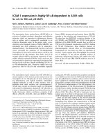
Repeated CS exposure increases KC, MIP-2, TNF-α and IL-1β in BAL fluidFigure 4
Repeated CS exposure increases KC, MIP-2, TNF-α
and IL-1β in BAL fluid. Mice were repeatedly exposed to
CS for up to 10 days and the levels of KC (A), MIP-2 (B),
TNF-α (C) and IL-1β (D) in the BAL fluid were determined by
ELISA. Data represent the average concentration per ml BAL
fluid ± SEM from eight mice. Statistical significance: * = p <
0.05; ** = p < 0.01; *** = p < 0.001.
Repeated CS exposure induces inflammatory cell recruit-mentFigure 3
Repeated CS exposure induces inflammatory cell
recruitment. Mice were repeatedly exposed to CS for up
to 10 days and the cell content in the BAL fluid was identified
as described in Materials and Methods. Data represent the
average number of total cells (A), macrophages (B), neu-
trophils (C), and lymphocytes (D) per ml BAL fluid ± SEM
from eight mice. Statistical significance: * = p < 0.05; ** = p <
0.01; *** = p < 0.001.
Respiratory Research 2008, 9:7 />Page 7 of 12
(page number not for citation purposes)
hrs. Although the expression of KC and MIP-2 in bronchi-
olar epithelial cells of mice after 10 days of repeated expo-
sure was elevated before the final CS exposure, a transient
increase was not observed after CS exposure.
Similar to KC and MIP-2, but to a much lesser extent (2-
fold), an increase in IL-1β expression was detected in the
bronchiolar epithelium within 1 hr following a single CS
exposure which returned close to baseline levels within 3
hrs (Figure 6D). Also similar to KC and MIP-2, the level of
IL-1β expression following repeated CS exposure was ele-
vated before the final CS exposure as compared to base-
line levels of CS-naïve mice. However, unlike KC and
MIP-2, which were not upregulated in response to the
final CS exposure, IL-1β expression slowly rose over the
24 hr period following the final CS exposure.
In contrast to KC, MIP-2, and IL-1β, bronchiolar expres-
sion of TNF-α failed to return to baseline by 3 hr after the
initial CS exposure (Figure 6C) and after 10 days of
repeated exposure, there was a slight, slow increase in
TNF-α expression by the bronchiolar epithelium follow-
ing the final CS exposure. These data indicate that the
kinetic patterns of expression of different cytokines by the
bronchiolar epithelium following CS exposure vary.
Inflammatory cells in BAL fluid during long-term CS
exposure
Thereafter, we addressed whether the pattern of inflam-
matory response of the lung to CS exposure observed after
10 days persists following long-term CS exposure. We
found that as the exposure of CS to the mice was extended
to 4, 12, 18 and 24 weeks, a further increase in the total
cell number in BAL fluid was observed (Figure 7A). Simi-
larly, the elevated number of neutrophils in BAL fluid that
developed during the short-term CS exposure persisted in
the long-term CS exposure, showing over 50% neu-
trophils out of the total BAL cells at 24 weeks (Figure 7B).
KC and MIP-2 in BAL fluid during long-term CS exposure
In contrast to the parallel increase in the number of neu-
trophils and the levels of KC and MIP-2 in BAL fluid in the
short-term CS exposure experiment, KC and MIP-2 levels
in BAL fluid declined by 4 weeks of CS exposure com-
pared to the levels at 10 days despite the persistent
increase of neutrophils (Figure 8A and 8B).
Bronchiolar KC and MIP-2 expression during long-term CS
exposure
As described above, we detected enhanced bronchiolar
expression of KC and MIP-2 after 10 consecutive days of
CS exposure (Figure 5A and 5B). However, as the exposure
of CS to the mice was extended to 4, 12, 18 and 24 weeks,
bronchiolar KC and MIP-2 mRNA were nearly back to
baseline after 4 weeks of CS exposure and did not change
with continued CS exposure up to 24 weeks (Figure 9A
and 9B). Bronchiolar KC and MIP-2 expressions exhibited
a similar pattern to those levels in BAL fluid (Figure 8A
and 8B).
Discussion
Prior animal studies have established the expression of
pro-inflammatory cytokines in various types of experi-
mental lung injury including CS-induced models [30,35-
39]. However, the role of bronchiolar epithelial cells, spe-
cifically, in producing pro-inflammatory cytokines and
their inflammatory sequela in vivo remains to be eluci-
dated. Several approaches might be used to detect
cytokine expression. In situ hybridization can provide cell-
specific information regarding gene expression, but it is
not quantitative. Real-time RT-PCR provides quantitative
measure of gene expression, but using RNA from homog-
enized tissue has the disadvantage of averaging-out sig-
nals, in that signals from small, but potentially critical,
cell populations could go undetected. The use of LCM to
selectively isolate a defined cell population improves the
sample preparation for gene expression analysis. Further-
more, the predominance of Clara cells in the distal air-
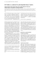
Repeated CS exposure upregulates KC, MIP-2, TNF-α, and IL-1β expression in whole lung homogenate and in LCM-retrieved bronchiolar epitheliumFigure 5
Repeated CS exposure upregulates KC, MIP-2, TNF-
α, and IL-1β expression in whole lung homogenate
and in LCM-retrieved bronchiolar epithelium. Mice
were repeatedly exposed to CS for up to 10 days, and the
expression of KC (A), MIP-2 (B), TNF-α (C), and IL-1β (D) in
whole lung homogenates (white bars) and LCM-retrieved
bronchiolar epithelium (black bars) were determined by real-
time RT-PCR. Data represent the average expression rela-
tive to β2-MG ± SEM from at least six mice. Statistical signifi-
cance: * = p < 0.05; ** = p < 0.01; *** = p < 0.001.
Respiratory Research 2008, 9:7 />Page 8 of 12
(page number not for citation purposes)
ways of mice [40] enables us to harvest a relatively
homogeneous population of cells by LCM and a confir-
mation method that we harvested distal bronchiolar epi-
thelium, the expression of Clara cell-specific protein
(CCSP). In these studies, CCSP expression was more than
6,000-fold higher in LCM-retrieved bronchiolar epithe-
lium compared to that in whole lung homogenate (data
not shown). Although the sample was highly enriched in
Clara cells, it should be noted that LCM harvests all the
cells present at a given site. Thus, migrating inflammatory
cells within the bronchiolar epithelium may have influ-
enced the changes in gene expression. However, we
detected minimal, if any, Gr-1 stained neutrophils within
the bronchiolar epithelial region following 10 days of
repeated CS exposure (data not shown). These data sug-
gest that the cytokine expression in the LCM-retrieved
samples were derived from the airway epithelium.
The present study indicates that the acute effects of single
CS exposure cannot easily be extrapolated to the effects of
repeated smoking for short or long term. The effects of CS
exposure on bronchiolar epithelial cells over time may
result from several processes having different time frames:
(a) direct toxic interaction with constituents of CS
(including free radicals) that have penetrated the protec-
tive antioxidant shield of epithelial lining fluid [41]; (b)
damage to cells by toxic reactive products such as hydro-
gen peroxide generated by interaction between CS and
epithelial cells [42] or epithelial lining fluid, which con-
tains oxidized proteins, such as oxidized glutathione and
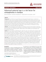
Kinetics in bronchiolar expression of KC and MIP-2 over 24 hrs is different after single vs. repeated CS exposureFigure 6
Kinetics in bronchiolar expression of KC and MIP-2 over 24 hrs is different after single vs. repeated CS expo-
sure. Mice were exposed to a single CS exposure (closed circles) or repeatedly exposed to CS for 10 days (open circles). For the
10 day exposure the time point before CS represents 24 hrs after 9 days exposure. At various times up to 24 hrs following last
CS exposure, the bronchiolar epithelial cells were harvested by LCM and the expression of KC (A), MIP-2 (B), IL-1β (C), and
TNF-α (D) were determined by real-time RT-PCR. Data represent the average expression relative to β2-MG ± SEM from at
least six mice. Statistical significance: *** = p < 0.001 vs. before CS exposure at each time point.
Respiratory Research 2008, 9:7 />Page 9 of 12
(page number not for citation purposes)
protein carbonyls [43]; and (c) reactions occurring subse-
quent to the activation of inflammatory-immune proc-
esses initiated by (a) and/or (b). Bronchiolar gene
expression in vivo may thus be affected not only by exoge-
nous CS, but also by the local microenvironment in bron-
chioles, such as infiltration of inflammatory cells, which
cannot be replicated in vitro.
CS has been implicated in initiating a lung inflammatory
response by activating transcription factors, such as NF-κB
and AP-1, and chromatin unwinding (histone acetyla-
tion/deacetylation), that lead to upregulation of pro-
inflammatory genes [44-46]. Di Stefano et al. demon-
strated an increase in NF-κB p65 (A) protein in bronchial
epithelium from COPD patients and from smokers with
normal lung function [11]. Skerrett et al. reported that the
cell-targeted inhibition of NF-κB activation in distal air-
way epithelial cells under the Clara cell 10-kDa protein/
uteroglobin promoter in mice suppresses the inflamma-
tory response to inhaled lipopolysaccharide, providing
direct evidence that NF-κB activation in these cells and the
subsequent signal transduction play a critical role in lung
inflammation in vivo [47]. Elizur et al. also demonstrated
that Clara cells, a predominant cell type in the distal air-
ways of mice, were the predominant source of KC and
MCP-1 in the early response to lipopolysaccharide [48]. In
the present study, we observed 8-OHdG formation, a
major reactive oxygen species (ROS)-induced DNA stress
product, at 1 hr, but not 24 hr after single exposure to CS
(Figure 2), which was mirrored by the expression pattern
of KC and MIP-2 by bronchiolar epithelial cells (Figure 6).
These data suggest that bronchiolar epithelial cells are
capable of repairing oxidative DNA stress rapidly, and the
temporal DNA stress in bronchiolar epithelial cells is
involved in the rapid surge of KC and MIP-2 induction
Long-term of CS exposure does not enhance KC and MIP-2 in BAL fluidFigure 8
Long-term of CS exposure does not enhance KC and
MIP-2 in BAL fluid. Mice were exposed to CS (black bars)
or to air (hatched bars) for 4, 12, 18 and 24 weeks and the
levels of KC (A) and MIP-2 (B) in the BAL fluid were deter-
mined by ELISA. Data represent the average concentration
per ml BAL fluid ± SEM from eight mice. The data set of Fig.
4A and 4B are shown for comparison. Statistical significance:
* = p < 0.05; ** = p < 0.01; *** = p < 0.001 vs. before CS
exposure at day 0.
Long-term of CS exposure induces inflammatory cell recruit-mentFigure 7
Long-term of CS exposure induces inflammatory cell
recruitment. Mice were exposed to CS (black bars) or to
air (hatched bars) for 4, 12, 18 and 24 weeks, and the cell con-
tent in the BAL fluid was identified as described in Materials
and Methods. Data represent the average number of total
cells (A) and neutrophils (B) per ml BAL fluid ± SEM from
eight mice. The data set of Fig. 3A and 3C are also included
for comparison. Statistical significance: * = p < 0.05; ** = p <
0.01; *** = p < 0.001 vs. before CS exposure at day 0.
Respiratory Research 2008, 9:7 />Page 10 of 12
(page number not for citation purposes)
through redox-sensitive transcription factors, such as NF-
κB or AP-1. Thus, it should be further investigated how the
expression of KC and MIP-2 in those cells following CS
challenge is associated with activation of NF-κB and/or
AP-1 in vivo.
There is apparently marked diversity in the mechanisms of
CS-induced inflammatory responses, even between in
vitro experiments [8,49,50], which precludes further repli-
cation of the molecular dynamics in primary cells in vivo.
It should be noted that rapid bronchiolar induction
occurs selectively for KC and MIP-2, but not for TNF-α
and IL-1β in response to initial CS exposure, suggesting
that these genes are regulated by diverse pathways. All of
these cytokines are eventually upregulated in bronchiolar
epithelium at 10 days, however, the source of those
increased cytokines in BAL fluid would become more
complex at later time points, considering many other cell
types involved.
In the 10 consecutive day CS exposure experiments, we
have found that the response of bronchiolar epithelium to
CS varies depending on prior exposure. There are marked
differences in the response of the distal airway epithelial
cells elicited by the very first CS exposure compared to
what happens after repeated CS exposure. After repeated
CS exposures, we did not detect DNA oxidative stress or a
surge in KC and MIP-2 expression. These data suggest that
repeated CS exposure elicits a mechanism in airway epi-
thelial cells to protect against DNA oxidative stress, which
in turn affects redox-mediated cytokine production. How-
ever, although the surge of KC and MIP-2 expression in
response to CS was lost, there was a continual rise in
expression of KC, MIP-2, TNF-α, and IL-1β by the bron-
chiolar epithelial cells upon repeated CS exposure, which
was mirrored by their levels in BAL fluids and the influx of
neutrophils into the lung up to 10 days.
The comparison of short and long CS exposure models
highlighted the complexity of the inflammatory response
of the lungs to exposure to CS. The mechanisms by which
the long-term CS exposure dampens bronchiolar expres-
sions of KC and MIP-2 need further investigation. Interest-
ingly, neutrophil accumulation in BAL fluid becomes
independent of KC and MIP-2 levels during long-term CS
exposure. Possible explanations are: (a) other chemoat-
tractants, such as MIP-3α/CCL20, replace KC and MIP-2
to recruit neutrophils [51], (b) extracellular matrix frag-
ments resulting from damage after chronic CS exposure
could be pro-inflammatory [52,53], and/or (c) the parti-
tioning of neutrophils between tissue and alveolar spaces
changes, possibly due to changes in adhesion and/or
development of more channels for neutrophil egress into
alveolar spaces.
Taken together, the successful collection of bronchiolar
epithelium by LCM and the comparative gene expression
analyses has revealed the detailed kinetic profiles of
cytokine expression following CS exposure in bronchiolar
epithelium. Our data suggest that airway epithelial cells
play a role in the recruitment of inflammatory cells in
response to CS exposure, and that there are multiple
mechanisms by which CS exposure induces cytokine pro-
duction by bronchiolar epithelial cells. It is to be empha-
sized that the CS model used in this study is only intended
as a bridge between in vitro and in vivo studies of neu-
trophil recruitment in response to CS. Extrapolations of
current findings to the other experimental CS models or
to the human should be made with caution.
Long-term CS exposure dampens KC and MIP-2 expressions in LCM-retrieved bronchiolar epitheliumFigure 9
Long-term CS exposure dampens KC and MIP-2
expressions in LCM-retrieved bronchiolar epithe-
lium. Mice were exposed to CS for 4, 12, 18 and 24 weeks
and the expression of KC (A) and MIP-2 (B) in LCM-retrieved
bronchiolar epithelium (black bars) were determined by real-
time RT-PCR. Data represent the average expression rela-
tive to β2-MG ± SEM from at least six mice. The part of data
in Fig. 5A and 5B are also used for comparison. Statistical sig-
nificance: * = p < 0.05; ** = p < 0.01; *** = p < 0.001 vs.
before CS exposure at day 0.
Respiratory Research 2008, 9:7 />Page 11 of 12
(page number not for citation purposes)
Conclusion
In this study, we described the variable patterns of bron-
chiolar epithelial cytokine expression depending on the
duration of CS exposure, and these findings indicate that
complex mechanisms govern bronchiolar molecular
dynamics in vivo.
Competing interests
The authors declare that they have no competing interests.
The study has not been supported by tobacco industry.
Authors' contributions
TB conceived of the study, participated in its design, and
drafted the manuscript. IH and HT smoked mice to CS,
collected lung samples, and carried out ELISA and part of
RT-PCR. JH carried out laser capture microdissection and
immunohistochemistry and part of RT-PCR, and per-
formed the statistical analysis. TA participated in the study
design and drafted the manuscript. HM, RS and MN
supervised the study, and helped to draft the manuscript.
All authors have read and approved the final manuscript.
Acknowledgements
The authors wish to thank to Ms. Yoko Suzuki for technical assistance, and
Ms. Naomi Matsui for care and treatment of the mouse CS model. This
work was supported by the scientific research grants from the Ministry of
Education, Science, Culture and Sports of Japan (13470125 to MN,
14570532 to TB), Francis Family Foundation (TLA-K), Teijin Pharma Ltd.,
NHLBI/NIH P50 HL084922 (RMS, TB), and respiratory failure research
group of the Ministry of Health, Labor, and Welfare of Japan.
References
1. Jeffery PK: Structural and inflammatory changes in COPD: a
comparison with asthma. Thorax 1998, 53:129-136.
2. Saetta M, Di Stefano A, Turato G, Facchini FM, Corbino L, Mapp CE,
Maestrelli P, Ciaccia A, Fabbri LM: CD8+ T-lymphocytes in
peripheral airways of smokers with chronic obstructive pul-
monary disease. Am J Respir Crit Care Med 1998, 157:822-826.
3. Saetta M: Airway inflammation in chronic obstructive pulmo-
nary disease. Am J Respir Crit Care Med 1999, 160:S17-S20.
4. Barnes PJ: Small airways in COPD. N Engl J Med 2004,
350:2635-2637.
5. Chung KF: Cytokines in chronic obstructive pulmonary dis-
ease. Eur Respir J Suppl 2001, 34:50s-59s.
6. Brusselle GG, Bracke KR, Maes T, D'hulst AI, Moerloose KB, Joos GF,
Pauwels RA: Murine models of COPD. Pulm Pharmacol Ther 2006,
19:155-165.
7. Pettersen CA, Adler KB: Airways inflammation and COPD: epi-
thelial-neutrophil interactions. Chest 2002, 121:142S-150S.
8. van der Vaart H, Postma DS, Timens W, ten Hacken NH: Acute
effects of cigarette smoke on inflammation and oxidative
stress: a review. Thorax 2004, 59:713-721.
9. Moodie FM, Marwick JA, Anderson CS, Szulakowski P, Biswas SK,
Bauter MR, Kilty I, Rahman I: Oxidative stress and cigarette
smoke alter chromatin remodeling but differentially regu-
late NF-kappaB activation and proinflammatory cytokine
release in alveolar epithelial cells. FASEB J 2004, 18:1897-1899.
10. Yang SR, Chida AS, Bauter MR, Shafiq N, Seweryniak K, Maggirwar SB,
Kilty I, Rahman I: Cigarette smoke induces proinflammatory
cytokine release by activation of NF-kappaB and posttrans-
lational modifications of histone deacetylase in macro-
phages. Am J Physiol Lung Cell Mol Physiol 2006, 291:L46-L57.
11. Di Stefano A, Caramori G, Oates T, Capelli A, Lusuardi M, Gnemmi
I, Ioli F, Chung KF, Donner CF, Barnes PJ, Adcock IM: Increased
expression of nuclear factor-kappaB in bronchial biopsies
from smokers and patients with COPD. Eur Respir J 2002,
20:556-563.
12. Bates DV: The respiratory bronchiole as a target organ for the
effects of dusts and gases. J Occup Med 1973, 15:177-180.
13. Shaw RJ, Djukanovic R, Tashkin DP, Millar AB, du Bois RM, Orr PA:
The role of small airways in lung disease. Respir Med 2002,
96:67-80.
14. Beisswenger C, Platz J, Seifart C, Vogelmeier C, Bals R: Exposure of
differentiated airway epithelial cells to volatile smoke in
vitro. Respiration 2004, 71:402-409.
15. Kode A, Yang SR, Rahman I: Differential effects of cigarette
smoke on oxidative stress and proinflammatory cytokine
release in primary human airway epithelial cells and in a vari-
ety of transformed alveolar epithelial cells. Respir Res 2006,
7:132.
16. Mio T, Romberger DJ, Thompson AB, Robbins RA, Heires A, Rennard
SI: Cigarette smoke induces interleukin-8 release from
human bronchial epithelial cells. Am J Respir Crit Care Med 1997,
155:1770-1776.
17. de Boer WI, Sont JK, van Schadewijk A, Stolk J, van Krieken JH, Hiem-
stra PS: Monocyte chemoattractant protein 1, interleukin 8,
and chronic airways inflammation in COPD. J Pathol 2000,
190:619-626.
18. Fuke S, Betsuyaku T, Nasuhara Y, Morikawa T, Katoh H, Nishimura
M: Chemokines in bronchiolar epithelium in the develop-
ment of chronic obstructive pulmonary disease. Am J Respir
Cell Mol Biol 2004, 31:405-412.
19. Profita M, Chiappara G, Mirabella F, Di Giorgi R, Chimenti L, Cos-
tanzo G, Riccobono L, Bellia V, Bousquet J, Vignola AM: Effect of
cilomilast (Ariflo) on TNF-alpha, IL-8, and GM-CSF release
by airway cells of patients with COPD. Thorax 2003,
58:573-579.
20. Suzuki M, Betsuyaku T, Nagai K, Fuke S, Nasuhara Y, Kaga K, Kondo
S, Hamamura I, Hata J, Takahashi H, Nishimura M: Decreased air-
way expression of vascular endothelial growth factor in ciga-
rette smoke-induced emphysema in mice and COPD
patients. Inhal Toxicol in press.
21. Dhar P: Measuring tobacco smoke exposure: quantifying nic-
otine/cotinine concentration in biological samples by color-
imetry, chromatography and immunoassay methods. J Pharm
Biomed Anal 2004, 35:155-168.
22. Betsuyaku T, Fukuda Y, Parks WC, Shipley JM, Senior RM: Gelati-
nase B is required for alveolar bronchiolization after intrat-
racheal bleomycin. Am J Pathol 2000, 157:525-535.
23. Betsuyaku T, Griffin GL, Watson MA, Senior RM: Laser capture
microdissection and real-time reverse transcriptase/
polymerase chain reaction of bronchiolar epithelium after
bleomycin. Am J Respir Cell Mol Biol 2001, 25:278-284.
24. Toyokuni S, Tanaka T, Hattori Y, Nishiyama Y, Yoshida A, Uchida K,
Hiai H, Ochi H, Osawa T: Quantitative immunohistochemical
determination of 8-hydroxy-2'-deoxyguanosine by a mono-
clonal antibody N45.1: its application to ferric nitrilotriace-
tate-induced renal carcinogenesis model. Lab Invest 1997,
76:365-374.
25. Betsuyaku T, Senior RM: Laser capture microdissection and
mRNA characterization of mouse airway epithelium: meth-
odological considerations. Micron 2004, 35:229-234.
26. Cataisson C, Pearson AJ, Tsien MZ, Mascia F, Gao JL, Pastore S, Yuspa
SH: CXCR2 ligands and G-CSF mediate PKCalpha-induced
intraepidermal inflammation. J Clin Invest 2006, 116:2757-2766.
27. d'Empaire G, Baer MT, Gibson FC 3rd: The K1 serotype capsular
polysaccharide of Porphyromonas gingivalis elicits chemok-
ine production from murine macrophages that facilitates
cell migration. Infect Immun 2006, 74:6236-6243.
28. Nakazawa T, Nakazawa C, Matsubara A, Noda K, Hisatomi T, She H,
Michaud N, Hafezi-Moghadam A, Miller JW, Benowitz LI: Tumor
necrosis factor-alpha mediates oligodendrocyte death and
delayed retinal ganglion cell loss in a mouse model of glau-
coma. J Neurosci 2006, 26:12633-12641.
29. Lu JY, Sadri N, Schneider RJ: Endotoxic shock in AUF1 knockout
mice mediated by failure to degrade proinflammatory
cytokine mRNAs. Genes Dev 2006, 20:3174-3184.
30. Obot C, Lee K, Fuciarelli A, Renne R, McKinney W: Characteriza-
tion of mainstream cigarette smoke-induced biomarker
responses in ICR and C57Bl/6 mice. Inhal Toxicol 2004,
16:701-719.
Publish with BioMed Central and every
scientist can read your work free of charge
"BioMed Central will be the most significant development for
disseminating the results of biomedical researc h in our lifetime."
Sir Paul Nurse, Cancer Research UK
Your research papers will be:
available free of charge to the entire biomedical community
peer reviewed and published immediately upon acceptance
cited in PubMed and archived on PubMed Central
yours — you keep the copyright
Submit your manuscript here:
/>BioMedcentral
Respiratory Research 2008, 9:7 />Page 12 of 12
(page number not for citation purposes)
31. Jarvis MJ, Primatesta P, Erens B, Feyerabend C, Bryant A: Measuring
nicotine intake in population surveys: comparability of saliva
cotinine and plasma cotinine estimates. Nicotine Tob Res 2003,
5:349-355.
32. Subramaniam S, Whitsett JA, Hull W, Gairola CG: Alteration of
pulmonary surfactant proteins in rats chronically exposed to
cigarette smoke. Toxicol Appl Pharmacol 1996, 140:274-280.
33. Van Miert E, Dumont X, Bernard A: CC16 as a marker of lung
epithelial hyperpermeability in an acute model of rats
exposed to mainstream cigarette smoke. Toxicol Lett 2005,
159:115-123.
34. Aoshiba K, Koinuma M, Yokohori N, Nagai A: Immunohistochem-
ical evaluation of oxidative stress in murine lungs after ciga-
rette smoke exposure. Inhal Toxicol 2003, 15:1029-1038.
35. Belperio JA, Keane MP, Burdick MD, Londhe V, Xue YY, Li K, Phillips
RJ, Strieter RM: Critical role for CXCR2 and CXCR2 ligands
during the pathogenesis of ventilator-induced lung injury. J
Clin Invest 2002, 110:1703-1716.
36. Belperio JA, Keane MP, Burdick MD, Gomperts BN, Xue YY, Hong K,
Mestas J, Zisman D, Ardehali A, Saggar R, Lynch JP 3rd, Ross DJ, Stri-
eter RM: CXCR2/CXCR2 ligand biology during lung trans-
plant ischemia-reperfusion injury. J Immunol 2005,
175:6931-6939.
37. Call DR, Nemzek JA, Ebong SJ, Bolgos GR, Newcomb DE, Wollen-
berg GK, Remick DG: Differential local and systemic regulation
of the murine chemokines KC and MIP2. Shock 2001,
15:278-284.
38. Kubo S, Kobayashi M, Masunaga Y, Ishii H, Hirano Y, Takahashi K,
Shimizu Y: Cytokine and chemokine expression in cigarette
smoke-induced lung injury in guinea pigs. Eur Respir J 2005,
26:993-1001.
39. Thatcher TH, McHugh NA, Egan RW, Chapman RW, Hey JA, Turner
CK, Redonnet MR, Seweryniak KE, Sime PJ, Phipps RP: Role of
CXCR2 in cigarette smoke-induced lung inflammation. Am J
Physiol Lung Cell Mol Physiol 2005, 289:L322-L328.
40. Plopper CG: Clara cells. In Lung Growth and Development. Lung Biol-
ogy in Health and Disease Edited by: McDonald JA. Marcel Dekker.
New York; 1997:181-209.
41. Pryor WA, Stone K: Oxidants in cigarette smoke. Radicals,
hydrogen peroxide, peroxynitrate, and peroxynitrite. Ann N
Y Acad Sci 1993, 686:12-27. discussion 27–28
42. Lavigne MC, Eppihimer MJ: Cigarette smoke condensate induces
MMP-12 gene expression in airway-like epithelia. Biochem Bio-
phys Res Commun 2005, 330:194-203.
43. Nagai K, Betsuyaku T, Kondo T, Nasuhara Y, Nishimura M: Long
term smoking with age builds up excessive oxidative stress
in bronchoalveolar lavage fluid. Thorax 2006, 61:496-502.
44. Everhart MB, Han W, Sherrill TP, Arutiunov M, Polosukhin VV, Burke
JR, Sadikot RT, Christman JW, Yull FE, Blackwell TS: Duration and
intensity of NF-kappaB activity determine the severity of
endotoxin-induced acute lung injury. J Immunol 2006,
176:4995-5005.
45. Rahman I, Adcock IM: Oxidative stress and redox regulation of
lung inflammation in COPD. Eur Respir J 2006, 28:219-242.
46. Zhou L, Tan A, Iasvovskaia S, Li J, Lin A, Hershenson MB: Ras and
mitogen-activated protein kinase kinase kinase-1 coregulate
activator protein-1- and nuclear factor-kappaB-mediated
gene expression in airway epithelial cells. Am J Respir Cell Mol
Biol 2003, 28:762-729.
47. Skerrett SJ, Liggitt HD, Hajjar AM, Ernst RK, Miller SI, Wilson CB:
Respiratory epithelial cells regulate lung inflammation in
response to inhaled endotoxin. Am J Physiol Lung Cell Mol Physiol
in press.
48. Elizur A, Adair-Kirk TL, Kelley DG, Griffin GL, Demello DE, Senior
RM: Clara cells impact the pulmonary innate immune
response to LPS. Am J Physiol Lung Cell Mol Physiol 2007. 2007 May
25, 101152/ajplung.00024.2007
49. Hellermann GR, Nagy SB, Kong X, Lockey RF, Mohapatra SS: Mech-
anism of cigarette smoke condensate-induced acute inflam-
matory response in human bronchial epithelial cells. Respir
Res 2002, 3:22.
50. Laan M, Bozinovski S, Anderson GP: Cigarette smoke inhibits
lipopolysaccharide-induced production of inflammatory
cytokines by suppressing the activation of activator protein-
1 in bronchial epithelial cells. J Immunol 2004, 173:4164-4170.
51. Bracke KR, D'hulst AI, Maes T, Moerloose KB, Demedts IK, Lebecque
S, Joos GF, Brusselle GG: Cigarette smoke-induced pulmonary
inflammation and emphysema are attenuated in CCR6-defi-
cient mice. J Immunol 2006, 177:4350-4359.
52. Houghton AM, Quintero PA, Perkins DL, Kobayashi DK, Kelley DG,
Marconcini LA, Mecham RP, Senior RM, Shapiro SD: Elastin frag-
ments drive disease progression in a murine model of
emphysema. J Clin Invest 2006, 116:753-759.
53. Weathington NM, van Houwelingen AH, Noerager BD, Jackson PL,
Kraneveld AD, Galin FS, Folkerts G, Nijkamp FP, Blalock JE: A novel
peptide CXCR ligand derived from extracellular matrix deg-
radation during airway inflammation. Nat Med 2006,
12:317-323.