Báo cáo y học: " Maximal respiratory static pressures in patients with different stages of COPD severity" potx
Bạn đang xem bản rút gọn của tài liệu. Xem và tải ngay bản đầy đủ của tài liệu tại đây (261.69 KB, 7 trang )
BioMed Central
Page 1 of 7
(page number not for citation purposes)
Respiratory Research
Open Access
Research
Maximal respiratory static pressures in patients with different
stages of COPD severity
Claudio Terzano, Daniela Ceccarelli*, Vittoria Conti, Elda Graziani,
Alberto Ricci and Angelo Petroianni
Address: Department of Cardiovascular and Respiratory Sciences, UOC Malattie Respiratorie, University of Rome "La Sapienza", Italy
Email: Claudio Terzano - ; Daniela Ceccarelli* - ; Vittoria Conti - ;
Elda Graziani - ; Alberto Ricci - ; Angelo Petroianni -
* Corresponding author
Abstract
Background: In this study, we analyzed maximal inspiratory pressure (MIP) and maximal
expiratory pressure (MEP) values in a stable COPD population compared with normal subjects.
We evaluated the possible correlation between functional maximal respiratory static pressures and
functional and anthropometric parameters at different stages of COPD. Furthermore, we
considered the possible correlation between airway obstruction and MIP and MEP values.
Subject and methods: 110 patients with stable COPD and 21 age-matched healthy subjects
were enrolled in this study. Patients were subdivided according to GOLD guidelines: 31 mild, 39
moderate and 28 severe.
Results: Both MIP and MEP were lower in patients with severe airway impairment than in normal
subjects. Moreover, we found a correlation between respiratory muscle function and some
functional and anthropometric parameters: FEV
1
(forced expiratory volume in one second), FVC
(forced vital capacity), PEF (peak expiratory flow), TLC (total lung capacity) and height. MIP and
MEP values were lower in patients with severe impairment than in patients with a slight reduction
of FEV
1
.
Conclusion: The measurement of MIP and MEP indicates the state of respiratory muscles, thus
providing clinicians with a further and helpful tool in monitoring the evolution of COPD.
Background
In several diseases, the evaluation of respiratory muscle
strength can prove to be very useful. The measurement of
the maximum static mouth pressures made against an
occluded airway (maximal expiratory pressure and maxi-
mal inspiratory pressure) is the most widely used and is a
simple way to gauge respiratory muscle strength and to
quantify its severity [1-3].
When we analyze maximal respiratory pressure, we
should consider both the difficulty that some subjects
have in performing a maximal effort and the normal bio-
logical variability of respiratory muscle strength [4].
Maximal inspiratory pressure (MIP) is the maximum neg-
ative pressure that can be generated from one inspiratory
effort starting from functional residual capacity (FRC) or
residual volume (RV) [5,6]. Maximal expiratory pressure
Published: 21 January 2008
Respiratory Research 2008, 9:8 doi:10.1186/1465-9921-9-8
Received: 26 June 2007
Accepted: 21 January 2008
This article is available from: />© 2008 Terzano et al; licensee BioMed Central Ltd.
This is an Open Access article distributed under the terms of the Creative Commons Attribution License ( />),
which permits unrestricted use, distribution, and reproduction in any medium, provided the original work is properly cited.
Respiratory Research 2008, 9:8 />Page 2 of 7
(page number not for citation purposes)
(MEP) measures the maximum positive pressure that can
be generated from one expiratory effort starting from total
lung capacity (TLC) or FRC. Unlike inspiratory muscles,
expiratory muscles (abdominal and thoracic muscles)
reach their optimal force-length relationship at elevate
pulmonary volumes [7].
During normal breathing, most of the respiratory work
depends on the diaphragm function and the accessory res-
piratory muscles become necessary only during deep
inspiration [8].
The mouth pressures recorded during these maneuvers are
assumed to reflect respiratory muscle strength [9].
It is known that a reduction of MIP and MEP has been
associated with several neuromuscular diseases, but it is
also possible to point up lower values in patients with
chronic obstructive pulmonary disease (COPD) [10-12].
The factors contributing to respiratory muscle weakness in
many patients with COPD are: a) malnutrition related to
biochemical, anatomical and physiological changes; b)
muscular atrophy; c) steroid-induced myopathy; d) pul-
monary hyperinflation with increased residual volume; e)
reduced blood flow to the respiratory muscles [13-19].
The measurement of MIP and MEP is indicated in any of
these situations or when dyspnea or hypercapnia are not
proportional to FEV
1
reduction [6].
Age and sex could influence MIP and MEP values; these
are lower in females than in males and quite constant
until seventy years of age when they start to decrease [6].
The objectives of this study were: (1) to describe MIP and
MEP values in a stable COPD population, (2) to explore
the effect of varying degrees of obstructive ventilatory
impairment on MIP and MEP measurement, (3) to evalu-
ate the possible correlation between functional maximal
respiratory pressures and functional parameters at differ-
ent stages of COPD.
Methods
During 1 year 110 patients suffering from COPD were
enrolled in this study. All patients were in a clinically sta-
ble condition and the mean age was 70 ± 8 years (Table
1).
At the first examination, we excluded those patients
whose FEV
1
improvement, after a bronchodilatory test,
was ≥ 12% and 200 mL of the baseline value and with a
history of asthma. Other patients, with clinically signifi-
cant diseases such as fibrothorax, bronchiectasis, tubercu-
losis or neuromuscular diseases were also excluded. Some
patients were excluded because of lack of compliance dur-
ing the forced expiratory test or during the MIP and MEP
maneuvers.
All patients gave their written informed consent before the
study and the Ethics Committee approved the protocol.
At the enrolment, all the subjects were examined. Anthro-
pometric measurements (age, height and weight) were
taken and pulmonary function tests, including flow/vol-
ume spirometry and N2 Washout, were conducted. Maxi-
mal static inspiratory and expiratory mouth pressures
were measured using a portable mouth pressure meter
(Spirovis – COSMED – Pavona – Italy); this had a dispos-
Table 1: Characteristics of patients
CONTROL GROUP MILD MODERATE SEVERE p VALUE* COPD PATIENTS
N° 21 31 39 28 98
Age 67 ± 5 67 ± 8 71 ± 6 72 ± 7 p > 0.05 70 ± 8
Body weight (Kg) 72 ± 6 76 ± 11 78 ± 12 75 ± 20 p > 0.05 77 ± 15
Body height (cm) 165 ± 8 168 ± 9 166 ± 8 167 ± 6 p > 0.05 167 ± 8
MIP (cmH
2
O) 99 ± 18 84 ± 22 80 ± 34 65 ± 20 p = 0.0002 77 ± 28
MEP (cmH
2
O) 102 ± 26 93 ± 29 90 ± 32 75 ± 21 p = 0.01 89 ± 31
FEV
1
(L) 2.7 ± 0,5 2.4 ± 0,6 1.5 ± 0.4 0.9 ± 6.3 p < 0.0001 1.7 ± 0.7
FEV
1
% 102 ± 12 91 ± 11 65 ± 8 38 ± 7 p < 0.0001 65 ± 22
FVC (L) 3.3 ± 0.9 3.2 ± 0.8 2.4 ± 0.7 1.9 ± 0.6 p < 0.0001 2.5 ± 0.9
FVC % 101 ± 12 96 ± 9 76 ± 10 59 ± 14 p < 0.001 77 ± 18
PEF (L/sec) 6.5 ± 2 6.6 ± 1.6 4.9 ± 1.7 3.1 ± 1.2 p < 0.0001 4.9 ± 2
PEF % 102 ± 12 91 ± 15 71 ± 18 45 ± 15 p < 0.0001 70 ± 24
TLC (L) 4.9 ± 1.3 5.9 ± 1.1 5.4 ± 1.3 5.6 ± 1.4 p = 0.037 5.7 ± 1.3
TLC % 98 ± 20 98 ± 13 94 ± 14 97 ± 18 p = 0.0007 97 ± 16
RV (L) 2 ± 0.5 2.5 ± 0.7 3 ± 0.8 3.9 ± 1.2 p < 0.0001 3 ± 1
RV % 97 ± 20 111 ± 29 124 ± 32 151 ± 47 p < 0.0001 128 ± 39
RV/TLC % 40 ± 20 41 ± 11 50 ± 20 66 ± 23 p < 0.0001 49.7 ± 19
*Comparison between the four groups (control, mild, moderate, severe) have been made by analysis of variance (ANOVA).
Respiratory Research 2008, 9:8 />Page 3 of 7
(page number not for citation purposes)
able mouthpiece, and a small leak to prevent glottic clo-
sure. MIP was obtained at the level of RV and MEP was
measured at the level of TLC. The measurements were
made in standing position. The subjects were verbally
encouraged to achieve maximal strength. The measure-
ments were repeated until five values varying by less than
5% and sustained for at least 1 s were obtained; the best
value achieved was considered in the data analysis.
Forced expiratory tests were recorded with a Cosmed
Quark spirometer (PFT4 SUITE – COSMED – Pavona –
Italy) which was calibrated each morning using the same
3 L precision syringe.
We divided patients into three groups on the basis of air-
way obstruction: mild (FEV1 > 80%), moderate (FEV1
between 80 and 50%), and severe (FEV1 < 50%) with
FEV1/FVC ratio <70% in all the groups.
The control group included 21 age-matched normal sub-
jects who were non-smokers, free of respiratory symptoms
and disease and with normal functional parameters
(Table 1).
Statistical analysis was performed for all measured func-
tional parameters using GraphPad Prism version 4.00 for
Windows (GraphPad Software, San Diego California
USA). Standards descriptive statistics based on mean and
standard deviation (SD) for quantitative variables were
used. The mean difference between MIP/MEP in healthy
subjects and MIP/MEP in patients with COPD was deter-
mined by the unpaired Student's t test. Statistical differ-
ences for respiratory muscle strength and different stage of
COPD were analyzed by analysis of variance (ANOVA).
The Pearson's test was performed to assess possible corre-
lation between MIP and MEP values and anthropometric
and functional parameters.
All p values were two sided and values below 0.05 were
considered statistically significant.
Results
During this study, 12 COPD patients were withdrawn: 5
because of a contemporary restrictive deficit and 7
because of their insufficient compliance during the forced
expiratory test or during the MIP and MEP maneuvers.
Therefore, 98 patients took part in this study. Table 1
shows the anthropometric parameters and lung function
of the patients studied.
The MIP and MEP values of healthy subjects were used as
a control group for the comparison with patients with dif-
ferent stages of COPD.
MEP was significantly lower (p = 0.0014) in patients with
severe airway obstruction than in the control group; no
differences were observed in mild and moderate patients
(p > 0.05).
At the same time, MIP was significantly lower at all the
stages of COPD than in the control group: mild p = 0.010;
moderate p = 0.018; severe p < 0.0001.
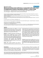
Statistical analysis performed on the total of COPD
patients to assess the possible correlation between MIP
and MEP values and anthropometric and functional
parameters showed significant (p < 0.05) positive correla-
tions among maximal static inspiratory pressure and FEV
1
(L, r
2
= 0.13, Figure 1), FVC (L, r
2
= 0.20), PEF (L/sec, r
2
=
0.19), TLC (L, r
2
= 0.11) and height (r
2
= 0.09).
Similar results were showed between MEP and the same
functional and anthropometric parameters (FEV1 r
2
=
0.13, figure 2; FVC r
2
= 0.19; PEF r
2
= 0.22; TLC r
2
= 0.12;
height r
2
= 0.15).
Respiratory muscle strength, however, had no significant
correlation with RV and RV/TLC (Figures 3, 4, 5, 6),
weight and age.
Moreover, we evaluated whether there was a possible cor-
relation between COPD stage and respiratory muscular
strength. The analysis of variance (ANOVA) showed no
statistically significant difference between mild and mod-
erate patients (p > 0.005), while the difference between
mild and severe was significant as well as that between
moderate and severe patients.
Relationship between MIP and FEV
1
(p = 0.0002; r
2
= 0.13)Figure 1
Relationship between MIP and FEV
1
(p = 0.0002; r
2
= 0.13).
Respiratory Research 2008, 9:8 />Page 4 of 7
(page number not for citation purposes)
Discussion
To our knowledge, this is the first study that analyzes MIP
and MEP variation in the different stages of COPD severity
to understand when MIP and MEP start to decrease.
The main finding of this work is that airway obstruction
may be closely associated with decreased respiratory pres-
sures in patients with COPD. In fact, both MIP and MEP
values were lower in patients with severe obstruction com-
pared with healthy subjects. MIP decreased also in patient
with mild and moderate functional impairment: this
could suggest an earlier deterioration of the inspiratory
muscles in this sort of patients.
Similar results have been showed in a recent work that
limited the evaluations only to the inspiratory muscle
strength [20].
Dynamic functional parameters are a measure of the res-
piratory muscular strength as well as maximal respiratory
pressures. In fact with the reduction of FEV
1
, PEF and FVC,
as in severe COPD, we have observed a similar decrease in
MIP and MEP. Interestingly, our study highlights the
importance of the predictivity of functional parameters
(FEV
1
, PEF, FVC, and TLC) on MIP and MEP reduction in
COPD patients.
Relationship between MIP and RV/TLC (p > 0.05; r
2
= 0.01)Figure 5
Relationship between MIP and RV/TLC (p > 0.05; r
2
= 0.01).
Relationship between MIP and RV (p > 0.05; r
2
= 0.01)Figure 3
Relationship between MIP and RV (p > 0.05; r
2
= 0.01).
Relationship between MEP and FEV
1
(p = 0.0002; r
2
= 0.13)Figure 2
Relationship between MEP and FEV
1
(p = 0.0002; r
2
= 0.13).
Relationship between MEP and RV (p > 0.05; r
2
= 0.0009)Figure 4
Relationship between MEP and RV (p > 0.05; r
2
= 0.0009).
Respiratory Research 2008, 9:8 />Page 5 of 7
(page number not for citation purposes)
Nishimura and colleagues showed a similar relation
between respiratory muscle force and FEV
1
[18].
Our findings could be explained by several factors.
Patients with severe disease probably have a decrease in
tension produced by inspiratory muscle shortening. How-
ever, our study did not show a correlation between maxi-
mal respiratory pressure and residual volume at any of the
different stages even though air-trapping was different
among patients.
As reported by Rochester, chronic airflow limitation per-
manently shortens the diaphragm, in this way muscles
lose sarcomeres, but the length of individual sarcomeres is
restored to normal and the force-length relationship of
the shortened diaphragm is reset to a new and shorter
muscle length so, the relation of diaphragm length to lung
volume is the same as in normal subjects [12].
Further support for the concept that COPD does not per-
manently alter diaphragm muscle length in human COPD
comes from another study by the same author who
reported that the diaphragm undergoes an adaptation to
compensate for the mechanical stresses that pulmonary
hyperinflation places on it [21].
Similarly, Nishimura and colleagues showed no signifi-
cant correlation between respiratory muscular strength
and RV [18].
Similowsky et al. demonstrated that the diaphragm of
COPD patients undergoes some structural adaptation
which preserves or even increases its capacity to generate
pressure even if the muscular function is impaired because
of an alteration in chest wall geometry [22,23].
Walsh and coworkers found that the size of the rib cage
and the arrangement of the ribs where not different
between severely hyperinflated patients with COPD and
healthy subjects [24].
McKenzie et al observed that at resting functional residual
capacity the curvature of the diaphragm is only 3.5%
smaller in patients with severe COPD than in healthy sub-
jects [25].
Additional mechanisms of muscular impairment in
COPD may include malnutrition, which predisposes the
diaphragm to a greater loss in muscle mass in proportion
to a patient's body-weight reduction [26-30]. Prolonged
malnutrition can lead to skeletal and respiratory muscle
wasting with severe effects on the contractile and fatigue
properties of the diaphragm [26-30]. This suggests that a
nutritional supplementation should be a primary inter-
vention in patients with lean body mass [31].
Corticosteroids, routinely used to manage chronic inflam-
mation, have negative consequences, including steroid
myopathy of respiratory and skeletal muscles, even at low
doses [26-28].
Further, electrolyte imbalance and hypoxemia alter mus-
cle function and should be corrected, when possible
[26,27].
Oxidative stress, disuse, and systemic inflammation may
contribute to the observed muscle abnormalities and each
factor has its own potential for innovative treatment
approaches [26,27].
These processes contribute to the reduced capacity of the
respiratory muscles in COPD and translate to measurable
decreases in maximal pressure generation, exhibited as
lower values for maximal inspiratory pressure (MIP),
maximal expiratory pressure (MEP), sniff testing, maxi-
mal voluntary ventilation (MVV), and exercise tolerance.
There is also a strength correlation between thoracic mor-
phology dimension and anthropometric variables even if
some studies have obtained conflicting results with age,
weight and height [32].
Wilson and co-workers reported that MIP and MEP in
men were significantly correlated with age and weight,
whereas in women they were correlated with height and
weight, while Leech et al found that age had no consistent
effect on respiratory muscular strength [33,34].
Relationship between MEP and RV/TLC (p > 0.05; r
2
= 0.01)Figure 6
Relationship between MEP and RV/TLC (p > 0.05; r
2
= 0.01).
Respiratory Research 2008, 9:8 />Page 6 of 7
(page number not for citation purposes)
In contrast with the study of Enright et al [35], our study
did not find any correlation with weight and age, proba-
bly because our patients had reached approximately 60
years of age with the same normal body weight.
For these reasons our study shows a significant linear rela-
tionship between respiratory muscles pressure and height.
This is likely to reflect an association between stature, dia-
phragm position and inspiratory muscle strength.
Nishimura showed that only lean body mass and abnor-
mal body weight were associated with decreased respira-
tory strength [18].
To conclude, when we treat a patient with severe airflow
obstruction we should consider the possible respiratory
muscle deterioration that could affect this sort of patients.
The periodical evaluation of the respiratory muscle
strength could represent a further and helpful tool in
monitoring the disease severity.
Our study is only a "snapshot" of the maximal respiratory
pressures and their correlation with functional parameters
at different stages of COPD severity in a hundred patients
living in Rome.
Further studies on a larger population sample are needed
to confirm our result.
Abbreviations
COPD: Chronic Obstructive Pulmonary Disease
FEV
1
: Forced Expiratory Volume in one second
FRC: Functional Residual Capacity
FVC: Forced Vital Capacity
MIP: Maximal Inspiratory Pressure
MEP: Maximal Expiratory Pressure
PEF: Peak Expiratory Flow
RV: Residual Volume
TLC: Total Lung Capacity
VC: Vital Capacity
Competing interests
The author(s) declare that they have no competing inter-
ests.
Authors' contributions
CT conceived the trial, participated in its design, study
procedures, interpretation of results, performed the statis-
tical analysis and helped to draft the manuscript. DC par-
ticipated in the study procedures, in its design,
interpretation of results, performed the statistical analysis
and helped to draft the manuscript. VC participated in the
study procedures, interpretation of results and helped to
draft the manuscript. EG participated in study design and
helped to draft the manuscript. AR participated in the
study procedures and helped to draft the manuscript. AP
participated in study design, study procedures, interpreta-
tion of results and helped to draft the manuscript. All of
the authors read and approved the final manuscript.
Acknowledgements
Our study was sponsored by the First School of Specialization in Respira-
tory Diseases, University of Rome "La Sapienza".
References
1. Black LF, Hyatt RE: Maximal respiratory pressure: normal val-
ues and relationship to age and sex. Am Rev Respis Dis 1969,
99:696-702.
2. Karvonen J, Saarelainen S, Nieminen MM: Measurements of respi-
ratory muscle forces based on maximal inspiratory and
expiratory pressures. Respiration 1994, 61:28-31.
3. Syabbalo N: Assessment of respiratory muscle function and
strength. Postgrad Med J 1998, 74:208-215.
4. Neder JA, Andreoni S, Lerario MC, Nery LE: Reference values for
lung function test, II. Maximal respiratory pression and vol-
untary ventilation. Brazilian Journal of Medical and Biological
Research 1999, 32:719-727.
5. ATS/ERS statement on respiratory muscle testing. Am J
Respir Crit Care Med 2002, 166:518-624.
6. Terzano C: Il Polmone nelle malattie neuromuscolari. In
Malattie dell'apparato respiratorio Springer-Verlag; 2006:666-667.
7. Rochester DF: The diaphragm: contractile properties and
fatigue. J Clin Invest 1985, 75:1397-1402.
8. Polla B, D'Antona G, Bottinelli R, Reggiani C: Respiratory muscle
fibres: specialisation and plasticity. Thorax 2004, 59:808-817.
9. Epstein SK: An overview on respiratory muscle function. Clin
Chest Med 1994, 15:619-39.
10. Iandell I, Gorini M, Misuri G, Gigliotti F, Rosi E, Duranti R, Scano G:
Assessing inspiratory muscle strenght in patients with neu-
rologic and neuromuscolar disease. Comparative evaluation
of two noninvasive techiques. Chest 2001, 119:1108-1113.
11. Cheng BC, Chang WN, Chang CS, Tsai NW, Chang CJ, Hung PL,
Wang KW, Chen JB, Tsai CY, Hsu KT, Chang HW, Lu CH: Predic-
tive factors and long term outcome of respiratory failure
after Guillain-Barre syndrome.
Am J Med Sci 2004, 327:336-340.
12. Rochester DF: The respiratory muscle in COPD. Chest 1984,
85:47S-50S.
13. American Thoracic Society/European Respiratory Society: Dysfunc-
tion of skeletal muscle in patients with chronic obstructive
pulmonary disease. Am J Respir Crit Care Med 1999,
159(Suppl):1-40.
14. Rochester DF: Malnutrition and the respiratory muscles. Clin
Chest Med 1986, 7:91-99.
15. Openbrier DR, Irwin MM, Rogers RM, Gottlieb GP, Dauber JH, Van
Thiel DH, Pennock BE: Nutritional status and lung function in
patients with emphysema and chronic bronchitis. Chest 1983,
83:17-22.
16. Decramer M, Stas KJ: Corticosteroids induced myophaty
involving respiratory muscles in patient with chronic obsc-
tuctive pulmonary disease and asthma. A Rev Respir Dis 1992,
146:800-802.
17. Van Balkom RHH, Zhan WZ: Corticosteroids effects on isotonic
contractile properties of rat diaphragm muscle. J Appl Physiol
1997, 83:1062-1067.
Publish with BioMed Central and every
scientist can read your work free of charge
"BioMed Central will be the most significant development for
disseminating the results of biomedical research in our lifetime."
Sir Paul Nurse, Cancer Research UK
Your research papers will be:
available free of charge to the entire biomedical community
peer reviewed and published immediately upon acceptance
cited in PubMed and archived on PubMed Central
yours — you keep the copyright
Submit your manuscript here:
/>BioMedcentral
Respiratory Research 2008, 9:8 />Page 7 of 7
(page number not for citation purposes)
18. Nishimura Y, Tsutsumi M, Nakata H, Tsunenari T, Maeda H,
Yokoyama M: Relationship between respiratory muscle
strenght and lean body mass in men with COPD. Chest 1995,
107:1232-1236.
19. Heijdra YF, Dekhuijzen PN, Van Herwaarden CL, Folgering HT:
Effects of body position, hyperinflation, and blood gas ten-
sions on maximal respiratory pressures in patients with
chronic obstructive pulmonary disease. Thorax 1994,
49:453-458.
20. Kabitz HJ, Walterspacher S, Walker D, Windisch W: Inspiratory
muscle strength in chronic obstructive pulmonary disease
depending on disease severity. Clin Sci (Lond) 2007, 113:243-249.
21. Rochester DF, Braun NMT, Arora NS: Respiratory muscle
strength in chronic obstructive pulmonary disease. Am Rev
Respir Dis 1979, 119:151-154.
22. Orozco-Levi M: Structure and function of the respiratory mus-
cles in patients with COPD: impairment or adaptation? Eur
Respir J 2003, 22:41S-51S.
23. Similowski T, Yan S, Gauthier AP, Macklem PT, Bellemare F: Con-
tractile properties of the human diaphragm during chronic
hyperinflation. N Engl J Med 1991, 325:917-923.
24. Walsh JM, Webber CL Jr, Fahey PJ, Sharp JT: Structural change of
the thorax in chronic obstructive pulmonary disease. J Appl
Physiol 1992, 72:1270-1278.
25. McKenzie DK, Gorman RB, Tolman J, Pride NB, Gandevia SC: Esti-
mation of diaphragm length in patients with severe chronic
obstructive pulmonary disease. Respir Physiol 2000,
123:225-234.
26. Gosker HR, Wouters EF, van der Vusse GJ, Schols AM: Skeletal
muscle dysfunction in chronic obstructive pulmonary dis-
ease and chronic heart failure: underlying mechanisms and
therapy perspectives. Am J Clin Nutr 2000, 71:1033-47.
27. Laghi F, Tobin MJ: Disorders of the Respiratory Muscles. Am J
Respir Crit Care Med 2003, 168:10-48.
28. Marchand E, Decramer M: Respiratory muscle function and
drive in chronic obstructive pulmonary disease. Clin Chest Med
2000, 1:679-692.
29. Lewis MI, Sieck GC, Fournier M, Belman MJ: Effects of nutritional
deprivation on diaphragm contractility and muscle fiber size.
J Appl Physiol 1986, 60:596-603.
30. Lewis MI, Belman MJ: Nutrition and respiratory muscles. Clin
Chest Med 1988, 9:337-358.
31. Gosselink R, Trooster T, Decramer M: Distrbution of muscle
weakness in patients with stable chronic obstructive pulmo-
nary disease. J Cardiopulm Rehabil 2000, 20:353-360.
32. Bellemare JF, Cordeau MP, Leblanc P, Bellemare F: Thoracic dimen-
sion at maximum lung inflation in normal subject and in
patients with obstructive and restrictive lung disease. Chest
2001, 119:376-386.
33. Wilson SH, Cooke NT, Edwards RHT, Spiro SG: Predicted normal
values for maximal respiratory pressures in Caucasian adults
and children. Thorax 1984, 39:535-538.
34. Leech JA, Ghezzo H, Stevens D, Becklake : Respiratory pressures
and function in young adults. Am Rev Respir Dis 1983, 128:17-23.
35. Enright PL, Kronmal RA, Manolio TA, Schenker MB, Hyatt RE: Res-
piratory muscle strength in the elderly: correlates and refer-
ence value. Cardiovascular Health Study Research Group.
Am J Respir Crit Care Med 1994, 149:430-438.