Báo cáo y học: " Endothelial cell apoptosis in chronically obstructed and reperfused pulmonary artery" pps
Bạn đang xem bản rút gọn của tài liệu. Xem và tải ngay bản đầy đủ của tài liệu tại đây (460.79 KB, 10 trang )
BioMed Central
Page 1 of 10
(page number not for citation purposes)
Respiratory Research
Open Access
Research
Endothelial cell apoptosis in chronically obstructed and reperfused
pulmonary artery
Edouard Sage*
1
, Olaf Mercier
1
, Frederic Van den Eyden
1
, Marc de Perrot
1
,
Anne Marie Barlier-Mur
2
, Philippe Dartevelle
1
, Saadia Eddahibi
2
,
Philippe Herve
1
and Elie Fadel
1
Address:
1
UPRES EA2705, Laboratoire de Chirurgie Expérimentale, Hôpital Marie Lannelongue, Le Plessis Robinson, France and
2
INSERM U841,
Hôpital H. Mondor, AP-HP, Créteil, France
Email: Edouard Sage* - ; Olaf Mercier - ; Frederic Van den Eyden - ;
Marc de Perrot - ; Anne Marie Barlier-Mur - ; Philippe Dartevelle - ;
Saadia Eddahibi - ; Philippe Herve - ; Elie Fadel -
* Corresponding author
Abstract
Background: Endothelial dysfunction is a major complication of pulmonary endarterectomy (PTE)
that can lead to pulmonary edema and persistent pulmonary hypertension. We hypothesized that
endothelial dysfunction is related to increased endothelial-cell (EC) death.
Methods: In piglets, the left pulmonary artery (PA) was ligated to induce lung ischemia then
reimplanted into the main PA to reperfuse the lung. Animals sacrificed 5 weeks after ligation (n =
5), 2 days after reperfusion (n = 5), or 5 weeks after reperfusion (n = 5) were compared to a sham-
operated group (n = 5). PA vasoreactivity was studied and eNOS assayed. EC apoptosis was
assessed by TUNEL in the proximal and distal PA and by caspase-3 activity assay in the proximal
PA. Gene expression of pro-apoptotic factors (thrombospondin-1 (Thsp-1) and plasminogen
activator inhibitor 1 (PAI-1)) and anti-apoptotic factors vascular endothelial growth factor (VEGF)
and basic fibroblast growth factor (bFGF) was investigated by QRT-PCR.
Results: Endothelium-dependent relaxation was altered 5 weeks after ligation (p = 0.04). The
alterations were exacerbated 2 days after reperfusion (p = 0.002) but recovered within 5 weeks
after reperfusion. EC apoptosis was increased 5 weeks after PA ligation (p = 0.02), increased
further within 2 days after reperfusion (p < 0.0001), and returned to normal within 5 weeks after
reperfusion. Whereas VEGF and bFGF expressions remained unchanged, TSP and PAI-1
expressions peaked 5 weeks after ligation (p = 0.001) and returned to normal within 2 days after
reperfusion.
Conclusion: Chronic lung ischemia induces over-expression of pro-apoptotic factors. Lung
reperfusion is followed by a dramatic transient increase in EC death that may explain the
development of endothelial dysfunction after PE. Anti-apoptotic agents may hold considerable
potential for preventing postoperative complications.
Published: 12 February 2008
Respiratory Research 2008, 9:19 doi:10.1186/1465-9921-9-19
Received: 10 November 2007
Accepted: 12 February 2008
This article is available from: />© 2008 Sage et al; licensee BioMed Central Ltd.
This is an Open Access article distributed under the terms of the Creative Commons Attribution License ( />),
which permits unrestricted use, distribution, and reproduction in any medium, provided the original work is properly cited.
Respiratory Research 2008, 9:19 />Page 2 of 10
(page number not for citation purposes)
Background
Chronic thromboembolic pulmonary hypertension
(CTEPH) is due to chronic obstruction of large pulmonary
arteries by organized blood clots after one or more epi-
sodes of acute pulmonary embolus [1,2]. CTEPH, initially
thought to be rare, is being increasingly diagnosed, prob-
ably because effective medical and surgical treatments
have been developed driven by progress in diagnostic
tools [3]. Pulmonary thromboendarterectomy (PTE) is
the treatment of choice for patients with CTEPH, as it
restores perfusion to previously occluded zones and nor-
malizes pulmonary vascular resistance [2]. However, PTE
is associated with two major complications, persistent
pulmonary hypertension and acute pulmonary edema,
both of which can be related to endothelial cell (EC) dys-
function [4-6].
ECs play a pivotal role in preserving vascular integrity and
preventing thrombosis. Good EC function is essential to
maintain vascular homeostasis in health and disease.
Apoptosis is among the biological processes that regulates
EC number. EC death has been documented after
ischemia and reperfusion in several organs [7,8]. Acute
lung ischemia and reperfusion induced apoptosis in over
30% of parenchymal lung cells in humans and animal
models after lung transplantation [9,10]. Thus, increased
apoptosis may contribute substantially to the develop-
ment of many of the adverse events seen after PTE, most
notably pulmonary edema and persistent pulmonary
hypertension.
The balance between pro-apoptotic and anti-apoptotic
factors determines the overall amount of apoptosis.
Therefore, we investigated whether expression of genes for
pro-apoptotic and anti-apoptotic factors was affected by
chronic lung ischemia and reperfusion. We studied two
pro-apoptotic factors, thrombospondin-1 (Thsp-1) and
plasminogen activator inhibitor-1 (PAI-1), and two anti-
apoptotic factors, vascular endothelial growth factor
(VEGF) and basic fibroblast growth factor (bFGF). Our
working hypothesis was that abnormal expression of
these pro-apoptotic and anti-apoptotic factors during
chronic lung ischemia and reperfusion was associated
with increased EC apoptosis and endothelial dysfunction.
To evaluate this hypothesis, we investigated whether
chronic pulmonary artery (PA) obstruction followed by
reperfusion in piglets altered EC function and pulmonary
vascular reactivity and/or EC apoptosis. Should such alter-
ations be documented, we planned to investigate their
mechanism, most notably the balance of pro-apoptotic
and anti-apoptotic gene expressions. Finally, we investi-
gated the effects of pentoxifylline, a nonselective anti-
apoptotic factor, on EC viability and function in our
ischemia/reperfusion model.
Methods
Experimental design
Groups
We used 50 piglets with a mean weight of 21.8 ± 3.9 kg.
All procedures were approved by our institutional animal
care committee.
In the first part of the study, we assessed EC apoptosis and
endothelial function during chronic ischemia-reperfusion
of the left lung induced by left PA ligation and re-anasto-
mosis, which were performed as previously described
[5,6]. The second part of the study investigated whether
pentoxifylline prevented EC apoptosis and improved
endothelial function after reperfusion.
The piglets were randomly divided into four groups of 10
animals. Animals were killed 5 weeks after ligation of the
left PA (ligated group), 2 days after re-anastomosis of the
left PA previously ligated for 5 weeks (acute reperfusion
group), 5 weeks after re-anastomosis of the left PA previ-
ously ligated for 5 weeks (chronic reperfusion group), or
5 weeks after left PA dissection without ligation (sham
group).
Tissue preparation
After heparin administration, the animals were killed by
exsanguination. The left lung was removed from each ani-
mal and the PA flushed with 500 ml of 0.9% normal
saline solution. The proximal PA was harvested down to
the sub-segmental division, and samples from the periph-
eral third of the lung parenchyma were examined. The
proximal and distal ECs of the left PA were examined sep-
arately.
Baseline wet-lung weight was estimated by measuring the
weight of the left lung at the end of the experiment. They
were then dried in an oven at 60°C. Dry weights were
obtained after the weights no longer changed on succes-
sive weighings (i.e., after about 30 d). The wet to dry-lung
weight ratio was then determinated.
Detection of apoptotic cells
TUNEL assay
Cells undergoing apoptosis were detected using the
ApopTag
®
Red In Situ Apoptosis Detection Kit (Qbiogene,
Illkirch, France), as specified by the manufacturer. Briefly,
paraffin-embedded sections were deparaffinized and pre-
treated with proteinase K (20 μg/ml) for 15 minutes.
Equilibration buffer was added directly onto the speci-
men, after which terminal deoxynucleotidyl transferase
(TdT) enzyme in reaction buffer was added for 1 hour at
37°C. Sections were washed in working strength Stop/
Wash buffer for 10 minutes. Pre-warmed working strength
anti-digoxigenin conjugate (rhodamine) was added to the
sections and incubated at room temperature for 30 min-
Respiratory Research 2008, 9:19 />Page 3 of 10
(page number not for citation purposes)
utes. The samples were washed with PBS and observed
under a fluorescence microscope after Hoechst staining
(Sigma, Saint-Quentin Fallavier, France). Then, ECs in the
proximal and distal PA were counted in a blinded fashion
by two investigators working independently from each
other, and the proportion of ECs undergoing apoptosis
was calculated. The mean of the two counts by the two
investigators was taken.
Caspase 3 Activity
Activity of the enzyme caspase 3 was measured using a
colorimetric assay kit (R&D Systems, Lille, France) accord-
ing to the manufacturer's instructions.
Endothelial cell function
Pulmonary artery reactivity
At the end of the study, intrapulmonary arterial segments
were dissected out, and endothelial relaxation was inves-
tigated as described previously [11]. Acetylcholine hydro-
chloride, calcium ionophore A23187, and sodium
nitroprusside were used for relaxation after precontraction
to phenylephrine.
Lung eNOS Protein
Endothelial nitric oxide synthase (eNOS) protein was
measured by Western blot in homogenized lung tissue as
described previously [11].
Gene expression analysis by real-time quantitative RTQ-
PCR
We used real-time quantitative polymerase-chain-reaction
technology (RTQ-PCR) to measure the expression of
genes for vascular endothelial growth factor (VEGF), basic
fibroblast growth factor (bFGF), thrombospondin-1
(Thps-1), and plasminogen activator inhibitor-1 (PAI-1).
RNA was extracted using Trizol reagent (Gibco Life Tech-
nologies, Maryland, USA). RNA concentration and quality
were determined by electrophoresis on agarose gel and
spectrophotometry. Then, reverse transcription was per-
formed using random hexamer primers and reverse tran-
scriptase (Biotech Ltd, UK). PCR primers were designed
using Primer Express Software (Applied Biosystems, Fos-
ter, CA). To avoid inappropriate amplification of residual
genomic DNA, intron-spanning primers were selected and
internal control 18S rRNA primers provided. For each
sample, the amplification reaction was performed in
duplicate using SyberGreen mix and specific primers. Sig-
nal detection and result analysis were achieved using ABI-
Prism 7000 sequence detection software (Applied Biosys-
tems, Foster, CA). Expression of the gene of interest was
computed relative to expression of the internal standard
mRNA, r18S, using the following formula: relative mRNA
= 1/2
(Ctgene of interest-Ctr18S)
.
Effect of pentoxifylline
To investigate whether an anti-apoptotic drug, pentoxifyl-
line [12], prevented EC damage and dysfunction, we stud-
ied 10 additional piglets, whose left PA was ligated for 5
weeks then reperfused for 2 days. A pentoxifylline bolus
(40 mg) was injected into the left PA immediately before
reperfusion. The results in this group (pentoxifylline
group) were compared to those in the acute reperfusion
group and sham group.
Statistical analysis
Data are expressed as mean ± SD. One-way analysis of var-
iance and simple linear regression studies were done
using the Statview software package version 5 (Abacus
Concept, Berkeley, CA). Probability values less than 0.05
were considered significant.
Results
Ischemia/reperfusion induced endothelial-cell apoptosis
Significant EC apoptosis was found in the proximal left
PA 5 weeks after ligation (14.8 ± 1% versus 8.1 ± 1.8% in
sham animals; p < 0.0001). The proportion of apoptotic
endothelial cells peaked 2 days after reperfusion (42.9 ±
1.9%) and returned to control values 5 weeks after reper-
fusion (9.8 ± 1.6%) (Figure 1A, B)
Caspase-3 activity in the proximal PA followed a similar
pattern, but the increase fell short of statistical significance
at the end of the ischemic period (Figure 1C). Caspase-3
activity increased significantly after reperfusion (0.101 ±
0.028 mg/ml versus 0.019 ± 0.008 mg/ml in the sham ani-
mals;p < 0.0001) and returned to control values within 5
weeks after reperfusion (0.034 ± 0.015 mg/ml).
Similarly, in the distal PAs, the proportion of apoptotic
ECs increased significantly during the ischemic period
(18.9 ± 3.9% versus 6.6 ± 1.8% in sham animals; p <
0.0001), peaked 2 days after reperfusion (46.4 ± 4.5%),
and returned to control values within 5 weeks after reper-
fusion (Figure 2A, B). The proportions of apoptotic ECs in
the proximal and distal PAs correlated significantly with
each other (p < 0.0001, R = 0.97).
Effect of ischemia/reperfusion on endothelial function
Neither PA contraction to phenylephrine nor PA relaxa-
tion response to sodium nitroprusside was affected by
ligation or reperfusion. However, maximal relaxation in
response to acetylcholine (Figure 3) was lower in the acute
reperfusion group than in the sham (p = 0.0001) groups,
although no significant differences in acetylcholine EC
50
were noted across the three groups. Moreover, maximal
relaxation in response to calcium ionophore (Figure 3)
was lower in the acute reperfusion group than in the sham
group. The EC
50
to calcium ionophore was lower in the
sham group than in the acute reperfusion group. The
Respiratory Research 2008, 9:19 />Page 4 of 10
(page number not for citation purposes)
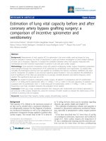
A: Proportion of endothelial cells undergoing apoptosis in the proximal pulmonary arteriesFigure 1
A: Proportion of endothelial cells undergoing apoptosis in the proximal pulmonary arteries. The proportion increased signifi-
cantly from 8.1 ± 1.8% in sham animals (S) to 14.8 ± 1% 5 weeks after ligation of the left pulmonary artery (L), peaked at 42.9
± 1.9% 2 days after reperfusion (AR), and returned to 9.8 ± 1.6% 5 weeks after reperfusion (CR). *p < 0.0001 versus sham, **p
< 0.0001 versus all other groups. S: sham group; L: ligated group; AR: acute reperfusion group; CR: chronic reperfusion group.
B: TUNEL staining of endothelial cells in the sham group, ligated group, acute reperfusion group after 2 days, and chronic
reperfusion group after 5 weeks. Apoptotic cells are yellow. S: sham group; L: ligated group; AR: acute reperfusion group; CR:
chronic reperfusion group. C: Caspase-3 activity in the proximal pulmonary arteries. Caspase-3 activity increased from 0.019 ±
0.008 mg/ml in sham animals (S) to 0.035 ± 0.015 mg/ml 5 weeks after ligation of the left pulmonary artery (L), peaked at 0.101
± 0.028 mg/ml 2 days after reperfusion (AR), and returned to 0.034 ± 0.015 mg/ml after 5 weeks of reperfusion (CR). *p <
0.0001 versus sham.
A C
Apoptotic endothelial cells %
0
5
10
15
20
25
30
35
40
45
50
SHAM L AR CR
*
**
Apoptotic endothelial cells %
0
5
10
15
20
25
30
35
40
45
50
SHAM L AR CR
*
**
0
5
10
15
20
25
30
35
40
45
50
SHAM L AR CR
*
**
B
Respiratory Research 2008, 9:19 />Page 5 of 10
(page number not for citation purposes)
impairment in endothelium-dependent relaxation
seemed correlated to the steady state of eNOS protein lev-
els: eNOS protein was significantly decreased in the acute
reperfusion group compared to the sham group (Figure
4).
Wet to dry ratios were not significantly different among
the four groups.
Gene expression in lung tissue from piglets with chronic
ischemia/reperfusion
No changes in levels of VEGF or bFGF mRNA were
detected during ischemia or reperfusion (Figure 5A).
However, Thps-1 and PAI-1 mRNAs peaked at the end of
the ischemic period (p = 0.0003 and p = 0.0025, respec-
tively when compared to sham group). After reperfusion,
expression levels decreased significantly (p = 0.0008 and p
= 0.0163, respectively compared to the values found after
the ischemic period) and returned towards normal values.
(Figure 5B).
Effect of pentoxifylline
Injecting pentoxifylline immediately before reperfusion
resulted in a significant decrease in the proportion of
apoptotic ECs in the distal PA, from 46.4 ± 4.5% to 36.3
± 2.9% (p < 0.0001). However, the proportion of apop-
totic cells remained higher than in the sham group (6.6 ±
1.8%; p < 0.0001) (Figure 6A). Neither contraction to phe-
nylephrine nor relaxation to sodium nitroprusside were
affected by PA ligation or reperfusion. However, maximal
relaxation in response to acetylcholine (Figure 6B) was
lower in the acute reperfusion group than in the pentoxi-
fylline (p = 0.0001) and sham (p = 0.0001) groups; it was
higher in the sham group than in the pentoxifylline group
(p = 0.01). No differences in acetylcholine EC
50
were seen
across the three groups. Maximal relaxation in response to
calcium ionophore was lower in the acute reperfusion
group than in the pentoxifylline (p = 0.002) and sham (p
= 0.0001) groups; it was higher in the sham group than in
the pentoxifylline group (p = 0.001). The EC
50
to calcium
ionophore was lower in the sham group than in the acute
reperfusion and pentoxifylline groups (1·35.10
-7
±
1.1·10
-7
M versus 3.39·10
-7
± 1.2·10
-7
M, p = 0.0001; and
versus 4.07·10
-7
± 1.9·10
-7
M, p = 0.0001). The EC
50
to
calcium ionophore was similar in the acute reperfusion
and pentoxifylline groups.
Reperfusion was followed by a significant decrease in
eNOS protein (acute reperfusion group versus sham
group). However, no significant difference was found
between the acute reperfusion group and the pentoxifyl-
line group (Figure 6C).
Discussion
This study provides the first evidence that chronic lung
ischemia followed by reperfusion is associated with signif-
icant EC apoptosis in proximal and distal PAs. EC apopto-
sis occurred during chronic lung ischemia, reached 46% 2
days after reperfusion, and returned to control values
within 5 weeks after reperfusion. EC apoptosis was associ-
ated with significant endothelial function impairment,
most notably regarding eNOS expression and nitric oxide
synthesis. Our gene expression studies provided a likely
explanation to the abnormalities in EC apoptosis and
endothelial function. We found overexpression of the
pro-apoptotic factors Thps-1 and PAI-1 after 5 weeks of PA
ligation. Moreover, pentoxifylline injection before lung
reperfusion protected against EC apoptosis and endothe-
lial dysfunction. These data suggest prevention of EC
death after reperfusion as a key target for treatments
A: Proportion of endothelial cells undergoing apoptosis in the distal pulmonary arteriesFigure 2
A: Proportion of endothelial cells undergoing apoptosis in
the distal pulmonary arteries. The proportion increased sig-
nificantly from 6.6 ± 1.8% in sham animals (S) to 18.9 ± 3.9%
5 weeks after ligation of the left pulmonary artery (L), peaked
at 46.4 ± 4.5% 2 days after reperfusion (AR), and returned to
5.4 ± 1.5% 5 weeks after reperfusion (CR). *p < 0.0001 ver-
sus sham animals, **p < 0.0001 versus all other groups. S:
sham group; L: ligated group; AR: acute reperfusion group;
CR: chronic reperfusion group. B: TUNEL staining of
endothelial cells in pulmonary artery branches ranging from
20 to 200 μm. Apoptotic cells are yellow (arrows). S: sham
group; L: ligated group; AR: acute reperfusion group; CR:
chronic reperfusion group.
A
B
Respiratory Research 2008, 9:19 />Page 6 of 10
(page number not for citation purposes)
aimed at preventing the development of pulmonary
edema and persistent pulmonary hypertension after PTE.
Apoptosis is the process of normal program to cell death
that allows normal cell turnover and wall remodelling in
blood vessels. Chronic PA occlusion, as occurs in CTEPH,
may result in chronic pulmonary EC ischemia. However,
the development of the bronchial circulation in areas of
chronic PA occlusion has been shown to preserve pulmo-
nary aerobic metabolism and to reduce the severity of
lung oedema after reperfusion [6,11]. Interestingly, we
showed that this systemic blood supply failed to prevent
EC apoptosis in the reperfused PA bed, since almost half
the ECs underwent apoptosis after reperfusion in our
model. EC apoptosis occurred in both the proximal and
the distal PA beds, as established not only by TUNEL assay
but also by the dramatic increase in caspase-3 activation 2
days after reperfusion. This finding corroborates previous
results made in the setting of acute ischemia-reperfusion
injury of the lung and other organs [7-10].
Apoptosis is a quiet and well-organized cell death modal-
ity that is associated with less inflammation, compared to
necrosis [13,14]. Apoptotic cells are phagocytized by mac-
rophages before their membrane breaks down, so that
their intracellular enzymes are not released. Apoptosis is
triggered and modulated by two pathways. The intrinsic
pathway involves the mitochondria and is activated by
reactive oxygen species; whereas the extrinsic pathway is
activated when ligands bind to their receptors, for
instance tumour necrosis factor alpha (TNF-α) to TNF-
receptors and Fas-ligand to Fas. Activation is rapid for the
intrinsic pathway but may take up to several hours for the
extrinsic pathway [14]. Increased apoptosis immediately
after reperfusion in human lung transplantation was first
reported by Fischer et al. in 2000, who found a time-
dependent increase in apoptotic cell numbers after trans-
plant reperfusion [9,10]. Although they found almost no
positive TUNEL staining after cold or warm ischemia, the
number of apoptotic cells increased over time after reper-
fusion. Subsequent studies established that cell death con-
sistently peaked after reperfusion, both when acute
ischemia was followed by transplantation and when
chronic ischemia was followed by PA revascularisation.
We found that nearly half the ECs were apoptotic 2 days
after reperfusion and that control levels of apoptosis were
recovered within 5 weeks. Similarly, in a rat lung trans-
plant model over 30% of cells in the lung parenchyma
were apoptotic 2 hours after reperfusion [10]. The peak in
apoptotic cell number coincided with the peak of caspase-
3 activity in our model, supporting a crucial role for the
caspase pathway in cell death activation.
Relative eNOS protein expression as assessed by densitometric quantification of endothelial nitric oxide synthase on immuno-blots prepared from lung preparations in the sham group (S) acute reperfusion group (AR)Figure 3
Relative eNOS protein expression as assessed by densitometric quantification of endothelial nitric oxide synthase on immuno-
blots prepared from lung preparations in the sham group (S) acute reperfusion group (AR).
0
20
40
60
80
100
120
10
-9
10
-8
10
-7
10
-6
LOG [A23187]
% RELAXATION
LOG [Acetylcholine]
0
10
20
30
40
50
60
70
80
10
-9
10
-8
10
-7
10
-6
10
-5
S
AR
S
AR
L
L
0
20
40
60
80
100
120
0
20
40
60
80
100
120
10
-9
10
-8
10
-7
10
-6
LOG [A23187]
% RELAXATION
LOG [Acetylcholine]
0
10
20
30
40
50
60
70
80
0
10
20
30
40
50
60
70
80
10
-9
10
-9
10
-8
10
-8
10
-7
10
-7
10
-6
10
-6
10
-5
10
-5
S
AR
S
AR
L
L
Respiratory Research 2008, 9:19 />Page 7 of 10
(page number not for citation purposes)
Increased EC death is the likely explanation to the
endothelial function impairment, most notably the defi-
cient nitric oxide production, documented in our study. A
role for pro-apoptotic and anti-apoptotic proteins in pul-
monary endothelial dysfunction has been hypothesized.
This hypothesis, however, had not been previously stud-
ied in PA endothelium exposed to chronic ischemia. We
found significant increases in mRNAs for two potent pro-
apoptotic factors, Thsp-1 and PAI-1, in lung tissue
exposed to chronic ischemia. In contrast, VEGF and bFGF
remained unchanged. After reperfusion, pro-apoptotic
protein expression returned to control values, although
EC apoptosis increased to its peak. These results suggest
that chronic lung ischemia induces overexpression of pro-
apoptotic factors and that PA reperfusion after chronic
ischemia may trigger massive EC apoptosis, leading to
endothelial damage.
Our results indicating overexpression of pro-apoptotic
proteins, including Thsp-1 and PAI-1, prompted us to
investigate the effects of a nonselective inhibitor of EC
apoptosis, pentoxifylline. Pentoxifylline is a potent anti-
inflammatory agent that attenuates neutrophil-mediated
lung injury and prevents EC dysfunction in several models
of acute lung injury [12]. Pentoxifylline administration to
patients with ischemic cardiomyopathy inhibited pro-
inflammatory cytokines and reduced apoptosis by
decreasing TNF-α and Fas concentrations in plasma [15].
Pentoxifylline probably prevents apoptosis both by inhib-
iting TNF-α production and by diminishing reactive oxy-
gen species in our model. The extrinsic pathway can be
activated during chronic ischemia via the release of
cytokines (e.g., TNF-α) and of other proinflammatory
inflammatory mediators in the lung, whereas the intrinsic
pathway can be triggered after reperfusion by the release
of reactive oxygen species.
Reperfusion pulmonary edema is a major cause of mor-
bidity and mortality after PTE for CTEPH that requires in
prolonged mechanical ventilation and may be fatal
[16,17]. Increased permeability of the small lung vessels is
the underlying mechanism. Onset is usually within 24
hours after reperfusion and is associated with neutrophil
activation and sequestration in the lung [18]. Arterial
hypoxemia and radiographic infiltrates in the reperfused
pulmonary segments are noted. Severity is variable, rang-
ing from mild reperfusion injury manifesting only as focal
infiltrates to severe alveolar flooding. Treatment is prima-
rily supportive, with mechanical ventilation and pharma-
cological support. Extracorporeal support has been used
in selected patients with overwhelming reperfusion
injury. Our results suggest that massive EC death shortly
after reperfusion may cause pulmonary edema and pul-
monary hypertension mediated by endothelial cell dys-
function [6,11]. Thus, EC apoptosis may be among the
primary causes of reperfusion pulmonary edema and per-
sistent pulmonary hypertension, the two main complica-
tions of PTE. Therefore, anti-apoptotic therapy holds
promise for improving outcomes after PTE for CTEPH.
Blocking the apoptotic cascade before reperfusion dimin-
ished ischemia-reperfusion lung injury and improved
graft function [19]. Similar results were obtained with
other organs, such as the heart and kidney [20,21]. Cas-
pase inhibitors are now available commercially and their
potential benefits in transplant patients are being evalu-
ated in clinical trials.
In conclusion, our results show that chronic lung
ischemia is associated with overexpression of pro-apop-
totic genes. A 46% rate of apoptosis and significant
endothelial dysfunction were noted 2 days after reper-
fusion of chronically ischemic PA endothelium. EC apop-
tosis was significantly reduced by pentoxifylline injected
Percent reduction of maximal contraction to phenylephrine produced by acetylcholine stimulation and calcium ionophore stimulation of pulmonary artery rings taken from the left lungs of sham animals (S), animals with chronic ischemia (left pulmonary artery ligation for 5 weeks, L), and animals with acute reperfusion (revascularisation 2 days earlier, after liga-tion for 5 weeks, AR)Figure 4
Percent reduction of maximal contraction to phenylephrine
produced by acetylcholine stimulation and calcium ionophore
stimulation of pulmonary artery rings taken from the left
lungs of sham animals (S), animals with chronic ischemia (left
pulmonary artery ligation for 5 weeks, L), and animals with
acute reperfusion (revascularisation 2 days earlier, after liga-
tion for 5 weeks, AR).
0
0.2
0.4
0.6
0.8
1
1.2
Sham Acute
Revascularized
eNOS Relative Quantity
Sham Acute
Revascularized
eNOS
ß-actin
P=0.0007
0
0.2
0.4
0.6
0.8
1
1.2
Sham Acute
Revascularized
eNOS Relative Quantity
Sham Acute
Revascularized
eNOS
ß-actin
P=0.0007
Respiratory Research 2008, 9:19 />Page 8 of 10
(page number not for citation purposes)
A: Relative expression of lung vascular endothelial growth factor and basic fibroblast growth factor mRNA in control piglets (sham) and animals exposed to chronic ligation (L) alone or followed by reperfusion for 2 days (acute reperfusion group, AR) or 5 weeks (chronic reperfusion group, CR)Figure 5
A: Relative expression of lung vascular endothelial growth factor and basic fibroblast growth factor mRNA in control piglets
(sham) and animals exposed to chronic ligation (L) alone or followed by reperfusion for 2 days (acute reperfusion group, AR)
or 5 weeks (chronic reperfusion group, CR). B: Relative expression of thrombospondin-1 and plasminogen activator inhibitor-
1 mRNA in control piglets (sham) and animals exposed to chronic ligation (L) alone or followed by reperfusion for 2 days
(acute reperfusion group, AR) or 5 weeks (chronic reperfusion group, CR). Expression levels were significantly increased 5
weeks after ligation of the left pulmonary artery (L) (*p = 0.0003 and *p = 0.0025, respectively) and significantly decreased after
acute reperfusion (AR) ($p = 0.0008 and $p = 0.0163, respectively).
A
Vascular endothelial growth factor mRNA levels
AR CRLSHAM
AR CRL
SHAM
0,00
,25
,50
,75
1,00
1,25
1,50
1,75
2,00
2,25
Basic fibroblast growth factor mRNA levels
0
1
2
3
4
5
6
7
Vascular endothelial growth factor mRNA levels
AR CRLSHAM
AR CRL
SHAM
0,00
,25
,50
,75
1,00
1,25
1,50
1,75
2,00
2,25
Basic fibroblast growth factor mRNA levels
0
1
2
3
4
5
6
7
ARAR CRCRLLSHAMSHAM
AR CRL
SHAM
0,00
,25
,50
,75
1,00
1,25
1,50
1,75
2,00
2,25
Basic fibroblast growth factor mRNA levels
0
1
2
3
4
5
6
7
ARAR CRCRCRLL
SHAMSHAM
0,00
,25
,50
,75
1,00
1,25
1,50
1,75
2,00
2,25
Basic fibroblast growth factor mRNA levels
0
1
2
3
4
5
6
7
B
AR
CR
L
SHAM
0
5
10
15
20
25
30
35
40
45
*
Plasminogen activator inhibitor-1 mRNA levels
AR
CR
L
SHAM
45
0
5
10
15
20
25
30
35
40
*
Thrombospondin-1 mRNA levels
$
$
ARARAR
CRCR
LL
SHAMSHAM
0
5
10
15
20
25
30
35
40
45
*
Plasminogen activator inhibitor-1 mRNA levels
ARAR
CRCR
LL
SHAMSHAMSHAM
45
0
5
10
15
20
25
30
35
40
*
Thrombospondin-1 mRNA levels
$
$
Respiratory Research 2008, 9:19 />Page 9 of 10
(page number not for citation purposes)
A: Proportion of endothelial cells undergoing apoptosis in the distal pulmonary arteries in the sham group and in piglets exposed to ligation for 5 weeks followed by reperfusion for two days, with (AR P+) or without (AR P-) pentoxifylline adminis-tration immediately before reperfusionFigure 6
A: Proportion of endothelial cells undergoing apoptosis in the distal pulmonary arteries in the sham group and in piglets
exposed to ligation for 5 weeks followed by reperfusion for two days, with (AR P+) or without (AR P-) pentoxifylline adminis-
tration immediately before reperfusion. Typical results from two animals of each group are shown. B: Percent reduction of
maximal contraction to phenylephrine produced by acetylcholine stimulation and calcium ionophore stimulation of pulmonary
artery rings taken from left lungs 2 days after reperfusion with (AR P+) or without (AR P-) pentoxifylline administration (pen-
toxifylline+ and pentoxifylline-groups, respectively). Values are means ± SEM. The concentration-response curves were signifi-
cantly flattened in the pentoxifylline-group and partially restored by administration of pentoxifylline. S: sham group; P-: acute
reperfusion without pentoxifylline; P+: acute reperfusion with pentoxifylline. C: Western blot of eNOS protein content (mean
± SEM) 2 days after reperfusion with pentoxifylline (AR P+ group) or without pentoxifylline (AR P- group) compared to the
sham group. Typical results from two animals of each group are shown.
A C
SHAM
AR P- AR P+
0
10
20
30
40
50
60
p<0,0001p<0,0001
SHAM
AR P- AR P+
0
10
20
30
40
50
60
0
10
20
30
40
50
60
p<0,0001p<0,0001p<0,0001p<0,0001
0
0.2
0.4
0.6
0.8
1
1.2
Sham AR P- AR P+
eNOS Relat iv e Quantity
Sham
AR P-
AR P+
eNOS
ß-actin
p=0.0007
p=0.0007
p=NS
0
0.2
0.4
0.6
0.8
1
1.2
Sham AR P- AR P+
eNOS Relat iv e Quantity
Sham
AR P-
AR P+
eNOS
ß-actin
p=0.0007
p=0.0007
p=NS
B
0
20
40
60
80
100
120
10
-9
10
-8
10
-7
10
-6
LOG [A23187]
% RELAXATION
LOG [Acet ylcholine]
0
10
20
30
40
50
60
70
80
10
-9
10
-8
10
-7
10
-6
10
-5
S
P
AR
S
P
0
20
40
60
80
100
120
0
20
40
60
80
100
120
10
-9
10
-8
10
-7
10
-6
LOG [A23187]
% RELAXATION
LOG [Acet ylcholine]
0
10
20
30
40
50
60
70
80
0
10
20
30
40
50
60
70
80
10
-9
10
-9
10
-8
10
-8
10
-7
10
-7
10
-6
10
-6
10
-5
10
-5
S
S
P
AR
AR P+
AR P-
AR P-
P
AR P+
Publish with BioMed Central and every
scientist can read your work free of charge
"BioMed Central will be the most significant development for
disseminating the results of biomedical research in our lifetime."
Sir Paul Nurse, Cancer Research UK
Your research papers will be:
available free of charge to the entire biomedical community
peer reviewed and published immediately upon acceptance
cited in PubMed and archived on PubMed Central
yours — you keep the copyright
Submit your manuscript here:
/>BioMedcentral
Respiratory Research 2008, 9:19 />Page 10 of 10
(page number not for citation purposes)
immediately before lung reperfusion, which significantly
improved endothelial function. EC death may explain
reperfusion pulmonary edema and persistent pulmonary
hypertension occurring immediately after PTE. Therefore,
anti-apoptotic therapy holds considerable promise for
preventing the severe complications in patients undergo-
ing PTE.
Acknowledgements
This work was supported by a grant from the Association chirurgicale pour
le Développement et l’amélioration des Techniques de dépistage et de
Traitement des Maladies Cardio-vasculaires (ADETEC).
References
1. Fedullo PF, Auger WR, Kerr KM, Rubin LJ: Chronic thromboem-
bolic pulmonary hypertension. N Engl J Med 2001,
345:1465-1472.
2. Dartevelle P, Fadel E, Mussot S, Chapelier A, Herve P, de Perrot M,
Cerrina J, Ladurie FL, Lehouerou D, Humbert M, et al.: Chronic
thromboembolic pulmonary hypertension. Eur Respir J 2004,
23:637-648.
3. Pengo V, Lensing AW, Prins MH, Marchiori A, Davidson BL, Tiozzo F,
Albanese P, Biasiolo A, Pegoraro C, Iliceto S, et al.: Thromboem-
bolic Pulmonary Hypertension Study Group. Incidence of
chronic thromboembolic pulmonary hypertension after pul-
monary embolism. N Engl J Med 2004, 350:2257-2264.
4. Fadel E, Riou JY, Mazmanian M, Brenot P, Dulmet E, Detruit H, Serraf
A, Bacha EA, Herve P, Dartevelle P: Pulmonary thromboendar-
terectomy for chronic thromboembolic obstruction of the
pulmonary artery in piglets. J Thorac Cardiovasc Surg 1999,
117:787-793.
5. Fadel E, Michel RP, Eddahibi S, Bernatchez R, Mazmanian GM, Baudet
B, Dartevelle P, Herve P: Regression of postobstructive vascu-
lopathy after revascularization of chronically obstructed pul-
monary artery. J Thorac Cardiovasc Surg 2004, 127:1009-1017.
6. Fadel E, Mazmanian GM, Chapelier A, Baudet B, Detruit H, de
Montpreville V, Libert JM, Wartski M, Herve P, Dartevelle P: Lung
reperfusion injury after chronic or acute unilateral pulmo-
nary artery occlusion. Am J Respir Crit Care Med 1998,
157:1294-1300.
7. Hall AV, Jevnikar AM: Significance of endothelial cell survival
programs for renal transplantation. Am J Kidney Dis 2003,
41:1140-1154.
8. Natori S, Selzner M, Valentino KL, Fritz LC, Srinivasan A, Clavien PA,
Gores GJ: Apoptosis of sinusoidal endothelial cells occurs dur-
ing liver preservation injury by a caspase-dependent mecha-
nism. Transplantation 1999, 68:89-96.
9. Fischer S, Cassivi SD, Xavier AM, Cardella JA, Cutz E, Edwards V, Liu
M, Keshavjee S: Cell death in human lung transplantation:
apoptosis induction in human lungs during ischemia and
after transplantation. Ann Surg 2000,
231:424-431.
10. Fischer S, Maclean AA, Liu M, Cardella JA, Slutsky AS, Suga M, Moreira
JF, Keshavjee S: Dynamic changes in apoptotic and necrotic
cell death correlate with severity of ischemia-reperfusion
injury in lung transplantation. Am J Respir Crit Care Med 2000,
162:1932-1939.
11. Fadel E, Mazmanian GM, Baudet B, Detruit H, Verhoye JP, Cron J, Fat-
tal S, Dartevelle P, Herve P: Endothelial nitric oxide synthase
function in pig lung after chronic pulmonary artery obstruc-
tion. Am J Respir Crit Care Med 2000, 162:1429-1434.
12. Reignier J, Mazmanian M, Detruit H, Chapelier A, Weiss M, Libert JM,
Herve P: Reduction of ischemia-reperfusion injury by pentoxi-
fylline in the isolated rat lung. Paris-Sud University Lung
Transplantation Group. Am J Respir Crit Care Med 1994,
150:342-7.
13. Denecker G, Vercammen D, Declercq W, Vandenabeele P: Apop-
totic and necrotic cell death induced by death domain recep-
tors. Cell Mol Life Sci 2001, 58:356-370.
14. Kuwano K, Hara N: Signal transduction pathways of apoptosis
and inflammation induced by the tumor necrosis factor
receptor family. Am J Respir Cell Mol Biol 2000, 22:147-149.
15. Sliwa K, Woodiwiss A, Kone VN, Candy G, Badenhorst D, Norton G,
Zambakides C, Peters F, Essop R: Therapy of ischemic cardiomy-
opathy with the immunomodulating agent pentoxifylline:
results of a randomized study. Circulation 2004, 109:750-5.
16. Jamieson SW, Kapelanski DP, Sakakibara N, Manecke GR,
Thistlethwaite PA, Kerr KM, Channick RN, Fedullo PF, Auger WR:
Pulmonary endarterectomy: experience and lessons learned
in 1,500 cases. Ann Thorac Surg 2003, 76:1457-1462.
17. Dartevelle P, Fadel E, Chapelier A, Macchiarini P, Cerrina J, Parquin F,
Simonneau F, Simonneau G: Angioscopic video-assisted pulmo-
nary endarterectomy for post-embolic pulmonary hyperten-
sion. Eur J Cardiothorac Surg 1999, 16:38-43.
18. de Perrot M, Liu M, Waddell TK, Keshavjee S: Ischemia-reper-
fusion-induced lung injury. Am J Respir Crit Care Med 2003,
167:490-511.
19. Quadri SM, Segall L, de Perrot M, Han B, Edwards V, Jones N, Wad-
dell TK, Liu M, Keshavjee S:
Caspase inhibition improves
ischemia-reperfusion injury after lung transplantation. Am J
transplant 2005, 5:292-9.
20. Yaoita H, Ogawa K, Maehara K, Maruyama Y: Attenuation of
ischemia/reperfusion injury in rats by a caspase inhibitor. Cir-
culation 1998, 97:276-281.
21. Daemen MA, van't Veer C, Denecker G, Heemskerk VH, Wolfs TG,
Clauss M, Vandenabeele P, Buurman WA: Inhibition of apoptosis
induced by ischemia-reperfusion prevents inflammation. J
Clin Invest 1999, 104:541-549.