Báo cáo y học: "Distinct synovial immunopathology in Behçet disease and psoriatic arthritis" pot
Bạn đang xem bản rút gọn của tài liệu. Xem và tải ngay bản đầy đủ của tài liệu tại đây (2.25 MB, 7 trang )
Open Access
Available online />Page 1 of 7
(page number not for citation purposes)
Vol 11 No 1
Research article
Distinct synovial immunopathology in Behçet disease and
psoriatic arthritis
Juan D Cañete
1
, Raquel Celis
1
, Troy Noordenbos
2
, Conchita Moll
1
, Jose A Gómez-Puerta
1
,
Pilar Pizcueta
3
, Antonio Palacin
4
, Paul P Tak
2
, Raimon Sanmartí
1
and Dominique Baeten
2
1
Arthritis Unit, Department of Rheumatology, Hospital Clinic de Barcelona and IDIBAPS, Villaroel 170, Barcelona, 08036, Spain
2
Clinical Immunology and Rheumatology, Academic Medical Center/University of Amsterdam, Meibergdreef 9, Amsterdam, 1105 AZ, The
Netherlands
3
Immunology Unit, Department of Cell Biology and Pathology, Calle Casanova 143, Universidad de Barcelona, 08036, Spain
4
Department of Pathology, Hospital Clinic de Barcelona, Villaroel 170, Barcelona, 08036, Spain
Corresponding author: Dominique Baeten,
Received: 22 Sep 2008 Revisions requested: 10 Nov 2008 Revisions received: 29 Dec 2008 Accepted: 6 Feb 2009 Published: 6 Feb 2009
Arthritis Research & Therapy 2009, 11:R17 (doi:10.1186/ar2608)
This article is online at: />© 2009 Cañete et al.; licensee BioMed Central Ltd.
This is an open access article distributed under the terms of the Creative Commons Attribution License ( />),
which permits unrestricted use, distribution, and reproduction in any medium, provided the original work is properly cited.
Abstract
Introduction The aim of the study was to investigate synovial
immunopathology differences between early Behçet disease
(BD) and psoriatic arthritis (PsA).
Methods Needle arthroscopy of an inflamed knee joint was
performed in patients with early untreated BD (n = 8) and PsA
(n = 9). Synovial fluid (SF) was collected for cytokines, perforin,
and granzyme analysis. Eight synovial biopsies per patient were
obtained for immunohistochemical analysis of the cellular
infiltrate (T cells, natural killer cells, macrophages, B cells,
plasma cells, mast cells, and neutrophils), blood vessels as well
as expression of perforin and granzyme. The stained slides were
evaluated by digital image analysis.
Results The global degree of synovial inflammation was similar
in the two types of arthritis. In the analysis of the innate immune
cell infiltration, there was a striking neutrophilic inflammation in
BD synovitis whereas PsA displayed significantly higher
numbers of cells positive for c-kit, a marker of mast cells. As for
lymphocytes, CD3
+
T cells, but neither CD20
+
B cells nor
CD138
+
plasma cells, were significantly increased in BD versus
PsA. Further analysis of the T-lymphocyte population showed no
clear shift in CD4/CD8 ratio or Th1/Th2/Th17 profile. The SF
levels of perforin, an effector molecule of cytotoxic cells,
displayed a significant four- to fivefold increase in BD.
Conclusions This systematic comparative analysis of early
untreated synovitis identifies neutrophils and T lymphocytes as
important infiltrating cell populations in BD. Increased levels of
perforin in BD suggest the relevance of cytotoxicity in this
disease.
Introduction
Behçet disease (BD) is a systemic inflammatory disorder with
oral and/or genital ulcerations, uveitis, and skin lesions as pro-
totypic clinical symptoms [1]. The systemic nature of the dis-
ease is emphasized by the potential involvement of the central
nervous system, the vascular system, the gut, and the kidney.
Up to half of the patients with BD also display rheumatic fea-
tures. The arthritis is usually monoarticular, intermittent, and
not deforming and affects mainly knees or ankles. Less com-
mon rheumatic features are enthesitis, spondylitis, and sacro-
iliitis. This pattern of rheumatic inflammation as well as the
association with eye, gut, and skin involvement display clinical
similarities with spondyloarthritis (SpA) in general and psori-
atic arthritis (PsA) in particular. Although the pathogenesis of
BD is still poorly understood, the association with class I major
histocompatibility complex molecules (HLA-B51 in BD and
HLA-B27 in SpA) and the response to tumor necrosis factor
(TNF) blockers in both diseases further support the possibility
of common pathophysiological pathways.
Previous studies in psoriatic and non-psoriatic SpA and rheu-
matoid arthritis (RA) have indicated that detailed synovial his-
BD: Behçet disease; ELISA: enzyme-linked immunosorbent assay; IFN-γ: interferon-gamma; IL: interleukin; NK: natural killer; PMN: polymorphonuclear
neutrophil; PsA: psoriatic arthritis; RA: rheumatoid arthritis; SF: synovial fluid; SpA: spondyloarthritis; TNF: tumor necrosis factor.
Arthritis Research & Therapy Vol 11 No 1 Cañete et al.
Page 2 of 7
(page number not for citation purposes)
topathology can help to reveal differences in cellular and
molecular immunopathology which are of interest for differen-
tial diagnosis, classification, and pathogenesis of the different
types of arthritis [2-4]. Although joint involvement is clinically
well recognized, few histologic studies have focused on the
synovial features in BD [5-7]. Here, to explore the immunopa-
thology of BD, we performed a detailed comparative study of
early untreated synovitis in BD versus PsA.
Materials and methods
Patients and samples
Eight patients with early untreated BD and nine patients with
early untreated PsA gave written informed consent to partici-
pate in the study as approved by the local ethics committee.
All untreated patients from our early arthritis clinic undergoing
diagnostic or therapeutic knee arthroscopy and fulfilling either
the International Study Group for Behçet's disease criteria for
BD [8] or the CASPAR (classification of psoriatic arthritis) cri-
teria for PsA [9] were included. All BD patients had recurrent
oral and genital aphtosis and folliculitis, five had a positive
pathergy test, two had uveitis, and one developed retinal vas-
culitis. All patients of both study cohorts had early disease (val-
ues of median [range] of disease duration since diagnosis was
made in the early arthritis clinic were 1.0 [0.2 to 7.9] months
in BD and 4.0 [1.7 to 13.5] months in PsA) and were disease-
modifying antirheumatic drug (DMARD)-naïve and corticoster-
oid-naïve. All patients had active joint disease with at least one
swollen knee joint (five monoarthritides and three oligoar-
thritides in BD and two monoarthritides and seven oligoar-
thritides in PsA). Median erythrocyte sedimentation rate
(millimeters per hour) values were 50 (range of 12 to 113) in
BD and 32 (10 to 85) in PsA. Median C-reactive protein (mil-
ligrams per deciliter) values were 6.8 (2 to 15.5) in BD and 2.3
(1.5 to 9.4) in PsA. Needle arthroscopy of the clinically
inflamed knee joint was performed in all patients, with a 2.7-
mm arthroscope (Olympus
®
; Barcelona, Spain). Synovial fluid
(SF) was collected for cytokine analysis, and eight synovial
biopsies per patient were obtained for immunohistochemical
analysis.
Immunohistochemistry
The synovial biopsies were embedded in paraffin, sectioned,
and subjected to antigen retrieval by cooking when required.
The slides were subsequently stained with an automated
immunostainer (TechMate 500 Plus; Dako, Cambridge, UK)
using the following monoclonal antibodies: anti-CD3 (clone
PS1; Novocastra, Newcastle, UK), anti-CD4 (clone 1F6;
Novocastra), anti-CD8 (clone 4B11; Novocastra), anti-CD20
(clone L26; Dako), anti-CD15 (clone BY87; Novocastra), anti-
CD31 (clone JC70A; Dako), anti-CD56 (clone 123C3; Mon-
osan, Uden, The Netherlands), anti-CD68 (clone KP-1; Dako),
anti-CD117 (mast cells, rabbit anti-human polyclonal antibody;
Dako), anti-CD138 (clone B-B4; Santa Cruz Biotechnology,
Inc., San Diego, CA, USA), anti-granzyme B (clone GrB7;
Monosan), and anti-perforin (clone 5B10; Novocastra). As a
negative control, the primary antibodies were substituted by
isotype- and concentration-matched control antibodies. The
primary antibodies were subsequently detected by an avidin-
biotin-peroxidase-based method (Envision System; Dako) and
an aminoethylcarbazole color reaction (Sigma-Aldrich, St.
Louis, MO, USA) as described previously in detail [2-4,10].
Finally, the slides were counterstained with hematoxylin.
Digital image analysis
The stained slides were scored by digital image analysis by an
independent observer (RC) who was blinded to diagnosis and
clinical data. Each stained slide in its entirety was scored by
dividing it in different regions. Within each region, the number
of stained cells per area as well as the percentage of stained
cells were measured in at least 20 high-power fields using the
AnalySIS
®
Imaging processing program (Olympus
®
) as
described previously in detail [11]. The presence of grade-3
lymphoid aggregates, as defined previously [12-15], was
assessed on consecutive sections stained for CD3 and
CD20.
Synovial fluid analysis
SF was collected at the time of arthroscopy for assessment of
total cellular count and number of neutrophils. SF levels of
interferon-gamma (IFN-γ), TNF-α, interleukin (IL)-2, IL-4, IL-6,
and IL-10 were quantified using a multiplex assay in accord-
ance with the instructions of the manufacturer (Cytometric
Bead Assay; BD Biosciences, San Jose, CA, USA). Perforin
(Abcam, Cambridge, UK), granzyme B (Abcam), IL-8 (R&D
Systems, Abingdon, UK), and IL-17 (R&D Systems) levels in
SF were assessed by enzyme-linked immunosorbent assay
(ELISA) as indicated by the manufacturer.
Statistical analysis
Data were analyzed using the SPSS 10.0 statistical program
(SPSS Inc., Chicago, IL, USA). As the data are non-paramet-
ric, they are represented as median (range). Comparisons
were performed with the non-parametric Mann-Whitney U
test. The level of statistical significance was established at a P
value of less than 0.05.
Results
Neutrophilic infiltration in Behçet disease synovitis
The results of the immunohistochemical analysis of the syno-
vial tissue samples are summarized in Table 1. The number of
CD68
+
macrophages, which reflects global synovial inflamma-
tion [16], was similar in the intimal lining layer as well as the
synovial sublining of BD and PsA, indicating that there is no
systematic bias in local inflammation between the two study
groups (Figure 1). Further analysis of the synovial leukocyte
infiltration revealed two striking differences between BD and
PsA in non-lymphocytic innate immune populations. First,
there was an important infiltration with CD15
+
neutrophils in
BD, representing approximately 13% of the total synovial cel-
lularity compared with approximately 4% in PsA (Figure 1).
Available online />Page 3 of 7
(page number not for citation purposes)
This neutrophilic infiltration in BD synovitis was seen essen-
tially in the intimal lining layer (P = 0.036 versus PsA), with dif-
ferences that were less pronounced in the synovial sublining.
This prominent neutrophilic infiltration was not related to
higher levels of IL-8, a major chemoattractant for neutrophils,
in BD versus PsA (Table 2). Second, this increase in neu-
trophils was a specific rather than a general phenomenon
related to innate immune cells as the number of cells express-
ing CD117 (c-kit), which is abundantly expressed on mast
cells, was decreased in BD versus PsA (P = 0.046). Finally,
there was no difference in the number of CD31
+
blood vessels
between BD and PsA. There were no signs of neutrophilic or
cytoclastic vasculitis.
Accumulation of T lymphocytes in Behçet disease
synovitis
In the analysis of the lymphocytic infiltration (Table 1), the num-
bers of CD20
+
B lymphocytes, lymphoid aggregates, and
large grade-3 aggregates were similar in the two diseases,
with a trend toward lower CD138
+
plasma cell numbers in BD
versus PsA (P = 0.071) (Figure 2). In sharp contrast, there
was a threefold increase of CD3
+
T lymphocytes in BD versus
PsA (P = 0.015) (Figure 2). Though not statistically significant,
this increase was seen in both the CD4
+
and CD8
+
T-lym-
phocyte subsets.
Synovial fluid cytokine profiles
As the immunohistochemical analysis indicated a striking dif-
ference in the number of infiltrating T cells between BD and
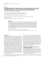
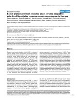
Figure 1
Microscopic analysis of synovial inflammation with innate immune cells in early untreated Behçet synovitis (BD) versus psoriatic synovitis (PsA)Microscopic analysis of synovial inflammation with innate immune cells in early untreated Behçet synovitis (BD) versus psoriatic synovitis (PsA).
Whereas the global degree of synovial inflammation, reflected by the number of CD68
+
macrophages, was similar in the two diseases, there were
significant increases of CD15
+
neutrophils in BD and of CD117
+
mast cells in PsA. Original magnification ×20.
Arthritis Research & Therapy Vol 11 No 1 Cañete et al.
Page 4 of 7
(page number not for citation purposes)
PsA synovitis, we next aimed to analyze the Th1/Th2/Th17
profile by assessing SF cytokines. As shown in Table 2, BD
and PsA had similar SF leukocyte and neutrophil counts,
reflecting a similar degree of local inflammatory reaction in the
two diseases. This was further confirmed by the similar levels
of IL-6. In an investigation of skewing toward Th1 (IFN-γ, TNF-
α, and IL-2), Th2 (IL-4 and IL-10), or Th17 (IL-17) cytokine pro-
files, only IL-2 showed significantly higher levels in PsA than in
BD (P = 0.017) (Table 2).
Increased synovial fluid perforin levels in Behçet disease
As we observed no clear skewing of the Th1/Th2 profile, we
next investigated whether the infiltrating T cells displayed fea-
tures of cytotoxic cells. CD56, a marker expressed by cyto-
toxic T cells and natural killer (NK) cells, was slightly but not
significantly increased in BD versus PsA (Figure 2). Immunos-
taining of perforin and granzyme B, both cytotoxic effectors,
showed only few positive cells without differences between
BD and PsA (Table 1), but the SF levels of perforin showed a
highly significant four- to fivefold increase in BD (1,168 pg/mL,
range 428 to 2,010 pg/mL) versus PsA (278 pg/mL, range
132 to 642 pg/mL) (P = 0.005) (Table 2). The SF levels of
granzyme were below the detection limit of the ELISA in all
samples.
Discussion
Although BD is a relatively common and potentially severe sys-
temic disease, the pathophysiology of the disease remains
poorly understood. Microbial infections, sterile neutrophil-
mediated inflammation, and autoimmune lymphocytes have all
been implicated in the recurrent attacks of inflammation char-
acteristic of this disease [1], leading to an ongoing debate on
the autoimmune versus autoinflammatory origin of BD [17]. As
we and others demonstrated previously that detailed analysis
of the histopathology of the target lesions can yield important
pathophysiological information in rheumatic conditions [2-
4,10], we took advantage here of the predilection of the dis-
ease for knee joints and the availability of biopsy sampling by
needle arthroscopy to revisit in detail the synovial immunopa-
thology of BD. None of the arthroscopies, performed primarily
for diagnostic and/or therapeutical reasons, leads to cutane-
ous or joint complications, including pathergy-like reactions
[18]. This observation indicates that it is medically and ethi-
cally acceptable to perform histology studies in BD.
Table 1
Immunohistochemical analysis of synovial tissue biopsies from early untreated Behçet disease and psoriatic arthritis
Behçet disease Psoriatic arthritis P value
n = 8 n = 9
Synovial immunopathology (cells/mm
2
)
CD68 lining 3,681 (877–6,831) 2,947 (541–4,834) NS
CD68 sublining 529 (311–2,339) 739 (183–2,636) NS
CD3 1,077 (354–1,427) 336 (164–1,036) 0.015
CD4 516 (31–1,306) 196 (3–1,061) NS
CD8 524 (2–832) 113 (3–669) NS
CD20 126.5 (61–703) 106 (38–510) NS
Lymphoid aggregates 0.93 (0.34–6.8) 0.43 (0–3.04) NS
Grade-3 lymphoid aggregates 0.08 (0–1.38) 0 (0–0.44) NS
CD138 45 (11–432) 173 (8–1,490) 0.071
CD56 17 (1–144) 9 (2–77) NS
Granzyme B 12 (2–93) 9 (2–20) NS
Perforin 8.5 (3–26) 6 (1–22) NS
CD15 lining 919 (101–4,436) 275 (11–1,015) 0.036
CD15 sublining 305.5 (31–1,050) 203 (25–467) NS
CD117 60 (21–286) 162 (29–365) 0.046
CD31 (vessels/mm
2
) 80 (60–255) 133 (63–166) NS
After immunostaining of paraffin or frozen sections, the number of positive cells per square millimeter was assessed by digital image analysis. Data
are presented as median (range). NS, non-significant.
Available online />Page 5 of 7
(page number not for citation purposes)
Almost 30 years ago, a study a study assessed for the first
time the histology of synovitis in six BD patients [5]. That study
found no evidence of infection but a dense mixed inflammatory
infiltrate with the prominent presence of neutrophils and scar-
city of plasma cells in most samples. Similarly, a second study
found a paucity of plasma cells in 12 BD synovial tissues but
did not describe the presence of neutrophils [7]. Strikingly,
they found ectopic lymphoid neogenesis, defined as the devel-
opment of large lymphoid aggregates resembling secondary
lymphoid organs [12-15], in 5 out of 12 samples. Unfortu-
nately, the interpretation of both data sets is hampered by the
inclusion of patients with long disease duration and the lack of
a control group with non-BD synovitis. Including RA synovial
tissue as control, a third study could not confirm the prominent
infiltration with neutrophils or any other distinctive feature in
seven BD cases but indicated that the findings may be influ-
enced by the longer disease duration [6]. The present study
was designed to overcome the limitations of these previous
analyses by systematic comparison with a clinically related
inflammatory arthritis, PsA, and by stringent selection of active,
early, and untreated disease for both groups. As local disease
activity is an important determinant of synovial histopathology
[10], this design additionally allowed us to match BD and PsA
for local disease activity as measured by SF cell count, SF IL-
6 levels, and synovial tissue CD68
+
macrophage numbers
[16]. As such, this stringent study design obviously limits the
number of cases that can be included but minimizes the risk of
systematic biases and false-positive findings.
In the analysis of the infiltrating leukocytes from the innate
immune system, a first striking feature of BD synovitis was the
marked infiltration with polymorphonuclear neutrophil (PMN).
This neutrophilic infiltration cannot be explained by disease
duration as both BD and PsA had very early, untreated dis-
ease. It was also not related to a difference in levels of IL-8, a
major chemokine for PMN. Moreover, this increase did not
extend more broadly to innate immunity in general as cells pos-
itive for the mast cell marker CD117 appeared to be
decreased in BD versus PsA. This relative increase in BD ver-
sus PsA most likely reflects a true increase in BD as previous
studies showed that the number of infiltrating PMN was
already high in PsA compared with RA synovial tissue [3,4].
Increased numbers of PMN in BD inflammation have been
reported not only in synovium [5] but also in other target
organs such as skin [19,20] and the central nervous system
[21]. Moreover, the function of PMN has been reported to be
altered in BD [22,23]. Taken together, these data are consist-
ent with a prominent role of PMN in the disease process and
may suggest some degree of similarity between the patho-
physiology of the synovitis and skin reactions such as the
pathergy test in BD. In contrast, however, we did not find any
evidence for neutrophilic vasculitis or any other form of vessel
pathology in the synovial tissue. Synovitis is thus clearly differ-
ent from cutaneous lesions in this respect as leukocytoclastic
vasculitis is a prominent feature of skin lesions in BD [24,25].
Table 2
Synovial fluid analysis in early untreated Behçet disease and psoriatic arthritis
Behçet disease Psoriatic arthritis P value
n = 8 n = 9
Cells/mm
3
15,840 (7,600–30,720) 15,000 (2,250–35,500) NS
Neutrophils/mm
3
10,865 (4,200–28,000) 10,000 (562–23,450) NS
Percentage neutrophils 72.5 (53.75–91.15) 74.07 (24.98–94) NS
IL-6 8,850 (6,484–12,157) 8,678 (6,107–11,022) NS
IL-8 272 (115–4,500) 241 (102–3,033) NS
TNF-α 13.5 (9–44) 11 (0.1–32) NS
IFN-γ 103.5 (9–323) 89 (62–134) NS
IL-2 26.5 (9–34) 38 (31–78) 0.017
IL-4 80.5 (66–111) 86 (77–107) NS
IL-10 17.5 (5–22) 10 (2–28) NS
IL-17 <25 <25 NS
Perforin 1168 (428–2,010) 278 (132–642) 0.005
Granzyme B <15 (<15–56) <15 (<15–41) NS
The cytokines as well as perforin and granzyme B were detected by enzyme-linked immunosorbent assay and are expressed as picograms per
milliliter. Data are presented as median (range). IFN-γ, interferon-gamma; IL, interleukin; NS, non-significant; TNF-α, tumour necrosis factor-alpha.
Arthritis Research & Therapy Vol 11 No 1 Cañete et al.
Page 6 of 7
(page number not for citation purposes)
A second important finding was the selective increase of syn-
ovial T lymphocytes in BD. Again, this was specific for T cells
rather than related to a global increase in lymphocyte infiltra-
tion as we found similar numbers of B lymphocytes and a sim-
ilar degree of organization in lymphoid aggregates in both
diseases. Moreover, previous studies showed no difference in
T-cell infiltration between PsA and RA, suggesting that the
increase in BD is actually specific [3,4]. Confirming previous
studies on BD synovitis [5,7], plasma cells were even lower in
BD versus PsA synovitis. Although we did not investigate the
mechanism underlying the selective increase in T lym-
phocytes, previous studies indicated that BD T cells are par-
tially protected against apoptosis [26]. Moreover, a recent
report indicates that specific T-cell subsets are associated
with sterile neutrophil-rich inflammation as observed in BD
synovitis, which may explain the simultaneous increase of both
T cells and PMN [27].
As to the potential functional contribution of these T cells to
the pathology, both Th1 skewing [28,29] and cytotoxicity by
classical CD8
+
cells, NK T cells, or gamma-delta cells [28-30]
have been proposed. Our SF analysis failed to demonstrate a
clear Th1 skewing, with lower IL-2, but not for IFN-γ and TNF-
α, levels in BD versus PsA. There was also no significant dif-
ference in the number of CD8
+
, CD56
+
, perforin
+
, or granzyme
B
+
cells, defining cytotoxic cells, in BD versus PsA synovitis.
However, the levels of perforin released in the SF were fivefold
increased in BD versus PsA, suggesting the relevance of cyto-
toxicity in BD synovitis [30-32].
Conclusion
This systematic comparative analysis of early untreated syno-
vitis identifies both lymphocytes and PMN as important infil-
trating cell populations and indicates an increase of cytotoxic
molecules in BD versus PsA. Further studies should clarify
their functional contribution to the pathogenesis of the dis-
ease.
Competing interests
The authors declare that they have no competing interests.
Authors' contributions
JDC helped to design the study, to collect the samples, to per-
form the experiments, to analyze the data, and to prepare the
manuscript. DB helped to design the study, to analyze the
data, and to prepare the manuscript. RC, TN, CM, JAG-P, PP,
and AP helped to collect the samples and to perform the
experiments. PPT and RS helped to prepare the manuscript.
All authors read and approved the final manuscript.
Acknowledgements
The research of JDC was supported by a grant from Fundación Españo-
la de Reumatología (DIB-SER) and by Ministerio de Ciencia e Inno-
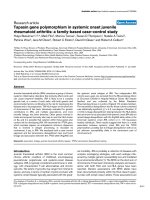
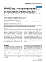
Figure 2
Microscopic analysis of synovial inflammation with lymphocytes in early untreated Behçet synovitis (BD) versus psoriatic synovitis (PsA)Microscopic analysis of synovial inflammation with lymphocytes in early untreated Behçet synovitis (BD) versus psoriatic synovitis (PsA). Represent-
ative pictures are shown of the immunostainings for CD20
+
B lymphocytes; CD138
+
plasma cells; CD3
+
, CD4
+
, and CD8
+
T lymphocytes; and
CD56
+
natural killer cells. The number of infiltrating CD3
+
T lymphocytes was significantly increased in BD versus PsA. Original magnification ×20.
Available online />Page 7 of 7
(page number not for citation purposes)
vación (FIS PI080206). TN is supported by a human immunology grant
from the Dana Foundation. DB is supported by the Dutch Arthritis Foun-
dation and by a Vidi Grant from the Netherlands Organisation for Scien-
tific Research (NWO).
References
1. Sakane T, Takeno M, Suzuki N, Inaba G: Behçet's disease. N
Engl J Med 1999, 341:1284-1291.
2. Baeten D, Demetter P, Cuvelier CA, Kruithof E, Van Damme N, De
Vos M, Veys EM, De Keyser F: Macrophages expressing the
scavenger receptor CD163: a link between immune alterations
of the gut and synovial inflammation in spondyloarthropathy.
J Pathol 2002, 196:343-350.
3. Baeten D, Kruithof E, De Rycke L, Vandooren B, Wyns B, Boullart
L, Hoffman IE, Boots AM, Veys EM, De Keyser F: Diagnostic clas-
sification of spondylarthropathy and rheumatoid arthritis by
synovial histopathology: a prospective study in 154 consecu-
tive patients. Arthritis Rheum 2004, 50:2931-2941.
4. Kruithof E, Baeten D, De Rycke L, Vandooren B, Foell D, Roth J,
Canete JD, Boots AM, Veys EM, De Keyser F: Synovial histopa-
thology of psoriatic arthritis, both oligo- and polyarticular,
resembles spondyloarthropathy more than it does rheumatoid
arthritis. Arthritis Res Ther 2005, 7:R569-580.
5. Vernon-Roberts B, Barnes CG, Revell A: Synovial pathology in
Behçet's syndrome. Ann Rheum Dis 1978, 37:139-145.
6. Gibson T, Laurent R, Highton J, Wilton M, Dyson M, Millis R: Syn-
ovial histopathology of Behçet's syndrome. Ann Rheum Dis
1981, 40:376-381.
7. Yurdakul S, Yazici H, Tüzün Y, Pazarli H, Yalçin B, Altaç M,
Ozyazgan Y, Tüzüner N, Müftüoğlu A: The arthritis of Behçet's
disease: a prospective study. Ann Rheum Dis 1983,
42:505-515.
8. International Study Group for Behçet's disease: Criteria for diag-
nosis of Behçet's disease. Lancet 1990, 335:1078-1080.
9. Taylor W, Gladman D, Helliwell P, Marchesoni A, Mease P, Mie-
lants H, CASPAR Study Group: Classification criteria for psori-
atic arthritis: development of new criteria from a large
international study. Arthritis Rheum 2006, 54:2665-2673.
10. Baeten D, Demetter P, Cuvelier C, Bosch F Van Den, Kruithof E,
Van Damme N, Verbruggen G, Mielants H, Veys EM, De Keyser F:
Comparative study of the synovial histology in rheumatoid
arthritis, spondyloarthropathy, and osteoarthritis: influence of
disease duration and activity. Ann Rheum Dis 2000,
59:945-953.
11. Haringman JJ, Vinkenoog M, Gerlag DM, Smeets TJ, Zwinderman
AH, Tak PP: Reliability of computerized image analysis for the
evaluation of serial synovial biopsies in randomized controlled
trials in rheumatoid arthritis. Arthritis Res Ther 2005,
7:R862-867.
12. Cañete JD, Santiago B, Cantaert T, Sanmartí R, Palacin A, Celis R,
Graell E, Gil-Torregrosa B, Baeten D, Pablos JL: Ectopic lym-
phoid neogenesis in psoriatic arthritis. Ann Rheum Dis 2007,
66:720-726.
13. Cantaert T, Kolln J, Timmer T, van der Pouw Kraan TC, Vandooren
B, Thurlings RM, Cañete JD, Catrina AI, Out T, Verweij CL, Zhang
Y, Tak PP, Baeten D: B lymphocyte autoimmunity in rheuma-
toid synovitis is independent of ectopic lymphoid neogenesis.
J Immunol 2008, 181:785-794.
14. Thurlings RM, Wijbrandts CA, Mebius RE, Cantaert T, Dinant HJ,
Pouw-Kraan TC van der, Verweij CL, Baeten D, Tak PP: Synovial
lymphoid neogenesis does not define a specific clinical rheu-
matoid arthritis phenotype. Arthritis Rheum 2008,
58:1582-1589.
15. Cañete JD, Celis R, Moll C, Izquierdo E, Marsal S, Sanmartí R, Pal-
acín A, Lora D, de la Cruz J, Pablos JL: Clinical significance of
synovial lymphoid neogenesis and its reversal after anti-TNF-
α therapy in rheumatoid arthritis. Ann Rheum Dis 2008 in
press.
16. Haringman JJ, Gerlag DM, Zwinderman AH, Smeets TJ, Kraan MC,
Baeten D, McInnes IB, Bresnihan B, Tak PP: Synovial tissue mac-
rophages: a sensitive biomarker for response to treatment in
patients with rheumatoid arthritis. Ann Rheum Dis 2005,
64:834-838.
17. Direskeneli H: Autoimmunity vs autoinflammation in Behçet's
disease: do we oversimplify a complex disorder? Rheumatol-
ogy (Oxford) 2006, 45:1461-1465.
18. Humby F, Gullick N, Kelly S, Pitzalis C, Oakley SP: A synovial
pathergy reaction leading to a pseudo-septic arthritis and a
diagnosis of Behçet's disease. Rheumatology (Oxford) 2008,
47:1255-1256.
19. Ergun T, Gürbüz O, Harvell J, Jorizzo J, White W: The histopathol-
ogy of pathergy: a chronologic study of skin hyperreactivity in
Behçet's disease. Int J Dermatol 1998, 37:929-933.
20. Nijsten TE, Meuleman L, Lambert J: Chronic pruritic neutrophilic
eccrine hidradenitis in a patient with Behçet's disease. Br J
Dermatol 2002, 147:797-800.
21. Arai Y, Kohno S, Takahashi Y, Miyajima Y, Tsutusi Y: Autopsy case
of neuro-Behçet's disease with multifocal neutrophilic
perivascular inflammation.
Neuropathology 2006, 26:579-585.
22. Takeno M, Kariyone A, Yamashita N, Takiguchi M, Mizushima Y,
Kaneoka H, Sakane T: Excessive function of peripheral blood
neutrophils from patients with Behçet's disease and from
HLA-B51 transgenic mice. Arthritis Rheum 1995, 38:426-433.
23. Eksioglu-Demiralp E, Direskeneli H, Kibaroglu A, Yavuz S, Ergun T,
Akoglu T: Neutrophil activation in Behçet's disease. Clin Exp
Rheumatol 2001, 19:S19-24.
24. Demirkesen C, Tüzüner N, Mat C, Senocak M, Büyükbabani N,
Tüzün Y, Yazici H: Clinicopathologic evaluation of nodular cuta-
neous lesions of Behçet syndrome. Am J Clin Pathol 2001,
116:341-346.
25. Ilknur T, Pabuççuoglu U, Akin C, Lebe B, Gunes AT: Histopatho-
logic and direct immunofluorescence findings of the papulo-
pustular lesions in Behçet's disease. Eur J Dermatol 2006,
16:146-150.
26. Todaro M, Zerilli M, Triolo G, Iovino F, Patti M, Accardo-Palumbo
A, di Gaudio F, Turco MC, Petrella A, de Maria R, Stassi G: NF-
kappaB protects Behçet's disease T cells against CD95-
induced apoptosis up-regulating antiapoptotic proteins.
Arthritis Rheum 2005, 52:2179-2191.
27. Keller M, Spanou Z, Schaerli P, Britschgi M, Yawalkar N, Seitz M,
Villiger PM, Pichler WJ: T cell-regulated neutrophilic inflamma-
tion in autoinflammatory diseases. J Immunol 2005,
175:7678-7686.
28. Suzuki N, Nara K, Suzuki T: Skewed Th1 responses caused by
excessive expression of Txk, a member of the Tec family of
tyrosine kinases, in patients with Behçet's disease. Clin Med
Res 2006, 4:147-151.
29. Melikoglu M, Uysal S, Krueger JG, Kaplan G, Gogus F, Yazici H,
Oliver S: Characterization of the divergent wound-healing
responses occurring in the pathergy reaction and normal
healthy volunteers. J Immunol 2006, 177:6415-6421.
30. Verjans GM, van Hagen PM, Kooi A van der, Osterhaus AD,
Baarsma GS: Vgamma9Vdelta2 T cells recovered from eyes of
patients with Behçet's disease recognize non-peptide prenyl
pyrophosphate antigens. J Neuroimmunol 2002, 130:46-54.
31. Yasuoka H, Okazaki Y, Kawakami Y, Hirakata M, Inoko H, Ikeda Y,
Kuwana M:
Autoreactive CD8
+
cytotoxic T lymphocytes to
major histocompatibility complex class I chain-related gene A
in patients with Behçet's disease. Arthritis Rheum 2004,
50:3658-3662.
32. Ahn JK, Chung H, Lee DS, Yu YS, Yu HG: CD8brightCD56
+
T
cells are cytotoxic effectors in patients with active Behçet's
uveitis. J Immunol 2005, 175:6133-6142.