Báo cáo y học: " IL-13 induces a bronchial epithelial phenotype that is profibrotic" ppt
Bạn đang xem bản rút gọn của tài liệu. Xem và tải ngay bản đầy đủ của tài liệu tại đây (741.52 KB, 12 trang )
BioMed Central
Page 1 of 12
(page number not for citation purposes)
Respiratory Research
Open Access
Research
IL-13 induces a bronchial epithelial phenotype that is profibrotic
Nikita K Malavia
1
, Justin D Mih
2
, Christopher B Raub
2
, Bao T Dinh
2
and
Steven C George*
1,2
Address:
1
Department of Chemical Engineering and Materials Science, University of California – Irvine, Irvine, CA, USA and
2
Department of
Biomedical Engineering, University of California – Irvine, Irvine, CA, USA
Email: Nikita K Malavia - ; Justin D Mih - ; Christopher B Raub - ;
Bao T Dinh - ; Steven C George* -
* Corresponding author
Abstract
Background: Inflammatory cytokines (e.g. IL-13) and mechanical perturbations (e.g. scrape injury)
to the epithelium release profibrotic factors such as TGF-β
2
, which may, in turn, stimulate
subepithelial fibrosis in asthma. We hypothesized that prolonged IL-13 exposure creates a plastic
epithelial phenotype that is profibrotic through continuous secretion of soluble mediators at levels
that stimulate subepithelial fibrosis.
Methods: Normal human bronchial epithelial cells (NHBE) were treated with IL-13 (0, 0.1, 1, or
10 ng/ml) for 14 days (day 7 to day 21 following seeding) at an air-liquid interface during
differentiation, and then withdrawn for 1 or 7 days. Pre-treated and untreated NHBE were co-
cultured for 3 days with normal human lung fibroblasts (NHLF) embedded in rat-tail collagen gels
during days 22–25 or days 28–31.
Results: IL-13 induced increasing levels of MUC5AC protein, and TGF-β
2
, while decreasing β-
Tubulin IV at day 22 and 28 in the NHBE. TGF-β
2
, soluble collagen in the media, salt soluble collagen
in the matrix, and second harmonic generation (SHG) signal from fibrillar collagen in the matrix
were elevated in the IL-13 pre-treated NHBE co-cultures at day 25, but not at day 31. A TGF-β
2
neutralizing antibody reversed the increase in collagen content and SHG signal.
Conclusion: Prolonged IL-13 exposure followed by withdrawal creates an epithelial phenotype,
which continuously secretes TGF-β
2
at levels that increase collagen secretion and alters the bulk
optical properties of an underlying fibroblast-embedded collagen matrix. Extended withdrawal of
IL-13 from the epithelium followed by co-culture does not stimulate fibrosis, indicating plasticity of
the cultured airway epithelium and an ability to return to a baseline. Hence, IL-13 may contribute
to subepithelial fibrosis in asthma by stimulating biologically significant TGF-β
2
secretion from the
airway epithelium.
Background
Asthma is a disease characterized by inflammation and
chronic repetitive bouts of reversible bronchoconstriction
[1]. As the disease progresses there are well-documented
structural and phenotypic changes in the airways that
have been termed 'airway remodeling'. These structural
changes include epithelial damage, goblet cell metaplasia
in the airway epithelium, subepithelial fibrosis in the lam-
Published: 18 March 2008
Respiratory Research 2008, 9:27 doi:10.1186/1465-9921-9-27
Received: 27 December 2007
Accepted: 18 March 2008
This article is available from: />© 2008 Malavia et al; licensee BioMed Central Ltd.
This is an Open Access article distributed under the terms of the Creative Commons Attribution License ( />),
which permits unrestricted use, distribution, and reproduction in any medium, provided the original work is properly cited.
Respiratory Research 2008, 9:27 />Page 2 of 12
(page number not for citation purposes)
ina reticularis, smooth muscle cell hyperplasia and hyper-
trophy, and hyperemia. It is generally thought that these
structural changes are the result of inflammation and air-
way injury and contribute to the chronic progression of
the disease. Therapies using corticosteroids and β2 ago-
nists alleviate inflammation and improve pulmonary air-
flow in mild to moderate asthma; however, their efficacy
in reversing structural remodeling in the airways of
chronic asthmatics has been limited leading to an
impaired quality of life, significant airflow obstruction,
bronchial hyperresponsiveness, and decline in lung func-
tion [2-10]. The mechanisms underlying these airway
structural changes are complex, and only partially under-
stood.
The role of the epithelium in orchestrating subepithelial
structural changes in asthma is of keen interest [11-13]. In
embryogenesis, the epithelium can dictate mesenchymal
differentiation and growth [14]. In asthma, the epithe-
lium is injured in a repetitive fashion, and also exposed to
chronic inflammation [15-17]. The result is an altered
phenotype which may modulate subepithelial tissue dif-
ferentiation and growth by influencing the phenotype of
numerous neighboring cells including the fibroblast,
endothelial cell, and smooth muscle cell through the
secretion of various cytokines, chemokines and growth
factors [18-20]. Furthermore, most forms of asthma are
characterized by abundant Th2 cytokine secretion (e.g IL-
4, 5, 9 and, in particular, IL-13), and the over-expression
of these cytokines in transgenic mouse models has been
shown to reproduce numerous features of asthma includ-
ing subepithelial fibrosis.
In separate and isolated experiments, it is known that 1)
mechanical perturbations and Th2-type cytokine expo-
sure (e.g., IL-13) to the bronchial epithelium can cause the
release of profibrotic factors (e.g., TGF-β
2
) [21-29], and
induce goblet cell metaplasia [21-23]; and 2) that exoge-
nous TGF-β
2
stimulates collagen production and secretion
from fibroblasts. However, it is not known whether IL-13
can induce phenotypic changes in the airway epithelium
which result in TGF-β
2
secretion at levels that impact col-
lagen secretion and the bulk properties (e.g., optical) of
the subepithelial matrix. Furthermore, the ability and
time course of the airway epithelium to recover from a
repeated inflammatory insult and return to a baseline
phenotype has not been described. Finally, over expres-
sion of IL-13 has also been implicated in the development
of other fibrotic diseases including idiopathic pulmonary
fibrosis (IPF) [24]. An impaired signaling between the epi-
thelium and stroma has been suggested, however these
mechanisms are only partially understood [25,26].
We hypothesized that phenotypic changes induced by IL-
13 create an epithelium that is profibrotic; that is, an IL-
13-treated epithelium could secrete soluble mediators in
a continuous fashion to induce changes in a subepithelial
fibroblast-embedded matrix consistent with fibrosis, but
in the absence of the Th2 cytokine. Using a co-culture
model of fully mucociliary-differentiated normal human
bronchial epithelial cells and normal human lung fibrob-
lasts embedded in a collagen gel [27-29], we found that
prolonged (14 days) exposure to IL-13 during the differ-
entiation phase induced an increase in MUC5AC expres-
sion which persisted for up to seven days following
withdrawal of IL-13. Furthermore, this altered epithelial
phenotype stimulated soluble collagen release in the
media, increased deposition of salt soluble collagen in the
matrix, and enhanced the second harmonic generation
(SHG) signal from fibrillar collagen in the subepithelial
matrix. The enhanced collagen content and changes in the
optical properties are due, in part, to the continuous secre-
tion of epithelial-derived TGF-β
2
. Furthermore, the return
to a baseline phenotype of the airway epithelium was
observed following withdrawal of IL-13 for a period of ten
days, demonstrating plasticity of the airway epithelial
phenotype.
Methods
Materials
Recombinant human IL-13 (Cat # 213-IL), TGF-β
2
(Cat #
302-B2), and polyclonal goat TGF-β
2
neutralizing anti-
body (Cat # AB-112-NA) were purchased from R&D Sys-
tems (Minneapolis, MN). Human active and total TGF-β
2
was measured in the media using ELISA per manufactur-
ers instructions (Cat # DB250, R&D Systems). Sircol™ sol-
uble collagen assay was obtained from Accurate Chemical
(Westbury, N.Y., USA) and performed following the man-
ufacturers instructions. Purified goat IgG and all other
chemicals were purchased from Sigma (St. Louis, MO).
Cell culture
Cryopreserved passage 1 normal human bronchial epithe-
lial (NHBE) cells from three different donors (donor 1:
4F1624, donor 2: 4F1430, donor 3: 5F1387) were
obtained from Lonza (formerly Cambrex, Walkersville,
MD). Cells were thawed and passaged twice in T-75 cm
2
flasks (Corning, Fisher) in a 37°C, 5% CO
2
/95% air incu-
bator in bronchial epithelial growth medium (BEGM)
supplemented with growth factors supplied in the Sin-
gleQuot
®
kit (Lonza). Cells were trypsinized and seeded
(day 0) at passage 3 onto uncoated Costar Transwells
®
inserts with 0.4 μm pore size (Corning, Fisher) at a density
of 1.5 × 10
5
cells/cm
2
in media comprised of 50% BEBM
and 50% DMEM-F12 low glucose (Invitrogen). This
media was then supplemented with the growth factors
provided in the SingleQuot
®
kits and retinoic acid at 50
nM as previously described [27], and will be referred to as
"50:50 media".
Respiratory Research 2008, 9:27 />Page 3 of 12
(page number not for citation purposes)
Once the cells were confluent (approximately seven days
after seeding), they were switched to an air-liquid inter-
face for 2 weeks (days 7–21) to achieve mucociliary differ-
entiation. In some cases, a varying concentration of IL-13
(0.1, 1 or 10 ng/ml) was added to the basal medium from
day 7 to day 21(14 day treatment). In vivo IL-13 concen-
trations are not known; thus, we selected concentrations
based on previous reports that induced mucus cell meta-
plasia in the bronchial epithelium [21,22,30-33].
Primary normal human lung fibroblasts (NHLF, Lonza)
were used between passage 3 and 7. Cells were routinely
cultured as a monolayer in T-75 cm
2
flasks (Corning,
Fisher) in fibroblast growth media (FGM-2) supple-
mented with growth factors supplied in the correspond-
ing SingleQuot
®
kit (Lonza). The NHLF were seeded in 1.7
mg/ml rat-tail tendon collagen 1 (Collaborative, Bedford,
MA), 5× DMEM (Gibco, Invitrogen) and 10× reconstitu-
tion buffer (25 mM NaHCO
3
, 20 mM HEPES, and 5 mM
NaOH). 800 μl aliquots of the mixture containing 25,000
cells per ml were pipetted into 12 well plates, and some
into glass bottom plates specially designed for Confocal
microscopy (Mattek Corp.). The collagen mixture was
then non-covalently crosslinked in 5% CO
2
for 1 h at
37°C, after which 1 ml of 50:50 media was added to each
well.
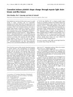
Fig. 1 describes the different experimental conditions
schematically. After the 14 days treatment with varying IL-
13 concentrations, in one group (Fig. 1B), IL-13 media is
withdrawn (removed) at day 21. At day 22, some of these
Transwells
®
were moved to new 12 well plates and co-cul-
tured with NHLF embedded in rat tail collagen gels for 3
days (days 22–25) in 50:50 media without IL-13, with/
without TGF-β
2
neutralizing antibody or goat IgG control.
In a second group (Fig. 1C), the IL-13 media was with-
drawn at day 21 and cultured in 50:50 media without IL-
13 until day 28, and then co-cultured with NHLF embed-
ded rat tail collagen gels for 3 days (days 28–31), with/
without TGF-β
2
neutralizing antibody or goat IgG control.
A third group served as the control (Fig. 1A), where the co-
cultures were performed at the same time points, but the
cells were never exposed to IL-13. A final condition con-
sisted of NHLF-embedded collagen gels alone (not co-cul-
tured with NHBE) with 1 ml of 50:50 media on top, and
is referred to as "NHLF only". Media was collected from
all conditions and stored at -80°C for active and total
TGF-β
2
analysis by ELISA. A critical feature of the study
design is treatment of the NHBE with IL-13 for 14 days
followed by withdrawal and subsequent culture in the
absence of IL-13 all throughout the withdrawal and 'co-
culture with NHLF' periods.
Immunofluorescence microscopy
At day 22 (i.e. after 14 days IL-13 treatment and 1 day
withdrawal of IL-13 containing media) and day 28 (i.e. 14
day treatment and 7 day withdrawal of IL-13 media), the
NHBE were fixed using 4% formaldehyde (Sigma) in PBS
at 4°C for 20 minutes. Non-specific binding was blocked
by addition of Abdil (2% BSA in TBS-0.1% Triton-X) for 1
hr at 4°C. Samples were incubated in mouse monoclonal
anti-MUC5AC (Clone 45M1, Neomarkers, Fremont, CA,
diluted 1:500 in Abdil) or anti-β-Tubulin IV (Sigma
Aldrich, St. Louis, MO, diluted 1:1000 in Abdil) overnight
followed by wash and incubation with Alexa Fluor 488
anti-mouse secondary antibody (Molecular probes,
Eugene, OR) at 1:500 in Abdil for 2 hr at 4°C. Cell nuclei
were stained with 4', 6-diamidino-2-phenylindole dihy-
drocholride hydrate (1 μg/ml, DAPI, Sigma) in PBS for 5
minutes. After staining, the Transwell
®
membrane was
removed by a scalpel, placed on a microscope slide with a
drop of Vectashield, and visualized using a Nikon Eclipse
E800 epifluorescence microscope.
SDS-PAGE and western blot
At day 22, 28, 25 and 31 some of the NHBE monolayers
alone or from co-cultures were lysed using RIPA buffer on
ice (50 mM Tris, pH 8.0, 150 mM NaCl, 1% Nonidet P-40,
0.1% SDS, 0.5% sodium deoxycholate, and 0.1 mM
sodium orthovanadate) supplemented with protease
inhibitor cocktail (Sigma, P8340) by repetitive scraping.
Protein concentrations were determined using BCA pro-
tein assay (Pierce Biotechnology) following the manufac-
turers directions. Laemmli buffer was added to 40 μg
equal protein in gel running buffer and then boiled for 5
minutes. Samples were subjected to SDS-PAGE and trans-
ferred onto nitrocellulose (0.1 A, overnight). Western
blotting was performed using appropriate primary (mon-
oclonal mouse anti-MUC5AC or anti-β-Tubulin-IV were
diluted in 5% milk in TBS-0.1% Tween at 1:500 and
1:1000 respectively) and horseradish peroxidase (HRP)
conjugated secondary antibodies (1:10,000, Santa Cruz
biotechnology), and visualized using an enhanced chemi-
luminescence system (Amersham Biosciences and Biorad
Imaging system). The blots were also probed with mouse
monoclonal anti-β-actin (Abcam) as a loading control.
Sircol™ soluble collagen assay
At day 25 and day 31, collagen gels from the co-culture
and NHLF only conditions were collected in different 15
ml centrifuge tubes (6 gels per condition, per time point
from two donors), weighed and stored at -80°C. At the
start of the experiment for extracting salt soluble collagen,
0.05 M Tris buffer (pH 7.5) containing 1 M NaCl with
1:100 protease inhibitor cocktail (P8340, Sigma) was
added to the gels. A ratio of 5 volumes of solvent to wet
tissue weight was used. The sample was then stirred over-
night at 4°C. The next day they were centrifuged at 1,000
Respiratory Research 2008, 9:27 />Page 4 of 12
(page number not for citation purposes)
g for 5 minutes to obtain a colorless supernatant. Sircol™
soluble collagen assay was then performed following the
manufacturers directions. In brief, 1 ml of Sircol dye was
added to 200 μl of supernatant extracted from collagen
gels, as described above, or 200 μl of cell culture media.
The tubes were mixed on a shaker for 30 minutes and then
centrifuged at 10,000 g for 10 minutes to obtain a well-
compacted pellet composed of precipitated collagen-dye
complex, followed by inverting and draining the tubes on
tissue paper to remove unbound dye. 1 ml of alkali rea-
gent was added to the pellet in each tube, and the tubes
were thoroughly vortexed to dissolve the bound dye pel-
let. 200 μl of this mixture for the sample (either extracted
from collagen gels or cell culture media), a corresponding
blank and standard (conc. 1 mg/ml used to generate a
standard curve, supplied in the Sircol assay kit) was then
analyzed in triplicate using a 96-well plate in a spectro-
photometer at 540 nM in a Benchmark Microplate Reader
(Biorad). All values are expressed as a percentage of NHLF
embedded in rat-tail collagen gels levels ("NHLF only").
Multiphoton microscopy
Some co-cultures were developed in 12 well glass bottom
plates (Mattek Corp.) for imaging the NHLF-embedded
Protocol for IL-13 treatment and withdrawalFigure 1
Protocol for IL-13 treatment and withdrawal. NHBE cells are seeded on Transwells
®
as described in the Materials and
Methods, treatment with IL-13 followed by its withdrawal is carried out as shown. (A) For the control case, the NHBE are cul-
tured in 50:50 epithelial media without any IL-13 all throughout and co-cultured with NHLF embedded in rat tail collagen gel
from days 22 to day 25 and day 28 to day 31. (B) NHBE are treated for 14 days from day 7 to day 21 with varying IL-13 con-
centrations (0.1, 1, 10 ng/ml), then the IL-13 media is withdrawn and replaced with 50:50 epithelial media for 1 day from day 21
to day 22. On day 22 the NHBE are co-cultured with NHLF embedded in a rat-tail collagen gel for a period of 3 days till day 25.
(C) The NHBE are treated for 14 days from day 7 to day 21 with varying IL-13 concentrations (0.1, 1, 10 ng/ml), then the IL-13
media is withdrawn and replaced with 50:50 media for a period of 7 days from day 21 to day 28. On day 28 the NHBE are co-
cultured with NHLF embedded in a rat-tail collagen gel for 3 days till day 31.
Respiratory Research 2008, 9:27 />Page 5 of 12
(page number not for citation purposes)
collagen gels at day 25 and day 31. The network of colla-
gen fibers was studied in the extracellular matrix of the co-
culture model using multiphoton microscopy (MPM) as
previously described [34,35]. Briefly, a Zeiss LSM 510
Meta multiphoton microscope (Zeiss, Jena, Germany)
was used. A Mai Tai laser was tuned to 780 nm and
focused on the sample with an EC Plan-Neofluar 40×/1.3
NA oil DIC objective (Zeiss). Power before the objective
was 250 mW. Resolution was ~0.4μm in x-, y- and 1 μm
in z- image planes, with an area of 0.019 μm
2
per pixel.
Each image was 512 × 512 pixels. Pixel intensity histo-
grams showed minimal pixel saturation. The meta chan-
nel was used to collect emitted light in an
epiconfiguration at wavelength 390 nm using a narrow
bandpass filter (380–400 nm), which exclusively repre-
sented the second harmonic generation (SHG) signal
from fibrillar collagen [36,37]. Image stacks were col-
lected at 10-micron intervals between 20–120 microns
from the coverslip. Three image stacks were collected per
gel at random locations with each stack at least 1 mm
apart, and for three gels per condition and per timepoint.
The LSM 510 software (Zeiss) was used to quantify images
using the average segmented pixel intensity.
Statistics
Experiments were performed using three NHBE donors,
repeated twice, with 3–6 wells/gels per condition per time
point. Data are reported as mean ± SD and InStat 2.01 for
Macintosh software package was used for all analysis.
Data were analyzed using one-way analysis of variance
(ANOVA) with Student Newman Keuls post-test analysis
for multiple comparisons. Data were considered signifi-
cant at p < 0.05.
Results
IL-13 treatment and plasticity of the NHBE
At day 22, after 14 days treatment with 1 or 10 ng/ml of
IL-13 followed by 1-day withdrawal of IL-13, the NHBE
demonstrate a increase in MUC5AC protein as detected by
immunofluorescence (Fig. 2A). At day 28, when the IL-13
treatment has been withdrawn for 7 days, the treated
NHBE (10 ng/ml) still demonstrate elevated levels of
MUC5AC over the untreated control (i.e. NHBE cultured
in media without any IL-13) condition (Fig. 2A). Chronic
treatment with the lowest concentration (0.1 ng/ml) of IL-
13 did not increase MUC5AC staining over control (0 ng/
ml IL-13) either at day 22 or day 28 (Fig. 2A).
Immunoblotting for MUC5AC protein mirrored the
trends of immunofluorescence at days 22 and day 28. At
day 25 (1 day withdrawal of IL-13 treatment followed by
3 day co-culture with NHLF), MUC5AC protein by immu-
noblot increased in a dose-response fashion with IL-13
treatment concentration (Fig. 2B, C). In contrast, by day
31 (7 day withdrawal of IL-13 treatment followed by 3
day co-culture with NHLF) the MUC5AC protein levels in
NHBE for all treatment concentrations were no different
than control levels.
The trends in the protein level of β-Tubulin-IV are the
opposite of MUC5AC (Fig. 2B, D). As IL-13 exposure con-
centration increases, the amount of β-Tubulin-IV
decreases. This effect of IL-13 is observed both 1-day (day
22) and 7-days (day 28) following withdrawal of the IL-
13, but is not observed in the conditions following 3-days
of co-culture with the NHLF (days 25 and 31). Images are
from one representative donor.
IL-13 stimulates TGF-
β
2
release from NHBE
Fig. 3A, B demonstrates that the IL-13 treatment induces
active and total TGF-β
2
release from the airway epithelium
in the media (as measured by ELISA) over baseline levels
secreted from the untreated NHBE and the 10 ng/ml pre-
treated NHBE levels remain elevated at day 28 (7 days
post withdrawal of IL-13 treatment). Fig. 3C–D shows
that active and total TGF-β
2
release remains significantly
(p < 0.01) elevated at day 25, following 14 days IL-13
treatment at 10 ng/ml, 1 day withdrawal and co-culture
with NHLF, although at day 31 following 7 day with-
drawal after treatment, the levels of TGF-β
2
are no differ-
ent from control (i.e. untreated NHBE co-cultured with
NHLF). NHLF embedded in a rat tail collagen gel, "NHLF
only" (Fig. 3C–D) secrete negligible levels of active and
total TGF-β
2
. Chronic treatment with the lowest concen-
tration (0.1 ng/ml) of IL-13 did not increase levels of
active and total TGF-β
2
over control at any of the time
points and thus was not used in the following experi-
ments.
IL-13 treated epithelium secretes biologically relevant
TGF-
β
2
that stimulates collagen secretion
To investigate further the physiological relevance of epi-
thelial-derived TGF-β
2
on collagen secretion from the
NHLF and its ability to modulate the optical properties of
the matrix, we co-cultured pre-treated and untreated
NHBE with NHLF embedded in collagen gels. At day 25
the NHLF embedded collagen gels demonstrate signifi-
cantly (p < 0.01) elevated levels of soluble collagen
secreted in the media (Fig. 4A) and matrix (Fig. 4B), in the
co-culture of NHBE pre- treated with 1 or 10 ng/ml IL-13
as compared to control (i.e. untreated NHBE co-cultured
with NHLF). This increase is abolished on incubation
with TGF-β
2
neutralizing antibody (10 μg/ml) in the 3-
day co-culture period (levels were unaffected upon incu-
bation with purified goat IgG control). Furthermore the
SHG signal (Fig. 4C–D), which is an index of collagen
fibril organization and density from the matrix (20 μm
from the surface), is augmented from the 10 ng/ml IL-13
pre-treated NHBE-NHLF co-culture. This increase is also
inhibited upon incubation with TGF-β
2
neutralizing anti-
Respiratory Research 2008, 9:27 />Page 6 of 12
(page number not for citation purposes)
IF staining and Western blot for MUC5AC/β-Tubulin IV in the NHBEFigure 2
IF staining and Western blot for MUC5AC/β-Tubulin IV in the NHBE. IL-13 mediated a concentration dependent
increase in MUC5AC protein levels in the NHBE as seen by (A) Immunofluorescence, where at day 22 and day 28, 1 and 7 days
after withdrawal of 14 day treatment with IL-13 (1,10 ng/ml for day 22 and 10 ng/ml for day 28) the staining for MUC5AC is
higher compared to the untreated NHBE (0 ng/ml IL-13) (n = 3 donors of NHBE; grown in duplicate; with 3–6 wells per condi-
tion; scale bar = 20 μm). DAPI staining of the nuclei showed similar number of cells in all conditions (data not shown). (B) Lev-
els of MUC5AC protein show a dose dependent increase via western blot at day 22 and day 28. Also during co-culture with
the NHLF the dose dependent increase of MUC5AC is maintained at day 25 and not at day 31. Levels of β-Tubulin IV protein
in the NHBE shown an inverse dependence on IL-13 concentration at days 22 and day 28 with levels remaining constant at day
25 and day 31 of co-culture with NHLF. Images are representative from 3 NHBE donors. (C, D) Quantification of MUC5AC/β-
Actin and β-Tubulin IV/β-Actin levels relative to IL-13 concentration of 0 ng/ml at day 22 condition, show a dose dependent
increase with IL-13 concentration at day 22,28 and 25 for MUC5AC and dose dependent decrease at day 22, 28 and 31 for β-
Tubulin IV (Statistical difference between conditions by ANOVA # p < 0.01).
Respiratory Research 2008, 9:27 />Page 7 of 12
(page number not for citation purposes)
ELISA for active and total TGF-β
2
in the media (A, B)Figure 3
ELISA for active and total TGF-β
2
in the media (A, B). At day 22, the concentration of active and total TGF-β
2
in the
media of IL-13 pre-treated NHBE at 1 and 10 ng/ml is significantly higher as compared to untreated NHBE (0 ng/ml of IL-13)
media; * p < 0.01. At day 22 and day 28, the concentration of active TGF-β
2
in the IL-13 pre-treated NHBE at 10 ng/ml is ele-
vated compared to pre-treated NHBE at 1 ng/ml; # p < 0.01. At day 28 active and total TGF-β
2
in IL-13 pre-treated NHBE at
10 ng/ml is increased compared to untreated NHBE; * p < 0.01. (C, D) At day 25, the NHBE pre-treated with IL-13 at 10 ng/
ml, has higher levels of active and total TGF-β
2
in the media as compared to untreated and pre-treated NHBE at 1 ng/ml co-cul-
tured with NHLF (*, # p < 0.01 compared to 0 and 1 ng/ml IL-13 pre-treated NHBE co-cultured with NHLF, respectively). At
day 31, there is no significant difference in the levels of active and total TGF-β
2
between treatment conditions. NHLF repre-
sents levels of active and total TGF-β
2
in media of fibroblasts in a collagen gel without NHBE co-culture. All experiments were
performed using 3 donors, grown in duplicate, with 3–6 wells per condition.
Respiratory Research 2008, 9:27 />Page 8 of 12
(page number not for citation purposes)
Quantification of soluble collagen content in the media and matrixFigure 4
Quantification of soluble collagen content in the media and matrix. (A) Sircol soluble collagen assay was performed
as described in the Materials and Methods, which quantifies the amount of soluble collagen in the cell culture supernatant and
newly synthesized salt soluble collagen in the matrix. The amount of soluble collagen secreted in the media at day 25 in the IL-
13 pre-treated NHBE at 1 and 10 ng/ml co-cultured with NHLF is augmented as compared to the untreated NHBE co-culture;
* p < 0.01 and addition of TGFβ
2
neutralizing antibody (10 μg/ml) abolishes this increase (
§
p < 0.01 compared to respective
condition without TGFβ
2
neutralizing antibody). (B) At day 25 there is an increase in newly synthesized salt soluble collagen
content in the matrix in the IL-13 pre-treated NHBE at 1 and 10 ng/ml followed by co-culture with NHLF as compared to the
untreated NHBE co-culture; * p < 0.01 and the IL-13 pre-treated NHBE at 10 ng/ml co-culture collagen levels are elevated as
compared to the IL-13 pretreated NHBE at 1 ng/ml co-culture; # p < 0.01. Also, addition of the TGFβ
2
neutralizing antibody
abolishes this increase (
§
p < 0.01 compared to respective condition without TGFβ
2
antibody). The media and matrix collagen
levels are normalized to respective levels obtained from NHLF embedded in collagen gels ("NHLF only"). (C, D) Representa-
tive Second harmonic generated (SHG) images (scale bar = 50 μm) of collagen fibrils at day 25 are shown along with the quan-
tification of signal intensities. The SHG signals from the collagen secreted by NHLF embedded in rat tail collagen gels which
were co-cultured with the IL-13 pre-treated NHBE at 10 ng/ml are elevated compared to the untreated NHBE co-culture; * p
< 0.01 and this increase is inhibited on incubation with TGFβ
2
neutralizing antibody in the 3 day co-culture period (
§
p < 0.01
compared to respective condition without TGFβ
2
antibody). Addition of goat IgG did not alter the increased levels of collagen
in the matrix and media in the pre-treated NHBE-NHLF co-culture. (E) Exogenous active TGF-β
2
at 0.05, 0.1, 0.5, 1 and 10 ng/
ml is added in 50:50 epithelial media to NHLF embedded in collagen gels for a period of 3 days. There is a significant increase in
the newly synthesized salt soluble collagen content in the matrix with addition of increasing concentration of active TGF-β
2
(*
p < 0.01 compared to only NHLF condition). All values are normalized to those obtained from "NHLF only" condition. All
experiments were performed using 3 donors, grown in duplicate, with 3–6 wells for each condition.
Respiratory Research 2008, 9:27 />Page 9 of 12
(page number not for citation purposes)
body (10 μg/ml). At day 31 there is no significant differ-
ence in the levels of collagen secreted or SHG signal from
the IL-13 treated and untreated NHBE co-cultures (data
not shown).
Exogenous active TGF-β
2
at 0.05, 0.1, 0.5, 1, and 10 ng/ml
(Fig. 4E) was added in epithelial media to NHLF embed-
ded in collagen gels for a period of 3 days and collagen
secretion assayed. The amount of collagen secreted in the
matrix as compared to "NHLF only" was increased, in a
dose-dependent manner. All values are expressed as a per-
centage of NHLF embedded in rat-tail collagen gels levels
("NHLF only").
Discussion
Asthma affects 8%–10% of the population, and is charac-
terized by chronic airway inflammation, repetitive bron-
choconstriction, and marked structural changes in the
airway wall including goblet cell metaplasia in the airway
epithelium, and collagen deposition in the lamina reticu-
laris (sub-epithelial fibrosis) [9,38,39]. Furthermore,
other fibrotic diseases in the lungs (e.g., IPF) share similar
characteristics [24-26,40]. Mechanisms linking these fea-
tures of the disease are only partially understood. Our
study demonstrates that a prominent TH2-type inflamma-
tory mediator, IL-13, can alter the differentiated pheno-
typic features of the epithelium. Furthermore, the altered
epithelium alone (i.e., in the absence of IL-13) secretes
biologically significant TGF-β
2
levels, which stimulates
features of fibrosis (e.g., collagen secretion) in the subep-
ithelial matrix and alters the bulk optical properties of the
matrix. In addition, following extended withdrawal of IL-
13, the epithelium is capable of reverting back to its base-
line phenotype.
The inflammatory process in asthma has a prominent
allergic component which involves Th2-type cytokines
including interleukin(IL)-4, -5 and -13 [1]. Both in vivo
and in vitro model systems have been employed to deter-
mine the role of IL-13 in modulating features of the dis-
ease [41,42]. In vivo, transgenic mice which selectively
over express IL-13 or mice which do not express IL-13
have been used to demonstrate critical roles of IL-13 in
airway hyperresponsivness, fibrosis, and mucus cell meta-
plasia [43,44]. However, the source of IL-13 leading to
these findings cannot be isolated as multiple cells types
are capable of producing IL-13. IL-13 mediated changes to
the bronchial epithelium could be paracrine (Th2 lym-
phocytes as the source) or autocrine (epithelium as the
source) [45].
In vitro, treatment of the airway epithelium with IL-13
during the differentiation phase has been shown to stim-
ulate mucus production [22,23,30] and acute (48 hours)
treatment of the epithelium with IL-13 can stimulate the
release TGF-β
2
[32,33]. Both of these observations are
consistent with our results. Nonetheless, the biological
consequence(s) of these IL-13-induced changes to the epi-
thelium have not been described.
The current study demonstrates that chronic treatment of
the epithelium with IL-13 during the differentiation phase
results in enhanced MUC5AC expression (Fig. 2A–C),
reduced β-Tubulin-IV expression (Fig. 2B, D) and elevated
TGF-β
2
secretion (Fig. 3A–D). These changes are observed
for up to seven days following withdrawal of the IL-13.
When the IL-13 pre-treated epithelium was co-cultured
with a fibroblast-embedded collagen gel in the absence of
IL-13 at day 25, the IL-13 concentration dependent
increase in MUC5AC was maintained but not at day 31.
However, the down regulation of β-Tubulin-IV expression
with increasing IL-13 concentration was suppressed dur-
ing the co-culture with fibroblasts at day 25, and this trend
was also observed at day 31. While we did not pursue the
mechanism of this observation at this time, it seems clear
that the fibroblasts influence the epithelium through as
yet unidentified mediators. This observation lends sup-
port to co-culture models as they provide unique insight
into epithelial and mesenchyme communication.
Although there is some donor to donor variability, the
trends for all these proteins remain the same [46]. In addi-
tion, IL-13 can stimulate cell proliferation [31], which
may account for increased levels of TGF-β
2
and MUC5AC,
we did not observe a significant increase in cell number
on staining nuclei (data not shown). These phenotypic
changes are consistent with the loss of ciliated epithelial
cells (reduced β-Tubulin-IV), and goblet cell metaplasia
(enhanced expression of MUC5AC).
At day 25, when the IL-13 treated epithelium was co-cul-
tured with a fibroblast-embedded collagen gel in the
absence of IL-13, the fibroblasts increased both the secre-
tion of soluble collagen in the media and matrix (Fig.
4A–B), and the second harmonic generated signal in the
extracellular matrix (Fig. 4C–D). Upon incubation with a
TGF-β
2
neutralizing antibody this increase is abolished
suggesting that biologically relevant levels of TGF-β
2
levels
are continuously secreted by the epithelium. Furthermore,
at day 31 when the levels of TGF-β
2
are the same in the
treated and untreated NHBE co-cultures, there is no
increase in collagen content in the media or matrix condi-
tion nor were there any differences in the SHG signals.
Thickening of the reticular layer in asthma has been
termed subepithelial fibrosis, and is due to deposition of
fibrillar collagens (types I, III, and V), tenascin C, and
fibronectin [9]. The Sircol collagen assay is a quantitative
dye-binding method that can measure collagens from
type I-V in a soluble form. Only newly secreted collagen
Respiratory Research 2008, 9:27 />Page 10 of 12
(page number not for citation purposes)
into the matrix is soluble. Over time, collagen becomes
insoluble due to intermolecular crosslinking. Our results
demonstrate at day 25 that soluble collagen levels are ele-
vated when the fibroblast-embedded collagen gel is co-
cultured with the airway epithelium compared to levels
without co-culture, and an additional increase when the
airway epithelium has been differentiated in the presence
of IL-13. This observation is consistent with enhanced col-
lagen secretion by the fibroblasts due to soluble mediators
produced by the airway epithelium [28,29,47,48]. It has
been shown that IL-13 can induce the secretion of matrix
metalloproteases that could potentially degrade the rat-
tail collagen gels. We tested this possibility by taking the
media at day 25 and day 31 from the varying concentra-
tion IL-13 pretreated NHBE-NHLF co-culture conditions
and exposing it to acellular collagen gels for 3 days. We
did not observe a significant change in the levels of colla-
gen in the media before and after exposure to the gels
(data not shown), suggesting that, although IL-13 may
induce secretion of matrix metalloproteases, the type or
magnitude did not impact or degrade the rat tail collagen
in our system.
In addition to the Sircol soluble collagen assay, we quan-
tified structural changes in the matrix using multiphoton
microscopy and second harmonic generation (SHG).
SHG in the extracellular matrix is specific to fibrillar colla-
gen, and is generated by non-linear interactions of the
near-infrared light with the non-centrosymmetric features
of collagen. SHG is largely forward propagated from col-
lagen fibers at exactly 1/2 the wavelength of the excitation
wavelength [35,49]. In our experiment, we utilized an
excitation wavelength of 780 nm, and detected the subse-
quently backward scattered SHG signal (390 nm) from
collagen using a narrow bandpass filter (380–400 nm) in
an epiconfiguration. The intensity of the SHG signal is a
positive function of the concentration of collagen, but can
also increase when the organization of collagen at second-
ary and tertiary levels increases [37]. Thus, at day 25 our
observation of an enhanced SHG signal in the extracellu-
lar matrix following co-culture of the airway epithelium
with the fibroblast-embedded collagen gel is consistent
with either an increased amount of collagen, and/or an
increased organization of the collagen.
We hypothesized that the increase in collagen secretion
and the enhanced SHG signal from the matrix was due, in
part, to epithelial-derived TGF-β
2
. TGF-β has three iso-
forms (TGF-β
1
, TGF-β
2
, TGF-β
3
) in mammalian systems
[50], and are pleiotropic mediators of cell growth and dif-
ferentiation [51]. All three isoforms are present in the
lungs, can be produced by epithelial cells, and have been
shown to contribute to fibrosis. For example, our group
has recently demonstrated that scrape-injured airway epi-
thelial cells release active TGF-β
2
at concentrations similar
to the current study (50–100 pg/ml), and that the epithe-
lial-derived TGF-β
2
enhances the SHG signal from an
underlying fibroblast-embedded collagen gel [28]. More-
over, relatively small deviations (i.e., 30–50 pg/ml) above
or below the basal production of TGF-β
2
, that are similar
in magnitude observed in our study, result in altered SHG
from the matrix suggesting that tight regulation of TGF-β
2
is required for normal matrix homeostasis. Similarly, it
has been shown that compression of airway epithelial
cells stimulates collagen secretion from fibroblasts in co-
culture [47], as well as TGF-β
2
release [29]. The role of bio-
logically relevant epithelial-derived TGF-β
2
in subepithe-
lial fibrosis is also strongly supported by our observation
that a neutralizing antibody negates the observed
increases in collagen secretion and SHG, and that addi-
tion of exogenous active TGF-β
2
(Fig. 4E) at concentra-
tions observed in the media reproduces the increase in
collagen secretion.
TGF-β
1
was measured in the media in our co-culture
model (60–70 pg/ml, data not shown), but the concentra-
tion was not impacted by IL-13 treatment or co-culture.
Transgenic mice bred to over express IL-13 demonstrate
tissue fibrosis and stimulate TGF-β
1
production. Although
the major source of TGF-β
1
in the mouse model are mac-
rophages, alveolar epithelial cells, and eosinophils, we
cannot rule out the role of TGF-β
1
in subepithelial fibrosis
[52,53].
Finally, the normal human bronchial epithelial cells are
commercially (Lonza) purchased primary cells from nor-
mal human healthy and non-smoking individuals who
are tested and found non-reactive by an FDA approved
method for the presence of HIV-1 and other viruses. The
asthmatic airway epithelium in vivo displays signs of
structural damage, it is more susceptible to oxidant-
induced apoptosis and has marked mucus metaplasia. It is
widely accepted that the epithelium in asthmatics is bio-
chemically abnormal due to its ability to release greater
amounts of pro-inflammatory cytokines and express ele-
vated levels of transcription factors both in vivo and fol-
lowing in vitro culture [12,54,55]. Thus, the asthmatic
airway epithelium may respond differently to IL-13 than
the normal airway epithelium; nonetheless, the current
study forms the basis for observing similar endpoints in
future studies using asthmatic epithelial cells.
Conclusion
IL-13 enhances MUC5AC expression and TGF-β
2
secre-
tion, and decreases β-Tubulin-IV expression in the airway
epithelium when present during the 14-day differentia-
tion phase at an air-liquid interface. The altered pheno-
typic features of the airway epithelium in vitro are
consistent with those observed in asthma. Co-culturing
this altered epithelial phenotype with a lung fibroblast-
Respiratory Research 2008, 9:27 />Page 11 of 12
(page number not for citation purposes)
embedded collagen gel in the absence of IL-13 results in
enhanced collagen secretion and second harmonic gener-
ation signal from the extracellular matrix, both of which
are dependent on biologically significant levels of epithe-
lial-derived TGF-β
2
. However, upon withdrawal for a
period of ten days, the levels of MUC5AC, β-Tubulin-IV
and TGF-β
2
secretion are similar in the treated and
untreated case indicating plasticity of the cultured airway
epithelium, and its ability to return to a baseline pheno-
type. We conclude that IL-13 may contribute to subepithe-
lial fibrosis in asthma by stimulating the continuous
release TGF-β
2
from the airway epithelium.
Competing interests
The author(s) declare that they have no competing inter-
ests.
Authors' contributions
NKM designed, planned, and performed all of the experi-
ments, and wrote the manuscript; JDM performed some
initial studies regarding NHBE culture and IL-13 expo-
sure; CBR and BTD assisted with the design and interpre-
tation of multiphoton microscopy and SHG imaging; and
SCG provided overall guidance for the study, assisted in
the experimental design, analysis and interpretation of the
data, and writing of the manuscript. All authors have read
and approved the final manuscript.
Acknowledgements
This work was funded by a grant from the National Heart Lung and Blood
Institute (R01-HL067954). We also acknowledge support from the Laser
Medical and Microbream Program (LAMMP, P41-RR001192), and the Air
Force Office of Scientific Research (FA9550-04-1-0101). The U.S. Govern-
ment is authorized to reproduce and distribute reprints for Governmental
purposes notwithstanding any copyright notation thereon. The views and
conclusions contained herein are those of the authors and should not be
interpreted as necessarily representing the official policies or endorse-
ments, either expressed or implied, of the Air Force Research Laboratory
or the U.S. Government. Finally, we acknowledge the expert technical
assistance of Mr. Chirag Khatiwala.
References
1. Cohn L, Elias JA, Chupp GL: Asthma: mechanisms of disease
persistence and progression. Annu Rev Immunol 2004,
22:789-815.
2. Busse W, Elias J, Sheppard D, Banks-Schlegel S: Airway remodeling
and repair. Am J Respir Crit Care Med 1999, 160(3):1035-1042.
3. Davies DE, Wicks J, Powell RM, Puddicombe SM, Holgate ST: Airway
remodeling in asthma: new insights. J Allergy Clin Immunol 2003,
111(2):215-25; quiz 226.
4. Elias JA, Zhu Z, Chupp G, Homer RJ: Airway remodeling in
asthma. J Clin Invest 1999, 104(8):1001-1006.
5. Fixman ED, Stewart A, Martin JG: Basic mechanisms of develop-
ment of airway structural changes in asthma. Eur Respir J 2007,
29(2):379-389.
6. Homer RJ, Elias JA: Airway remodeling in asthma: therapeutic
implications of mechanisms. Physiology (Bethesda) 2005,
20:28-35.
7. James A: Airway remodeling in asthma. Curr Opin Pulm Med
2005, 11(1):1-6.
8. James AL, Maxwell PS, Pearce-Pinto G, Elliot JG, Carroll NG: The
relationship of reticular basement membrane thickness to
airway wall remodeling in asthma. Am J Respir Crit Care Med
2002, 166(12 Pt 1):1590-1595.
9. Jeffery PK: Remodeling in asthma and chronic obstructive
lung disease. Am J Respir Crit Care Med 2001, 164(10 Pt 2):S28-38.
10. Vignola AM, Gagliardo R, Siena A, Chiappara G, Bonsignore MR,
Bousquet J, Bonsignore G: Airway remodeling in the pathogen-
esis of asthma. Curr Allergy Asthma Rep 2001, 1(2):108-115.
11. Davies DE, Holgate ST: Asthma: the importance of epithelial
mesenchymal communication in pathogenesis. Inflamma-
tion and the airway epithelium in asthma. Int J Biochem Cell Biol
2002, 34(12):1520-1526.
12. Knight DA, Holgate ST: The airway epithelium: structural and
functional properties in health and disease.
Respirology 2003,
8(4):432-446.
13. Mullin JM: Epithelial barriers, compartmentation, and cancer.
Sci STKE 2004, 2004(216):pe2.
14. Warburton D, Schwarz M, Tefft D, Flores-Delgado G, Anderson KD,
Cardoso WV: The molecular basis of lung morphogenesis.
Mech Dev 2000, 92(1):55-81.
15. Hackett TL, Knight DA: The role of epithelial injury and repair
in the origins of asthma. Curr Opin Allergy Clin Immunol 2007,
7(1):63-68.
16. Holgate ST: The inflammation-repair cycle in asthma: the piv-
otal role of the airway epithelium. Clin Exp Allergy 1998, 28
Suppl 5:97-103.
17. Knight D: Epithelium-fibroblast interactions in response to
airway inflammation. Immunol Cell Biol 2001, 79(2):160-164.
18. Holgate ST, Davies DE, Puddicombe S, Richter A, Lackie P, Lordan J,
Howarth P: Mechanisms of airway epithelial damage: epithe-
lial-mesenchymal interactions in the pathogenesis of
asthma. Eur Respir J Suppl 2003, 44:24s-29s.
19. Holgate ST, Holloway J, Wilson S, Bucchieri F, Puddicombe S, Davies
DE: Epithelial-mesenchymal communication in the patho-
genesis of chronic asthma. Proc Am Thorac Soc 2004, 1(2):93-98.
20. Lazaar AL, Panettieri RA Jr.: Airway smooth muscle: a modula-
tor of airway remodeling in asthma. J Allergy Clin Immunol 2005,
116(3):488-95; quiz 496.
21. Kuperman DA, Huang X, Koth LL, Chang GH, Dolganov GM, Zhu Z,
Elias JA, Sheppard D, Erle DJ: Direct effects of interleukin-13 on
epithelial cells cause airway hyperreactivity and mucus over-
production in asthma. Nat Med 2002, 8(8):885-889.
22. Laoukili J, Perret E, Willems T, Minty A, Parthoens E, Houcine O,
Coste A, Jorissen M, Marano F, Caput D, Tournier F: IL-13 alters
mucociliary differentiation and ciliary beating of human res-
piratory epithelial cells. J Clin Invest 2001,
108(12):1817-1824.
23. Ordonez CL, Khashayar R, Wong HH, Ferrando R, Wu R, Hyde DM,
Hotchkiss JA, Zhang Y, Novikov A, Dolganov G, Fahy JV: Mild and
moderate asthma is associated with airway goblet cell
hyperplasia and abnormalities in mucin gene expression. Am
J Respir Crit Care Med 2001, 163(2):517-523.
24. Hancock A, Armstrong L, Gama R, Millar A: Production of inter-
leukin 13 by alveolar macrophages from normal and fibrotic
lung. Am J Respir Cell Mol Biol 1998, 18(1):60-65.
25. Noble PW, Homer RJ: Idiopathic pulmonary fibrosis: new
insights into pathogenesis. Clin Chest Med 2004, 25(4):749-58, vii.
26. Selman M, King TE, Pardo A: Idiopathic pulmonary fibrosis: pre-
vailing and evolving hypotheses about its pathogenesis and
implications for therapy. Ann Intern Med 2001, 134(2):136-151.
27. Gray TE, Guzman K, Davis CW, Abdullah LH, Nettesheim P: Muco-
ciliary differentiation of serially passaged normal human tra-
cheobronchial epithelial cells. Am J Respir Cell Mol Biol 1996,
14(1):104-112.
28. Thompson HG, Mih JD, Krasieva TB, Tromberg BJ, George SC: Epi-
thelial-derived TGF-beta2 modulates basal and wound-heal-
ing subepithelial matrix homeostasis. Am J Physiol Lung Cell Mol
Physiol 2006, 291(6):L1277-85.
29. Tschumperlin DJ, Shively JD, Kikuchi T, Drazen JM: Mechanical
stress triggers selective release of fibrotic mediators from
bronchial epithelium. Am J Respir Cell Mol Biol 2003,
28(2):142-149.
30. Atherton HC, Jones G, Danahay H: IL-13-induced changes in the
goblet cell density of human bronchial epithelial cell cul-
tures: MAP kinase and phosphatidylinositol 3-kinase regula-
tion. Am J Physiol Lung Cell Mol Physiol 2003, 285(3):L730-9.
31. Booth BW, Adler KB, Bonner JC, Tournier F, Martin LD: Inter-
leukin-13 induces proliferation of human airway epithelial
Publish with BioMed Central and every
scientist can read your work free of charge
"BioMed Central will be the most significant development for
disseminating the results of biomedical research in our lifetime."
Sir Paul Nurse, Cancer Research UK
Your research papers will be:
available free of charge to the entire biomedical community
peer reviewed and published immediately upon acceptance
cited in PubMed and archived on PubMed Central
yours — you keep the copyright
Submit your manuscript here:
/>BioMedcentral
Respiratory Research 2008, 9:27 />Page 12 of 12
(page number not for citation purposes)
cells in vitro via a mechanism mediated by transforming
growth factor-alpha. Am J Respir Cell Mol Biol 2001, 25(6):739-743.
32. Richter A, Puddicombe SM, Lordan JL, Bucchieri F, Wilson SJ, Dju-
kanovic R, Dent G, Holgate ST, Davies DE: The contribution of
interleukin (IL)-4 and IL-13 to the epithelial-mesenchymal
trophic unit in asthma. Am J Respir Cell Mol Biol 2001,
25(3):385-391.
33. Wen FQ, Liu XD, Terasaki Y, Fang QH, Kobayashi T, Abe S, Rennard
SI: Interferon-gamma reduces interleukin-4- and interleukin-
13-augmented transforming growth factor-beta2 produc-
tion in human bronchial epithelial cells by targeting Smads.
Chest 2003, 123(3 Suppl):372S-3S.
34. Agarwal A, Coleno ML, Wallace VP, Wu WY, Sun CH, Tromberg BJ,
George SC: Two-photon laser scanning microscopy of epithe-
lial cell-modulated collagen density in engineered human
lung tissue. Tissue Eng 2001, 7(2):191-202.
35. Wong BJ, Wallace V, Coleno M, Benton HP, Tromberg BJ: Two-pho-
ton excitation laser scanning microscopy of human, porcine,
and rabbit nasal septal cartilage. Tissue Eng 2001, 7(5):599-606.
36. Raub CB, Suresh V, Krasieva T, Lyubovitsky J, Mih JD, Putnam AJ,
Tromberg BJ, George SC: Noninvasive assessment of collagen
gel microstructure and mechanics using multiphoton micro-
scopy. Biophys J 2007, 92(6):2212-2222.
37. Zoumi A, Yeh A, Tromberg BJ: Imaging cells and extracellular
matrix in vivo by using second-harmonic generation and
two-photon excited fluorescence. Proc Natl Acad Sci U S A 2002,
99(17):11014-11019.
38. Redington AE: Fibrosis and airway remodelling. Clin Exp Allergy
2000, 30 Suppl 1:42-45.
39. Roberts CR: Is asthma a fibrotic disease? Chest 1995, 107(3
Suppl):111S-117S.
40. Keating DT, Sadlier DM, Patricelli A, Smith SM, Walls D, Egan JJ,
Doran PP: Microarray identifies ADAM family members as
key responders to TGF-beta1 in alveolar epithelial cells.
Respir Res 2006, 7:114.
41. Batra V, Musani AI, Hastie AT, Khurana S, Carpenter KA, Zangrilli JG,
Peters SP: Bronchoalveolar lavage fluid concentrations of
transforming growth factor (TGF)-beta1, TGF-beta2, inter-
leukin (IL)-4 and IL-13 after segmental allergen challenge
and their effects on alpha-smooth muscle actin and collagen
III synthesis by primary human lung fibroblasts. Clin Exp Allergy
2004, 34(3):437-444.
42. Chu HW, Balzar S, Seedorf GJ, Westcott JY, Trudeau JB, Silkoff P,
Wenzel SE: Transforming growth factor-beta2 induces bron-
chial epithelial mucin expression in asthma. Am J Pathol 2004,
165(4):1097-1106.
43. Chen Q, Rabach L, Noble P, Zheng T, Lee CG, Homer RJ, Elias JA: IL-
11 receptor alpha in the pathogenesis of IL-13-induced
inflammation and remodeling. J Immunol 2005,
174(4):2305-2313.
44. Walter DM, McIntire JJ, Berry G, McKenzie AN, Donaldson DD,
DeKruyff RH, Umetsu DT: Critical role for IL-13 in the develop-
ment of allergen-induced airway hyperreactivity. J Immunol
2001, 167(8):4668-4675.
45. Allahverdian S, Harada N, Singhera GK, Knight DA, Dorscheid DR:
Secretion of IL-13 by airway epithelial cells enhances epithe-
lial repair via HB-EGF. Am J Respir Cell Mol Biol 2008,
38(2):153-160.
46. Suresh V, Mih JD, George SC: Measurement of IL-13-induced
iNOS-derived gas phase nitric oxide in human bronchial epi-
thelial cells. Am J Respir Cell Mol Biol 2007, 37(1):97-104.
47. Swartz MA, Tschumperlin DJ, Kamm RD, Drazen JM: Mechanical
stress is communicated between different cell types to elicit
matrix remodeling. Proc Natl Acad Sci U S A 2001,
98(11):6180-6185.
48. Olman MA: Epithelial cell modulation of airway fibrosis in
asthma. Am J Respir Cell Mol Biol 2003, 28(2):125-128.
49. Williams RM, Zipfel WR, Webb WW: Interpreting second-har-
monic generation images of collagen I fibrils. Biophys J 2005,
88(2):1377-1386.
50. Sheppard D: Transforming growth factor beta: a central mod-
ulator of pulmonary and airway inflammation and fibrosis.
Proc Am Thorac Soc 2006, 3(5):413-417.
51. Fine A, Goldstein RH: The effect of transforming growth factor-
beta on cell proliferation and collagen formation by lung
fibroblasts.
J Biol Chem 1987, 262(8):3897-3902.
52. Lee CG, Homer RJ, Zhu Z, Lanone S, Wang X, Koteliansky V, Shipley
JM, Gotwals P, Noble P, Chen Q, Senior RM, Elias JA: Interleukin-
13 induces tissue fibrosis by selectively stimulating and acti-
vating transforming growth factor beta(1). J Exp Med 2001,
194(6):809-821.
53. Zhu Z, Homer RJ, Wang Z, Chen Q, Geba GP, Wang J, Zhang Y, Elias
JA: Pulmonary expression of interleukin-13 causes inflamma-
tion, mucus hypersecretion, subepithelial fibrosis, physio-
logic abnormalities, and eotaxin production. J Clin Invest 1999,
103(6):779-788.
54. Bayram H, Devalia JL, Khair OA, Abdelaziz MM, Sapsford RJ, Sagai M,
Davies RJ: Comparison of ciliary activity and inflammatory
mediator release from bronchial epithelial cells of nonatopic
nonasthmatic subjects and atopic asthmatic patients and the
effect of diesel exhaust particles in vitro. J Allergy Clin Immunol
1998, 102(5):771-782.
55. Devalia JL, Bayram H, Abdelaziz MM, Sapsford RJ, Davies RJ: Differ-
ences between cytokine release from bronchial epithelial
cells of asthmatic patients and non-asthmatic subjects: effect
of exposure to diesel exhaust particles. Int Arch Allergy Immunol
1999, 118(2-4):437-439.