Báo cáo y học: " Pathogenic effect of interleukin-17A in induction of Sjögren’s syndrome-like disease using adenovirus-mediated gene transfer" docx
Bạn đang xem bản rút gọn của tài liệu. Xem và tải ngay bản đầy đủ của tài liệu tại đây (627.84 KB, 11 trang )
RESEARC H ARTIC LE Open Access
Pathogenic effect of interleukin-17A in induction
of Sjögren’s syndrome-like disease using
adenovirus-mediated gene transfer
Cuong Q Nguyen
1,2,3,4*
, Hongen Yin
5
, Byung Ha Lee
3
, Wendy C Carcamo
3
, John A Chiorini
5
, Ammon B Peck
3,4,6
Abstract
Introduction: Sjögren’s syndrome (SS) involves a chronic, progressive inflammation primarily of the salivary and
lacrimal glands leading to decreased levels of saliva and tears resulting in dry mouth and dry eye diseases. Seminal
findings regarding T
H
17 cell populations that secrete predominantly interleukin (IL)-17A have been shown to play
an important role in an increasing number of autoimmune diseases, including SS. In the present study, we
investigated the function of IL-17A on the development and onset of SS.
Methods: Adenovirus serotype 5 (Ad5) vectors expressing either IL-17A or LacZ were infused via retrograde
cannulation into the salivary glands of C57BL/6J mice between 6 and 8 weeks of age or between 15 and 17 weeks
of age. The mice were characterized for SS phenotypes.
Results: Disease profiling indicated that SS-non-susceptible C57BL/6J mice whose salivary glands received the Ad5-
IL17A vector developed a SS-like disease profile, including the appearance of lymphocytic foci, increased cytokine
levels, changes in antinuclear antibody profiles, and temporal loss of saliva flow.
Conclusions: Induction of SS pathology by IL-17A in SS-non-susceptible mice strongly suggests that IL-17A is an
important inflammatory cytokine in salivary gland dysfunction. Thus, localized anti-IL17 therapy may be effective in
preventing glandular dysfunction.
Introduction
Sjögren’s syndrome (SS) is a chronic, systemic autoim-
mune disease characterized most notably by development
of dry eyes and dry mouth manifestations, indicative of
secretory dysfunction of the lacrimal and salivary glands
[1-3]. Although the etiology of SS remains unknown,
intensive studies of an ever-expanding number of animal
models is beginning to unravel the genetic, molecular
and immunological basis for this disease [1]. Previous
studies have implicated critical roles for both interferon-g
(IFN-g) and interleukin ( IL)-4 in the development and
onset of SS-like disease in NOD/LtJ and C57BL/6.NOD-
Aec1Aec2 mice [4,5], strongly suggesting involvement of
T
H
1andT
H
2 cell populations, respectively. While IFN-g
regulates cell-mediated immunity through activation of
macrophages, NK cells and CD8
+
T cells, this cytokine
appears to predispose these SS-susceptible mice by
retarding salivary gland organogenesis, especially prolif-
eration of acinar tissue [5]. This delay in acinar cell
maturation has been postulated to prevent expression of
cellular an tigens at the c ritical time of self-to lerance,
resulting in inefficient clonal deletion of acinar tissue-
reactive T cells. In contrast to the role of IFN-g both
prior to and during development of SS, IL-4 appears to
be essential during development of adaptive immunit y
and subsequent onset of glandular dysfunction. Specifi-
cally, IL-4 was sho wn to be necessary for pro per isotypic
switching, regulating B lymphocyte sy nthesis of patho-
genic IgG1 anti-muscarinic acetylcholine type III rec ep-
tor (M3R) autoantibodies [6,7].
Although these earlier studies have implicated both
T
H
1andT
H
2 cell-associated functions in the develop-
ment and onset of clinical SS, recent identification of
the CD4
+
T
H
17 memory cells within the lymphocytic
focus (LF) of l acrimal and salivary glands of SS
s
C57BL/
6.NOD-Aec1Aec2 mice, as well as minor salivary glands
* Correspondence:
1
Eli and Edythe L. Broad Institute, 7 Cambridge Center, Cambridge, MA
02142, USA
Full list of author information is available at the end of the article
Nguyen et al. Arthritis Research & Therapy 2010, 12:R220
/>© 2010 Nguyen et al.; licensee BioMed Central Ltd. This is an open acce ss article distri buted under the te rms of the Creative Co mmons
Attribution License ( .0), which permits unrestricted use, distribution, and reprod uction in
any medium, provided the origina l work is properly cited.
of human SS patients, greatly expands the potential
complexity in deciphering the autoimmune response
underlying SS [8,9]. The T
H
17 cell population, while
clearly a subset of CD4
+
memory effector T cells,
appears to be distinct from, and unrelated to, either the
T
H
1orT
H
2 cell lineages [10-14]. T
H
17 effector cells
secrete at least one of the six cytokines belonging to the
IL-17 family, that is, IL-17A, IL-17B, IL-17C, IL-17D,
IL-25 and/or IL-17F; however, IL-17A, the signature
cytokine, has received the greatest attention in studies
of autoimmune diseases [15 ]. The IL-17 cytokines are
potent pro-inflammatory molecules, actively involved in
tissue inflammation via induction of pro-inflammatory
cytokine and chemokine expressions [16]. In addition,
IL-17 is involved in the mobilization, maturation and
migration of neutrophils via the release of IL-8 at the
site of injury [17]. Interestingly, IL-17A is known to reg-
ulate Foxp3+ T
Reg
cells and vice versa [18].
While T
H
17 cells have been implicated in several
autoimmune diseases (for example, Crohn’ sdisease
[19,20], experimental autoimmune encephalomyelitis
(EAE) [21], collagen-induced arthritis (CIA) [21 ], SS [8]
and others [2,3]), this characteristi c may require signal-
ing from T
H
1 cells already present in the lesion [3]. In
any event, recent observational studies in SS patients
and animal models of primary SS have identified the
presence of IL-17A and it s activating cytokine IL-23 in
the lymphocytic infiltrates of the exocrine glands, as
well as higher levels of circulating IL-17A in both sera
and saliva [8], raising the question of the importance of
IL-17 in SS. Thus, the goals of the present study were
to determine whether IL-17A can directly influence the
pathology leading to the onset of SS-like disease by
administrating exogenous IL-17A to the salivary glands
at specific time points.
Materials and methods
Animals
SS non-susceptible C57BL/6J mice were bred and main-
tained under specific pathogen-free conditions. The ani-
mals were maintained on a 12-hr light-dark schedule
andprovidedfoodandacidifiedwaterad libitum.At
times indicated in the text, mice were euthanized by
cervical dislocation following deep anesthetization with
isoflurane, after which organs were freshl y explanted for
analyses. Both the breeding and use of these animals for
the present studies were appr oved by the University o f
Florida’s IACUC and IBC. Salivary glands of mice were
cannulated with mouse IL-17A-expressing Ad5-IL17A
vector using retrograde injections at either 7 w eeks
(wks) of age (n = 11) or 16 wks of age (n =8).Inaddi-
tion, mice at 6 wks (n = 4) and 15 wks (n = 4) were ran-
domly selected and used as pre-treated or baseline
analysis. Age- and sex-matched control C57BL/6J mice
(n = 10 per age group) received the Ad5-LacZ control
vector using the same protocols.
Production of Ad5-LacZ and Ad5-IL17A vectors
The recombinant adenovirus vectors used in this study
were gener ously provided by Dr. Jay K. Kolls (Children’s
Hospital of Pittsburgh, Pittsburgh, PA, USA). These vec-
tors are based on the first generation adenovirus sero-
type 5 (Ad5) and shown to produce their appropriate
and functional mouse IL- 17A and LacZ products
[22-24]. To obtain sufficient viral vectors for the present
studies, each recombinant vector was amplified in
HEK293 cells, purified by two rounds of CsCl gradient
centrifugation, then dialyzed against 100 mM Tris-HCl
(pH 7.4), 10 mM MgCl
2
and 10% (v/v) glycerol, as
described elsewhere [25].
Retrograde salivary gland cannulation of Ad5-LacZ or
Ad5-IL17A vectors
Previous studies have demonstrated that retrograde sali-
vary gland cannulation is an effective method to direct
local gene expression in t he salivary glands [26-28]. I n
brief, prior to cannulation, each mouse was anest hetized
with a ketamine:xylazine mixture (100 mg/mL, 1 mL/kg
body weight; Fort Dodge Animal Health, Fort Dodge,
IA, USA) and xylazine (20 mg/mL, 0.7 mL/kg body
weight; Phoenix Scientific, St. Joseph, MO, USA)) intra-
muscularly. Stretched PE-10 polyethylene tubes were
inserted into each of the two openings of the salivary
ducts. After securing the cannulas, the mouse received
an intramuscular injection of atropine (1 mg/kg), fol-
lowed 10 minutes later by a slow, steady injection of
viral vector. Each salivary gland received 50 μlofvector
solution containing 10
7
viral particles). The cannulas
were removed five minutes later to ensure successful
cannulation.
Measurement of saliva flow
To measure stimulated saliva flow, individual non-
anesthetized mice were weighed and given an intraperi-
toneal injection of 100 μl of phosphate-buffered saline
(PBS) containing isoproterenol (0.02 mg/ml) and pilo-
carpine (0.05 mg/ml). Saliva was collected for 10 min-
utes from the oral cavity of individual mice using a
micropipette starting 1 minute after injection of the
secretagogue. The volume of each saliva sample was
measured. Prior to vector cannulation and again at each
time-point designated in the text, saliva and sera were
collected from each mouse. Samples w ere stored at
-80°C until analyzed.
Determination of cytokines levels
Measurements of IL-6 and IL-17A cytokine levels in
sera samples were performed by an independent
Nguyen et al. Arthritis Research & Therapy 2010, 12:R220
/>Page 2 of 11
contractor (Millipore, Billerica, MA, USA) using Lumi-
nex
®
platform.
Intracellular cytokine staining and flow cytometric
analysis
Spleens were freshly explanted, gently minced through
stainless steel sieves, suspended in PBS and centrifuged
(1,200 rpm for five minutes). Erythrocytes were lysed by
seven-minute incubation in 0.84% NH4Cl. The resulting
leukocyte suspensions were washed two times in PBS,
counted and resuspended inculture media (RPMI 1640
medium, 10% FBS, 2 mM L-glutamine, 0.05 mM b-
mercaptoethanol) at a density of 2 × 10
6
cells/ml.
One million cells were pipetted to individual wells of a 24-
well microtiter plate pre-coated with anti-CD3 (10 μg/ml)
and anti-CD 28 antibodies (2 μg/ml) for T cell activation.
Cells were incubated for five hours with Leukocyte Activa-
tion Cocktail containing GolgiPlug (2 μl/ml). Collected
cells were fixed and permeabilized using Cytofix/Cyto-
perm Fixation/Perme abil izati on. Flow cytometric acquisi-
tion for intracellular staining was performed following
staining with PE-Cy5-conjugated anti-mouse CD4, FITC-
conjugated a nti-IFN-g and PE-conjugated anti-I L-17AA.
The cells were counted on a FACSCalibur (BD, Franklin
Lakes, NJ, USA) and analyzed by FCS Express (De Novo
Software, Los Angeles, CA, USA).
Histology
Following euthanasia, whole salivary glands containing
submandibular, sublingual, and parotid glands were sur-
gically removed from each mouse and placed in 10%
phosphate-buffered formalin for 24 hrs. Fixed tissues
were embedded in paraffin and sectioned at 5 μmthick-
ness. Paraffin-embedded sections were de-paraffinized
by immersing in xylene, followe d by dehydrating in
ethanol. The tissue sections were prepared and stained
with hematoxylin and eosin (H&E) dye. Stained sections
were observed under a micro scope for glandular struc-
ture and leukocyte infiltration determi nation. A double-
blinded procedure was used to enumerate leukocytic
infiltrations (lymphocytic foci) in the histological sec-
tions of salivary glands. Lymphocytic foci (LF) were
defined as aggregates of >50 leukocytes quantified per
each histological section. Calculations were based on
one histological section per mouse.
Immunofluorescent staining for CD3+T cells and B220+B
cells
Histological sections of salivary glands were incubated
with rat anti-mouse B220 (BD Pharmingen, San Jose,
CA, USA) and goat anti-mouse CD3 (Santa Cruz Bio-
technology, Santa Cruz, CA, USA), followed by incuba-
tion with Texas Red-conjugatedrabbitanti-ratIgG
(Biomeda, Foster City, CA, USA) and FITC-conjugated
rabbit anti-goat IgG (Sigma-Aldrich, St. Louis, MO,
USA). The slides were mounted with DAPI-mounting
medium (Vector Laboratories, Burlingame, CA, USA).
Sections were observed at 200X magnification using a
Zeiss Axiovert 200 M microscope.and images were
obtained with AxioVs40 software (Ver. 4.7.1.0, Zeiss)
(Carl Zeiss, Thornwood, NY, USA). Enumeration of B,
T cells and total number of nuclei in the LF were per-
formed using Mayachitra imago software (Mayachitra,
Inc, Santa Barbara, CA, USA).
Immunohistochemical staining for IL17A in salivary
glands
Immunohistochemical staining for IL17A were carried
out as previously described [8]. In brief, paraffin-
embedded salivary glands were deparaffinized by immer-
sion in xylene, followed by antigen retrieval with 10 mM
citrate buffer, pH 6.0. Tissue sections were incubated
ove rnight at 4°C wit h ant i-IL-17A antibody (Santa Cruz
Biotechnology). Isotype controls were done with rabbit
IgG. The slides were incubated with biotinylated goat
anti-rabbit IgG followed by horseradish peroxidase-
conjugated strepavidin incubation using the Vectastain
ABC kit. The staining was developed by using diamino-
benzidine substrate (Vector Laboratories), and counter-
staining was performed with hematoxylin. Sections were
observed at 20 0X magnification using a Zeiss Axiovert
200 M microscope. And images were obtained with
AxioVs40 software (Ver. 4.7.1.0, Zeiss) (Carl Zeiss). Enu-
meration of IL17A-positive cells was performed on the
entire histological sections of the whole salivary glands
using Mayachitra imago software (Mayachitra, Inc.),
although lymphocytic infiltrations are normally seen
only in the submandibular glands.
Detection of antinuclear antibodies (ANA) in the sera
ANA in the sera of mice were detected using HEp-2
ANA kit (INOVA Diagnostics, Inc., San Diego, CA,
USA). All procedures were performed per manufac-
turer’s instructions. In brief, HEp-2 fixed substrate slides
were overlaid with appropriate mouse sera diluted 1:40,
1:80 and 1:160. Slides were incubated for one hour at
room temperature in a h umidified chamber. After three
washes for five minutes with P BS, the substrate slides
werecoveredwithAlexa488-conjugatedgoatanti-
mouse IgG (H/L) (Invitrogen Inc, Carlsbad, CA, USA)
diluted 1:100 for 45 minutes at room temperature. After
three washes, fluorescence was detected by fluorescence
microscopy at 200X magnification using a Zeiss Axio-
vert 200 M microscope and all images were obtained
with AxioVs40 software with constant exposure of 0.3
seconds (Carl Zeiss). Negative controls are secondary
antibody only and positive controls are standard serum
with nuclear speckled pattern provided with the kits.
Nguyen et al. Arthritis Research & Therapy 2010, 12:R220
/>Page 3 of 11
Data presented in the results are from slides using 1:40
dilutions of sera from each experimental group.
Statistical analyses
Statistical evaluations were determined by using the
Mann-Whitney U test generated by the GraphPad InStat
software (GraphPad S oftware, La Jolla, CA, USA). The
two-tailed P-value < 0.05 was considered significant.
Results
Induction of IL-17A and IL-6 cytokine levels in sera
following transduction with Ad5-IL17A vector
Adenoviral vectors have been reported to show peak gene
expressions around Day 5 post-infusion and then persist for
approximately two weeks [2 9]. In the current stu dy, immu-
nohistochemical staining for the presence of LacZ protein
in the infused salivary glands demonstrated that optimal
transduction efficiency was approximately 26 ± 5% at two
weeks post-infusion which decreased to 15 ± 3% by nine
weeks post-infusion. The cells within the salivary glands
positive for LacZ expression were predominantly ductal
cells, as expected, and acinar cells (data not shown), indicat-
ing the virus was capable of passing through the ducts.
To determine if transduction of salivary glands with
IL-17A alters the serum cytokine profiles, serum pre-
parations were assessed for temporal changes in pro-
inflammatory cytokine levels. Sera of treated mice were
collected at Days 5 and 12 post-treatment to determine
the efficacy of the IL-17A expressing viral vectors to
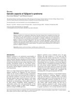
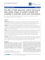
affect cytokine secretions. As shown in Figure 1, C57BL/
6J mice treated with the Ad5-IL17A vector at 10
7
viral
particles per salivary gland exhibited a marked increase
in the levels of serum IL-17A compared to baseline
levels or wit h C57BL/6J mice receiving the control Ad5-
LacZ vector at 10
7
viral particles per salivary gland,
demonstrating the efficacy of this viral vector to produce
IL-17A. In addition, Ad5-IL17A-treated C57BL/6J mice
also secreted elevated amounts of the IL-17A-related
cytokine IL-6 following cannulation. Thus, the vectors
gain access into the glands and appare ntly secrete IL-
17A in quantities that elevate systemic levels.
Increased numbers of IL-17A-producing CD4+ T cells in
the spleens of Ad5-IL17A transduced mice
As mentioned previously, salivary glands were cannu-
lated with Ad5-IL17A vector at either 7 wks or 16 wks
of age. The time points chosen are based on extensive
studies of the development and onset of disease in
our C57BL/6.NOD-Aec1Aec2 mouse model of SS
[1-3,30,31]. The t wo time points selected represent the
innate and adaptive immune response phases, respec-
tively, in the disease model, thus they were chosen to
mimic these changes in the parental C57BL/6 mouse.
Microarray analyses examined the temporal differential
gene expression of salivary and lacrimal glands of
C57BL/6 mice revealed gradual change in pathophysio-
logical related genes from 16 to 20 wks of age, concomi-
tantly, leukocyte infiltration in the exocrine glands is
oftenobservedattheseages[32,33].Thus,itisimpor-
tant to examine the role of IL17A in the development of
SS prior and post to any pathophysiological changes.
Mice treated with Ad5-IL17A or Ad5-LacZ at either
7 wks or 16 wks of age were euthanized at 26 and 27
wks of age, that is, 19 wks and 11 wks post-treatment,
respectively. Splenocytes were isolated from individual
miceandexaminedforthenumberofIFN-g and
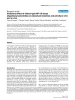
IL-17A secreting CD4+T cells. Representative data, pre-
sented in Figure 2b, c, revealed that the number of IL-
17A secreting CD4+T cells in the spleens of mice
receiving the Ad5-IL17A vector at seven weeks of age
was approximately two-fold higher than mice receiving
the control Ad5-LacZ vector, while the number of IFN-g
secreting CD4+T cells was approximately half at time of
analysis. Similarly, the number of IL-17A secreting CD4
+T cells in the spleens of mice receiving the Ad5-IL17A
vector at 16 wks of age was approximately seven-fold
higher than mice receiving the control Ad5-LacZ vector,
while the number of IFN-g secreting CD4+T cells was
less than 50% at time of analysis (Figure 2e, f). Results
of a similar analysis with untreated mice performed one
week prior to vector cannulations are presented in
Figures 2a, d. These data suggest that even though the
Ad5 vector is considered locally restricted, the effec t in
C57BL/6 J mice appeared systematic. More importantly,
the systemic effects of IL17A in Ad5 appears to be cor-
related with the duration of ge ne expression after vector
Figure 1 Rapid changes in IL-17A and IL-6 serum cytokine
concentrations in C57BL/6J mice following vector cannulations.
Sera were prepared from blood collected from individual five-week
old mice (n = 4) randomly chosen one week prior to vector
treatment (Day 0 on the graph). Mice were allowed to acclimate for
seven days, followed by vector instillation of each salivary gland
with 50 μl of vector solution containing 10
7
viral particles of either
Ad5-LacZ or Ad5-IL17A vector. Sera were again prepared from
blood collected from individual mice (n = 11) at Day 5 and Day 12
post-treatment. Concentrations of cytokines were determined using
the Luminex platform. To ensure sufficient quantities for testing, the
sera of three individual mice of each experimental group were
pooled. ND, not detected indicates levels below threshold
detection.
Nguyen et al. Arthritis Research & Therapy 2010, 12:R220
/>Page 4 of 11
cannulation as evidenced by the two-fold increase in the
levels of IL-17A secreting cells at 19 wks post-treatment
in younger mice but a seven-fold increase at 11 wks
post-treatment in the older group. However, one cannot
rule out the possibility that different efficacies are
achieved based on the stat us of disease development in
different ages of mice.
Induction of SS immune-pathology in C57BL/6 mice
following treatment with Ad5-IL17A vector
Lymphocyte infiltration of the salivary and/or lacrimal
glands is a critical criterion for identification of the
autoimmune phase of SS in both human and animal
models. Although the number of LF present in the sali-
vary and lacrimal glands does not often c orrelate
directly with disease or its severity, SS patients and
NOD-derived mouse strains exhibiting SS-like disease
typically have lymphocytic infiltrates in their salivary
glands. IL-17A appears to play a critical role in
the development of LF and has recently been found to
be present within LF in both SS patients and animal
models [8]. Salivary glands of C57BL/6J mice following
cannulation with Ad5-IL17A vector were examined for
thepresenceofinfiltratingleukocytes. Salivary glands
retrieved from C57BL/6J mice treated with Ad5-LacZ
vector at either 7 or 16 wks of age revealed that 10% (1
of 10) in each group had evidence of glandular infiltra-
tions(Figure3a,b,g,h;Table1).Thisobservationis
consistent with the number of healthy, untreated
C57BL/6J mice expected to have infiltration of the
Figure 2 Intracellular staining for IL-17A and IFN-g secreting CD4
+
T cells in spleens of Ad5-IL17A-treated mice. Splenic leukoc ytes
prepared from C57BL/6J mice (n = 4) at 6 wks of age (one wk prior to vector treatment) and 26 wks old (19 wks post vector treatment),
considered early treatment (a-c), or splenic leukocytes prepared from C57BL/6J mice (n = 4) at 15 wks of age (1 wk prior to vector treatment)
and 27 wks old (11 wks post vector treatment), considered late treatment (d-f) were examined for the presence of intracellular IL-17A and IFN-g
gated on CD4
+
T cells following a 5-hr in-vitro activation with anti-CD3ε and anti-CD28 in Leukocyte Activation Cocktail containing GolgiPlug.
Flow cytometric acquisition was performed by staining with PE-Cy5-conjugated rat anti-CD4, FITC-conjugated rat anti-IFNg and/or PE-conjugated
rat anti-IL-17A. Data were analyzed by FCS Express. Flow cytometric images shown are from one representative analysis of two independent
experiments that examined two different mice within each experiment. Data presented as mean ± SEM for n = 4 per group and statistical
analyses were performed comparing the means of the Ad-LacZ and Ad-IL17A treated groups at 26 wks and 27 wks of early and late treatment,
respectively. (*) indicates P < 0.5 using the Mann-Whitney U test.
Nguyen et al. Arthritis Research & Therapy 2010, 12:R220
/>Page 5 of 11
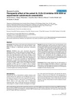
salivary glands [8]. In contrast, salivary glands from
C57BL/6J mice treated with Ad5-IL17A vector at seven
weeks of age showed infiltrations in 91% (10 of 11) with
the mean LF per histological section numbering 4 ±
1.32, while salivary glands from C57BL/6J mice treated
with Ad5-IL17A vector at 16 wks of age revealed
infiltrations in 75% (6 of 8) with a mean LF number per
histological section of 2 ± 0.83 (Table 1).
Besides the number of LF detected in the salivary glands
of the experimental ani mals, immunofluorescent stai ning
to detect B and T cells revealed further differences in the
cellular composition of the infiltrations between mice
administered Ad5-IL17A at an early or late stage. At time
of euthanasia, C57BL/6J mice treated with Ad5-IL17A
vector at 7 wks of age generally exhibited smaller foci con-
taining fewer IL-17 positive cells compared to mice receiv-
ing the vector at 16 wks of age (Figure 3c-f, i-l). Consistent
with previous observation, the smaller foci in mice treated
at 7 wks of age may have resulted from the l onger dura-
tion of time after cannulation (19 wks) reflecting the
decreases in IL-17A serum levels and IL-17A- positive cell
numbers. Detailed examination of IL-17A-positive cells
revealed that a majority of IL-17A cells are present in the
LF and ductal c ells with smaller percentage of positive
Figure 3 Histological examination of salivary glands. Salivary gland histology was examined at 19 wks post-vector infusions of mice treated
at 7 wks of age (early treatment) or at 11 wks post-vector infusions of mice treated at 16 wks of age (late treatment). Panels show
representative H&E staining of salivary gland tissue from mice receiving early treatment with Ad5-LacZ (n = 10) (a), or Ad5-IL17A (n = 11)
(b); fluorescent staining and enumeration of B and T cells in Ad5-IL17A treated mice (c and d) and immunohistochemical staining and
enumeration of IL-17A-positive cells in Ad5-IL17A treated mice (e and f); H&E staining of salivary gland tissue from mice receiving late treatment
with Ad5-LacZ (n = 10) (g), or Ad5-IL17 (n =8)(h); and fluorescent staining and enumeration of B and T cells in Ad5-IL17A treated mice
(i and j) and immunohistochemical staining and enumeration of IL-17A-positive cells in Ad5-IL17A treated mice (k and l). Black arrows indicate
representative lymphocytic infiltrate.
Table 1 Quantification of lymphocytic foci (LF) in salivary
glands
Ad5:LacZ Ad5:IL17A
No LF LF Mean LF No LF LF Mean LF
Early 9
a
(90%)
b
1 (10%) 1 1 (9%) 10 (91%) 4 ± 1.32
c
Late 9 (90%) 1 (10%) 1 2 (25%) 6 (75%) 2 ± 0.83
a
number of mice.
b
percentage of mice.
c
mean number of LF ± SEM per histological salivary gland section.
Ad5, Adenovirus serotype 5; IL, interleukin; LF, lymphocytic foci.
Nguyen et al. Arthritis Research & Therapy 2010, 12:R220
/>Page 6 of 11
cells found in the epithelium and acinar cells. Neverthe-
less, these data support the concept that formation and
maintenance of LF are due, in part, to the expression levels
of IL17A in the salivary glands.
Changes in ANA profiles following instillation of the
Ad5-IL-17A vector
With the appearance of B and T lymphocytes within the
salivary glands of Ad5-IL17A treated C57BL/6 mice, plus
the significant changes within their splenic T
H
17 and
T
H
1 cell populations, the presence of circulating autoan -
tibodies, specifically ANA, detectable by staining of HEp-
2 cells was examined. To id entify the presence of ANA,
the sera prepared from blood samples collected from
each C57BL/6J mouse b oth pre- and post-cannulation
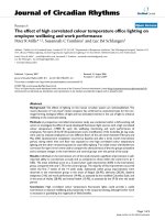
were tested for reactivity on HEp-2 cells. As presented in
Figure 4a, the sera collected from C57BL/6J mice at six
weeks of age or one week prior to vector treatment
showed a general weakly diffused cytoplasmic and
nuclear background staining of the individual target cells.
However, sera collected 19 wks post-treatment from
mice treated with Ad5-IL17A vector at 7 w ks of age
showed no cytoplasmic staining with course speckled
staining and negative nucleoli, while Ad5-LacZ treated
mice exhibited diffused cytoplasmic staining, weak but
fine speckled nucleoplasmic staining with negative
nucleoli (Figures 4b, c). Similar results were seen in
C7BL/6J mice whose salivary glands were transduced
with Ad5-IL17A vector at 16 wks of age in which the pat-
tern was pronounced course speckled staining with no
cytoplasmic staining and negative nucleoli at 29 wks of
age, or 11 wks post-treatment (Figures 4d-f). Considering
the functions of IL-17A, it is interesting to see a gradual
and subtle change in ANA profile from diffused cytoplas-
mic/nuclea r pattern to a distinct course nuclear speck led
pattern, suggesting influence of IL-17A on the B cells
repertoire.
Induction of salivary gland dysfunction in C57BL/6J mice
following cannulation with Ad5-IL17A vector
To determine if the expression of exogenous IL-17A can
induce salivary gland dysfunction, saliva volumes for each
Figure 4 Identification of the antinuclear antibodies in sera of C57BL/6J mice. Representative patterns of cellular staining of HEp-2 cells by
sera diluted at 1:40 prepared from sera taken from C57BL/6 mice cannulated with Ad5-LacZ or Ad5-IL17A vectors at 7 wks of age with pre-
treated mice (baseline) at 6 wks of age (n =4)(a-c), and cannulated at 16 wks of age with Ad5-LacZ or Ad5-IL17A and pre-treated mice
(baseline) at 15 wks of age (n =4)(d-f) with negative control using secondary antibody only (g) and positive control with standard nuclear
speckled serum (h). Representative patterns were determined with n = 4 for two baselines and n = 7 for each time point presented in the
figure. Fixed HEp-2 substrate slides were incubated with individual mouse sera diluted 1:40, 1:80 and 1:160 followed by development with FITC-
conjugated goat anti-mouse IgG. Fluorescent patterns were detected by fluorescence microscopy at 400X magnification.
Nguyen et al. Arthritis Research & Therapy 2010, 12:R220
/>Page 7 of 11
mouse were measured at one week prior to treatment,
then at three- to five-wee k intervals post-cannulation.
C57BL/6J mice that received control Ad5-L acZ vector at
seven weeks of age exhibited stable stimulated saliva
volumes at seven weeks post treatment with a statistically
non-significant increase in saliva volumes at 11 weeks
post treatment. Nevertheless, C57BL/6J mice whose sali-
vary glands were cannulated at seven weeks of age with
Ad5-IL17A exhibited a significa nt and relatively ra pid
decrease in stimulated sa liva volumes that was most pro-
nounced at seven weeks post treatment, and this observa-
tion is seen even if the saliva volumes are converted to
saliva flow rates based on weights of the mice. After
seven weeks post treatment, t hese mice showed a slight
recovery (Figure 5a). Similar results were observed with
C57BL/6J mice cannulated at 16 wks of age with Ad5-
LacZ and Ad5-I L17A vec tors; however, no saliva volume
recovery was observed at time of euthanization (that is,
11 wks post-treatment) (Figure 5b). Whether a reversal
of this inhibition would occur in these older an imals will
require further studies. Thus, saliva secretions of mice
receiving the Ad5-IL17A vector were significantly
decreased one to two months post-treatment when com-
pared to secretions of mice receiving the Ad5-LacZ
vector.
Discussion
The T
H
17-derived IL-17A cytokine is a potent inflam-
matory cytokine that has been implicated in a growing
list of autoimmune diseases, for example, multiple
sclerosis, Crohn’s disease, rheumatoid arthritis, psoriasis,
systemic lupus e rythematosus, and SS, as well as auto-
immunity in animal models [3]. As the T
H
17/IL-17A
system is considered to be an important factor in innate
immunity that, in turn, regulates development of the
adaptive immune response, it is not surprising that
environmental microflora trigger IL-17A responses [34].
The consequence of T
H
17/IL-17A activation includes, in
addition to the production the IL-17A family of
cytokines, the production of IL-21, IL-22, chemokines
(MIP-2, CXCL1, CXCL2, CXCL5), and matrix metallo-
proteases (MMP3 and MMP13) [16] all actively involved
in tissue inflammation. Interact ion of the IL-17A with
its receptors evokes activation of IL-8, resulting in
recruitment of neutrophils to the site of injur y. How-
ever, the relationship between such early innate/inflam-
matory events mediated by the T
H
17/IL-17A system and
theroleT
H
17 cells play in subsequent autoimmunity
remains unknown, especially in light of the multiple
functions now associated with the T
H
17 cell popula-
tions. Thus, in the present study, we have attempted to
elucidate the importance of the cytokine IL-17A per se
in the development of SS and whether its function may
be dependent on when it is expressed.
Results in which SS-non-susceptible C57BL/6J mice
were cannulated with the Ad5-IL17A vector revealed that
increased IL-17A expression could induce several patho-
logical features of SS, irrespective of whether the mice
Figure 5 Stimulated saliva flow in treated C57BL/6J mice. One week prior to salivary gland cannulations with either Ad5-LacZ or Ad5-IL17A
vector, stimulated saliva volumes were determined for individual mice within each of the four experimental groups: early treatment with Ad5-
LacZ (n = 10) or Ad5-IL17A (n = 11) at 7 wks of age (a) or late treatment with Ad5-LacZ (n = 10) or Ad5-IL17A (n = 8) at 16 wks of age (b).
Saliva was collected every three to five weeks post-treatment until the mice were euthanized. Statistical analysis was used to determine the
significance between the Ad5-LacZ and Ad5-IL17A treated mice at each time point. (NS: not significant, P = *< 0.05, P = **< 0.01, P = ***<
0.001). Arrows indicate the initial time point of vector cannulation.
Nguyen et al. Arthritis Research & Therapy 2010, 12:R220
/>Page 8 of 11
received the vector at 7 or 16 wks of age, two time points
corresponding to innate and adaptive im mune responses
in SS-susceptible C57BL/6.NOD-Aec1Aec2 mice. This
was noted by decreases in saliva produ ction compared to
control vector, elevated production of specific pro-
inflammatory cytokines detected in sera, changes in the
weak cytoplasmic/nuclear ANA patterns to nuclear
specked staining on HEp2 cells and increased numbers of
LF and IL17A positive cells present in the salivary glands
at time of euthanasia. Interestingly, mice received Ad5-
IL17A at 7 wks of age showed a sli ght recovery of saliva
secretion at 7 wks of treatment in contrast to mice
received Ad5-IL17A at 16 wks of age. This observation
might be supported by the differential immunological or
biological response of mice at different ages and the
effect of Ad5-IL17A exerted on the mice.
Previous studies have indicated that genes placed
within Ad5 vectors are generally expressed transiently
and locally restricted (that is, 7 to 14 days) [29]. The
present study demonstrates that a rapid and significant
increase in the levels of plasma IL-17A was affected at
12 days post-cannulation by the A d5-IL17A transgene
vector. Interestingly, this systemic increase in IL17 cyto-
kine levels correlated with significant increases in sple-
nic IL-17A secreting CD4+T cells that persisted at least
19 wks for mice treated at 7 wks of age and 11 wks for
mice treated at 16 wks of age. These observations indi-
cated that the Ad5 vector effect was longer t han antici-
pated. Whether this effect might be due to an indirect
secondary effect of the Ad5-IL17 vector is unknown . In
addition, the systemic increase in IL17A production by
local treatment of Ad5-IL17A presented in this study is
consistent wi th previous studies by Bruce Baum’ s
laboratory [35-38]. Adesanya et al.[39]hasdemon-
strated that acinar cells can be punctured by retrograde
salivary gland cannulation at a certain vector dosage.
The injured acinar cells, which have compromised
mucosa l barrier integrity, allow for leakage of the vector
systemically. Further studies by Kagami et al.[37]and
He et al. [40] provided evidence that ductal cannulation
of salivary glands can also have systemic effects due to
the secretory nature of the salivary glands which are
well endowed with protein synthesis organelles and
secretory machinery.
Nevertheless, these observations are consistent with
the concept that SS develops along specific biological
processes in a sequential fashion and interference with
this process alters development of disease [1-3]. There-
fore, this study clearly indicates the pathogenic nature
of IL-17A in inducing SS-like phenotypes when cannu-
lated in the salivary glands.
Previous data have shown that lymphocytic infiltrates
in the salivary glands secreting IL-17A and its related
cytokines are more important in local glandular
destruction. Staining salivary glands for IL-17A revealed
that C57BL/6J mice receiving Ad5-IL17A vector not
only expressed significant levels of IL-17A, but that IL-
17A levels correlated with recruitment of inflammatory
cells, specifically B and T cells, to the glands. This
observation is important in light of the recent study sug-
gesting IL-17A is a critical factor in the a daptive
immune response by inducing the formation of germinal
centers for the production of autoreactive antibodies
[24]. Autoantibodies represent a major component in
the onset of SS, thus the changes in the ANA profiles
observed with sera of C57BL/6J mice cannulated with
the Ad5-IL17A vector indicate that IL-17A affects even
the B cell compartment in SS-non-susceptible mice. The
presence of LF and loss of saliva secretion raises an
interestin g question about the possible role of IL-17A in
B cell activation. As BAFF is capable of inducing T
H
17
cell differentiation in addition to regulating B cell activa-
tion [41], the possible role of BAFF and IL17A in this
phenomenon needs to be better defined in SS
pathogenesis.
Conclusions
The capability of IL-17A to induce features of SS in SS-
non-susceptible mice demonstrates the major role this
cytokine plays in the development, and possible onset,
of the autoimmune process. How this one cytokine
affects the various features of autoimmunity, and at
what level or time point, will require additional studies.
More importantly, the study demonstrates that IL-17A
might be a potential therapeutic target for SS.
Abbreviations
Ad5: adenovirus serotype 5; ANA: antinuclear antibodies; BAFF: B cell
activating factor; CIA: collagen-induced arthritis; CXCL1: chemokine (C-X-C
motif) ligand; EAE: experimental autoimmune encephalomyelitis; IFN-γ:
interferon-γ; IL: interleukin; LF: lymphocytic focus; MIP-2: macrophage
inflammatory protein-2; MMP: matrix metalloproteases; SS: Sjögren’ s
syndrome.
Acknowledgements
The authors would like to thank Dr. Jay K. Kolls and Dr. Julie Bindas
(Children’s Hospital of Pittsburgh) for generously providing the Ad5-LacZ
and Ad5-IL17A vectors and Dr. Phil Cohen for his critical reading of the
manuscript and helpful suggestions. We greatly appreciate the assistance of
Dr. Craig Meyers and Dr. Nicholas Muzyczka for the use of the microscope.
Publication of this article was funded in part by the University of Florida
Open-Access publishing Fund.
Funding: This work was supported by PHS grants K99DE018958 (CQN) from
NIDCR, R21AI081952 (ABP) from NIAID and funds from the Sjögren’s
Syndrome Foundation and Center for Orphan Autoimmune Disorders. HY
and JAC were supported by an NIH, NIDCR intramural research grant.
Author details
1
Eli and Edythe L. Broad Institute, 7 Cambridge Center, Cambridge, MA
02142, USA.
2
Department of Chemical Engineering, Massachusetts Institute
of Technology, 77 Massachusetts Ave, E25-545, Cambridge MA 02139, USA.
3
Department of Oral Biology, University of Florida College of Dentistry, 1600
SW Archer Rd, Gainesville, FL 32610, USA.
4
Center for Orphan Autoimmune
Disorders, University of Florida College of Dentistry, 1600 SW Archer Rd,
Nguyen et al. Arthritis Research & Therapy 2010, 12:R220
/>Page 9 of 11
Gainesville, FL 32610, USA.
5
National Institute of Dental and Craniofacial
Research, NIH, 10 Center Drive MSC 1190, Bethesda, MD 20892, USA.
6
Department of Pathology, Immunology & Laboratory Medicine, University of
Florida College of Medicine, 1600 SW Archer Rd, Gainesville, FL 32610, USA.
Authors’ contributions
JAC produced and determined the titers of the Ad5-LacZ and Ad5-IL17A
viral vectors. HY and BL performed retrograde ductal cannulations/
instillations of the vectors into the salivary glands. CQN designed the study,
performed saliva flow, flow cytometry, histology and statistical analyses, and
prepared the manuscript. WC carried out the ANA staining. ABP assisted in
the manuscript preparation. All authors read and approved the final
manuscript.
Competing interests
The authors declare that they have no competing interests.
Received: 20 September 2010 Revised: 30 November 2010
Accepted: 23 December 2010 Published: 23 December 2010
References
1. Nguyen CQ, Cha SR, Peck AB: Sjögren’s syndrome (SjS)-like disease of
mice: the importance of B lymphocytes and autoantibodies. Frontiers in
Bioscience 2007, 12:1767-1789.
2. Nguyen CQ, Peck AB: Unraveling the pathophysiology of Sjogren
syndrome-associated dry eye disease. Ocul Surf 2009, 7:11-27.
3. Lee BH, Tudares MA, Nguyen CQ: Sjogren’s syndrome: an old tale with a
new twist. Arch Immunol Ther Exp (Warsz) 2009, 57:57-66.
4. Brayer JB, Cha S, Nagashima H, Yasunari U, Lindberg A, Diggs S, Martinez J,
Goa J, Humphreys-Beher MG, Peck AB: IL-4-dependent effector phase in
autoimmune exocrinopathy as defined by the NOD.IL-4-gene knockout
mouse model of Sjogren’s syndrome. Scand J Immunol 2001, 54:133-140.
5. Cha S, Brayer J, Gao J, Brown V, Killedar S, Yasunari U, Peck AB: A dual role
for interferon-gamma in the pathogenesis of Sjogren’s syndrome-like
autoimmune exocrinopathy in the nonobese diabetic mouse. Scand J
Immunol 2004, 60:552-565.
6. Nguyen KH, Brayer J, Cha S, Diggs S, Yasunari U, Hilal G, Peck AB,
Humphreys-Beher MG: Evidence for antimuscarinic acetylcholine receptor
antibody-mediated secretory dysfunction in nod mice. Arthritis Rheum
2000, 43:2297-2306.
7. Nguyen CQ, Gao JH, Kim H, Saban DR, Cornelius JG, Peck AB: IL-4-STAT6
signal transduction-dependent induction of the clinical phase of
Sjogren’s syndrome-like disease of the nonobese diabetic mouse. J
Immunol 2007, 179:382-390.
8. Nguyen CQ, Hu MH, Li Y, Stewart C, Peck AB: Salivary gland tissue
expression of interleukin-23 and interleukin-17 in Sjogren’s syndrome:
Findings in humans and mice. Arthritis Rheum 2008, 58:734-743.
9. Sakai A, Sugawara Y, Kuroishi T, Sasano T, Sugawara S: Identification of IL-
18 and Th17 cells in salivary glands of patients with Sjogren’s
syndrome, and amplification of IL-17-mediated secretion of
inflammatory cytokines from salivary gland cells by IL-18. J Immunol
2008, 181:2898-2906.
10. Harrington LE, Hatton RD, Mangan PR, Turner H, Murphy TL, Murphy KM,
Weaver CT: Interleukin 17-producing CD4+ effector T cells develop via a
lineage distinct from the T helper type 1 and 2 lineages. Nat Immunol
2005, 6:1123-1132.
11. Park H, Li Z, Yang XO, Chang SH, Nurieva R, Wang YH, Wang Y, Hood L,
Zhu Z, Tian Q, Dong C: A distinct lineage of CD4 T cells regulates tissue
inflammation by producing interleukin 17. Nat Immunol 2005,
6
:1133-1141.
12. Veldhoen M, Hocking RJ, Atkins CJ, Locksley RM, Stockinger B: TGFbeta in
the context of an inflammatory cytokine milieu supports de novo
differentiation of IL-17-producing T cells. Immunity 2006, 24:179-189.
13. Bettelli E, Carrier Y, Gao W, Korn T, Strom TB, Oukka M, Weiner HL,
Kuchroo VK: Reciprocal developmental pathways for the generation of
pathogenic effector TH17 and regulatory T cells. Nature 2006,
441:235-238.
14. Mangan PR, Harrington LE, O’Quinn DB, Helms WS, Bullard DC, Elson CO,
Hatton RD, Wahl SM, Schoeb TR, Weaver CT: Transforming growth factor-
beta induces development of the T(H)17 lineage. Nature 2006,
441:231-234.
15. Ivanov II, McKenzie BS, Zhou L, Tadokoro CE, Lepelley A, Lafaille JJ, Cua DJ,
Littman DR: The orphan nuclear receptor RORgammat directs the
differentiation program of proinflammatory IL-17+ T helper cells. Cell
2006, 126:1121-1133.
16. Weaver CT, Hatton RD, Mangan PR, Harrington LE: IL-17 family cytokines
and the expanding diversity of effector T cell lineages. Annu Rev
Immunol 2007, 25:821-852.
17. Kastelein RA, Hunter CA, Cua DJ: Discovery and biology of IL-23 and IL-27:
related but functionally distinct regulators of inflammation. Annu Rev
Immunol 2007, 25:221-242.
18. Zhou L, Lopes JE, Chong MM, Ivanov II, Min R, Victora GD, Shen Y, Du J,
Rubtsov YP, Rudensky AY, Ziegler SF, Littman DR: TGF-beta-induced Foxp3
inhibits T(H)17 cell differentiation by antagonizing RORgammat function.
Nature 2008, 453:236-240.
19. Duerr RH, Taylor KD, Brant SR, Rioux JD, Silverberg MS, Daly MJ,
Steinhart AH, Abraham C, Regueiro M, Griffiths A, Dassopoulos T, Bitton A,
Yang H, Targan S, Datta LW, Kistner EO, Schumm LP, Lee AT, Gregersen PK,
Barmada MM, Rotter JI, Nicolae DL, Cho JH: A genome-wide association
study identifies IL23R as an inflammatory bowel disease gene. Science
2006, 314:1461-1463.
20. Hue S, Ahern P, Buonocore S, Kullberg MC, Cua DJ, McKenzie BS, Powrie F,
Maloy KJ: Interleukin-23 drives innate and T cell-mediated intestinal
inflammation. J Exp Med 2006, 203:2473-2483.
21. Cua DJ, Sherlock J, Chen Y, Murphy CA, Joyce B, Seymour B, Lucian L, To W,
Kwan S, Churakova T, Zurawski S, Wiekowski M, Lira SA, Gorman D,
Kastelein RA, Sedgwick JD: Interleukin-23 rather than interleukin-12 is the
critical cytokine for autoimmune inflammation of the brain. Nature 2003,
421:744-748.
22. Ye P, Rodriguez FH, Kanaly S, Stocking KL, Schurr J, Schwarzenberger P,
Oliver P, Huang W, Zhang P, Zhang J, Shellito JE, Bagby GJ, Nelson S,
Charrier K, Peschon JJ, Kolls JK: Requirement of interleukin 17 receptor
signaling for lung CXC chemokine and granulocyte colony-stimulating
factor expression, neutrophil recruitment, and host defense. J Exp Med
2001, 194:519-527.
23. Schwarzenberger P, La Russa V, Miller A, Ye P, Huang W, Zieske A, Nelson S,
Bagby GJ, Stoltz D, Mynatt RL, Spriggs M, Kolls JK: IL-17 stimulates
granulopoiesis in mice: use of an alternate, novel gene therapy-derived
method for in vivo evaluation of cytokines. J Immunol 1998,
161:6383-6389.
24. Hsu HC, Yang P, Wang J, Wu Q, Myers R, Chen J, Yi J, Guentert T,
Tousson A, Stanus AL, Le TV, Lorenz RG, Xu H, Kolls JK, Carter RH,
Chaplin DD, Williams RW, Mountz JD: Interleukin 17-producing T helper
cells and interleukin 17 orchestrate autoreactive germinal center
development in autoimmune BXD2 mice. Nat Immunol 2008, 9:166-175.
25. Zheng C, Baum BJ: Evaluation of promoters for use in tissue-specific
gene delivery. Methods Mol Biol 2008, 434:205-219.
26. Kok MR, Yamano S, Lodde BM, Wang J, Couwenhoven RI, Yakar S,
Voutetakis A, Leroith D, Schmidt M, Afione S, Pillemer SR, Tsutsui MT,
Tak PP, Chiorini JA, Baum BJ: Local adeno-associated virus-mediated
interleukin 10 gene transfer has disease-modifying effects in a murine
model of Sjogren’s syndrome. Hum Gene Ther 2003, 14:1605-1618.
27. Kok MR, Voutetakis A, Yamano S, Wang J, Cotrim A, Katano H, Bossis I,
Chiorini JA, Tran SD, Tak PP, Baum BJ: Immune responses following
salivary gland administration of recombinant adeno-associated virus
serotype 2 vectors. J Gene Med 2005, 7:432-441.
28. Lodde BM, Mineshiba F, Wang J, Cotrim AP, Afione S, Tak PP, Baum BJ:
Effect of human vasoactive intestinal peptide gene transfer in a murine
model of Sjogren’s syndrome. Ann Rheum Dis 2006, 65:195-200.
29. Delporte C, Redman RS, Baum BJ: Relationship between the cellular
distribution of the alpha(v)beta3/5 integrins and adenoviral infection in
salivary glands. Lab Invest 1997, 77:167-173.
30. Cha S, Nagashima H, Brown VB, Peck AB, Humphreys-Beher MG: Two NOD
Idd-associated intervals contribute synergistically to the development of
autoimmune exocrinopathy (Sjogren’s syndrome) on a healthy murine
background. Arthritis Rheum 2002, 46:1390-1398.
31. Brayer J, Lowry J, Cha S, Robinson CP, Yamachika S, Peck AB, Humphreys-
Beher MG: Alleles from chromosomes 1 and 3 of NOD mice combine to
influence Sjogren’s syndrome-like autoimmune exocrinopathy. J
Rheumatol 2000, 27:1896-1904.
32. Nguyen CQ, Sharma A, Lee BH, She JX, McIndoe RA, Peck AB: Differential
gene expression in the salivary gland during development and onset of
Nguyen et al. Arthritis Research & Therapy 2010, 12:R220
/>Page 10 of 11
xerostomia in Sjogren’s syndrome-like disease of the C57BL/6.NOD-
Aec1Aec2 mouse. Arthritis Res Ther 2009, 11:R56.
33. Nguyen CQ, Sharma A, She JX, McIndoe RA, Peck AB: Differential gene
expressions in the lacrimal gland during development and onset of
keratoconjunctivitis sicca in Sjogren’s syndrome (SJS)-like disease of the
C57BL/6.NOD-Aec1Aec2 mouse. Exp Eye Res 2009, 88:398-409.
34. Bettelli E, Korn T, Oukka M, Kuchroo VK: Induction and effector functions
of T(H)17 cells. Nature 2008, 453:1051-1057.
35. Zheng C, Voutetakis A, Kok MR, Goldsmith CM, Smith GB, Elmore S,
Nyska A, Vallant M, Irwin RD, Baum BJ: Toxicity and biodistribution of a
first-generation recombinant adenoviral vector, in the presence of
hydroxychloroquine, following retroductal delivery to a single rat
submandibular gland. Oral Dis 2006, 12:137-144.
36. O’Connell BC, Zheng C, Jacobson-Kram D, Baum BJ: Distribution and
toxicity resulting from adenoviral vector administration to a single
salivary gland in adult rats. J Oral Pathol Med 2003, 32:414-421.
37. Kagami H, O’Connell BC, Baum BJ: Evidence for the systemic delivery of a
transgene product from salivary glands. Hum Gene Ther 1996,
7:2177-2184.
38. Wang J, Voutetakis A, Mineshiba F, Illei GG, Dang H, Yeh CK, Baum BJ:
Effect of serotype 5 adenoviral and serotype 2 adeno- associated viral
vector-mediated gene transfer to salivary glands on the composition of
saliva. Hum Gene Ther 2006, 17:455-463.
39. Adesanya MR, Redman RS, Baum BJ, O’Connell BC: Immediate
inflammatory responses to adenovirus-mediated gene transfer in rat
salivary glands. Hum Gene Ther 1996, 7:1085-1093.
40. He X, Goldsmith CM, Marmary Y, Wellner RB, Parlow AF, Nieman LK,
Baum BJ: Systemic action of human growth hormone following
adenovirus-mediated gene transfer to rat submandibular glands. Gene
Ther 1998, 5:537-541.
41. Doreau A, Belot A, Bastid J, Riche B, Trescol-Biemont MC, Ranchin B,
Fabien N, Cochat P, Pouteil-Noble C, Trolliet P, Durieu I, Tebib J, Kassai B,
Ansieau S, Puisieux A, Eliaou JF, Bonnefoy-Berard N: Interleukin 17 acts in
synergy with B cell-activating factor to influence B cell biology and the
pathophysiology of systemic lupus erythematosus. Nat Immunol 2009,
10:778-785.
doi:10.1186/ar3207
Cite this article as: Nguyen et al.: Pathogenic effect of interleukin-17A in
induction of Sjögren’s syndrome-like disease using adenovirus-
mediated gene transfer. Arthritis Research & Therapy 2010 12:R220.
Submit your next manuscript to BioMed Central
and take full advantage of:
• Convenient online submission
• Thorough peer review
• No space constraints or color figure charges
• Immediate publication on acceptance
• Inclusion in PubMed, CAS, Scopus and Google Scholar
• Research which is freely available for redistribution
Submit your manuscript at
www.biomedcentral.com/submit
Nguyen et al. Arthritis Research & Therapy 2010, 12:R220
/>Page 11 of 11