Báo cáo y học: "(Sub)clinical cardiovascular disease is associated with increased bone loss and fracture risk; a systematic review of the association between cardiovascular disease and osteoporosis" ppsx
Bạn đang xem bản rút gọn của tài liệu. Xem và tải ngay bản đầy đủ của tài liệu tại đây (471.47 KB, 19 trang )
den Uyl et al. Arthritis Research & Therapy 2011, 13:R5
/>
RESEARCH ARTICLE
Open Access
(Sub)clinical cardiovascular disease is associated
with increased bone loss and fracture risk; a
systematic review of the association between
cardiovascular disease and osteoporosis
Debby den Uyl1, Mike T Nurmohamed2,3*, Lilian HD van Tuyl1, Hennie G Raterman1, Willem F Lems1,3
Abstract
Introduction: Both cardiovascular disease and osteoporosis are important causes of morbidity and mortality in the
elderly. The co-occurrence of cardiovascular disease and osteoporosis prompted us to review the evidence of an
association between cardiovascular (CV) disease and osteoporosis and potential shared common
pathophysiological mechanisms.
Methods: A systematic literature search (Medline, Pubmed and Embase) was conducted to identify all clinical
studies that investigated the association between cardiovascular disease and osteoporosis. Relevant studies were
screened for quality according to guidelines as proposed by the Dutch Cochrane Centre and evidence was
summarized.
Results: Seventy studies were included in this review. Due to a large heterogeneity in study population, design
and outcome measures a formal meta-analysis was not possible. Six of the highest ranked studies (mean n = 2,000)
showed that individuals with prevalent subclinical CV disease had higher risk for increased bone loss and fractures
during follow-up compared to persons without CV disease (range of reported risk: hazard ratio (HR) 1.5; odds ratio
(OR) 2.3 to 3.0). The largest study (n = 31,936) reported a more than four times higher risk in women and more
than six times higher risk in men. There is moderate evidence that individuals with low bone mass had higher CV
mortality rates and incident CV events than subjects with normal bone mass (risk rates 1.2 to 1.4). Although the
shared common pathophysiological mechanisms are not fully elucidated, the most important factors that might
explain this association appear to be, besides age, estrogen deficiency and inflammation.
Conclusions: The current evidence indicates that individuals with prevalent subclinical CV disease are at increased
risk for bone loss and subsequent fractures. Presently no firm conclusions can be drawn as to what extent low
bone mineral density might be associated with increased cardiovascular risk.
Introduction
Cardiovascular (CV) disease and osteoporosis are both
important causes of morbidity and mortality in aging
men and women. They share common risk factors, such
as increased age and inactivity, and are frequently found
in the same individuals, suggesting a possible relationship. Results from epidemiological studies indicate an
* Correspondence:
2
Department of Internal Medicine, VU Medical Centre, De Boelelaan 1117,
1081 NV Amsterdam, The Netherlands
Full list of author information is available at the end of the article
association between CV disease and osteoporosis. Prevalent CV disease and subclinical atherosclerosis have
been found to be related to low bone mass and
increased fracture risk [1-4]. Similarly, low bone mineral
density (BMD) has been related to increased cardiovascular risk [5-8]. This relationship is often regarded as a
result of aging; however, recent evidence suggests a
direct association, independent of age and traditional
cardiovascular risk factors and accumulating evidence
from experimental research indicates a shared pathogenesis. A variety of factors that influence bone metabolism
© 2011 den Uyl et al.; licensee BioMed Central Ltd. This is an open access article distributed under the terms of the Creative Commons
Attribution License ( which permits unrestricted use, distribution, and reproduction in
any medium, provided the original work is properly cited.
den Uyl et al. Arthritis Research & Therapy 2011, 13:R5
/>
are involved in the development of vascular disease, for
example, atherosclerosis and vascular calcification. Interestingly, several bone-related proteins are implicated in
the calcification process resulting in mineral deposition
[9]. This is important as calcification of the arterial wall
may be a marker for CV disease and was shown to predict CV events [10]. Given the importance of identifying
a person at risk for CV events or fractures, evidence for
an association of CV disease with osteoporosis might
have implications for screening decisions in patients
with low bone mass and vice versa. This review aims to
summarize all the present clinical literature about the
association between CV disease and osteoporosis and to
describe common pathophysiological mechanisms. The
results of this review are grouped into two topics: clinical results, discussing the relationship between 1) cardiovascular disease and osteoporosis and 2) vice versa. In
addition, the possible pathophysiological links of CV disease and osteoporosis will be discussed.
Materials and methods
Search strategy
A systematic search (in Medline, Pubmed and Embase)
was conducted to identify all clinical studies from 1966
to January 2010 (last updated 8 June 2010) that investigated the association between cardiovascular disease and
osteoporosis. The following search terms for cardiovascular disease were used: cardiovascular diseases, cerebrovascular diseases and peripheral vascular diseases.
These searches were each combined with an osteoporosis search block and duplicates were removed. Searches
were limited to human studies in the English, Dutch
and German languages. The complete Medline search is
available in Additional file 1. In addition, references
from the retrieved articles were scanned for additional
relevant studies.
Selection criteria
Abstracts were screened by one reviewer (DdU) and
studies were included in the review if they fulfilled the
following inclusion criteria: epidemiological studies
(including prospective, cross-sectional, case-control, or
retrospective studies) reporting the association between
CV disease and osteoporosis in the general population
or in patients with prevalent CV disease or low bone
mass. Cardiovascular disease was defined as coronary
heart disease (CHD) (myocardial infarction, angina
pectoris, coronary insufficiency or ischemic heart disease), cerebrovascular disease (stroke, transient
ischemic attacks), peripheral arterial disease (PAD)
(lower extremity claudication, arterial thrombosis/
embolism, ankle brachial index (ABI) <0.90) or subclinical atherosclerosis measured as intima media thickness (IMT) or vascular calcification. In addition, bone
Page 2 of 19
mass had to be assessed as bone mineral density or
bone quality, and osteoporosis was defined as low
bone mass (T-score ≤-2.5) or increased fracture risk
(vertebral and non-vertebral). Exclusion criteria were:
reviews, letters, case-reports, intervention studies
and biomechanical studies. Studies in patients with
co-morbidity other than osteoporosis or CV disease
were also excluded. Finally, investigations using risk
factors of CV disease or osteoporosis as outcome measurements, such as hypertension, metabolic syndrome,
atrial fibrillation, bone markers, and calcium supplementation were not included.
Assessment of study quality
The quality of each manuscript was systematically
assessed with a checklist for cohort studies as proposed
by the Dutch Cochrane Collaboration [11] (Additional
file 2). Quality assessment included a scoring of the following components: definition of study population, the
likelihood of bias, adequate blinding, the accuracy of
outcome measurements, duration of follow-up and
selective loss-to follow-up, the appropriateness of the
statistical analysis and the clinical relevance. All items
had the following answer options: yes/no/too little information to answer the question. We considered incomplete information or data important criteria for study
quality. Therefore, if the answer could not be given
because the study provided too little information, a
negative score (for example, “no”) was given. Each “no”
was scored and an equal weight was given to each item.
A maximum of 10 points could be given. The scores of
each study are given in Tables 1 and 2.
Statistical analysis
A formal meta-analysis of the prospective studies investigating the association between bone mass and risk for
cardiovascular events and mortality was not possible
due to extended heterogeneity between studies with
respect to the study population and methods used.
Furthermore, the number of prospective studies that
were eligible for pooling was too small for analysis. For
this reason, narrative summaries are provided in the
results section and quantitatively presented in Tables 1
and 2. The heterogeneity between studies in terms of
study population and outcome measures is shown in
Tables 1 and 2. Moreover, cross-sectional studies are
shown in Table 3.
Results
Studies included
Our search strategy resulted in 2,886 references. The
search strategy resulted in 70 relevant articles, including
9 studies prospectively assessing the relationship
between CV disease and osteoporosis and 18 prospective
Table 1 Prospective studies investigating relationship CV disease and low BMD
Study population Number of
(years follow-up) cases (%
women)
Postmenopausal
women
CV
disease
excluded
Mean
age
Outcome CV disease
Outcome bone mass
Results #
Quality
Sennerby,
2009 [13]
Population-based
(20)
31,936
(NA)
NA
Yes
67.9
to
74.4
CV disease by National
patient registry, ICD 9
codes
Incident hip fracture by National
patient registry, ICD 9 codes
Women:
HR: 4.42 (95% CI 3.49 to 5.61)
Men:
HR: 6.65 (95% CI 4.82 to 9.19)
3
Szulc,
2008 [14]
Population-based
(10)
781
(0%)
No
No
65
AC by X-spine
Incident fracture by hospital records or OR: 2.54 to 3.04 (P < 0.005 to
X-ray
0.001)
3
Naves,
2008 [4]
Population-based
(4)
624
(51%)
NA
No
65
AC by X-spine
BMD lumbar spine and femur by DXA
Incident fracture by hospital record or
X-ray
3
Von
Muhlen,
2009 [15]
Population-based
(4)
1,332
(60%)
NA
No
73.8
PAD by ABI
BMD lumbar spine and hip by DXA
and incident fracture by X-ray
Collins,
2009 [2]
Population-based
(5.4)
4,302
(0%)
NA
No
73.5
PAD by ABI
Hak,
2000 [3]
Population-based
(9)
236
(100%)
No (100%)
No
49
AC by X-spine
Samelson,
2007 [12]
Population-based
(21)
2,499
(58%)
No
61
AC by X-spine
Bagger,
2006 [1]
Population-based
(7.5)
2,262
(100%)
Yes (100%)
No
65
AC by X-spine
BMD lumbar spine and hip and
incident fractures by hospital records
or X-ray
Schulz,
2004 [17]
Clinic-based
(8)
228
(100%)
Yes
No
65.2
AC by CT-scan of spine
BMD spine by CT-scan
Change BMD spine in
progression AC vs no
progression AC:
-1.48% vs 1.43% (P <.0001)
Change BMD hip in
progression AC and no
progression AC:
-0.48% vs 0.23% (P = 0.315)
Incident fracture:
OR: 2.13 (95% CI 0.85 to 5.31)
Women:
Change BMD in PAD vs no
PAD:
59.2% vs 43.5% (P < 0.05)
Incident non-vert fracture:
OR: 0.84 (95% CI 0.31 to 2.26)
Men :
Change BMD in PAD vs no
PAD :
43.5% vs 35.5% (P = 0.20)
Incident non-vert fracture:
OR: 1.52 (95% CI 0.30 to 7.45)
BMD hip by DXA
Change BMD in PAD vs no
Incident fractures by x-ray and hospital PAD:
records
-0.60% vs -0.32% (P < 0.001
PAD and non-vert fracture risk:
HR = 1.47 (95% CI 1.07 to 2.04)
MCA by radiogrammetry
MCA in patients with AC
progression vs no AC
progression
-3.5 mm vs -2.0 mm (P < 0.01)
Incident hip fracture by hospital
Women:
records and death certificates
HR: 1.4 (0.8 to 2.3)
Men:
HR: 1.2 (0.2 to 5.7)
3
3
4
4
6
#adjusted for confounders; NA, not available; AC, aortic calcification; BMD, bone mineral density; DXA, dual-energy x-ray absorptiometry; PAD, peripheral arterial disease; ABI, ankle brachial index; MCA, metacarpal cortical area.
Page 3 of 19
Change hip BMD AC score ≥3
vs <3:
-0.38% vs -0.25% (P < 0.001)
AC and hip fracture:
OR: 2.3 (95% CI 1.1 to 4.8)
AC and vert fracture:
OR: 1.2 (95% CI 1.0 to 1.5)
Change BMD AC vs no AC:
-5.3% vs -1.3% (P < 0.001)
3
den Uyl et al. Arthritis Research & Therapy 2011, 13:R5
/>
Study
Study
Study
population
(years
follow-up)
Number
of cases
(%
women)
Postmenopausal
women
CV
Mean Race
disease age
excluded (years)
Mussolino,
2007 [69]
Populationbased
(9)
5,272
(NA)
NA
Yes
60.9 to Caucasian (NA BMD proximal femur by DXA
69.4
%), black and
MexicanAmerican
Farhat,
2007 [6]
Populationbased
(5.4)
2,310
(55%)
Yes
Yes
73.5
Caucasian
(58%) and
black
BMD total hip, femoral neck and
trochanter by DXA
BMD spine by CT-scans
Incident CV disease by Women: BMD fem neck and
incident CV disease: HR: 1.24
hospital records and
(95% CI 1.02 to 1.52)
death certificates
Men: BMD fem neck and
incident CV disease:
HR: 1.04 (95% CI 0.89 to 1.21)
3
Tamaki,
2009 [75]
Populationbased
(10)
609
(100%)
Yes (60%)
No
55.9
Japanese
BMD lumbar spine and total hip
by DXA
IMT values
<10 YSM:
IMT OP vs normal bone mass:
1.55 vs 1.19 (P < 0.05)
≥YSM:
IMT OP vs normal bone mass:
1.53 vs 1.28 (P < 0.05)
3
Browner,
1991 [5]
Populationbased
(2.8)
9,704
(100%)
Yes
No
NA
Caucasian
(99%) and
Asian
BMD distal radius, prox radius
and calcaneus by single photon
absorptiometry
Overall mortality and
CV mortality by death
certificates
BMD and risk overall mortality: 3
RR: 1.22 (95% CI 1.01 to 1.47)
BMD and stroke mortality: RR:
1.75 (95% CI 1.15 to 2.65)
BMD and CV mortality: RR:
1.17 (95% CI 0.92 to 1.51)
Trone,
2007 [68]
Populationbased
(7.6)
1,580
(60%)
Yes (NA %)
No
71.9
Caucasian
Prevalence vertebral fracture by
lateral spine radiographs
Overall mortality by
death certificates
Kado,
2000 [64]
Populationbased
(3.5)
6,018
(100%)
Yes
No
76.5
Caucasian
BMD total hip by DXA
Overall and CV
mortality by death
certificates
Women: prevalent vertebral
3
fracture and overall mortality:
HR: 1.15 (95% CI 0.83 to 1.59)
Men: prevalent vertebral
fracture and overall mortality:
HR: 0.98 (95% CI 0.55 to 1.46)
BMD and overall mortality: RH: 4
1.3 (95% CI 1.1 to 1.4)
BMD and CV mortality: RH: 1.3
(95% CI 1.0 to 1.9)
Trivedi,
2001 [67]
Populationbased
(6.7)
1,002
(0%)
No women
included
No
69.7
NA
BMD total hip by DXA
Overall and CV
mortality by death
certificates
Tanko,
2005 [76]
Clinic-based
(4)
2,576
(100%)
Yes
No
66.5
NA
BMD lumbar spine and femoral
neck by DXA
Incidence CV events
self-reported and
confirmed by primary
documents
den Uyl et al. Arthritis Research & Therapy 2011, 13:R5
/>
Table 2 Prospective studies investigating relationship low BMD and CV disease
Outcome osteoporosis
Outcome CV disease
Results #
CV and stroke
mortality by death
certificates
Women:
3
BMD and CV mortality RR: 1.26
(95% CI 0.88 to 1.80)
BMD and stroke mortality: RR:
1.34 (95% CI 0.86 to 2.07)
Men:
BMD and CV mortality: RR:
1.05 (95% CI 0.79 to 1.39)
BMD and stroke mortality: RR;
0.73 (95% CI 0.43 to 1.23)
4
4
Page 4 of 19
BMD and overall mortality: RR:
0.79 (95% CI 0.65 to 0.97)
BMD and CV mortality: RR:
0.72 (95% CI 0.56 to 0.93)
HR: 3.9 (95% CI 2.0 to 7.7)
Quality
(x nee)
Pinheiro,
2006 [66]
Populationbased
(5)
208
(100%)
Yes
No
75.1
Caucasian
BMD lumbar spine, femoral neck
and trochanter by DXA
Overall and CV
mortality by death
certificates
BMD and overall mortality: HR: 4
1.44 (95% CI 1.06 to 2.21)
BMD and CV mortality: HR:
1.28 (95% CI 1.08 to 2.26)
Johansson,
1998 [7]
Populationbased
(7)
1,468
(56%)
Yes
No
74.0
Caucasian
BMD calcaneus by DPA
Overall mortality by
death certificates
Mussolino,
2003 [65]
Populationbased
(18.5)
3,402
(NA)
NA
Yes
NA
Caucasian
(87%) and
black
BMD phalangeal by single
photon absorption
Stroke mortality by
death certificates
Women: RR: 1.19 (95% CI 1.02
to 1.39)
Men: RR: 1.23 (95% CI 1.10 to
1.41)
Women: RR: 1.01 (95% CI 0.86
to 1.19)
Men: RR: 1.13 (95% CI 0.93 to
1.38)
Blacks: RR : 0.93 (95% CI 0.72
to 1.21)
Samelson,
2004 [70]
Populationbased
(30)
2,059
(60%)
Yes (85,3-94%)
Yes
60.2
NA
Second MCA by radiogrammatry
Kiel, 2001
[77]
Populationbased
(25)
554
(66%)
NA
No
54.4
NA
Second MCA by radiogrammetry
Incidence coronary
heart disease by
hospital records and
death certificates
AC by radiograph of
the lumbar spine
Women: HR: 0.73 (95% CI 0.53 4
to 1.00)
Men: HR: 1.14 (95% CI 0.84 to
1.56)
Women: Sign association %
4
change in MCA and change
AC index (P = 0.01)
Men: No association % change
MCA and change AC index
(P = 0.50)
Browner,
1993 [62]
Populationbased
(1.98)
4,024
(100%)
Yes
Yes
NA
Caucasian
BMD distal radius and calcaneus
by single photon absorptiometry
HR: 1.31 (95% CI 1.03 to 1.67)
5
Von der
Recke,
1999 [8]
Clinic-based
(17)
1,063
(100%)
Yes
Yes
50 and NA
70
BMD distal forearm by single
photon absorptiometry with 125I
source
Clinic-based
(3)
2,565
(100%)
Yes
No
67
Caucasian
(95.8%)
Prevalence vertebral fracture by
lateral spine radiographs
Early menopause: RR: 2.3 (95%
CI 1.0 to 5.3)
Late menopause: RR: 1.3 (95%
CI 0.9 to 1.8)
CV event rate women with
prevalent vertebral fracture vs
no vertebral fracture: 15.1 vs
8.3 (P = 0.55)
5
Silverman,
2004 [71]
Incident strokes by
hospital records and
death certificates
CV mortality by death
certificates, hospital
records and autopsy
reports
Incident CV event selfreported and
confirmed by primary
documents
Varosy,
2003 [73]
Clinic-based
(4.1)
2,763
(100%)
Yes
Yes
NA
NA
GonzalesMacias,
2009 [63]
Clinic-based
(3)
5,201
(100%)
Yes
No
72.3
Caucasian
Prevalent and incident skeletal
fracture self-reported. Incident
fractures were confirmed by
radiological reports
eBMD calcaneus by QUS
den Uyl et al. Arthritis Research & Therapy 2011, 13:R5
/>
Table 2 Prospective studies investigating relationship low BMD and CV disease (Continued)
4
4
5
Incident coronay event HR: 0.75 (95% CI 0.57 to 0.98)
by hospital records
5
Overall and CV
mortality by medical
records
6
eBMD and overall mortality:
HR: 1.19 (95% CI 0.97 to 1.45)
eBMD and CV mortality: HR:
1.39 (95% CI 1.15 to 1.66)
Page 5 of 19
#adjusted for age; AC, aortic calcification; BMD, bone mineral density; DPA, dual photon absorptiometry; DXA, dual-energy x-ray absorptiometry; IMT, intima media thickness; MCA, metacarpal relative cortical area;
NA, not available; QUS, quantitative ultrasonography; YSM, years since menopause.
den Uyl et al. Arthritis Research & Therapy 2011, 13:R5
/>
Page 6 of 19
Table 3 Cross-sectional studies investigating relationship CV disease and low BMD
Study
Study
Number
population of cases
%
Outcome bone mass
women
Outcome CV disease Main results #
Frye,
1992 [35]
Population- 200
based
100%
BMD lumbar spine and hip by
single photon absorptiometry
AC by x-ray
Association AC and BMD lumbar spine:
b-2.213 (P < 0.05)
Association AC and BMD hip: b-0.661
(NS)
Barengolts,
1998 [32]
Clinicbased
45
100%
BMD lumbar spine and hip by
DXA
Coronary calcium
score by EBT
Correlation BDM hip and calcium score:
r-0.34 (P = 0.022)
Correlation BMD spine and calcium
score: r-0.28 (P = 0.056)
Jorgensen,
2001 [27]
Clinicbased
63
52%
BMD femoral neck by DXA
Incident stroke
Women:
OR: 6.6 (95% CI 1.8 to 24.8)
Men:
OR: 0.6 (95% CI 0.1 to 2.3)
Aoyagi,
2001 [40]
Population- 524
based
100%
BMD distal and proximal radius, AC by x-ray
calcaneus single photon
absorptiometry by sinlge photon
absorptiometry
BMD distal radius and AC: OR: 1.1 (95%
CI 0.9 ro 1.3)
BMD calcaneus and AC: OR: 1.1 (0.9 to
1.3)
Van der Klift,
2002 [29]
Population- 5,268
based
57%
BMD lumbar spine and hip by
DXA
PAD by ABI
Women:
PAD and BMD hip: OR: 1.35 (95% CI
1.02 to 1.79)
Men:
PAD and BMD hip: OR: 0.89 (95% CI
0.64 to 1.23)
Tanko,
2003 [39]
Population- 963
based
100%
BMD hip and lumbar spine by
DXA
AC by x-ray
AC and BMD hip: b-0.10, 9 (P = 0.004)
Hirose,
2003 [56]
Clinicbased
7,865
9%
OSI calcaneus
baPWV
Women: b-0.11 (P < 0.01)
Men: b-0.07 (P < 0.01)
Pennisi,
2004 [50]
Clinicbased
36
44%
Population- 5,296
based
52%
IMT and presence of
plaque in carotid
artery
IMT and prevalent
plaque
63% patients with BMD spine T <-1
93% patients with BMD hip T <-1
Jorgensen,
2004 [47]
BMD total body, lumbar spine,
and hip by DXA and calcaneus
by QUS
BMD distal radius by single x-ray
absorptiometry
Montalcini,
2004 [49]
Magnus,
2005 [23]
Clinic157
based
Population- 5,050
based
100%
BMD calcaneus by QUS
IMT
36%
BMD hip by DXA
Self reported CV
events
Women:
OR: 1.22 (0.80 to 1.86)
Men:
OR: 1.39 (95% CI 1.03 to 1.87)
Bakhireva,
2005 [31]
Population- 366
based
51%
BMD lumbar spine and hip by
DXA
CAC by CT scan
Wong,
2005 [30]
Population- 3,998
based
50%
BMD lumbar spine and hip by
DXA
PAD by ABI
Women:
BMD hip and CAC: OR: 0.69 (95% CI
0.51 to 0.93)
Men:
BMD hip and CAC: OR: 1.03 (0.75 to
1.41)
Per SD increase in ABI sign associated
with hip BMD:
0.5 (95% CI 0.02 to 0.9)
Yamada,
2005 [53]
Clinicbased
59%
BMD lumbar spine by DXA and
OSI calcanues
IMT carotid artery and BMD lumbar spine and FA-IMT: r-0.117
femoral artery
(P < 0.005)
Farhat,
2006 [34]
Population- 490
based
100%
vBMD spine by CT scan
AC and CAC by CT
scan
Farhat,
2006 [19]
Population- 1,489
based
51%
BMD hip by DXA
vBMD lumbar spine by QCT
Prevalent CV disease
Women:
self reported Prevalent Prevalent CV disease and BMD hip: OR:
PAD by ABI
1.22 (95% CI 1.03 to 1.43)
PAD and BMD hip: NS Men:
Prevalent CV disease and BMD hip: NS
PAD and BMD hip: OR: 1.39 (95% CI
1.03 to 1.84)
260
BMD and IMT: NS
BMD and prevalent plaque: OR: 0.90
(95% CI 0.75 to 1.07)
BMD and echogenic plaque: OR: 0.51
(95% CI 0.31 to 0.83)
BMD and IMT: NS
AC and BMD: OR: 1.68 (95% CI 1.06 to
2.68)
CAC and BMD: OR: 1.19 (95% CI 0.81 to
1.74)
Page 7 of 19
den Uyl et al. Arthritis Research & Therapy 2011, 13:R5
/>
Table 3 Cross-sectional studies investigating relationship CV disease and low BMD (Continued)
Yamada,
2006 [54]
Population- 149
based
100%
BMD lumbar spine by DXA and
vBMD calcaneus by QCT
IMT and PWV
FA-IMT and BMD spine: b-0.067 (P <
0.05)
PWV and BMD spine: NS
Sumino,
2006 [60]
Clinicbased
315
100%
BMD lumbar spine by DXA
baPWV
Association baPWV and BMD: b-0.265 (P
= 0.002)
Sinnot,
2006 [43]
Clinicbased
480
65%
BMD lumbar spine by QCT
Calcium score by CTscan
No correlation CAD and BMD in
women and men
Shaffer,
2007 [51]
Population- 870
based
61%
BMD lumbar spine, hip and
distal radius by DXA
IMT
Women >60 years:
IMT and BMD spine: b-73.0 (P < 0.001)
IMT and BMD hip: b-62.4 (P < 0.001)
Men >60 years:
IMT and BMD radius: b-27.0 (P < 0.001)
Sumino,
2007 [61]
Clinicbased
85
100%
BMD lumbar spine by DXA
Brachial arterial
endothelial function
(FMD)
Correlation FMD and BMD: r .034 (P <
0.01)
Association FMD and BMD: b 0.40 (P <
0.01)
Hyder,
2007 [36]
Clinicbased
365
64%
BMD lumbar spine by CT-scan
Atherosclerotic
calcium in carotid,
coronary and iliac
arteries by CT-scan
Women:
Calcium score aorta and BMD: OR: 3.14
(95% CI 1.55 to 6.38) Calcium score iliac
arteries and BMD: OR: 2.20 (95% CI 1.13
to 4.29)
Men:
Calcium score carotid and BMD: OR:
2.85 (95% CI 1.02 to 7.96)
Calcium score aorta and BMD: OR: 5.90
(95% CI 1.78 to 19.6)
Shen,
2007 [42]
Population- 682
based
56%
BMD lumbar spine and hip by
DXA
CAC by CT scan
CAC and BMD spine: -0.105 ± 0.132
(NS)
CAC and BMD hip: 0.022 ± 0.142 (NS)
Sioka,
2007 [24]
Clinicbased
21
0%
BMD lumbar spine and hip by
DXA
CAD by angiography
Sumino,
2008 [52]
Kim,
2008 [48]
Clinicbased
Clinicbased
175
100%
BMD lumbar spine by DXA
IMT
BMD in severe CAD vs no CAD: 77.8%
vs 37.5%, P =?
BMD and IMT b-0.313 (P = 0.001)
194
100%
BMD lumbar spine and hip by
DXA
Prevalent vertebral fracture
IMT and prevalent
plaque
BMD and IMT: NS
BMD and plaque: NS
Vertebral fracture and plaque: OR: 2.8
(95% CI 1.17 to 7.12)
Frost,
2008 [45]
Clinicbased
54
100%
Lumbar spine and hip by DXA
IMT and PWV
BMD spine and IMT: r -.025 (P = 0.26)
BMD hip and IMT: r-0.17 (NS)
BMD and PWV: NS
Mangiafico,
2008 [57]
Clinicbased
182
100%
BMD lumbar spine and hip DXA
PWA (AIx and PWV)
BMD hip and AIx: b-5.46 (P < 0.0001)
BMD spine and Aix: b-3.29 (P < 0.0001)
Tekin,
2008 [25]
Clinicbased
227
100%
BMD lumbar spine by DXA
Prevalence CAD
CAD and low BMD: OR: 0.68 (95% CI
0.39 to 1.28)
Broussard,
2008 [18]
Population- 3,881
based
51%
BMD total femur by DXA
Framingham CHD risk
score by Framingham
CHD prediction
model
Women:
moderate CHD risk and low BMD: OR:
1.45 (95% CI 1.03 to 2.06)
high CHD risk and low BMD: OR: 1.73
(95% CI 1.12 to 2.66)
Men: NS
Chow,
2008 [41]
Population- 693
based
54%
vBMD lumbar spine and hip by
QCT and vBMD distal radius by
HRpQCT
AC by CT-scan
Women: NS
Men: NS
Hyder,
2009 [37]
NA
1,909
50%
vBMD lumbar spine by CT scan
CAC and AAC score
Women:
vBMD and CAC (P-trend <0.002) vBMD
AND AAC (P-trend <0.004)
Men:
vBMD and CAC (P-trend <0.034)
vBMD and AAC (P-trend <0.001)
Hmamouchi,
2009 [46]
Clinicbased
72
100%
BMD lulmbar spine and hip by
DXA
IMT in carotid artery
and femoral artery
CA-IMT and BMD hip: r-0.330 (P < 0.05)
FA-IMT and BMD hip: NS
IMT and BMD lumbar spine: NS
Mikumo,
2009 [58]
Clinicbased
143
100%
BMD lumbar spine by DXA
PWV
BMD and PWV: r-99.78 (NS)
den Uyl et al. Arthritis Research & Therapy 2011, 13:R5
/>
Page 8 of 19
Table 3 Cross-sectional studies investigating relationship CV disease and low BMD (Continued)
Marcowitz,
2005 [20]
Clinicbased
209
88%
Lumbar spine, hip and distal
radius by DXA
CAD
Osteoporosis: OR: 5.58 (95% CI 2.59 to
12.0) for CAD
Ness,
2006 [38]
Clinicbased
1,000
100%
Diagnosis osteoporosis or
AVD
osteopenia by electronic medical
records
Prevalence AVD osteoporotis vs
osteopenia:
60% vs 35% (P < 0.001)
Prevalence AVD osteoporis vs normal
bone mass:
60% vs 22% (P < 0.001)
Gupta,
2006 [78]
Clinicbased
101
100%
BMD lumbar spine and total hip
by DXA
Prevalent CV disease
Prevalent CV disease in low BMD vs
normal BMD:
61% vs 38% (P < 0.025)
Mangifico,
2006 [28]
Clinicbased
345
100%
BMD lumbar spine and femoral
neck by DXA
PAD by ABI
PAD and BMD lumbar spine: OR: 1.01
(95% CI 0.97 to 1.05)
PAD and BMD hip: OR: 0.20 (95% CI
0.05 to 0.70)
Erbilen,
2007 [33]
Clinicbased
74
0%
BMD lumbar spine and hip by
DXA
CAD
Association BMD and CAD:
OR: 5.4 (95% CI 1.66 to 17.49)
Sennerby,
2007 [21]
Clinicbased
1,327
100%
Incident hip fracture by X-ray
and hospital record
Prevalent CV disease
by questionnaire
OR: 2.38 (95% CI 1.92 to 2.94)
Varma,
2008 [22]
Clinicbased
198
74%
Lumbar spine and hip by DXA
Obstructive CAD
Prevalence CAD osteoporosis vs
osteopenia:
76% vs 68% (P < 0.01)
Prevalence CAD osteoporosis vs normal
bone mass:
76% vs 47% (P < 0.005)
Seo,
2009 [59]
Clinicbased
253
100%
BMD lumbar spine and hip by
DXA
baPWV
Sign association BMD hip and baPWV:
Β-0.123 (P < 0.05)
Pouwels,
2009 [16]
Clinicbased
6,763
73%
Incident hip fracture
Incident stroke by ICD Risk hip fracture after stroke
9 code
Women: OR: 2.12 (95% CI 1.73 to 2.59)
Men: OR: 1.63 (95% CI 1.17 to 2.28)
#adjusted for confounders; BMD, bone mineral density; AC, aortic calcification; DXA, dual-energy x-ray absorptiometry; PAD, peripheral arterial disease; ABI, ankle
brachial index; OSI, osteosono assessment index; baPWV, brachial-ankle pulse wave velocity; IMT, intimal medial thickness; CAC, coronary artery calcium; QCT,
quantitative computerized tomography; PWV, pulse wave velocity; CAD, coronary artery disease; PWA, pulse wave analysis; AIx, augmentation index; CHD,
coronary hearth disease; AVD, atherosclerotic vascular disease.
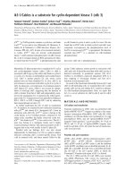
studies about the inverse relationship. Figure 1 shows
the flow-chart of included and excluded studies.
Study results
The relationship between CV disease and osteoporosis
Cardiovascular disease and fracture risk Seven population-based cohort studies assessed the relationship
between CV disease and fracture risk [1,2,4,12-15]
(Table 1). An increased risk of incident fractures was
observed in four studies with risk rates ranging from 1.2
to 6.7 [1,2,13,14].
The largest study included more than 30,000 twins
with a follow-up duration of 20 years [13]. In this study,
twins, without prevalent CV disease, were included at
the age of 50 years and followed up until a first hip fracture, death or end of follow-up period. Twins were considered unexposed until the first CV event. An increased
hip fracture risk was found after all diagnoses of CV disease in both men (hazard ratio (HR) 6.65; 95% CI 4.82
to 9.19) and women (HR 4.42; 95% CI 3.49 to 5.61).
Furthermore, this study showed that CHD was associated with an increased fracture risk (HR 2.32; 95% CI
1.91 to 2.84) as was cerebral vascular disease (HR 5.09
95% CI 4.18 to 6.20) [13]. This was confirmed in a large
population case-control study. This case-control study
was conducted using the Dutch PHARMO Record Linkage System database. Patients (n = 6,763) with a hip
fracture were compared with age- and sex-matched
patients without a hip fracture (n = 26,341), with the
objective to evaluate the association between stroke and
risk of hip fracture [16]. The prevalence of stroke was
3.3% in cases versus 1.5% in control patients. The risk
for a hip fracture was increased in patients who experienced a stroke before the index date (OR 1.96; 95% CI
1.65 to 2.33).
Three studies looked at the association between PAD
and fracture risk. PAD was associated with increased
risk for non-vertebral fractures (HR 1.47; 95% CI 1.07
to 2.04) [2] and hip fractures (HR 3.20; 95% CI 2.28 to
4.50) [13]. In contrast, a smaller study in men and
women, with shorter follow-up time, did not find an
association between PAD and non-vertebral fracture
risk [15]. Time of follow-up might be an important factor explaining different results, for the risk of fractures
den Uyl et al. Arthritis Research & Therapy 2011, 13:R5
/>
Page 9 of 19
Figure 1 Flow-chart of the systematic review.
was highest more than 10 years after the diagnosis of
PAD [13].
Longitudinal analysis in healthy postmenopausal
women (n = 2,262) showed that aortic calcifications
(AC) represented a strong predictor for fragility fractures: AC predicted a 2.3-fold increased risk for hip
fracture [1]. Not only women, but also men with
advanced AC have a two- to three-fold increased fracture risk [14]. However, a large population-based study
with 21 years follow-up, found no evidence that severity
of vascular calcification, measured as AC, is associated
with an increased risk of incident hip fracture [12]. Conflicting results might be due to differences in population
and methodology. The incident fracture rates were
equal in comparison to the other studies.
Hence, although heterogeneity makes it difficult to
draw firm conclusions, there is evidence that subjects
with atherosclerotic disease are at an increased risk for
frailty fractures. There are insufficient data to draw conclusions about fracture risk in patients with prevalent
coronary or cerebral CV disease.
Cardiovascular disease and bone loss Longitudinal
data about CV disease and bone loss were available
from six studies [1-4,15,17]. All studies showed that prevalent CV disease was associated with an increased bone
loss during follow-up, independent of age and traditional risk factors. In addition, several cross-sectional
studies similarly reported that prevalent CV disease is
associated with low BMD [18-22]. In the next section
the results are presented per subcategory of CV disease.
The association of CHD and BMD was only addressed
in cross-sectional studies and all but one found an association with low BMD [20,22-25]. Several studies
reported increased bone loss after an incident stroke.
Particularly patients who are wheelchair-bound or have
paretic limbs as a result of the stroke have significant
bone loss within months after the stroke [26]. These
studies were not included in this review, for the underlying pathogenesis is obvious. One study looked at bone
density immediately after the stroke and found that
female stroke patients have lower BMD than controls
[27]. Since the BMD measurement was assessed within
den Uyl et al. Arthritis Research & Therapy 2011, 13:R5
/>
six days after the stroke, one may assume that the possible differences are not a result of immobilisation.
A large prospective study found that men with prevalent PAD had an increased rate of hip bone loss compared with men without PAD (-0.6% vs -0.3%, P <
0.001) [2]. In another, smaller, study the association
between PAD and bone loss in women was weaker and
not observed in men [15]. In addition, a number of
cross-sectional studies showed that women and/or men
with PAD have decreased BMD [19,28-30].
Numerous reports have looked at the association
between subclinical atherosclerosis and osteoporosis.
Men and women with progression of AC have significantly higher bone loss in the lumbar spine compared
with subjects without AC progression (-1.5% vs 1.4%)
[4]. This is in line with other studies where AC progression is associated with higher rates of bone loss in the
proximal femur and metacarpal bones [1,3]. Furthermore, several studies confirmed the prospective data
and showed that subjects with calcifications in the
aorta, coronary arteries, carotid arteries or femoral
arteries have significant lower BMD compared with
controls [31-39]. Only a few studies fail to find an association [40-43]. In recent years, many studies have
examined the association between atherosclerosis and
osteoporosis. An increased IMT has been associated
with severity of atherosclerosis and increased cardiovascular risk and considered useful in identifying subjects
with increased risk [44]. An association between IMT
and BMD was studied intensively and most of the studies reported an association of increased IMT with low
bone density [45-54]. Endothelial dysfunction is considered to be an early phase of atherosclerosis and one
way to measure this is to focus on arterial compliance.
The endothelium plays an important role in determining vascular tone and dysfunction will result in
increased arterial stiffness [55]. In line with earlier discussed results, an increased arterial stiffness is associated with low BMD [45,54,56-61].
Altogether, the results strongly suggest that subjects
with subclinical atherosclerosis and early CV disease are
at increased risk of bone loss. Again, there were insufficient data to reach conclusions about bone loss in
patients with prevalent coronary or cerebral CV disease.
The relationship between osteoporosis and CV disease
Eighteen studies, most of moderate quality, reporting
about the relationship between osteoporosis and CV disease were included. Results will be discussed per subcategory of CV disease, when possible.
Low bone mineral density and cardiovascular mortality The association of osteoporosis with CV mortality
was studied in 10 prospective studies [5,7,8,62-68]
(Table 2). Low bone mass was inversely related with
CV mortality in seven studies [5,7,8,62-64,66,67].
Page 10 of 19
Postmenopausal women with a low BMD had a 1.2- to
2.3-fold increased risk of dying from CV events, independent of traditional CV risk factors [7,8,66]. Similar
results were found in elderly men [7,67]. Studies in
postmenopausal women with relative short follow-up
periods (around three years) showed no or minimally
significant elevated mortality rates [5,63,64]. Two large
population-based studies in elderly men and women did
not reveal a significant association between low bone
mass and CV mortality [65,69]. The most recent and
largest study determined the risk of CV mortality in
5,272 persons [69]. Women with low BMD had higher
risk for CV mortality; however, this did not reach significance (relative risk (RR) 1.26; 95% CI 0.88 to 1.80). No
association was found in men.
Focusing on the few studies that reported the results
per CV subcategory, women with low bone mass had no
or a small increased risk for mortality by coronary heart
disease (RR 1.17; 95% CI 0.92 to 1.51) and (relative
hazard 1.3; 95% CI 1.0 to 1.8), respectively [5,64] and
two out of three studies showed that men and women
with low BMD had a 1.3- to 1.7-fold increased risk for
stroke mortality [5,62,65].
Low bone mineral density and incident cardiovascular disease A total of six studies assessed the risk of
incident CV events in persons with osteoporosis
[6,62,70-73]. Most of them show a significant inverse
relationship between BMD and incident CV events in
women (HR 1.23 to 3.9) [6,39,62,70] but not in men
[6,70]. Two studies related the prevalence of vertebral
fractures with future CV events and were unable to find
any association [68,71]. Surprisingly, one study showed
that women with prevalent fractures and known CHD
had a reduced risk for CV events [73].
Few articles assessed incident CV events separated per
CV category. Three studies assessed the risk for CHD.
Two studies showed an association with increased risk
for CHD in postmenopausal women [72,73]. One study
could not find an association in elderly men and women
[70]. Cerebrovascular events were studied in two articles. Both found an increased risk for stroke in postmenopausal women with low BMD with hazard ratios of
1.31 and 4.1 [62,72].
There was a considerable heterogeneity in measurement of osteoporosis. It is shown that the specificity and
sensitivity of the densitometry tests differs greatly, and
the site of measurement plays an important role in diagnosing osteoporosis as well [74]. Only six studies used
dual energy absorptiometry (DXA) measurements to
assess BMD [6,64,66,67,69,75,76], while in the other studies BMD was measured with older techniques such as
single photon absorptiometry, dual photon absorptiometry (DPA) or quantitative ultrasonography (QUS). Most
studies measured BMD of the hip and lumbar spine, but
den Uyl et al. Arthritis Research & Therapy 2011, 13:R5
/>
also distal radius and heel were measured and in some
the phalangeals.
Low bone mineral density and subclinical atherosclerosis In addition to associations with CV events, low
BMD has also been shown to be associated with surrogate markers of CV disease, such as vascular calcification. In women with the largest decrease in metacarpal
cortical area during a 25-year follow-up, the most severe
progression of aortic calcification was observed [77] and
women with a prevalent vertebral fracture had a higher
IMT measured 10 years later [75]. Moreover, results
from several cross-sectional studies confirmed that both
women and men with low bone mass, compared to subjects with normal bone mass, have significantly more
subclinical atherosclerosis [20,28,31-34,37,38,45,48,
49,51,52,78,79], increased risk of peripheral arterial disease [28,29,34,54] and other surrogate end markers for
CV disease [57,60,61].
Taken together, there is some evidence that persons
with low BMD are at increased risk for CV events and
subsequent CV mortality. However, variations in study
design, for example, study population and outcome
measures, limits interpretation. Since only a few studies
assessed the CV outcome divided per CV subcategory,
no conclusions can be drawn concerning a relationship
between osteoporosis and specific categories of CV
disease.
Links between CV disease and osteoporosis
Common pathogenesis
CV disease is preceded by atherosclerosis, for example,
arterial disease. Atherosclerosis is a long-term process
in which deposits of cholesterol, cellular waste products and calcium accumulates in the arterial wall
causing it to thicken. Clinically, atherosclerosis is manifested by coronary heart disease, cerebrovascular disease and peripheral arterial disease. Endothelial
dysfunction is the first step in the pathogenesis of
atherosclerosis and predicts future CV events [80]. Calcification in the aorta and coronary arteries, for example, vascular calcification, may be a surrogate marker
for atherosclerosis and increased CV risk [81]. In a
recent meta-analysis patients with calcifications were
found to have an increased risk for CV mortality and
events [10]. Presently, vascular calcification is regarded
as an active process, regulated by factors known to be
involved in the process of osteogenesis, such as bone
morphogenetic protein (BMP), alkaline phosphatase
(ALP), osteopontin (OPN) and matrix GLA protein
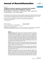
(MGP) [82-85] (Figure 2). Accumulating evidence suggests that calcification is a consequence of active bone
formation by osteoblast-like cells [86]. Vascular
smooth muscle cells (VSMCs) are able to re-differentiate towards osteoblast-like cells and a subpopulation,
Page 11 of 19
that is, calcifying vascular cells (CVCs), were shown to
form nodules and mineralisation spontaneously [87].
In vitro, these osteoblastic cells produce hydroxyapatite, a mineral important in bone formation [88]. In
the following paragraphs some of the bone-related factors that are involved in vascular calcification will be
discussed in more detail.
BMPs are members of the transforming growth factorb superfamily and important factors in the regulation of
osteoblast differentiation. BMP acts through upregulation of transcription factors important in bone metabolism, such as core binding factor-a1 (Cbfa1), also
known as runt-related transcription factor 2 (Runx2),
and msh homeobox 2 (Msx2). BMP appears to be an
important mediator in vascular calcification. An
increased expression of BMP2 and BMP4 is found in
atherosclerotic lesions in endothelial cells, foam cells
and VSMCs [88,89]. In vitro studies showed that several
factors that are known to induce CV disease, such
as oxidative stress, oxidized low-density lipoprotein
(ox-LDL) and tumor necrosis factor alpha (TNF-a),
are able to upregulate BMP expression in endothelial
cells [90,91].
MGP is a calcium-binding protein and requires vitamin K to function. MGP is found to be expressed in
areas with arterial calcification [92] and may be an
important calcification inhibitor. MGP knock-out mice
developed extensive calcification in coronary arteries
[93]. Recently the mechanism by which MGP inhibits
calcification has become clear. In vitro, MGP has been
shown to inhibit calcification by binding to BMP2,
thereby blocking the induction of osteoblasts [94].
OPN is a glycoprotein that accumulates in the extracellular matrix of bone tissue where it binds to hydroxyapatite and calcium. In bone, OPN is expressed by
(pre-) osteoblasts and osteoclasts and is also found to be
highly expressed in the atherosclerotic artery [89,92].
Whether it promotes or inhibits calcification in the
arterial wall is not completely clear [95]. While high
OPN serum levels are associated with vascular calcification [96] and vitamin increases OPN and subsequent
calcification in bovine VSMC’s [97], OPN is also shown
to inhibit calcification by inhibiting de novo hydroxyapatite production [98].
ALP is found on the surface of osteoblasts and is often
used as a marker for bone turnover. ALP is an enzyme
that catalyses the hydrolysis of phosphate esters. Hydrolysis of pyrophosphate, which is an inhibitor of hydroxyapatite formation, is especially needed to facilitate
normal mineralisation [99]. In vitro studies in VSMC’s
showed that the ALP expression is increased in response
to inflammatory markers, LDL and oxidative stress and
this increased expression was associated with increased
mineralisation [100-102].
den Uyl et al. Arthritis Research & Therapy 2011, 13:R5
/>
Page 12 of 19
Figure 2 Vascular calcification. Vascular calcification is an active process regulated by factors known to be involved in the process of
osteogenesis. Vascular smooth muscle cells are able to differentiate towards osteoblast-like cells, promoted by a variety of stimuli, including
BMP, RANKL, oxidative stress, inflammation and estrogen deficiency. These osteoblastic cells produce osteocalcin and ALP, important factors in
mineralisation. # Excessive vitamin D promotes mineralisation. * It is not clear whether OPN promotes or inhibits calcification in the arterial wall,
in bone mineralisation it is a known mineralisation inhibitor. Abbreviations: ALP, alkaline phosphatase; BMP, bone morphogenetic protein; Cbfa1,
core binding factor-a1; MGP, matrix GLA protein; Msx2, msh homeobox 2; OPG, osteoprotegerin; OPN, osteopontin; ox-LDL, oxidized low density
lipoprotein; RANKL, receptor activator of nuclear factor-B ligand; VSMC, vascular smooth muscle cell; Wnt, combination of wingless and Int.
The recent identification of receptor activator of
nuclear factor-kB (RANK), osteoprotegerin (OPG) and
RANK ligand (RANKL) provides more insight into bone
metabolism [103]. Most interestingly, there is increasing
evidence that OPG is a key regulator in the pathogenesis
of osteoporosis and vascular calcification. OPG production by osteoblastic cells is regulated by a number of
factors, including BMP-2, inflammation, estrogen, vitamin D and oxidative stress [104]. OPG is expressed in
various tissues, including the skeleton and vascular wall,
and serves as a soluble decoy for RANKL [105]. Interestingly, OPG knock-out mice show, in addition to
early-onset osteoporosis, increased vascular calcification
[106]. In vitro studies have shown that OPG appears to
be important for endothelial cell survival [107] and may
inhibit active calcification [108]. Surprisingly, while
experimental studies showed that OPG might protect
against vascular calcification, OPG levels appear to be
elevated in patients with CV disease. Several, but not all,
clinical studies found a correlation of high OPG serum
levels and more severe CV disease [45,50,62,109-111].
Other pathways interacting with OPG might explain this
discrepant finding. Estrogen deficiency results in an
increased vascular OPG/RANKL ratio with subsequent
increased calcification in an animal model [112].
Furthermore, pro-inflammatory cytokines are shown to
elevate OPG levels in patients with CV disease [113].
Thus, while OPG appears to play a role in the pathogenesis of atherosclerosis, the exact mechanism remains
to be elucidated.
Another important mechanism linking CV disease and
osteoporosis is Wnt signalling, a combination of the
genes Wg (wingless) and Int. Animal models showed the
important role of Wnt signalling in bone formation
den Uyl et al. Arthritis Research & Therapy 2011, 13:R5
/>
through lipoprotein receptor-related protein 5 (LRP5),
lipoprotein receptor-related protein 6 (LRP6) and
b-catenin [114]. Wnt signalling is suggested to play an
important role in bone formation and bone adaptation
to mechanical loading [115,116]. Interestingly, TNF-a
[117], oxidative stress [118] and vitamin D [119]
are shown to promote vascular calcification through
the Wnt signalling pathway and this supports the
hypothesis that Wnt signalling is an interesting new
molecular mechanism that influences bone and vascular
metabolism.
Common risk factors
CV disease and osteoporosis are both common diseases
in elderly men and women. While the increased prevalence of both conditions is often attributed to aging,
most of the associations found in observational studies
remain significant after adjustment for age. Other
important traditional risk factors are also shared, such
as inactivity, smoking, estrogen deficiency and chronic
inflammation, explaining part of the link between CV
disease and osteoporosis [9].
Estrogen deficiency is considered an important risk
factor for osteoporosis [120] and some studies suggest
estrogen deficiency to be a cardiovascular risk factor
[121-123]. Estrogen regulates bone turnover and the CV
system directly and indirectly through the effects on the
immune system, antioxidant system and other risk factors. After menopause, estrogen levels decrease rapidly
resulting in an upregulated osteoclast formation and differentiation, inducing high bone turnover and accelerated bone loss [124]. Furthermore, following estrogen
withdrawal the production and secretion of the proinflammatory cytokines interleukin-6 (IL-6), interleukin1 and TNF-a is increased [116,125].
Presently, inflammation is considered to play an
important role in the process of atherosclerosis
[126,127]. Both cellular and humoral pathways of the
immune response contribute to an important part in the
pathogenesis of atherosclerosis [128]. Markers of inflammation, such as pro-inflammatory cytokines and C-reactive protein (CRP), are involved in the development of
atherosclerosis and CRP predicts cardiovascular events
independently of other CV risk factors [129,130]. There
is accumulating evidence that inflammation influences
bone metabolism and is considered to be the most
important cause of postmenopausal osteoporosis. Proinflammatory cytokines enhance bone resorption
directly through an induction of osteoclastogenesis or
through the OPG pathway [116,131].
Recent research has identified new common mediators
for vascular calcification and bone loss, such as hyperlipidemia, oxidative stress and vitamin D deficiency. An
abnormal lipid profile, that is, high levels of total cholesterol, LDL and triglycerides and low levels of high-
Page 13 of 19
density lipoprotein (HDL), is known to play a key role
in development of atherosclerosis and CV disease
[132,133]. Interestingly, HDL is able to regulate the calcification of VSMCs [134]. HDL inhibited the spontaneous and cytokine induced osteogenic differentiation of
CVCs in vitro. The role of lipids in the regulation of
bone mass is more complicated. While experimental
studies showed that ox-LDL influences bone metabolism
[135], results in observational studies are contradictory
[1,136-138].
Oxidative stress is believed to increase with age and is
associated with hypertension and atherosclerosis [139].
Free radicals have important effects on osteoclast differentiation and function [140] and oxidative stress markers are significantly associated with BMD [141]. In
vitro, minimally oxidized low-density lipoprotein (MMLDL) enhances the differentiation of VSMC’s towards
osteoblastic cells. Interestingly, antioxidants inhibited
these effects [100].
The prevalence of vitamin D deficiency is high
among elderly men and women [142] and associated
with osteoporosis and increased fracture risk [143].
Observational studies showed an inverse association of
vitamin D deficiency with hypertension and CV events,
suggesting a role for low vitamin D [144-148]. Proposed mechanisms are effects on myocardial gene
expression, the renin-angiotensin axis or through secondary hyperparathyroidism. Important risk factors as
physical condition and immobility were rarely assessed.
Animal models and in vitro studies on the other hand,
demonstrated that toxic levels of vitamin D induce
vascular calcification [97,149]. Interestingly, osteoprotegerin has been shown to inhibit the vitamin-induced
calcifications in an animal model [150]. It has been
suggested that vitamin D has a biphasic relation with
vascular calcification and that both vitamin D deficiency and vitamin D excess results in increased vascular calcification.
Genetic studies
In complex, multifactorial diseases genetic factors are
believed to play an important role in the pathogenesis in
addition to environmental influences. Identifying candidate genes offers opportunities to gain more insight into
possible shared pathogenesis and common risk factors
in CV disease and osteoporosis. Many candidate genes
have been examined, mainly genes coding for known
factors, such as cytokines, bone-associated factors and
receptors. The genes that might be involved in both diseases will be discussed here.
Polymorphism in the IL-6 gene, a cytokine involved in
bone metabolism and CV disease, might be an interestingly candidate gene. A G174C polymorphism in the promoter region of the IL-6 gene was shown to be associated
with low bone mass in the radius in postmenopausal
den Uyl et al. Arthritis Research & Therapy 2011, 13:R5
/>
women [151] and with a high blood pressure and
increased CV risk in men [152].
Vitamin D receptor polymorphisms have been associated in many studies with bone density [153,154].
Although this could not be replicated in a large metaanalysis, it did show that the Cdx2 polymorphism was
associated with risk for vertebral fractures [155]. In
addition, the BsmI polymorphism was associated with
IMT and myocardial infarction (MI) [156,157], strengthening the possible role of vitamin D in linking CV disease and osteoporosis.
One of the most interesting candidate genes to mention is the OPG gene, located on chromosome 8 and
several single nucleotide polymorphisms (SNPs) are
identified in this gene. So far, studies were able to
associate different SNPs with either bone density or vascular disease. SNPs A163G and T245G were associated
with osteoporotic fractures [158]. The linked polymorphisms T950C and C1181C within the promoter
region of the OPG gene were associated with an
increased risk for CAD in men [159]. In addition,
C1181C was also associated with first-ever intracerebral
haemorrhage [160]. Furthermore, another SNP in the
promoter region in the TATA box was related to vascular morphology and function [161].
A genetic defect in the Wnt signalling pathway was
recently discovered in a family with features of metabolic syndrome and early onset coronary artery disease
[162]. This rare mutation in the LRP6 gene is associated
with dyslipidemia, hypertension and diabetes. This finding supports further research for mutations in genes
involved in the Wnt signalling pathway.
Collagen type I is an important protein in the mineralisation matrix and connective tissue. Mutations in this
gene are associated with low BMD and fracture risk
[163]. Interestingly, besides low BMD, individuals with a
SNP in the COL1A gene (rs42524) had an increased prevalence of stroke and MI [164].
The calcium-sensing receptor (CASR) is a receptor
involved in the regulation of calcium homeostasis.
A SNP in the CARS gene (A986S) was associated with
higher serum calcium and increased prevalence of coronary artery disease (CAD) and MI [165]. This SNP was
also associated with low BMD in premenopausal women
[166]. However, the role in postmenopausal osteoporosis
is not clear, since several studies showed no association
of this SNP with BMD or fracture risk in postmenopausal women [167,168].
An interesting candidate gene to mention is the klotho
gene. Defects in the klotho gene have been shown to
result in arteriosclerosis and increased IMT in klotho
deficient mice [169]. A SNP in this gene (G395A) was
associated with CAD. Surprisingly, this same SNP was
associated with bone density [170] and was suggested to
Page 14 of 19
be involved in the pathophysiology of bone loss. This
SNP in the promoter region resulted in impaired function of the gene. What makes this gene interesting is
that it might offer a new treatment approach, because
the abnormalities seen in klotho-deficient mice can be
reversed by restoring the klotho expression [171].
Finally, polymorphisms in the apolipoprotein E
(APOE) gene has been studied intensively. It has been
associated with hypertension, atherosclerotic disease and
CV disease [172-174]. Furthermore, APOE gene polymorphisms have been suggested to be associated with
low BMD and fracture risk. However, a recent metaanalysis was unable to show a strong and consistent
association with BMD and fracture incidence [175].
Discussion
Our study is the first to systematically review the epidemiological literature about the association between CV
disease and osteoporosis. An extensive literature search
yielded 27 prospective studies addressing this relationship. Due to considerable heterogeneity in study design
and outcome measurements the results could not be
pooled. Focusing on the methodologically strongest studies (those with minimal selection bias and the appropriate assessments, that is, a methodological score of
more than 3), our review indicates that the prevalent
subclinical CV disease predicts future fractures and
bone loss [2-4,13-15] (Table 4).
Furthermore, there is some evidence that low
bone mass predicts CV mortality and CV events
[6,62,68,69,75].
Interestingly, several studies demonstrated shared risk
factors, supporting the existence of a direct association
between vascular calcification and bone biology.
Due to the substantial diversity of patients and study
methods, pooled analysis was not considered appropriate. Although numerous efforts were made to investigate
the association between CV disease and osteoporosis, a
vast majority of studies used secondary outcome measurements, while a limited number of studies used primary outcome measurements such as incident CV
events or osteoporosis. Furthermore, the population studied varied with respect to age, sex, baseline risk for CV
events or fractures and ethnicity. Larger prospective studies in elderly persons, men and women, are needed to
answer this question. To reduce heterogeneity we
encourage that in new studies well-defined outcome
Table 4 Summary of findings in high quality prospective
studies
Association
No association
CV disease and OP
N=6
N=0
Bone mass and CV events
N=3
N=2
den Uyl et al. Arthritis Research & Therapy 2011, 13:R5
/>
measures should be incorporated, such as incident CV
disease presented per subcategory of CV disease and
measurement of BMD by DXA-scans on regular interval
periods.
Conclusions
The current evidence indicates that individuals with prevalent (sub)clinical CV disease are at increased risk for
bone loss and subsequent fractures. Presently, no firm
conclusions can be drawn to which extent low BMD
might be associated with increased cardiovascular risk.
Age, estrogen deficiency and inflammation represent the
most important common risk factors and the discovery
of new pathways, for example, OGP/RANKL and Wnt
signalling, might provide interesting new therapeutic
options. Altogether our results suggest that bone density
screening could be recommended in patients with prevalent CV disease.
Additional material
Additional file 1: Medline search. Complete medline search on 8 June
2010.
Additional file 2: Quality assessment cohort studies. List of quality
assessment of cohort studies as proposed by the Dutch Cochrane
Collaboration.
Abbreviations
ABI: ankle brachial index; AC: aortic calcifications; ALP: alkaline phosphatase;
APOE: apolipoprotein E; BMD: bone mineral density; BMP: bone
morphogenetic protein; CAD: coronary artery disease; CASR: calcium-sensing
receptor; Cbfa1: core binding factor-α1; CDH: coronary heart disease; CRP: Creactive protein; CV: cardiovascular; CVC: calcifying vascular cells; DPA: dual
photon absorptiometry; DXA: dual energy absorptiometry; HDL: high density
lipoprotein; HR: hazard ratio; IL-6: interleukine-6; IMT: intima media thickness;
LRP5: lipoprotein receptor-related protein 5; LRP6: lipoprotein receptorrelated protein 6; MGP: matrix GLA protein; MI: myocardial infarction; MMLDL: minimally oxidized low-density lipoprotein; Msx2: msh homeobox 2;
OPG: osteoprotegerin; OPN: osteopontin; OR: odds ratio; ox-LDL: oxidized
low density lipoprotein; PAD: peripheral arterial disease; QUS: quantitative
ultrasonography; RANK: receptor activator of nuclear factor-B; RANKL:
receptor activator of nuclear factor-B ligand; RR: relative risk; Runx2: runtrelated transcription factor 2; SNP: single nucleotide polymorphism; TNF-α:
tumour necrosis factor alpha; VSMC: vascular smooth muscle cell; Wnt:
combination of wingless and Int.
Acknowledgements
We would like to thank Hans Ket (Clinical Library, VU medical centre,
Amsterdam) for his assistance in collecting the literature for this systematic
review.
Author details
Department of Rheumatology, VU Medical Centre, De Boelelaan 1117, 1081
HV Amsterdam, The Netherlands. 2Department of Internal Medicine, VU
Medical Centre, De Boelelaan 1117, 1081 NV Amsterdam, The Netherlands.
3
Department of Rheumatology, Jan van Breemen Research Institute/Reade,
Dr Jan van Breemenstraat 2, 1056 AB Amsterdam, The Netherlands.
1
Authors’ contributions
DU conducted the data collection, interpretation and analysis of the data
and drafted the manuscript. LT participated in interpretation and analysis of
the data and helped to draft the manuscript. WL conceived of the
Page 15 of 19
hypothesis of the manuscript and participated in study design and
coordination. MT, HR and WL helped to draft the manuscript. All authors
critically reviewed, contributed to and approved the final manuscript.
Competing interests
The authors declare that they have no competing interests.
Received: 17 June 2010 Revised: 12 November 2010
Accepted: 17 January 2011 Published: 17 January 2011
References
1. Bagger YZ, Tanko LB, Alexandersen P, Qin G, Christiansen C: Radiographic
measure of aorta calcification is a site-specific predictor of bone loss
and fracture risk at the hip. J Intern Med 2006, 259:598-605.
2. Collins TC, Ewing SK, Diem SJ, Taylor BC, Orwoll ES, Cummings SR,
Strotmeyer ES, Ensrud KE: Peripheral arterial disease is associated with
higher rates of hip bone loss and increased fracture risk in older men.
Circulation 2009, 119:2305-2312.
3. Hak AE, Pols HA, van Hemert AM, Hofman A, Witteman JC: Progression of
aortic calcification is associated with metacarpal bone loss during
menopause: a population-based longitudinal study. Arterioscler Thromb
Vasc Biol 2000, 20:1926-1931.
4. Naves M, Rodriguez-Garcia M, Diaz-Lopez JB, Gomez-Alonso C, CannataAndia JB: Progression of vascular calcifications is associated with greater
bone loss and increased bone fractures. Osteoporos Int 2008,
19:1161-1166.
5. Browner WS, Seeley DG, Vogt TM, Cummings SR: Non-trauma mortality in
elderly women with low bone mineral density. Study of Osteoporotic
Fractures Research Group. Lancet 1991, 338:355-358.
6. Farhat GN, Newman AB, Sutton-Tyrrell K, Matthews KA, Boudreau R,
Schwartz AV, Harris T, Tylavsky F, Visser M, Cauley JA: The association of
bone mineral density measures with incident cardiovascular disease in
older adults. Osteoporos Int 2007, 18:999-1008.
7. Johansson C, Black D, Johnell O, Oden A, Mellstrom D: Bone mineral
density is a predictor of survival. Calcif Tissue Int 1998, 63:190-196.
8. von der Recke P, Hansen MA, Hassager C: The association between low
bone mass at the menopause and cardiovascular mortality. Am J Med
1999, 106:273-278.
9. Doherty TM, Fitzpatrick LA, Inoue D, Qiao JH, Fishbein MC, Detrano RC,
Shah PK, Rajavashisth TB: Molecular, endocrine, and genetic mechanisms
of arterial calcification. Endocr Rev 2004, 25:629-672.
10. Rennenberg RJ, Kessels AG, Schurgers LJ, van Engelshoven JM, de
Leeuw PW, Kroon AA: Vascular calcifications as a marker of increased
cardiovascular risk: a meta-analysis. Vasc Health Risk Manag 2009,
5:185-197.
11. Checklist quality assessment cohort studies. Dutch Cochrane Centre;
2010.
12. Samelson EJ, Cupples LA, Broe KE, Hannan MT, O’Donnell CJ, Kiel DP:
Vascular calcification in middle age and long-term risk of hip fracture:
the Framingham Study. J Bone Miner Res 2007, 22:1449-1454.
13. Sennerby U, Melhus H, Gedeborg R, Byberg L, Garmo H, Ahlbom A,
Pedersen NL, Michaelsson K: Cardiovascular diseases and risk of hip
fracture. JAMA 2009, 302:1666-1673.
14. Szulc P, Kiel DP, Delmas PD: Calcifications in the abdominal aorta predict
fractures in men: MINOS study. J Bone Miner Res 2008, 23:95-102.
15. von Mühlen D, Allison M, Jassal SK, Barrett-Connor E: Peripheral arterial
disease and osteoporosis in older adults: the Rancho Bernardo Study.
Osteoporos Int 2009, 20:2071-2078.
16. Pouwels S, Lalmohamed A, Leufkens B, de Boer A, Cooper C, van Staa T, de
Vries F: Risk of hip/femur fracture after stroke: a population-based casecontrol study. Stroke 2009, 40:3281-3285.
17. Schulz E, Arfai K, Liu X, Sayre J, Gilsanz V: Aortic calcification and the risk
of osteoporosis and fractures. J Clin Endocrinol Metab 2004, 89:4246-4253.
18. Broussard DL, Magnus JH: Coronary heart disease risk and bone mineral
density among U.S. women and men. J Womens Health (Larchmt) 2008,
17:479-490.
19. Farhat GN, Strotmeyer ES, Newman AB, Sutton-Tyrrell K, Bauer DC, Harris T,
Johnson KC, Taaffe DR, Cauley JA: Volumetric and areal bone mineral
density measures are associated with cardiovascular disease in older
men and women: the health, aging, and body composition study. Calcif
Tissue Int 2006, 79:102-111.
den Uyl et al. Arthritis Research & Therapy 2011, 13:R5
/>
20. Marcovitz PA, Tran HH, Franklin BA, O’Neill WW, Yerkey M, Boura J,
Kleerekoper M, Dickinson CZ: Usefulness of bone mineral density to
predict significant coronary artery disease. Am J Cardiol 2005,
96:1059-1063.
21. Sennerby U, Farahmand B, Ahlbom A, Ljunghall S, Michaelsson K:
Cardiovascular diseases and future risk of hip fracture in women.
Osteoporos Int 2007, 18:1355-1362.
22. Varma R, Aronow WS, Basis Y, Singh T, Kalapatapu K, Weiss MB, Pucillo AL,
Monsen CE: Relation of bone mineral density to frequency of coronary
heart disease. Am J Cardiol 2008, 101:1103-1104.
23. Magnus JH, Broussard DL: Relationship between bone mineral density
and myocardial infarction in US adults. Osteoporos Int 2005, 16:2053-2062.
24. Sioka C, Goudevenos J, Pappas K, Bougias C, Papadopoulos A,
Grammatikopoulos K, Fotopoulos A: Bone mineral density and coronary
atherosclerosis. Calcif Tissue Int 2007, 81:333.
25. Tekin GO, Kekilli E, Yagmur J, Uckan A, Yagmur C, Aksoy Y, Turhan H,
Yetkin E: Evaluation of cardiovascular risk factors and bone mineral
density in post menopausal women undergoing coronary angiography.
Int J Cardiol 2008, 131:66-69.
26. Sato Y, Kuno H, Kaji M, Ohshima Y, Asoh T, Oizumi K: Increased bone
resorption during the first year after stroke. Stroke 1998, 29:1373-1377.
27. Jorgensen L, Engstad T, Jacobsen BK: Bone mineral density in acute stroke
patients: low bone mineral density may predict first stroke in women.
Stroke 2001, 32:47-51.
28. Mangiafico RA, Russo E, Riccobene S, Pennisi P, Mangiafico M, D’Amico F,
Fiore CE: Increased prevalence of peripheral arterial disease in
osteoporotic postmenopausal women. J Bone Miner Metab 2006,
24:125-131.
29. van der Klift M, Pols HA, Hak AE, Witteman JC, Hofman A, de Laet CE: Bone
mineral density and the risk of peripheral arterial disease: the Rotterdam
Study. Calcif Tissue Int 2002, 70:443-449.
30. Wong SY, Kwok T, Woo J, Lynn H, Griffith JF, Leung J, Tang YY, Leung PC:
Bone mineral density and the risk of peripheral arterial disease in men
and women: results from Mr. and Ms Os, Hong Kong. Osteoporos Int
2005, 16:1933-1938.
31. Bakhireva LN, Barrett-Connor EL, Laughlin GA, Kritz-Silverstein D: Differences in
association of bone mineral density with coronary artery calcification in
men and women: the Rancho Bernardo Study. Menopause 2005, 12:691-698.
32. Barengolts EI, Berman M, Kukreja SC, Kouznetsova T, Lin C, Chomka EV:
Osteoporosis and coronary atherosclerosis in asymptomatic
postmenopausal women. Calcif Tissue Int 1998, 62:209-213.
33. Erbilen E, Yazici S, Ozhan H, Bulur S, Ordu S, Yazici M: Relationship
between angiographically documented coronary artery disease and low
bone mass in men. Circ J 2007, 71:1095-1098.
34. Farhat GN, Cauley JA, Matthews KA, Newman AB, Johnston J, Mackey R,
Edmundowicz D, Sutton-Tyrrell K: Volumetric BMD and vascular
calcification in middle-aged women: the Study of Women’s Health
Across the Nation. J Bone Miner Res 2006, 21:1839-1846.
35. Frye MA, Melton LJ III, Bryant SC, Fitzpatrick LA, Wahner HW, Schwartz RS,
Riggs BL: Osteoporosis and calcification of the aorta. Bone Miner 1992,
19:185-194.
36. Hyder JA, Allison MA, Criqui MH, Wright CM: Association between
systemic calcified atherosclerosis and bone density. Calcif Tissue Int 2007,
80:301-306.
37. Hyder JA, Allison MA, Wong N, Papa A, Lang TF, Sirlin C, Gapstur SM,
Ouyang P, Carr JJ, Criqui MH: Association of coronary artery and aortic
calcium with lumbar bone density: the MESA Abdominal Aortic Calcium
Study. Am J Epidemiol 2009, 169:186-194.
38. Ness J, Aronow WS: Comparison of prevalence of atherosclerotic vascular
disease in postmenopausal women with osteoporosis or osteopenia
versus without osteoporosis or osteopenia. Am J Cardiol 2006,
97:1427-1428.
39. Tanko LB, Bagger YZ, Christiansen C: Low bone mineral density in the hip
as a marker of advanced atherosclerosis in elderly women. Calcif Tissue
Int 2003, 73:15-20.
40. Aoyagi K, Ross PD, Orloff J, Davis JW, Katagiri H, Wasnich RD: Low bone
density is not associated with aortic calcification. Calcif Tissue Int 2001,
69:20-24.
41. Chow JT, Khosla S, Melton LJ III, Atkinson EJ, Camp JJ, Kearns AE:
Abdominal aortic calcification, BMD, and bone microstructure: a
population-based study. J Bone Miner Res 2008, 23:1601-1612.
Page 16 of 19
42. Shen H, Bielak LF, Streeten EA, Ryan KA, Rumberger JA, Sheedy PF,
Shuldiner AR, Peyser PA, Mitchell BD: Relationship between vascular
calcification and bone mineral density in the Old-order Amish. Calcif
Tissue Int 2007, 80:244-250.
43. Sinnott B, Syed I, Sevrukov A, Barengolts E: Coronary calcification
and osteoporosis in men and postmenopausal women are
independent processes associated with aging. Calcif Tissue Int
2006, 78:195-202.
44. Lorenz MW, Markus HS, Bots ML, Rosvall M, Sitzer M: Prediction of clinical
cardiovascular events with carotid intima-media thickness: a systematic
review and meta-analysis. Circulation 2007, 115:459-467.
45. Frost ML, Grella R, Millasseau SC, Jiang BY, Hampson G, Fogelman I,
Chowienczyk PJ: Relationship of calcification of atherosclerotic plaque
and arterial stiffness to bone mineral density and osteoprotegerin in
postmenopausal women referred for osteoporosis screening. Calcif Tissue
Int 2008, 83:112-120.
46. Hmamouchi I, Allali F, Khazzani H, Bennani L, El Mansouri L, Ichchou L,
Cherkaoui M, Abouqal R, Hajjaj-Hassouni N: Low bone mineral density is
related to atherosclerosis in postmenopausal Moroccan women. BMC
Public Health 2009, 9:388.
47. Jorgensen L, Joakimsen O, Rosvold Berntsen GK, Heuch I, Jacobsen BK: Low
bone mineral density is related to echogenic carotid artery plaques: a
population-based study. Am J Epidemiol 2004, 160:549-556.
48. Kim SH, Kim YM, Cho MA, Rhee Y, Hur KY, Kang ES, Cha BS, Lee EJ, Lee HC,
Lim SK: Echogenic carotid artery plaques are associated with vertebral
fractures in postmenopausal women with low bone mass. Calcif Tissue Int
2008, 82:411-417.
49. Montalcini T, Emanuele V, Ceravolo R, Gorgone G, Sesti G, Perticone F,
Pujia A: Relation of low bone mineral density and carotid atherosclerosis
in postmenopausal women. Am J Cardiol 2004, 94:266-269.
50. Pennisi P, Signorelli SS, Riccobene S, Celotta G, Di Pino L, La Malfa T,
Fiore CE: Low bone density and abnormal bone turnover in patients
with atherosclerosis of peripheral vessels. Osteoporos Int 2004, 15:389-395.
51. Shaffer JR, Kammerer CM, Rainwater DL, O’Leary DH, Bruder JM, Bauer RL,
Mitchell BD: Decreased bone mineral density is correlated with increased
subclinical atherosclerosis in older, but not younger, Mexican American
women and men: the San Antonio Family Osteoporosis Study. Calcif
Tissue Int 2007, 81:430-441.
52. Sumino H, Ichikawa S, Kasama S, Takahashi T, Sakamoto H,
Kumakura H, Takayama Y, Kanda T, Murakami M, Kurabayashi M:
Relationship between carotid atherosclerosis and lumbar spine
bone mineral density in postmenopausal women. Hypertens Res
2008, 31:1191-1197.
53. Yamada S, Inaba M, Goto H, Nagata M, Ueda M, Nakatuka K, Tahara H,
Yokoyama H, Emoto M, Shoji T, Nishizawa Y: Significance of intima-media
thickness in femoral artery in the determination of calcaneus osteo-sono
index but not of lumbar spine bone mass in healthy Japanese people.
Osteoporos Int 2005, 16:64-70.
54. Yamada S, Inaba M, Goto H, Nagata-Sakurai M, Kumeda Y, Imanishi Y,
Emoto M, Ishimura E, Nishizawa Y: Associations between physical activity,
peripheral atherosclerosis and bone status in healthy Japanese women.
Atherosclerosis 2006, 188:196-202.
55. Vlachopoulos C, Aznaouridis K, Stefanadis C: Prediction of cardiovascular
events and all-cause mortality with arterial stiffness: a systematic review
and meta-analysis. J Am Coll Cardiol 2010, 55:1318-1327.
56. Hirose K, Tomiyama H, Okazaki R, Arai T, Koji Y, Zaydun G, Hori S,
Yamashina A: Increased pulse wave velocity associated with reduced
calcaneal quantitative osteo-sono index: possible relationship between
atherosclerosis and osteopenia. J Clin Endocrinol Metab 2003,
88:2573-2578.
57. Mangiafico RA, Alagona C, Pennisi P, Parisi N, Mangiafico M, Purrello F,
Fiore CE: Increased augmentation index and central aortic blood
pressure in osteoporotic postmenopausal women. Osteoporos Int 2008,
19:49-56.
58. Mikumo M, Okano H, Yoshikata R, Ishitani K, Ohta H: Association between
lumbar bone mineral density and vascular stiffness as assessed by pulse
wave velocity in postmenopausal women. J Bone Miner Metab 2009,
27:89-94.
59. Seo SK, Cho S, Kim HY, Choi YS, Park KH, Cho DJ, Lee BS: Bone mineral
density, arterial stiffness, and coronary atherosclerosis in healthy
postmenopausal women. Menopause 2009, 16:937-943.
den Uyl et al. Arthritis Research & Therapy 2011, 13:R5
/>
60. Sumino H, Ichikawa S, Kasama S, Takahashi T, Kumakura H, Takayama Y,
Kanda T, Sakamaki T, Kurabayashi M: Elevated arterial stiffness in
postmenopausal women with osteoporosis. Maturitas 2006, 55:212-218.
61. Sumino H, Ichikawa S, Kasama S, Takahashi T, Sakamoto H, Kumakura H,
Takayama Y, Kanda T, Murakami M, Kurabayashi M: Relationship between
brachial arterial endothelial function and lumbar spine bone mineral
density in postmenopausal women. Circ J 2007, 71:1555-1559.
62. Browner WS, Pressman AR, Nevitt MC, Cauley JA, Cummings SR:
Association between low bone density and stroke in elderly women.
The study of osteoporotic fractures. Stroke 1993, 24:940-946.
63. González-Macías J, Marín F, Vila J, Carrasco E, Benavides P, Castell MV,
Maga JE, Chavida F, Díez-Pérez A, ECOSAP: Relationship between bone
quantitative ultrasound and mortality: a prospective study. Osteoporos Int
2009, 20:257-264.
64. Kado DM, Huang MH, Karlamangla AS, Barrett-Connor E, Greendale GA:
Hyperkyphotic posture predicts mortality in older community-dwelling
men and women: a prospective study. J Am Geriatr Soc 2004,
52:1662-1667.
65. Mussolino ME, Madans JH, Gillum RF: Bone mineral density and stroke.
Stroke 2003, 34:e20-e22.
66. Pinheiro MM, Castro CM, Szejnfeld VL: Low femoral bone mineral density
and quantitative ultrasound are risk factors for new osteoporotic
fracture and total and cardiovascular mortality: a 5-year populationbased study of Brazilian elderly women. J Gerontol A Biol Sci Med Sci 2006,
61:196-203.
67. Trivedi DP, Khaw KT: Bone mineral density at the hip predicts mortality in
elderly men. Osteoporos Int 2001, 12:259-265.
68. Trone DW, Kritz-Silverstein D, von Muhlen DG, Wingard DL, BarrettConnor E: Is radiographic vertebral fracture a risk factor for mortality?
Am J Epidemiol 2007, 166:1191-1197.
69. Mussolino ME, Armenian HK: Low bone mineral density, coronary heart
disease, and stroke mortality in men and women: the Third National
Health and Nutrition Examination Survey. Ann Epidemiol 2007, 17:841-846.
70. Samelson EJ, Kiel DP, Broe KE, Zhang Y, Cupples LA, Hannan MT,
Wilson PW, Levy D, Williams SA, Vaccarino V: Metacarpal cortical area and
risk of coronary heart disease: the Framingham Study. Am J Epidemiol
2004, 159:589-595.
71. Silverman SL, Delmas PD, Kulkarni PM, Stock JL, Wong M, Plouffe L Jr:
Comparison of fracture, cardiovascular event, and breast cancer rates at
3 years in postmenopausal women with osteoporosis. J Am Geriatr Soc
2004, 52:1543-1548.
72. Tanko LB, Christiansen C, Cox DA, Geiger MJ, McNabb MA, Cummings SR:
Relationship between osteoporosis and cardiovascular disease in
postmenopausal women. J Bone Miner Res 2005, 20:1912-1920.
73. Varosy PD, Shlipak MG, Vittinghoff E, Black DM, Herrington D, Hulley SB,
Browner WS: Fracture and the risk of coronary events in women with
heart disease. Am J Med 2003, 115:196-202.
74. Nelson HD, Helfand M, Woolf SH, Allan JD: Screening for postmenopausal
osteoporosis: a review of the evidence for the U.S. Preventive Services
Task Force. Ann Intern Med 2002, 137:529-541.
75. Tamaki J, Iki M, Hirano Y, Sato Y, Kajita E, Kagamimori S, Kagawa Y,
Yoneshima H: Low bone mass is associated with carotid atherosclerosis
in postmenopausal women: the Japanese Population-based
Osteoporosis (JPOS) Cohort Study. Osteoporos Int 2009, 20:53-60.
76. Tanko LB, Qin G, Alexandersen P, Bagger YZ, Christiansen C: Effective doses of
ibandronate do not influence the 3-year progression of aortic calcification
in elderly osteoporotic women. Osteoporos Int 2005, 16:184-190.
77. Kiel DP, Kauppila LI, Cupples LA, Hannan MT, O’Donnell CJ, Wilson PW:
Bone loss and the progression of abdominal aortic calcification over a
25 year period: the Framingham Heart Study. Calcif Tissue Int 2001,
68:271-276.
78. Gupta G, Aronow WS: Atherosclerotic vascular disease may be associated
with osteoporosis or osteopenia in postmenopausal women: a
preliminary study. Arch Gerontol Geriatr 2006, 43:285-288.
79. Jorgensen L, Joakimsen O, Mathiesen EB, Ahmed L, Berntsen GK,
Fonnebo V, Joakimsen R, Njolstad I, Schirmer H, Jacobsen BK: Carotid
plaque echogenicity and risk of nonvertebral fractures in women: a
longitudinal population-based study. Calcif Tissue Int 2006, 79:207-213.
80. Suwaidi JA, Hamasaki S, Higano ST, Nishimura RA, Holmes DR Jr, Lerman A:
Long-term follow-up of patients with mild coronary artery disease and
endothelial dysfunction. Circulation 2000, 101:948-954.
Page 17 of 19
81. Agmon Y, Khandheria BK, Meissner I, Schwartz GL, Petterson TM,
O’Fallon WM, Gentile F, Whisnant JP, Wiebers DO, Seward JB: Independent
association of high blood pressure and aortic atherosclerosis: A
population-based study. Circulation 2000, 102:2087-2093.
82. Abedin M, Tintut Y, Demer LL: Vascular calcification: mechanisms and
clinical ramifications. Arterioscler Thromb Vasc Biol 2004, 24:1161-1170.
83. Hofbauer LC, Brueck CC, Shanahan CM, Schoppet M, Dobnig H: Vascular
calcification and osteoporosis–from clinical observation towards
molecular understanding. Osteoporos Int 2007, 18:251-259.
84. Mody N, Tintut Y, Radcliff K, Demer LL: Vascular calcification and its
relation to bone calcification: possible underlying mechanisms. J Nucl
Cardiol 2003, 10:177-183.
85. Tintut Y, Demer LL: Recent advances in multifactorial regulation of
vascular calcification. Curr Opin Lipidol 2001, 12:555-560.
86. Bostrom K, Watson KE, Stanford WP, Demer LL: Atherosclerotic
calcification: relation to developmental osteogenesis. Am J Cardiol 1995,
75:88B-91B.
87. Watson KE, Bostrom K, Ravindranath R, Lam T, Norton B, Demer LL: TGFbeta 1 and 25-hydroxycholesterol stimulate osteoblast-like vascular cells
to calcify. J Clin Invest 1994, 93:2106-2113.
88. Bostrom K, Watson KE, Horn S, Wortham C, Herman IM, Demer LL: Bone
morphogenetic protein expression in human atherosclerotic lesions.
J Clin Invest 1993, 91:1800-1809.
89. Dhore CR, Cleutjens JP, Lutgens E, Cleutjens KB, Geusens PP, Kitslaar PJ,
Tordoir JH, Spronk HM, Vermeer C, Daemen MJ: Differential expression of
bone matrix regulatory proteins in human atherosclerotic plaques.
Arterioscler Thromb Vasc Biol 2001, 21:1998-2003.
90. Cola C, Almeida M, Li D, Romeo F, Mehta JL: Regulatory role of
endothelium in the expression of genes affecting arterial calcification.
Biochem Biophys Res Commun 2004, 320:424-427.
91. Sorescu GP, Song H, Tressel SL, Hwang J, Dikalov S, Smith DA, Boyd NL,
Platt MO, Lassegue B, Griendling KK, Jo H: Bone morphogenic protein 4
produced in endothelial cells by oscillatory shear stress induces
monocyte adhesion by stimulating reactive oxygen species production
from a nox1-based NADPH oxidase. Circ Res 2004, 95:773-779.
92. Shanahan CM, Cary NR, Metcalfe JC, Weissberg PL: High expression of
genes for calcification-regulating proteins in human atherosclerotic
plaques. J Clin Invest 1994, 93:2393-2402.
93. Luo G, Ducy P, McKee MD, Pinero GJ, Loyer E, Behringer RR, Karsenty G:
Spontaneous calcification of arteries and cartilage in mice lacking matrix
GLA protein. Nature 1997, 386:78-81.
94. Sweatt A, Sane DC, Hutson SM, Wallin R: Matrix Gla protein (MGP) and
bone morphogenetic protein-2 in aortic calcified lesions of aging rats.
J Thromb Haemost 2003, 1:178-185.
95. Hamerman D: Osteoporosis and atherosclerosis: biological linkages and
the emergence of dual-purpose therapies. QJM 2005, 98:467-484.
96. Ohmori R, Momiyama Y, Taniguchi H, Takahashi R, Kusuhara M,
Nakamura H, Ohsuzu F: Plasma osteopontin levels are associated with
the presence and extent of coronary artery disease. Atherosclerosis 2003,
170:333-337.
97. Jono S, Nishizawa Y, Shioi A, Morii H: 1,25-Dihydroxyvitamin D3 increases
in vitro vascular calcification by modulating secretion of endogenous
parathyroid hormone-related peptide. Circulation 1998, 98:1302-1306.
98. Jono S, Peinado C, Giachelli CM: Phosphorylation of osteopontin is
required for inhibition of vascular smooth muscle cell calcification. J Biol
Chem 2000, 275:20197-20203.
99. Hessle L, Johnson KA, Anderson HC, Narisawa S, Sali A, Goding JW,
Terkeltaub R, Millan JL: Tissue-nonspecific alkaline phosphatase and
plasma cell membrane glycoprotein-1 are central antagonistic regulators
of bone mineralization. Proc Natl Acad Sci USA 2002, 99:9445-9449.
100. Mody N, Parhami F, Sarafian TA, Demer LL: Oxidative stress modulates
osteoblastic differentiation of vascular and bone cells. Free Radic Biol Med
2001, 31:509-519.
101. Shanahan CM, Cary NR, Salisbury JR, Proudfoot D, Weissberg PL,
Edmonds ME: Medial localization of mineralization-regulating proteins in
association with Monckeberg’s sclerosis: evidence for smooth muscle
cell-mediated vascular calcification. Circulation 1999, 100:2168-2176.
102. Shioi A, Katagi M, Okuno Y, Mori K, Jono S, Koyama H, Nishizawa Y:
Induction of bone-type alkaline phosphatase in human vascular smooth
muscle cells: roles of tumor necrosis factor-alpha and oncostatin M
derived from macrophages. Circ Res 2002, 91:9-16.
den Uyl et al. Arthritis Research & Therapy 2011, 13:R5
/>
103. Lacey DL, Timms E, Tan HL, Kelley MJ, Dunstan CR, Burgess T, Elliott R,
Colombero A, Elliott G, Scully S, Hsu H, Sullivan J, Hawkins N, Davy E,
Capparelli C, Eli A, Qian YX, Kaufman S, Sarosi I, Shalhoub V, Senaldi G,
Guo J, Delaney J, Boyle WJ: Osteoprotegerin ligand is a cytokine that
regulates osteoclast differentiation and activation. Cell 1998, 93:165-176.
104. Hofbauer LC, Dunstan CR, Spelsberg TC, Riggs BL, Khosla S:
Osteoprotegerin production by human osteoblast lineage cells is
stimulated by vitamin D, bone morphogenetic protein-2, and cytokines.
Biochem Biophys Res Commun 1998, 250:776-781.
105. Simonet WS, Lacey DL, Dunstan CR, Kelley M, Chang MS, Luthy R,
Nguyen HQ, Wooden S, Bennett L, Boone T, Shimamoto G, DeRose M,
Elliott R, Colombero A, Tan HL, Trail G, Sullivan J, Davy E, Bucay N,
Renshaw-Gegg L, Hughes TM, Hill D, Pattison W, Campbell P, Sander S,
Van G, Tarpley J, Derby P, Lee R, Boyle WJ: Osteoprotegerin: a novel
secreted protein involved in the regulation of bone density. Cell 1997,
89:309-319.
106. Bucay N, Sarosi I, Dunstan CR, Morony S, Tarpley J, Capparelli C, Scully S,
Tan HL, Xu W, Lacey DL, Boyle WJ, Simonet WS: osteoprotegerin-deficient
mice develop early onset osteoporosis and arterial calcification. Genes
Dev 1998, 12:1260-1268.
107. Malyankar UM, Scatena M, Suchland KL, Yun TJ, Clark EA, Giachelli CM:
Osteoprotegerin is an alpha vbeta 3-induced, NF-kappa B-dependent
survival factor for endothelial cells. J Biol Chem 2000, 275:20959-20962.
108. Min H, Morony S, Sarosi I, Dunstan CR, Capparelli C, Scully S, Van G,
Kaufman S, Kostenuik PJ, Lacey DL, Boyle WJ, Simonet WS: Osteoprotegerin
reverses osteoporosis by inhibiting endosteal osteoclasts and prevents
vascular calcification by blocking a process resembling
osteoclastogenesis. J Exp Med 2000, 192:463-474.
109. Omland T, Ueland T, Jansson AM, Persson A, Karlsson T, Smith C, Herlitz J,
Aukrust P, Hartford M, Caidahl K: Circulating osteoprotegerin levels and
long-term prognosis in patients with acute coronary syndromes. J Am
Coll Cardiol 2008, 51:627-633.
110. Schoppet M, Sattler AM, Schaefer JR, Herzum M, Maisch B, Hofbauer LC:
Increased osteoprotegerin serum levels in men with coronary artery
disease. J Clin Endocrinol Metab 2003, 88:1024-1028.
111. Siepi D, Marchesi S, Vaudo G, Lupattelli G, Bagaglia F, Pirro M, Brozzetti M,
Roscini AR, Mannarino E: Preclinical vascular damage in white
postmenopausal women: the relevance of osteoprotegerin. Metabolism
2008, 57:321-325.
112. Choi BG, Vilahur G, Cardoso L, Fritton JC, Ibanez B, Zafar MU, Yadegar D,
Speidl WS, Schaffler MB, Fuster V, Badimon JJ: Ovariectomy increases
vascular calcification via the OPG/RANKL cytokine signalling pathway.
Eur J Clin Invest 2008, 38:211-217.
113. Collin-Osdoby P, Rothe L, Anderson F, Nelson M, Maloney W, Osdoby P:
Receptor activator of NF-kappa B and osteoprotegerin expression by
human microvascular endothelial cells, regulation by inflammatory
cytokines, and role in human osteoclastogenesis. J Biol Chem 2001,
276:20659-20672.
114. Bodine PV, Komm BS: Wnt signaling and osteoblastogenesis. Rev Endocr
Metab Disord 2006, 7:33-39.
115. Johnson RC, Leopold JA, Loscalzo J: Vascular calcification: pathobiological
mechanisms and clinical implications. Circ Res 2006, 99:1044-1059.
116. Manolagas SC, Jilka RL: Bone marrow, cytokines, and bone remodeling.
Emerging insights into the pathophysiology of osteoporosis. N Engl J
Med 1995, 332:305-311.
117. Al-Aly Z, Shao JS, Lai CF, Huang E, Cai J, Behrmann A, Cheng SL, Towler DA:
Aortic Msx2-Wnt calcification cascade is regulated by TNF-alphadependent signals in diabetic Ldlr-/- mice. Arterioscler Thromb Vasc Biol
2007, 27:2589-2596.
118. Shao JS, Aly ZA, Lai CF, Cheng SL, Cai J, Huang E, Behrmann A, Towler DA:
Vascular Bmp Msx2 Wnt signaling and oxidative stress in arterial
calcification. Ann N Y Acad Sci 2007, 1117:40-50.
119. Shalhoub V, Shatzen E, Henley C, Boedigheimer M, McNinch J,
Manoukian R, Damore M, Fitzpatrick D, Haas K, Twomey B, Kiaei P, Ward S,
Lacey DL, Martin D: Calcification inhibitors and Wnt signaling proteins
are implicated in bovine artery smooth muscle cell calcification in the
presence of phosphate and vitamin D sterols. Calcif Tissue Int 2006,
79:431-442.
120. Meema HE: Menopausal and aging changes in muscle mass and bone
mineral content. A roentgenographic study. J Bone Joint Surg Am 1966,
48:1138-1144.
Page 18 of 19
121. Christian RC, Harrington S, Edwards WD, Oberg AL, Fitzpatrick LA: Estrogen
status correlates with the calcium content of coronary atherosclerotic
plaques in women. J Clin Endocrinol Metab 2002, 87:1062-1067.
122. van der Schouw YT, van der Graaf Y, Steyerberg EW, Eijkemans JC,
Banga JD: Age at menopause as a risk factor for cardiovascular mortality.
Lancet 1996, 347:714-718.
123. Vogt MT, San VR, Forrest KY, Nevitt MC, Cauley JA: Bone mineral density
and aortic calcification: the Study of Osteoporotic Fractures. J Am Geriatr
Soc 1997, 45:140-145.
124. Riggs BL: The mechanisms of estrogen regulation of bone resorption.
J Clin Invest 2000, 106:1203-1204.
125. Pfeilschifter J, Koditz R, Pfohl M, Schatz H: Changes in proinflammatory
cytokine activity after menopause. Endocr Rev 2002, 23:90-119.
126. Hansson GK: Inflammation, atherosclerosis, and coronary artery disease.
N Engl J Med 2005, 352:1685-1695.
127. Ross R: Atherosclerosis–an inflammatory disease. N Engl J Med 1999,
340:115-126.
128. Libby P, Ridker PM, Hansson GK: Inflammation in atherosclerosis: from
pathophysiology to practice. J Am Coll Cardiol 2009, 54:2129-2138.
129. Harris TB, Ferrucci L, Tracy RP, Corti MC, Wacholder S, Ettinger WH Jr,
Heimovitz H, Cohen HJ, Wallace R: Associations of elevated interleukin-6
and C-reactive protein levels with mortality in the elderly. Am J Med
1999, 106:506-512.
130. Ridker PM, Hennekens CH, Buring JE, Rifai N: C-reactive protein and other
markers of inflammation in the prediction of cardiovascular disease in
women. N Engl J Med 2000, 342:836-843.
131. Boyle WJ, Simonet WS, Lacey DL: Osteoclast differentiation and activation.
Nature 2003, 423:337-342.
132. Castelli WP, Garrison RJ, Wilson PW, Abbott RD, Kalousdian S, Kannel WB:
Incidence of coronary heart disease and lipoprotein cholesterol levels.
The Framingham Study. JAMA 1986, 256:2835-2838.
133. Kinosian B, Glick H, Garland G: Cholesterol and coronary heart
disease: predicting risks by levels and ratios. Ann Intern Med 1994,
121:641-647.
134. Parhami F, Basseri B, Hwang J, Tintut Y, Demer LL: High-density lipoprotein
regulates calcification of vascular cells. Circ Res 2002, 91:570-576.
135. Tintut Y, Morony S, Demer LL: Hyperlipidemia promotes osteoclastic
potential of bone marrow cells ex vivo. Arterioscler Thromb Vasc Biol 2004,
24:e6-10.
136. Adami S, Braga V, Zamboni M, Gatti D, Rossini M, Bakri J, Battaglia E:
Relationship between lipids and bone mass in 2 cohorts of healthy
women and men. Calcif Tissue Int 2004, 74:136-142.
137. Brownbill RA, Ilich JZ: Lipid profile and bone paradox: higher serum lipids
are associated with higher bone mineral density in postmenopausal
women. J Womens Health (Larchmt) 2006, 15:261-270.
138. Samelson EJ, Cupples LA, Hannan MT, Wilson PW, Williams SA, Vaccarino V,
Zhang Y, Kiel DP: Long-term effects of serum cholesterol on bone
mineral density in women and men: the Framingham Osteoporosis
Study. Bone 2004, 34:557-561.
139. Matthews C, Gorenne I, Scott S, Figg N, Kirkpatrick P, Ritchie A, Goddard M,
Bennett M: Vascular smooth muscle cells undergo telomere-based
senescence in human atherosclerosis: effects of telomerase and
oxidative stress. Circ Res 2006, 99:156-164.
140. van’t Hof RJ, Ralston SH: Nitric oxide and bone. Immunology 2001,
103:255-261.
141. Ozgocmen S, Kaya H, Fadillioglu E, Aydogan R, Yilmaz Z: Role of
antioxidant systems, lipid peroxidation, and nitric oxide in
postmenopausal osteoporosis. Mol Cell Biochem 2007, 295:45-52.
142. Lips P: Vitamin D deficiency and secondary hyperparathyroidism in the
elderly: consequences for bone loss and fractures and therapeutic
implications. Endocr Rev 2001, 22:477-501.
143. Gallacher SJ, McQuillian C, Harkness M, Finlay F, Gallagher AP,
Dixon T: Prevalence of vitamin D inadequacy in Scottish adults
with non-vertebral fragility fractures. Curr Med Res Opin 2005,
21:1355-1361.
144. Giovannucci E, Liu Y, Hollis BW, Rimm EB: 25-hydroxyvitamin D and risk of
myocardial infarction in men: a prospective study. Arch Intern Med 2008,
168:1174-1180.
145. Li YC, Qiao G, Uskokovic M, Xiang W, Zheng W, Kong J: Vitamin D: a
negative endocrine regulator of the renin-angiotensin system and blood
pressure. J Steroid Biochem Mol Biol 2004, 89-90:387-392.
den Uyl et al. Arthritis Research & Therapy 2011, 13:R5
/>
146. Pilz S, Marz W, Wellnitz B, Seelhorst U, Fahrleitner-Pammer A, Dimai HP,
Boehm BO, Dobnig H: Association of vitamin D deficiency with heart
failure and sudden cardiac death in a large cross-sectional study of
patients referred for coronary angiography. J Clin Endocrinol Metab 2008,
93:3927-3935.
147. Wang TJ, Pencina MJ, Booth SL, Jacques PF, Ingelsson E, Lanier K,
Benjamin EJ, D’Agostino RB, Wolf M, Vasan RS: Vitamin D deficiency and
risk of cardiovascular disease. Circulation 2008, 117:503-511.
148. Xiang W, Kong J, Chen S, Cao LP, Qiao G, Zheng W, Liu W, Li X,
Gardner DG, Li YC: Cardiac hypertrophy in vitamin D receptor knockout
mice: role of the systemic and cardiac renin-angiotensin systems. Am J
Physiol Endocrinol Metab 2005, 288:E125-E132.
149. Kitagawa S, Yamaguchi Y, Kunitomo M, Imaizumi N, Fujiwara M: Altered
vasoconstrictor responsiveness in vitamin D-induced arteriosclerotic rat
aortas. Jpn J Pharmacol 1993, 61:283-289.
150. Price PA, June HH, Buckley JR, Williamson MK: Osteoprotegerin inhibits
artery calcification induced by warfarin and by vitamin D. Arterioscler
Thromb Vasc Biol 2001, 21:1610-1616.
151. Garnero P, Borel O, Sornay-Rendu E, Duboeuf F, Jeffery R, Woo P,
Delmas PD: Association between a functional interleukin-6 gene
polymorphism and peak bone mineral density and postmenopausal
bone loss in women: the OFELY study. Bone 2002, 31:43-50.
152. Humphries SE, Luong LA, Ogg MS, Hawe E, Miller GJ: The interleukin-6-174
G/C promoter polymorphism is associated with risk of coronary heart
disease and systolic blood pressure in healthy men. Eur Heart J 2001,
22:2243-2252.
153. Cooper GS, Umbach DM: Are vitamin D receptor polymorphisms
associated with bone mineral density? A meta-analysis. J Bone Miner Res
1996, 11:1841-1849.
154. Gong G, Stern HS, Cheng SC, Fong N, Mordeson J, Deng HW, Recker RR:
The association of bone mineral density with vitamin D receptor gene
polymorphisms. Osteoporos Int 1999, 9:55-64.
155. Uitterlinden AG, Ralston SH, Brandi ML, Carey AH, Grinberg D, Langdahl BL,
Lips P, Lorenc R, Obermayer-Pietsch B, Reeve J, Reid DM, Amedei A,
Bassiti A, Bustamante M, Husted LB, Diez-Perez A, Dobnig H, Dunning AM,
Enjuanes A, Fahrleitner-Pammer A, Fang Y, Karczmarewicz E, Kruk M, van
Leeuwen JP, Mavilia C, van Meurs JB, Mangion J, McGuigan FE, Pols HA,
Renner W, et al: The association between common vitamin D receptor
gene variations and osteoporosis: a participant-level meta-analysis. Ann
Intern Med 2006, 145:255-264.
156. Kammerer CM, Dualan AA, Samollow PB, Perisse AR, Bauer RL, MacCluer JW,
O’Leary DH, Mitchell BD: Bone mineral density, carotid artery intimal
medial thickness, and the vitamin D receptor BsmI polymorphism in
Mexican American women. Calcif Tissue Int 2004, 75:292-298.
157. Ortlepp JR, Krantz C, Kimmel M, von Korff A, Vesper K, Schmitz F,
Mevissen V, Janssens U, Franke A, Hanrath P, Zerres K, Hoffmann R:
Additive effects of the chemokine receptor 2, vitamin D receptor,
interleukin-6 polymorphisms and cardiovascular risk factors on the
prevalence of myocardial infarction in patients below 65 years. Int J
Cardiol 2005, 105:90-95.
158. Langdahl BL, Carstens M, Stenkjaer L, Eriksen EF: Polymorphisms in the
osteoprotegerin gene are associated with osteoporotic fractures. J Bone
Miner Res 2002, 17:1245-1255.
159. Soufi M, Schoppet M, Sattler AM, Herzum M, Maisch B, Hofbauer LC,
Schaefer JR: Osteoprotegerin gene polymorphisms in men with coronary
artery disease. J Clin Endocrinol Metab 2004, 89:3764-3768.
160. Strand M, Soderstrom I, Wiklund PG, Hallmans G, Weinehall L, Soderberg S,
Olsson T: Polymorphisms at the osteoprotegerin and interleukin-6 genes
in relation to first-ever stroke. Cerebrovasc Dis 2007, 24:418-425.
161. Brandstrom H, Stiger F, Lind L, Kahan T, Melhus H, Kindmark A: A single
nucleotide polymorphism in the promoter region of the human gene
for osteoprotegerin is related to vascular morphology and function.
Biochem Biophys Res Commun 2002, 293:13-17.
162. Mani A, Radhakrishnan J, Wang H, Mani A, Mani MA, Nelson-Williams C,
Carew KS, Mane S, Najmabadi H, Wu D, Lifton RP: LRP6 mutation in a
family with early coronary disease and metabolic risk factors. Science
2007, 315:1278-1282.
163. Mann V, Ralston SH: Meta-analysis of COL1A1 Sp1 polymorphism in
relation to bone mineral density and osteoporotic fracture. Bone 2003,
32:711-717.
Page 19 of 19
164. Lindahl K, Rubin CJ, Brandstrom H, Karlsson MK, Holmberg A, Ohlsson C,
Mellstrom D, Orwoll E, Mallmin H, Kindmark A, Ljunggren O:
Heterozygosity for a coding SNP in COL1A2 confers a lower BMD and
an increased stroke risk. Biochem Biophys Res Commun 2009, 384:501-505.
165. Marz W, Seelhorst U, Wellnitz B, Tiran B, Obermayer-Pietsch B, Renner W,
Boehm BO, Ritz E, Hoffmann MM: Alanine to serine polymorphism at
position 986 of the calcium-sensing receptor associated with coronary
heart disease, myocardial infarction, all-cause, and cardiovascular
mortality. J Clin Endocrinol Metab 2007, 92:2363-2369.
166. Lorentzon M, Lorentzon R, Lerner UH, Nordstrom P: Calcium sensing
receptor gene polymorphism, circulating calcium concentrations and
bone mineral density in healthy adolescent girls. Eur J Endocrinol 2001,
144:257-261.
167. Bollerslev J, Wilson SG, Dick IM, Devine A, Dhaliwal SS, Prince RL: Calciumsensing receptor gene polymorphism A986S does not predict serum
calcium level, bone mineral density, calcaneal ultrasound indices, or
fracture rate in a large cohort of elderly women. Calcif Tissue Int 2004,
74:12-17.
168. Takacs I, Speer G, Bajnok E, Tabak A, Nagy Z, Horvath C, Kovacs K, Lakatos P:
Lack of association between calcium-sensing receptor gene “A986S”
polymorphism and bone mineral density in Hungarian postmenopausal
women. Bone 2002, 30:849-852.
169. Saito Y, Nakamura T, Ohyama Y, Suzuki T, Iida A, Shiraki-Iida T, Kuro-o M,
Nabeshima Y, Kurabayashi M, Nagai R: In vivo klotho gene delivery
protects against endothelial dysfunction in multiple risk factor
syndrome. Biochem Biophys Res Commun 2000, 276:767-772.
170. Kawano K, Ogata N, Chiano M, Molloy H, Kleyn P, Spector TD, Uchida M,
Hosoi T, Suzuki T, Orimo H, Inoue S, Nabeshima Y, Nakamura K, Kuro-o M,
Kawaguchi H: Klotho gene polymorphisms associated with bone density
of aged postmenopausal women. J Bone Miner Res 2002, 17:1744-1751.
171. Masuda H, Chikuda H, Suga T, Kawaguchi H, Kuro-o M: Regulation of
multiple ageing-like phenotypes by inducible klotho gene expression in
klotho mutant mice. Mech Ageing Dev 2005, 126:1274-1283.
172. Niu W, Qi Y, Qian Y, Gao P, Zhu D: The relationship between
apolipoprotein E epsilon2/epsilon3/epsilon4 polymorphisms and
hypertension: a meta-analysis of six studies comprising 1812 cases and
1762 controls. Hypertens Res 2009, 32:1060-1066.
173. Paternoster L, Martinez-Gonzalez NA, Charleton R, Chung M, Lewis S,
Sudlow CL: Genetic effects on carotid intima-media thickness: systematic
assessment and meta-analyses of candidate gene polymorphisms
studied in more than 5000 subjects. Circ Cardiovasc Genet 2010, 3:15-21.
174. Xin XY, Song YY, Ma JF, Fan CN, Ding JQ, Yang GY, Chen SD: Gene
polymorphisms and risk of adult early-onset ischemic stroke: A metaanalysis. Thromb Res 2009, 124:619-624.
175. Peter I, Crosier MD, Yoshida M, Booth SL, Cupples LA, Dawson-Hughes B,
Karasik D, Kiel DP, Ordovas JM, Trikalinos TA: Associations of APOE gene
polymorphisms with bone mineral density and fracture risk: a metaanalysis. Osteoporos Int 2010.
doi:10.1186/ar3224
Cite this article as: den Uyl et al.: (Sub)clinical cardiovascular disease is
associated with increased bone loss and fracture risk; a systematic
review of the association between cardiovascular disease and
osteoporosis. Arthritis Research & Therapy 2011 13:R5.
Submit your next manuscript to BioMed Central
and take full advantage of:
• Convenient online submission
• Thorough peer review
• No space constraints or color figure charges
• Immediate publication on acceptance
• Inclusion in PubMed, CAS, Scopus and Google Scholar
• Research which is freely available for redistribution
Submit your manuscript at
www.biomedcentral.com/submit