Báo cáo y học: " What have we learned from clinical trials in primary Sjögren’s syndrome about pathogenesis" ppsx
Bạn đang xem bản rút gọn của tài liệu. Xem và tải ngay bản đầy đủ của tài liệu tại đây (434.91 KB, 8 trang )
Introduction
Primary Sjögren’s syndrome (pSS) is a systemic auto-
immune disease primarily characterized by chronic
infl am mation of the exocrine glands, in particular the
salivary and lacrimal glands. Extraglandular manifes-
tations occur in many patients and may involve almost any
organ. B-lymphocyte hyperactivity in pSS is mani fested by
the presence of anti-SS-A and anti-SS-B antibodies,
rheumatoid factor, type 2 cryoglobulins, and hypergamma-
globulinemia. Prolonged B-cell survival and excessive
B-cell activity, probably related to increased production of
B-cell activating factor (BAFF) [1], may even lead to
mucosa-associated lymphoid tissue lympho mas occurring
in 5% of Sjögren’s syndrome (SS) patients [2,3].
Despite systemic B-cell hyperactivity, analysis of lesional
tissue in the salivary glands shows a predomi nance of T
lymphocytes surrounding ductal epithelial cells. e
majority of these T cells (70 to 80%) are CD4-positive and
show an activated phenotype. CD8-positive T cells with
cytotoxic activity, as manifested by their expression of
granzymes, constitute around 10% of infi l trating cells. e
remaining infi ltrating cells are B lymphocytes [4].
ese data demonstrate that, on the one hand, systemic
B-cell hyperactivity is a dominant feature of pSS, but that,
on the other, T lymphocytes targeting glandular epithelial
cells are involved in lesion development. As mentioned
above, the majority of these T cells are CD4-positive and
express cytokines, such as IFNγ and TNFα, classically
considered characteristic for 1 cells. Lesional tissue
also shows B-cell activity, however, among others in
terms of local production of anti-SS-A and anti-SS-B
autoantibodies and formation of ectopic germinal center-
like structures. 2 cytokines, such as IL-6 and IL-10, are
also present. Furthermore, local IFNα production has
been demonstrated that induces expression of BAFF by
both infi ltrating cells, such as monocytes and dendritic
cells, and resident epithelial cells. Local production of
BAFF may underlie B-cell hyperactivity and prolonged B-
cell survival.
e complexity of the pathogenetic pathways involved
in pSS as described above, and as further elaborated in a
number of excellent reviews [5-7], makes it diffi cult to
defi ne which eff ector mechanisms are fundamental for
development, persistence and progression of the infl am-
matory process in the exocrine glands of patients with
pSS. During the past two decades, biologicals have
become available that target specifi c cells or cytokines
that are instrumental in physiological or pathological
immune responses. Targeting and elimination of certain
cells or cytokines may indicate their specifi c role in
lesion development in pSS. e current review will
discuss what clinical trials with biologicals have taught
us about the pathogenesis of pSS. Attention will be
given not only to the direct clinical results of these
trials, but also to the mechanistic eff ects of these
biologicals on pathways considered to be involved in the
(immuno)pathogenesis of pSS. Table 1 presents a
Abstract
In vitro and in vivo experimental data have pointed
to new immunopathogenic mechanisms in primary
Sjögren’s syndrome (pSS). The availability of targeted
treatment modalities has opened new ways to
selectively target these mechanistic pathways in vivo.
This has taught us that the role of proin ammatory
cytokines, in particular TNFα, is not crucial in the
immunopathogenesis of pSS. B cells appear to play a
major role, as depletion of B cells leads to restoration
of salivary ow and is e cacious for treatment of
extraglandular manifestations and mucosa-associated
lymphoid tissue lymphoma. B cells also orchestrate
T-cell in ltration and ductal epithelial dearrangement
in the salivary glands. Gene pro ling of salivary
gland tissue in relation to B-cell depletion con rms
that the axis of IFNα, B-cell activating factor, B-cell
activation, proliferation and survival constitutes a major
pathogenic route in pSS.
© 2010 BioMed Central Ltd
What have we learned from clinical trials in
primary Sjögren’s syndrome about pathogenesis?
Cees GM Kallenberg
1
*, Arjan Vissink
2
, Frans GM Kroese
1
, Wayel H Abdulahad
1
and Hendrika Bootsma
1
REVIEW
*Correspondence:
1
Department of Rheumatology and Clinical Immunology, AA21, University Medical
Center Groningen, P.O. Box 30.001, 9700 RB Groningen, The Netherlands
Full list of author information is available at the end of the article
Kallenberg et al. Arthritis Research & Therapy 2011, 13:205
/>© 2011 BioMed Central Ltd
summary of the biologicals that are used, or potentially
might be used, to treat pSS.
Targeting tumor necrosis factor
As mentioned above, CD4-positive T cells – expressing,
among others, TNFα – are abundantly present in the
salivary glands of patients with pSS. Other pro-
infl ammatory cytokines are also over expressed in salivary
glandular tissue [8]. Furthermore, levels of various
proinfl ammatory cytokines, including TNFα, are elevated
in peripheral blood and tears of patients with pSS [9,10].
In addition to its proinfl ammatory and immuno-
modulatory function, TNFα is also involved in direct
induction of cell death. Indeed, in vitro studies have
demonstrated the potential of TNF inhibitors to block
TNFα-mediated apoptosis of salivary gland epithelial
cells [11]. e localization of TNFα-expressing CD4-
positive T cells around ductal epithelial cells also suggests
their involvement in epi thelial cell apoptosis. Targeting
TNFα in pSS thus seems justifi ed.
Infl iximab is a therapeutically applied chimeric mono-
clonal IgG
1
antibody directed against TNFα. A single-
center, open-label pilot study in 16 patients with pSS
treated with infl iximab (three infusions of 3 mg/kg at 0, 2,
and 6 weeks) showed improvement in subjective and
objective assessments of glandular function after
12weeks [12]. With the exception of a slight decrease in
the erythrocyte sedimentation rate, no signifi cant
changes in immuno logical parameters were noted. No
repeat biopsies were performed to demonstrate an eff ect
on glandular tissue. Ten out of the 16 patients were
treated with additional infusions of infl iximab for a
period of 1 year, resulting in a persistent positive eff ect
on global and local disease manifestations without proof
of histopathological or immunological changes in disease
activity [13].
In a further study, four patients underwent labial
salivary gland biopsies before and 10 weeks after infl ixi-
mab treatment. No change in focus score was reported,
but the distribution of aquaporin-5, abnormally localized
at the apical and basolateral membranes of the acinar
epithelial cells, was restored to localization mainly at the
apical membranes [14]. Aquaporin-5 is involved in
passage of cellular water to the lumen of the acinus, and
abnormal distribution of aquaporin-5 – as seen in pSS
patients – has therefore been suggested to contribute to
decreased salivary fl ow. Indeed, restoration of normal
aquaporin-5 distribution as a consequence of treatment
with TNF inhibitors coincided with increase of salivary
fl ow.
Following these pilot studies, a randomized controlled
trial with infl iximab was performed on 103 patients with
pSS [15]. Patients received 5 mg/kg infl iximab at weeks 0,
2, and 6, and were followed for 22 weeks. is trial did
not show any eff ect of infl iximab compared with placebo
on global and both subjective and objective manifes-
tations of pSS. No changes were seen in the erythrocyte
sedimentation rate and C-reactive protein levels. Only a
slight but signifi cant increase in levels of IgM was
observed in the infl iximab group. In 57 out of the 103
patients, labial salivary gland biopsies were performed at
baseline and week 10. No change in focus score was seen
although a detailed analysis of the histopathology was not
presented. Of note, also in patients with pSS of recent
onset, no changes were documented. Apparently, TNFα
does not play a signifi cant role in the pathogenesis of pSS,
not at the level of the exocrine glands nor on extra-
glandular manifestations including arthritis. Indeed, TNF
defi ciency fails to protect development of sicca features
in a murine model of pSS consisting of BAFF-transgenic
mice [16]. ese data confi rm that TNF, apparently, is not
a major pathogenic factor in pSS.
Table 1. Biologicals and targets that are used or potentially may be used in primary Sjögren’s syndrome
Target Biological Structure Results trials References
TNFα In iximab Chimeric IgG
1
mAb No e ect in RCT [15]
TNFα Etanercept TNF–Rec1–Fc IgG
1
fusion protein No e ect in (small-size) RCT [17,18]
IFNα Recombinant IFNα
2a
Increase in unstimulated whole saliva ow (RCT) [32]
IFNα Rontalizumab Recombinant human mAb Not performed
CD20 B cells Rituximab Chimeric IgG
1
mAb Subjective and objective improvement of salivary [39,40]
ow (RCT), decrease in fatigue (RCT)
CD22 B cells Epratizumab Recombinant human mAb Increase in unstimulated whole saliva, decrease in [45]
fatigue
BAFF Belimumab Recombinant human mAb In progress
BAFF Atacicept TACI–Fc IgG
1
fusion protein Not performed
BAFF Briobacept BAFF–Rec–Fc IgG
1
fusion protein Not performed
CD28-mediated co-stimulation Abatacept CTLA4–Fc IgG
1
fusion protein In progress
BAFF, B-cell activating factor; CTLA4, cytotoxic T-lymphocyte antigen 4; mAb, monoclonal antibody; RCT, randomized controlled trial; Rec, receptor ; TACI,
transmembrane activator and calcium-modulating cyclophilin ligand interactor.
Kallenberg et al. Arthritis Research & Therapy 2011, 13:205
/>Page 2 of 8
Two additional studies using another TNF blocking
agent, etanercept, in patients with pSS reached the same
conclusion: no eff ect of blocking TNF was seen in these
small-sized controlled studies [17,18]. To explain this lack
of effi cacy of etanercept, Moutsopoulos and colleagues
analyzed serum cytokine levels and cellular markers of
immune activation in pSS patients treated with etaner-
cept [19]. ey observed that serum TNFα levels were
not related to glandular focus scores and that etanercept
treatment did not restore abnormal immune parameters;
in contrast, levels of circulating TNFα increased follow-
ing treatment. In addition, IFNα activity and BAFF levels
also increased following treatment, which may explain
the lack of effi cacy of blocking TNFα in pSS [20]. e role
of IFNα is discussed in the next section.
IFNα in primary Sjögren’s syndrome: a double-
edged sword?
ere is increasing interest in the role of IFNα in pSS.
First, case reports have mentioned the development of
pSS following treatment of chronic viral infections, in
particular hepatitis B and hepatitis C, with IFNα [21],
suggesting a role for IFNα in the induction of pSS.
Indeed, IFNα levels have been reported to be increased
in plasma of patients with pSS; IFNα mRNA levels were
increased in their peripheral blood cells, and IFNα-
positive lymphocytes and epithelial cells were detected in
their labial salivary glands [22,23]. e source of inter-
feron is probably the recruitment of plasmacytoid
dendritic cells to the salivary glands, as shown by Gotten-
berg and colleagues [24]. Sera from pSS patients also
have high type 1 interferon bioactivity, demonstrated by
their capacity to induce expression of type 1 interferon-
regulated genes in a monocytic cell line, whereas
monocytes of pSS patients showed increased expression
of interferon-inducible genes [25].
e origin of this increased IFNα production is not
clear, but Lövgren and colleagues demonstrated that
immune complexes or liposomes containing hY1RNA,
the target of anti-SS-A antibodies, were able to induce
IFNα production by monocytes and plasmacytoid
dendritic cells [26]. Importantly, Ittah and colleagues
subse quently demonstrated that IFNα stimulation of
salivary gland epithelial cells of patients with pSS
increased BAFF mRNA expression in these cells signifi -
cantly more than in control salivary gland epithelial cells
[27]. Stimulation with proinfl ammatory cytokines
resulted in a comparable increase in mRNA expression of
BAFF in patient cells and control cells. ese data suggest
an increased susceptibility of pSS glandular epithelial
cells for IFNα. Increased BAFF production plays a major
role in pSS pathogenesis, as discussed later. Based on
these data, interference in pSS with monoclonal anti-
bodies to IFNα seems a rational approach. Monoclonal
antibodies to IFNα are currently available and clinical
trials in systemic lupus erythematosus and dermato-
myositis/polymyositis are underway. ere are strong
arguments, as discussed above, to design clinical trials
with these monoclonal antibodies in pSS.
Surprisingly, instead of targeting this proinfl ammatory
cytokine, IFNα itself has been used as a therapeutic agent
in pSS. Shiozawa and colleagues found an increase in
saliva production following IFNα treatment (1 x 10
6
U
intramuscularly weekly) for 3 months in six pSS patients
[28]. Comparable fi ndings were obtained in another
study on 20 pSS patients in which IFNα was compared
with hydroxy chloro quine; lacrimal and salivary function
improved by 67% and 61%, respectively, in the IFNα
group and by 15% and 18%, respectively, in the hydroxy-
chloroquine group [29]. In a second controlled study in
60 pSS patients, Shiozawa and colleagues used oral IFNα
(150 IU, three times daily) for 6 months [30]. A signifi cant
increase in saliva production was observed. Furthermore,
serial labial salivary gland biopsies in nine patients
showed a decrease in lymphocytic infi ltration. ese data
were confi rmed in a phase II clinical trial in which oral
IFNα (in lozenges of 150 IU three times daily) improved
stimulated whole saliva production during a 12-week
period [31].
is latter study was followed by a phase III random-
ized controlled trial on 497 subjects [32]. IFNα increased
unstimulated whole saliva fl ow but no signifi cant increase
was noted in stimulated whole saliva fl ow and oral
dryness. It is not clear how the increase in salivary fl ow
following IFNα treatment can be explained. e authors
refer to a study in which incubation of parotid glandular
tissue with IFNα led to increased expression of
aquaporin-5, which is involved, as discussed before, in
passage of water to the lumen of the acinus [33].
Improvement of the physiological routes involved in
saliva production via IFNα might therefore possibly
underlie the observed outcomes in IFNα trials. An
immuno modu latory eff ect of IFNα has not been
convincingly demonstrated.
B-cell-depleting treatment in primary Sjögren’s
syndrome
As noted before, B-cell hyperactivity is a major fi nding in
pSS. Although the direct pathophysiological role of
Bcells in glandular tissue destruction in pSS has not been
fully elucidated, B-cell-targeted treatment has been
proposed as a therapeutic modality in pSS [34]. Most
B-cell-depleting therapies target CD20, expressed on
B cells from the stage of pre-B cells until the stage of
activated B cells but not on plasma cells.
An open-label phase II study with the anti-CD20
monoclonal antibody rituximab (four weekly infusions of
375 mg/m
2
) in eight patients with early pSS and in seven
Kallenberg et al. Arthritis Research & Therapy 2011, 13:205
/>Page 3 of 8
patients with pSS and mucosa-associated lymphoid tissue
lymphoma showed improvement, both subjective and
objective, in salivary gland function [35]. An increase in
saliva secretion occurred only in patients with residual
saliva production (Figure 1). Despite full depletion of
CD19-positive B lymphocytes from the peripheral blood,
levels of immunoglobulins did not change – but a
signifi cant decrease in IgM rheumatoid factor was seen.
e percentage and state of activation of T-cell subsets
did not change. Peripheral blood B cells had returned
after 36 weeks (but were still below baseline) and salivary
fl ow, after initial signifi cant improvement, had declined
to just above baseline at 48 weeks [36].
Retreatment with rituximab resulted in a clinical and
biological response fully comparable with that of the
initial treatment eff ect [36]. In fi ve patients, four of whom
showed an increased salivary fl ow rate following treat-
ment, parotid biopsies were performed before and
12weeks after treatment [37]. Histopathological analysis
of the biopsies showed a strong reduction of the
lymphocytic infi ltrate with (partial) disappearance of
germinal center-like structures. e B cell/T cell ratio
decreased, indicating a higher reduction in B cells than in
T cells, but B cells were not completely depleted despite
full depletion from the peripheral blood. Intraepithelial
lymphocytes in the ducts and the amount and extent of
lymphoepithelial lesions decreased, demonstrating re-
duc tion in T lymphocytes as well. Most interestingly,
cellular proliferation of acinar parenchyma decreased
after treatment, sometimes resulting in normal acinar
structures (Figure 2). ese data demonstrate that B-cell
depletion via rituximab not only reduces B cells in the
diseased glands, but also infl uences the presence of
infi ltrated eff ector T cells – so allowing restoration, at
least in part, of the architecture of the ducts and acini.
is observation strongly argues for a major role, if not a
primary role, of B cells in the pathogenesis of pSS.
Following these initial studies several, in part
controlled, trials – although small in size – have
confi rmed the effi cacy of rituximab in pSS. Devauchelle-
Pensec and colleagues treated 16 pSS patients with two
infusions of rituximab (375 mg/m
2
) and noted a decrease
of subjective complaints of dryness, fatigue and arthralgia
[38]. B cells were strongly reduced in the peripheral
blood and labial salivary glands but the focus score in the
gland did not change and neither did the authors observe
an increase in salivary fl ow, possibly because of the
already long history of pSS in these patients. Lack of
salivary fl ow restor a tion following rituximab treatment
was also observed in the study by Pijpe and colleagues in
pSS patients with longstanding disease and low levels of
salivary fl ow [35]. Dass and colleagues performed a
controlled study on 17 pSS patients with rituximab (1 g
twice, 2 weeks apart) and noted a signifi cant decrease in
fatigue persisting for at least 6 months [39]. Unstimulated
salivary fl ow did not change in this group with
longstanding pSS (median disease duration 7.25 years).
Longstanding pSS leads to further decrease in saliva
production (Figure 3), and residual saliva production, as
mentioned before (Figure 1), is a prerequisite for an
increase in salivary fl ow following rituximab treatment.
B-cell depletion was accompanied by a reduction in
rheumatoid factor, but not in levels of immunoglobulins
or other autoantibodies. A controlled study on 30
patients with early pSS using two infusions of rituximab
(1 g) showed a signifi cant increase in stimulated and
unstimulated salivary fl ow. Again, a decrease in rheu ma-
toid factor but no change in levels of immunoglobulins
was noted [40].
All of these studies thus report effi cacy of rituximab in
reducing fatigue and extraglandular symptoms including
arthralgia, whereas an increase in salivary fl ow is depen-
dent on the residual function of the glands that is related
to disease duration. Since unpublished data from our
group show that rituximab treatment results in decreased
serum levels of proinfl ammatory cytokines, chemokines
and adhesion molecules, B cells may play a major role
also in the global symptoms and extraglandular mani fes-
tations of pSS.
As mentioned above, studying recurrence of B cells
after B-cell depletion by rituximab off ers an opportunity
to analyze the pathogenic events leading to recurrence of
symptoms. Lavie and colleagues reported the role of
BAFF in B-cell repopulation after rituximab treatment
[41]. ey observed an increase of serum BAFF and
BAFF mRNA in peripheral blood mononuclear cells. e
authors concluded that an increase of serum BAFF is
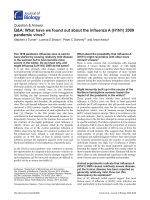
Figure 1. Stimulated whole saliva secretion following rituximab
treatment in patients with primary Sjögren’s syndrome.
Stimulated whole saliva secretion (SWS) at baseline and at 5 and
12weeks following rituximab treatment in 14 patients with primary
Sjögren’s syndrome; an increase in saliva secretion occurred only
in patients (n = 9) with baseline SWS >0.10 ml/minute and not
in patients (n = 5) with baseline secretion <0.10 ml/minute. SWS
consisted of submandibular and sublingual (SM/SL) salivary secretion.
Reprinted with permission from [35].
Kallenberg et al. Arthritis Research & Therapy 2011, 13:205
/>Page 4 of 8
related to disappearance of BAFF receptors after B-cell
depletion, and that B cells exert negative feedback on
BAFF production by monocytes – explaining the increase
of BAFF mRNA in monocytes following B-cell depletion.
e role of BAFF in recruiting (autoimmune) B cells in
pSS has been further explored by Pers and colleagues
[42]. ey observed that serum BAFF levels were
inversely correlated with the duration of B-cell depletion.
In some patients repeated labial salivary gland biopsies
were performed, showing that partial B-cell depletion in
the glands persisted for at least 12 months and B cells had
recurred at 24 months. Whereas repopulation of the
peripheral blood showed increased numbers of mature
naïve B cells (Bm2 cells) and decreased numbers of
memory B cells, repopulation of the salivary gland showed
memory B cells and transitional type 1 B cells as the fi rst
B cells to be identifi ed. ese memory B cells were specu-
lated to be autoreactive. We also observed delayed
recovery of CD27
+
memory B cells in the blood 48 weeks
after rituximab treatment, whereas the majority of emerg-
ing B cells had a phenotype of transitional B cells [43].
A recent study analyzed gene expression profi le of
labial salivary glands before and after rituximab treat-
ment and related these profi les to the clinical response on
rituximab [44]. Interestingly, the authors found two
groups of genes higher expressed in responders than in
nonresponders. e fi rst group consisted of genes
involved in the B-cell signaling pathway and the second
group was related to genes involved in the interferon
pathway. ese data fi t the concept of IFNα-induced
BAFF expression resulting in B-cell hyperactivity and
prolonged B-cell survival.
One open-label study targets CD22 on B cells [45]. is
molecule has a more or less similar distribution profi le to
CD20. Treatment of 16 patients with a monoclonal anti-
CD22 antibody, epratuzumab, resulted in improvement
of unstimulated whole saliva production and a decrease
in fatigue in one-half of the patients.
In summary, B cells seem to play a major role in
orchestrating the pathological immune response in pSS.
Depleting B cells off ers a unique possibility to study the
immunopathogenesis of pSS. BAFF appears as a strong
stimulant for B-cell activation and proliferation and for
B-cell survival in pSS.
Targeting BAFF in Sjögren’s syndrome
As mentioned before, BAFF plays a major role in pSS.
First, mice transgenic for BAFF develop with time a
clinical presentation of SS with lymphocytic infi ltration
of the salivary glands [46]. In these mice, marginal zone B
cells, part of them autoreactive, proliferate in the spleen
and later infi ltrate the salivary glands. Secondly, levels of
BAFF are increased in pSS and correlate with titers of
anti-SS-A and anti-SS-B antibodies [47]. irdly, BAFF is
overexpressed in the salivary glands in pSS [48], and
BAFF seems to determine B-cell repopulation in the
peripheral blood and salivary glands of pSS patients
following rituximab treatment [42].
Targeting BAFF in pSS therefore seems logical.
Currently, at least three drugs are available for targeting
BAFF in pSS. First, belimumab – a monoclonal antibody
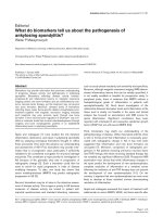
Figure 2. Histopathology of parotid gland before and after
treatment with rituximab in primary Sjögren’s syndrome.
Comparison of parotid biopsy specimens obtained from a primary
Sjögren’s syndrome (pSS) patient before rituximab therapy (A1 to
A4) and 12 weeks after therapy (B1 to AB4). (A1) Before treatment,
double staining illustrates intense in ammation (arrows) with highly
proliferating, large germinal center-like structures (GS; red nuclear
staining for Ki-67), fully developed lymphoepithelial lesions (LEL;
brown staining for cytokeratin 14 (CK14)), and reduced glandular
parenchyma (PAR). (B1) After treatment, in ammation was reduced
(arrows), with the absence of GS and the presence of regular
striated ducts (SD) devoid of lymphoepithelial lesions. (A2) Before
treatment, there was a dominance of B lymphocytes with GS (CD20)
in comparison with T lymphocytes (CD3) (A3). (B2) After treatment,
the lymphoid in ltrate overall was reduced, with a slight dominance
of T lymphocytes (CD3) (B3) compared with B lymphocytes
(CD20). (A4)Higher-magni cation view showing fully developed
lymphoepithelial lesions with many intraepithelial lymphocytes and
increased basal cell proliferation (arrows), in contrast to the SD after
therapy with CK14-positive basal cells (B4) (arrows) with regular
di erentiation into luminal ductal cells devoid of intraepithelial
lymphocytes (arrowheads). Original magni cation: A1 and B1, x120;
A2 and B2, x100; A3 and B3, x60; A4 and B4, x200. Reprinted with
permission from [37].
Kallenberg et al. Arthritis Research & Therapy 2011, 13:205
/>Page 5 of 8
to BAFF – is currently under trial (two studies) in
patients with pSS (NCT01160666 and NCT01008982)
but data are not yet available. Secondly, atacicept – a
fusion molecule of IgG–Fc and the extracellular domain
of TACI (the combined receptor for BAFF and A-
proliferation-inducing ligand)– has not yet been studied
in pSS. Finally, briobacept – a fusion protein of IgG–Fc
and the extracellular domain of the BAFF receptor – has
not yet been used in clinical trials in pSS. Targeting BAFF
using either belimumab, atacicept or briobacept could
reveal the pathogenic signifi cance of BAFF in pSS. A
hurdle to overcome, however, might be the heterogeneity
of BAFF presentation, either as monomers, homotrimers,
hetero trimers, splicoforms, or as membrane-bound
BAFF. Nevertheless this approach is promising. Further-
more, combining the targeting of BAFF with rituximab
treat ment could enhance and prolong the eff ect of
rituximab in pSS. Trials with belimumab, atacicept and
briobacept in pSS are eagerly awaited.
Targeting co-stimulation in Sjögren’s syndrome
Co-stimulation between antigen-presenting cells and
Tcells and between B cells and T cells is an essential step
in T-cell-dependent immune responses, including auto-
immune responses. Salivary gland epithelial cells in pSS
have been shown to express HLA class II and co-stimu-
latory molecules and may function as antigen-presenting
cells in pSS, besides dendritic cells and B cells [49].
Interfering in co-stimulation in pSS could, theoretically,
inhibit both systemic and local autoimmune responses in
pSS. Abatacept, a fusion molecule of IgG–Fc and cyto toxic
T-lymphocyte antigen 4, modulates CD28-mediated T-cell
co-stimulation. A controlled trial with abatacept in pSS
has been started in the authors’ department, but results
of treatment with abatacept in pSS are not yet available.
Conclusion
Treatment of SS has been only symptomatic for a long
time. e increasing availability of targeted treatment
modalities has created possibilities for intervention in
pathogenic pathways involved in the disease. is availa-
bility has not only opened new horizons for treatment,
but has also provided insight into the pathogenesis of SS.
In contrast to rheumatoid arthritis, the role of
proinfl ammatory cyto kines – in particular TNFα – is not
very outspoken in SS, as demonstrated by the lack of
effi cacy of TNF blocking. Otherwise, B cells appear to
play a major role in pSS. Depletion of B cells leads to
restoration of salivary fl ow and is eff ective for
extraglandular disease and mucosa-associated lymphoid
tissue lymphoma. B cells apparently also orchestrate T-
cell infi ltration and ductal epithelial dearrangement in
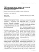
Figure 3. Relationship between disease duration and salivary ow rates in patients with primary Sjögren’s syndrome. The relationship
between disease duration (the time from rst complaints induced by or related to oral dryness until referral) and mean (standard error of the mean)
salivary ow rates in primary Sjögren’s syndrome (pSS) patients. Normal values are derived from historic controls (n = 36). SM/SL, submandibular/
sublingual glands; UWS, unstimulated whole saliva. *Signi cant di erence versus patients with early-onset pSS (≤1-year oral complaints; P <0.005)
by Mann–Whitney U test.
†
Signi cant di erence versus patients with early-onset pSS (P <0.05) by Mann–Whitney U test. Reprinted with permission
from [50].
Autoimmune Basis of Rheumatic Diseases
This article is part of a series on Sjögren’s syndrome, edited by Thomas
Dörner, which can be found online at />series/Sjögrens
This series forms part of a special collection of reviews covering major
autoimmune rheumatic diseases, available at:
/>Kallenberg et al. Arthritis Research & Therapy 2011, 13:205
/>Page 6 of 8
the glands, as deduced from histo patho logical studies. A
scenario in which the axis of IFNα, BAFF, B-cell
activation, proliferation and survival constitutes a basic
patho genic mechanism in pSS is supported by the results
of intervention studies currently available. Controlled
studies targeting IFNα and BAFF are eagerly awaited.
Abbreviations
BAFF, B-cell activating factor; IFN, interferon; IL, interleukin; pSS, primary
Sjögren’s syndrome; SS, Sjögren’s syndrome; TACI, transmembrane activator
and calcium-modulating cyclophilin ligand interactor; Th, T-helper type; TNF,
tumor necrosis factor.
Competing interests
The authors declare that they have no competing interests.
Author details
1
Department of Rheumatology and Clinical Immunology, AA21, University
Medical Center Groningen, University of Groningen, P.O. Box 30.001, 9700 RB
Groningen, The Netherlands.
2
Department of Oral and Maxillofacial Surgery,
University Medical Center Groningen, University of Groningen, 9700 RB
Groningen, The Netherlands.
Published: 28 February 2011
References
1. Varim MM, Le Pottier L, Youinou P, Saulep D, Mackay F, Pers JO: B-cell
tolerance breakdown in Sjögren’s syndrome: focus on BAFF. Autoimmun
Rev 2010, 9:604-608.
2. Kassan SS, Thomas TL, Moutsopoulos HM, Hoover R, Kimberly RP, Budman DR,
Costa J, Decker JL, Chused TM: Increased risk of lymphoma in sicca
syndrome. Ann Intern Med 1978, 88:888-892.
3. Baimpa E, Dahabreh U, Voulgarelis M, Moutsopoulos HM: Hematologic
manifestations and predictors of lymphoma development in primary
Sjögren’s syndrome: clinical and pathophysiological aspects. Medicine
2009, 88:284-293.
4. Tapinos NI, Polihronis M, Tzioufas AG, Skopouli FN: Immunopathology of
Sjögren’s syndrome. Ann Med Interne (Paris) 1998, 149:17-24.
5. Mavragani CP, Crow MK: Activation of the type I interferon pathway in
primary Sjogren’s syndrome. J Autoimmun 2010, 35:225-231.
6. Mariette X, Gottenberg JE: Pathogenesis of Sjögren’s syndrome and
therapeutic consequences. Curr Opin Rheumatol. 2010, 22:471-477.
7. Hansen A, Lipsky PE, Dörner T: Immunopathogenesis of primary Sjögren’s
syndrome: implications for disease management and therapy. Curr Opin
Rheumatol 2005, 17:558-565.
8. Yoon KC, Jeong IY, Park YG, Yang SY: Interleukin-6 and tumor necrosis
factor-alpha levels in tears of patients with dry eye syndrome. Cornea
2007, 26:431-437.
9. Koski H, Janin A, Humphreys-Beher MG, Sorsa T, Malmström M, Konttinen YT:
Tumor necrosis factor-alpha and receptors for it in labial salivary glands in
Sjögren’s syndrome. Clin Exp Rheumatol 2001, 19:131-137.
10. Baturone R, Soto MJ, Márquez M, Macías I, de Oca MM, Medina F, Chozas N,
García-Pérez S, Girón-González JA: Health-related quality of life in patients
with primary Sjögren’s syndrome: relationship with serum levels of
proin ammatory cytokines. Scand J Rheumatol 2009, 38:386-389.
11. Sisto M, D’Amore M, Caprio S, Mitolo V, Scagliusi P, Lisi S: Tumor necrosis
factor inhibitors block apoptosis of human epithelial cells of the salivary
glands. Ann N Y Acad Sci 2009, 1171:407-414.
12. Steinfeld SD, Demols P, Salmon I, Kiss R, Appelboom T: In iximab in patients
with primary Sjögren’s syndrome. Arthritis Rheum 2001, 44:2371-2375.
13. Steinfeld SD, Demols P, Appelboom T: In
iximab in primary Sjögren’s
syndrome. Arthritis Rheum 2002, 46:3301-3303.
14. Steinfeld SD, Appelboom T, Delporte C: Treatment with in iximab restores
normal aquaporin 5 distribution in minor salivary glands of patients with
Sjögren’s syndrome. Arthritis Rheum 2002, 46:2249-2251.
15. Mariette X, Ravaud P, Steinfeld S, Baron G, Goetz J, Hachulla E, Combe B,
Puéchal X, Pennec Y, Sauvezie B, Perdriger A, Hayem G, Janin A, Sibilia J:
Ine cacy of In iximab in primary Sjögren’s syndrome. Arthritis Rheum
2004, 50:1270-1276.
16. Batten M, Fletcher C, Ng LG, Groom J, Wheway J, Laâbi Y, Xin X, Schneider P,
Tschopp J, Mackay CR, Mackay F: TNF de ciency fails to protect BAFF
transgenic mice against autoimmunity and reveals a predisposition to
Bcell lymphoma. J Immunol 2004, 172:812-822.
17. Sankar V, Brennan MT, Kok MR, Leakan RA, Smith JA, Manny J, Baum BJ,
Pillemer SR: Etanercept in Sjögren’s syndrome. Arthritis Rheum 2004,
50:2240-2245.
18. Zandbelt MM, de Wilde P, van Damme P, Hoyng CB, van de Putte L, van den
Hoogen F: Etanercept in the treatment of patients with primary Sjögren’s
syndrome: a pilot study. J Rheumatol 2004, 31:96-101.
19. Moutsopoulos NM, Katsi s GE, Angelov N, Leakan RA, Sankar V, Pillemer S,
Wahl SM: Lack of e cacy of etanercept in Sjögren’s syndrome correlates
with failed suppression of tumour necrosis factor alpha and systemic
immune activation. Ann Rheum Dis 2008, 67:1437-1443.
20. Mavragani CP, Niewold TB, Moutsopoulos NM, Pillemer SR, Wahl SM, Crow
MK: Augmented interferon-α pathway activation in patients with Sjögren’s
syndrome treated with etanercept. Arthritis Rheum 2007, 50:3995-4004.
21. Onishi S, Nagashima T, Kimura H, Matsuyama Y, Yoshio T, Minota S: Systemic
lupus erythematosus and Sjögren’s syndrome induced in a case by
interferon-alpha used for the treatment of hepatitis C. Lupus 2010,
19:753-755.
22. Zheng L, Zhang Z, Yu C, Tu L, Zhong L, Yang C: Association between
IFN-alpha and primary Sjögren’s syndrome. Oral Surg Oral Med Oral Pathol
Oral Radiol Endod 2009, 107:e12-e18.
23. Båve U, Nordmark G, Lövgren T, Rönnelid J, Cajander S, Eloranta ML, Alm GV,
Rönnblom L: Activation of the type I interferon system in primary Sjögren’s
syndrome: a possible etiopathogenic mechanism. Arthritis Rheum 2005,
52:1185-1195.
24. Gottenberg JE, Cagnard N, Lucchesi C, Letourneur F, Mistou S, Lazure T,
Jacques S, Ba N, Ittah M, Lepajolec C, Labetoulle M, Ardizzone M, Sibilia J,
Fournier C, Chiocchia G, Mariette X: Activation of IFN pathways and
plasmacytoid dendritic cell recruitment in target organs of primary
Sjögren’s syndrome. Proc Natl Acad Sci U S A 2006, 103:2770-2775.
25. Wildenberg ME, van Helden-Meeuwsen CG, van de Merwe JP, Drexhage HA,
Versnel MA: Systemic increase in type I interferon activity in Sjögren’s
syndrome: a putative role for plasmacytoid dendritic cells. Eur J Immunol
2008, 38:2024-2033.
26. Lövgren T, Eloranta ML, Kastner B, Wahren-Herlenius M, Alm GV, Rönnblom L:
Induction of interferon-alpha by immune complexes or liposomes
containing systemic lupus erythematosus autoantigen- and Sjögren’s
syndrome autoantigen-associated RNA. Arthritis Rheum 2006,
54:1917-1927.
27. Ittah M, Miceli-Richard C, Gottenberg JE, Lavie F, Lazure T, Ba N, Sellam J,
Lepajolec C, Mariette X: B cell-activating factor of the tumor necrosis factor
family (BAFF) is expressed under stimulation by interferon in salivary
gland epithelial cells in primary Sjögren’s syndrome. Arthritis Res Ther 2006,
8:R51.
28. Shiozawa S, Morimoto I, Tanaka Y, Shiozawa K: A preliminary study on the
interferon-alpha treatment for xerostomia of Sjögren’s syndrome. Br J
Rheumatol 1993, 32:52-54.
29. Ferraccioli GF, Sala F, De Vita S, Casatta L, Avellini C, Carotti M, Beltrami CA,
Cervini C, Bartoli E: Interferon alpha-2 (IFNα2) increases lacrimal and
salivary function in Sjögren’s syndrome patients. Preliminary results of an
open pilot trial versus OH-chloroquine. Clin Exp Rheumatol 1996,
14:367-371.
30. Shiozawa S, Tanaka Y, Shiozawa K: Single-blinded controlled trial of low-
dose oral IFN-alpha for the treatment of xerostomia in patients with
Sjögren’s syndrome. J Interferon Cytokine Res 1998, 18:255-262.
31. Ship JA, Fox PC, Michalek JE, Cummins MJ, Richards AB: Treatment of
primary Sjögren’s syndrome with low-dose natural human interferon-
alpha administered by the oral mucosal route: a phase II clinical trial. IFN
Protocol Study Group. J Interferon Cytokine Res 1999, 19:943-951.
32. Cummins MJ, Papas A, Kammer GM, Fox PC: Treatment of primary Sjögren’s
syndrome with low-dose human interferon alfa administered by the
oromucosal route: combined phase III results. Arthritis Rheum 2003,
49:585-593.
33. Smith JK, Siddiqui AA, Modica LA, Dykes R, Simmons C, Schmidt J,
Krishnaswamy GA, Berk SL: Interferon-alpha upregulates gene expression
of aquaporin-5 in human parotid glands. J Interferon Cytokine Res 1999,
19:929-935.
34. Mariette X: Therapeutic potential for B-cell modulation in Sjögren’s
Kallenberg et al. Arthritis Research & Therapy 2011, 13:205
/>Page 7 of 8
syndrome. Rheum Dis Clin North Am 2008, 34:1025-1033.
35. Pijpe J, van Imho GW, Spijkervet FKL, Roodenburg JLN, Wolbink GJ, Mansour
K, Vissink A, Kallenberg CGM, Bootsma H: Rituximab treatment in patients
with primary Sjögren’s syndrome. Arthritis Rheum 2005, 52:2740-2750.
36. Meijer JM, Pijpe J, Vissink A, Kallenberg CGM, Bootsma H: Treatment of
primary Sjögren’s syndrome with rituximab: extended follow-up, safety
and e cacy of retreatment. Ann Rheum Dis 2009, 68:284-285.
37. Pijpe J, Meijer JM, Bootsma H, van der Wal JE, Spijkervet FKL, Kallenberg CGM,
Vissink A, Ihrler S: Clinical and histologic evidence of salivary gland
restoration supports the e cacy of rituximab treatment in Sjögren’s
syndrome. Arthritis Rheum 2009, 60:3251-3256.
38. Devauchelle-Pensec V, Pennec Y, Morvan J, Pers J-O, Daridon C, Jousse-Joulin
S, Roudaut A, Jamin C, Renaudineau Y, Quintin Roué I, Cochener B, Youinou P,
Saraux A: Improvement of Sjögren’s syndrome after two infusions of
rituximab (anti-CD20). Arthritis Rheum 2007, 57:310-317.
39. Dass S, Bowman SJ, Vital EM, Ikeda K, Pease CT, Hamburger J, Richards A, Rauz
S, Emery P: Reduction of fatigue in Sjögren’s syndrome with rituximab:
results of a randomised, double-blind, placebo-controlled pilot study.
AnnRheum Dis 2008, 67:1541-1544.
40. Meijer JM, Meiners PM, Vissink A, Spijkervet FKL, Abdulahad W, Kamminga N,
Brouwer E, Kallenberg CGM, Bootsma H: E ectiveness of rituximab
treatment in primary Sjögren’s syndrome. A randomized double-blind,
placebo-controlled trial. Arthritis Rheum 2010, 62:960-968.
41. Lavie F, Miceli-Richard C, Ittah M, Sellam J, Gottenberg JE, Mariette X: Increase
of B cell-activating factor of the TNF family (BAFF) after rituximab
treatment: insights into a new regulating system of BAFF production.
AnnRheum Dis 2007, 66:700-703.
42. Pers J-O, Devauchelle V, Daridon C, Bendaoud B, Le Berre R, Bordron A, Hutin
P, Renaudineau Y, Dueymes M, Loisel S, Berthou C, Saraux A, Youinou P:
BAFF-modulated repopulation of B lymphocytes in the blood and salivary
glands of rituximab-treated patients with Sjögren’s syndrome. Arthritis
Rheum 2007, 56:1464-1477.
43. Abdulahad WH, Meijer JM, Kroese FGM, Meiners PM, Vissink A, Spijkervet FKL,
Kallenberg CGM, Bootsma H: B-cell reconstitution and T-helper-cell balance
after rituximab treatment of active primary Sjögren’s syndrome. Arthritis
Rheum 2011, in press.
44. Devauchelle-Pensec V, Cagnard N, Pers JO, Youinou P, Saraux A, Chiocchia G:
Gene expression pro le in the salivary gland of primary Sjögren syndrome
patients, before and after treatment with Rituximab. Arthritis Rheum 2010,
62:2262-2271.
45. Steinfeld SD, Tant L, Burmester GR, Teoh NK, Wegener WA, Goldenberg DM,
Pradier O: Epratuzumab (humanised anti-CD22 antibody) in primary
Sjögren’s syndrome: an open-label phase I/II study. Arthritis Res Ther 2006,
8:R129.
46. Groom J, Kalled SL, Cutler AH, Olson C, Woodcock SA, Schneider P, Tschopp J,
Cachero TG, Batten M, Wheway J, Mauri D, Cavill D, Gordon TP, Mackay CR,
Mackay F: Association of BAFF/BLyS overexpression and altered B cell
di erentiation with Sjögren’s syndrome.
J Clin Invest 2002, 109:59-68.
47. Mariette X, Roux S, Zhang J, Bengoufa D, Lavie F, Zhou T, Kimberly R: The level
of BLyS (BAFF) correlates with the titre of autoantibodies in human
Sjögren’s syndrome. Ann Rheum Dis 2003, 62:168-171.
48. Lavie F, Miceli-Richard C, Quillard J, Roux S, Leclerc P, Mariette X: Expression
of BAFF (BLyS) in T cells in ltrating labial salivary glands from patients
with Sjögren’s syndrome. J Pathol 2004, 202:496-502.
49. Routsias JG, Tzioufas AG: Autoimmune response and target autoantigens in
Sjogren’s syndrome. Eur J Clin Invest 2010, 40:1026-1036.
50. Pijpe J, Kalk WW, Bootsma H, Spijkervet FK, Kallenberg CG, Vissink A:
Progression of salivary gland dysfunction in patients with Sjogren’s
syndrome. Ann Rheum Dis 2007, 66:107-112.
doi:10.1186/ar3234
Cite this article as: Kallenberg CGM, et al.: What have we learned from
clinical trials in primary Sjögren’s syndrome about pathogenesis? Arthritis
Research & Therapy 2011, 13:205.
Kallenberg et al. Arthritis Research & Therapy 2011, 13:205
/>Page 8 of 8