Báo cáo y học: "Dual role of interleukin-17 in pannus growth and osteoclastogenesis in rheumatoid arthritis" pps
Bạn đang xem bản rút gọn của tài liệu. Xem và tải ngay bản đầy đủ của tài liệu tại đây (2.78 MB, 11 trang )
RESEARC H ARTIC L E Open Access
Dual role of interleukin-17 in pannus growth and
osteoclastogenesis in rheumatoid arthritis
Hiroshi Ito
1
, Hidehiro Yamada
1*
, Toshiko N Shibata
1
, Hirofumi Mitomi
1
, So Nomoto
2
, Shoichi Ozaki
1
Abstract
Introduction: In a murine model, interleukin (IL)-17 plays a critical role in the pathogenesis of arthritis. There are
controversies, however, regarding whether IL-17 is a proinflammatory mediator in rheumatoid arthritis (RA). We
previously established an ex vivo cellular model using synovial tissue (ST)-derived inflammatory cells, which
reproduced pannus-like tissue growth and osteoclastic activity in vitro. Using this model, we investigated the
effects of IL-17 on pannus growth and osteoclastogenesis in RA.
Methods: Inflammatory cells that infiltrated synovial tissue from patients with RA were collected without enzyme
digestion and designated as ST-derived inflammatory cells. ST-derived inflammatory cells were cultured in the
presence or absence of IL-17 or indomethacin, and the morphologic changes were observed for 4 weeks.
Cytokines produced in the culture sup ernatants were measured by using enzyme-linked immunosorbent assay kits.
Osteoclastic activity was assessed by the development of resorption pits in calcium phosphate-coated slides.
Results: Exogenous addition of IL-17 dramatically enhanced the spontaneous production of IL-6 and prostaglandin
E
2
(PGE
2
) by the ST-derived inflammatory cells, while it had no effect on the production of tumor necrosis factor
(TNF)-a and macrophage colony-stimulating factor (M-CSF). Furthermore, IL-17 did not affect the spontaneous
development of pannus-like tissue growth and osteoclastic activity by the ST-derived inflammatory cells. On the
other hand, IL-17 enhanced pannus-like tissue growth, the production of TNF-a and M-CSF and the development
of osteoclastic activity in the presence of indomethacin, an inhibitor of endogenous prostanoid production, while
exogenous addition of PGE
1
suppressed their activities.
Conclusions: The present study suggests that IL-17 induces negative feedback regulation through the induction of
PGE
2
, while it stimulates proinfla mmatory pathways such as inflammatory cytokine production, pannus growth and
osteoclastogenesis in RA.
Introduction
Rheumatoid arthritis (RA) is chronic autoimmune
inflammatory disease that ultimately leads to the pro-
gressive destruction of cartilage and bone in nume rous
joints. Proinflammatory cytokines such as tumor necro-
sis factor (TNF)-a [1], interleukin (IL)-1 [2] and IL-6 [3]
were produced from synovial tissue (ST), which main-
tains it s inflammatory condition. Inflammation of syno-
vial membrane results in the development of a ggressive
granulation tissue, called pannus. Pannus tissue is
composed mainly of inflammatory cells such as macro-
phages and fibroblast-like synoviocytes (FLSs) [4].
At present, TNF-a and IL-6 are among the most
important targets of therapy, and blocking TNF-a results
in a rapid and sustained improvement of clinical signs
and symptoms [5-7]. Anti-TNF therapy also prevents
radiological progression of joint destructi on [8-10]. Anti-
IL-6 receptor monoclonal antibody (mAb) (tocilizumab)
has also proved to reduce disease activity, even in
patients who had an insufficient response to anti-TNF
therapy, and to inhibit the progression of structural joint
damage [11-13]. These clinical experiences suggest that
there are at least two pathways, TNF-a-dependent and
IL-6-dependent, leading to the progression of pannus
growth and joint destruction in RA.
* Correspondence:
1
Division of Rheumatology and Allergology, Department of Internal
Medicine, St. Marianna University School of Medicine, 2-16-1 Sugao,
Miyamae-ku, Kawasaki 216-8511, Japan
Full list of author information is available at the end of the article
Ito et al. Arthritis Research & Therapy 2011, 13:R14
/>© 2011 Ito et al.; licensee BioMed Central Ltd. This is an open access art icle distributed under the terms of the Creative Commons
Attribution License ( censes/by/2.0), which permits unrestricted use, distribution, and reproduction in
any medium, provided the original work is properly cited.
Recent studies have demonstrated critical roles of IL-
17, which is produced by a newly identified subset of
CD4
+
T cells, Th-17, in animal models of arthritis
[14,15]. In humans, IL-17 is a potent inducer of other
proinflammatory cytokines, such as TNF-a,IL-1b,IL-6
and IL-8 from monocytes and/or macrophages or syno-
vial fibroblas ts [16,17]. IL-17 has been detected in syno-
vial fluids of RA [18,19]. These findings suggest that
IL-17 is an important cytokine located upstream of the
two pathways, TNF-a-dependent and IL-6-dependent.
Preliminary clinical trial using humanized anti-IL-17 mAb
has shown an improvement of clinical signs and symptoms
of RA [20]. It is still unknown, however, whether inhibi-
tion of IL-17 prevents joint destruction in RA.
To further confirm the hy pothesis, the p resent study
was undertaken to clarify a role of IL-17 in RA using
our recently established ex vivo human cellular model,
where rheumatoid ST-derived inflammatory cells spon-
taneously develop pannus-like tissue in vitro and osteo-
clastic bone resorption [21].
Materials and methods
Reagents
IL-17 was purchased from PeproTech (Rocky Hill, NJ,
USA). PGE
1
was purchased from Sigma-Aldrich (St.
Louis, MO, USA). Indomethacin was obtained from
Wako (Osaka, Japan).
Synovial tissue specimens
ST specimens were obtained from patients who fulfilled
the American College of Rheumatology criteria for RA
who underwent knee join t replacement. In compliance
with institutional policies, informed consent was
obtained from all patients. The study was approved by
the ethics committee of each institution.
In vitro reconstruction of inflammatory tissue by ST-
derived inflammatory cells
ST-derived inflammatory cells were prepared as pre-
viously described [21]. In brief, ST specimens were cut
into small pieces and cultured in 100-mm dishes contain-
ing RPMI-1640 (Asahi Technoglass, Chiba, Japan) with
10% fetal calf serum ( FCS) and 1,000 U/ml penicillin G
sodium-streptomycin sulfate (Gibco BRL, Grand Island,
NY, USA). After 1 to 3 days’ incubation, tissue was
removed and single cells were c ollected by vigorous
pipetting. Cell suspensions were washed once, and viable
cells were collected into Lymphocyte Separation Medium
(Nacalai Tesque, Kyoto, Japan). Single suspensions of ST-
derived inflammatory cells were seeded at a density of
5×10
5
/well in 48-well culture plates and cultured in
Dulbecco’ s modified Eagle’ s medium (DMEM; Gibco
BRL) containing 10% FCS, 100 U/ml penicillin G sodium
and 100 μg/ml streptomycin sulfate. The c ulture was
observed for morphologic changes under an inverted
phase-contrast microscope twice a week for 4 weeks.
When cultured in DMEM and 10% FCS in the absence
or presence o f IL-17 (0.1 to 100 ng/ml) or indomethacin
(100 nM to 1 μ M), ST-derived inflammatory cells started
to aggregate, forming foci within a few days. Further cul-
turing resulted in three-dimensional (3-D) growth, which
ultimately produced macroscopic tissue 2 mm in size
within 4 weeks. Morphologic changes were semiquantita-
tively scored on a scale of 0 to 4, according to the degree
of tissue development, where 0 was no cellular foci or
aggregations, 1 was the formation of cellular foci or
aggregation, 2 was further growth of cellular aggrega-
tions, 3 was further 3-D growth with a multilayered
structure, and 4 was th e development of macroscopic tis-
sue. Cumulative tissue growth score was calculated by
the total sum of the tissue growth scores obtained twice
weekly for 4 weeks of culture. Half of the supernatants
were collected twice weekly and replaced with fresh med-
ium or the addition of a half dose of IL-17 or indometha-
cin. Supernatants were frozen at -80°C until assayed.
Cytokine assay
ST-derived inflammatory cells were seeded in 48-well
culture plates (5 × 10
5
/well) and cultured in DMEM
and 10% FCS.
Half of the supernatants were collected three times per
week and replaced with fresh medium. Supernatants were
frozen at -80°C until assayed, and level s of IL-6, PGE
2
,
TNF-a and M-CSF (all from R&D Systems, Minneapolis,
MN, USA) released into the culture supernatants were
measured using enzyme-linked immunosorbent assay kits
according to the manufacturers’ recommendations.
Bone resorption assay
ST-derived inflammatory cells were seeded (1 × 10
5
cells/ well) onto calcium phosphate-coated slides (Osteo-
logic; BD Biosciences, MA, USA) and incubated in
RPMI-1640 with 1% FCS, 50 μg/ml ascorbic acid
(Sigma) and 10 mM b-glycerophosphate (Sigma) for 7
to 14 days in a CO
2
incubator (5% CO
2
,100%humidity
at 37°C). Half of the supernatants were replaced with
fresh medium once weekly. The calcium phosphate-
coated slides were washed with distilled water and
bleach solution (6% NaOCl and 5.2% NaCl) and then
air-dried. The number of resorption pits were counted
under a microscope.
Results
IL-17 enhances IL-6 and PGE
2
production by ST-derived
inflammatory cells
Using a recently established ex vivo cellular model of
RA, we examined the eff ect of IL-17 on the production
of IL-6 and PGE
2
by the ST-derived inflammatory cells.
Ito et al. Arthritis Research & Therapy 2011, 13:R14
/>Page 2 of 11
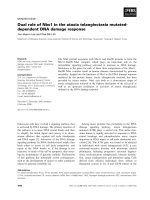
During the cell cultur e, ST-derived inflammatory cells
spontaneously produced IL-6 and PGE
2
in the superna-
tant as shown in Figure 1. Addition of IL-17 into the
culture resulted in the enhancement of both IL-6 and
PGE
2
production in a dose-dependent manner.
Effect of IL-17 on the development of pannus-like
inflammatory tissue in vitro by the ST-derived
inflammatory cells
We have reported that ST-derived inflammatory cells
showed spontaneous develop ment of pannu s-like tissue
in vitro [21]. The ST-derived inflammatory cells at the
beginning of the culture contained 1.6% to 4.2% FLSs
(mean, 2.6%), 35.8% to 65.7% macrophages (mean,
53.7%) and 32.4% t o 62.6% small lymphoc ytes (mean,
44.7%) when assessed by morphological observation.
During the culture of ST-derived inflammatory cells,
marked proli feration and migration of the FLSs into the
pannus-like tissue were observed. At the end o f culture,
pannus-like tissue contained more than 80% FLSs and
less than 10% of macrophages and T cells as assessed by
immunohistochemistry. As IL-17 enhanced IL-6 and
PGE
2
production by the ST-derived inflammatory cells,
we investigated the effect of IL-17 on the development
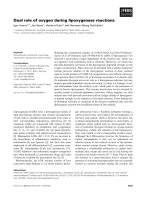
of pannus-like tissue in vitro. The cumulative tissue
growth score during 4 weeks of cult uring of ST-derived
inflammatory cells was not affected by the addition of
IL-17upto100ng/ml,whileitwassuppressedbythe
exogenous addition of 100 nM PGE
1
(Figure 2) as well
as 100 nM PGE
2
(data not shown).
These results suggested that the effect of IL-17 on the
development of pannus-like tissue w as modified by IL-
17-enhanced endogenous PGE
2
production. To confirm
this possibility, we investigated the effect of indomet ha-
cin, an inhibitor of endogenous prostanoids, on the pan-
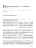
nus-like tissue growth in vitro. Addition of in domethacin
resulted in a significant enhancement of the in vitro
tissue growth by the ST-derived inflammatory cells
(Figure 3). In the presence of indomethacin, the in vitro
tissue growth was enhanced by the addition of IL-17 in a
dose-dependent manner.
IL-17 enhances M-CSF and TNF-a production by ST-
derived inflammatory cells in the presence of
indomethacin
Rheumatoid ST contains a number o f proinflammatory
cytokines that influence osteoclast formation and bone
resorption. Proinflammatory cytokines such as TNF-a and
IL-6 stimulate differentiation and activation of osteoclasts,
resulting in increased bone resorption. M-CSF is constitu-
tively produced by synov ial fibroblasts from RA patients
and contributes to the differentiation of synovial macro-
phages into osteoclasts. We investigated the effect of IL-17
on M-CSF and TNF-a production from ST-derived
medium IL-17 1ng/ml
IL-17 10 ng/ml IL-17 100ng/ml
(pg/ml)
1
10
100
1000
10000
100000
1W 2W 3
W
B
P
G
E2
0
125
250
375
500
1W 2W 3W
(ng/ml)
A
IL-6
Figure 1 Effect of interl eukin (IL)-17 on the production of (A) IL -6 (A) and (B) pro staglan din E
2
(PGE
2
) by the synovial tissue (ST)-
derived inflammatory cells. Cells were incubated in the absence or presence of increasing concentrations of IL-17 (0 to 100 ng/ml) for 3
weeks. IL-6 (n = 7) and PGE
2
(n = 3) in the culture supernatants were measured by enzyme-linked immunosorbent assay as described in
Materials and methods.
Ito et al. Arthritis Research & Therapy 2011, 13:R14
/>Page 3 of 11
inflammatory cells. During the cell c ulture, ST-derived
inflammatory cells spontaneously produced M-CSF and
TNF-a in the supernatant as described previously [21].
Contrary to our expectation, spontaneous production of
both M-CSF and TNF-a was not affected by the addition
of IL-17 up to100 ng/ml (Figures 4A and 4B).
As PGE
2
is known to inhibit the production of M-CSF
and TNF-a from macrophages and synovial fibroblasts
[22,23], respectively, we examined the effect of IL-17 on
the production of M-CSF and TNF-a in the presence of
indomethacin to block the effect of en dogenous PGE
2
.
In the presence of indomethacin, IL-17 significantly
enhanced the production of M-CSF and TNF-a in a
dose-dependent manner (Figures 4A and 4B), w hile IL-
17-induced IL-6 production was not affected by the
addition of indomethacin (Figure 4C).
IL-17 stimulates osteoclastic bone resorption
We previously showed that ST-derived inflammatory cells
in a 1% FCS-containing medium showed spontaneous
Figure 2 Effect of interleukin (IL)-17 and prostaglandin E
1
(PGE
1
) on pannus-like tissue growth in vitro.Synovialtissue(ST)-derived
inflammatory cells were incubated in the absence or presence of increasing concentrations of IL-17 (0 to 100 ng/ml) (n = 17) or PGE
1
(100 nM)
(n = 9). Morphologic changes were observed under an inverted phase contrast microscope twice weekly for 4 weeks and were scored
semiquantitatively on a scale of 0 to 4 according to the degree of tissue development as described in Materials and methods. Box and whisker
plots indicate 25th/75th percentile and minimum/maximum, respectively. Bar indicates the median value. *P < 0.001 (by Mann-Whitney U test).
Ito et al. Arthritis Research & Therapy 2011, 13:R14
/>Page 4 of 11
development of multinucleated giant cells within 2 weeks.
They were tartrate-resistant acid phosphatase-positive
multinucleated cells and developed numerous resorption
pits when incubated on a calcium phosphate-coated slide
[21]. Exogenous additio n of IL-17 tended to increase the
number of resorption pits, but the difference did not reach
statistical significance (Figure 5). Indomethacin signifi-
cantly enhanced the development of resorption pits by the
ST-derived inflammatory cells. In the presence of indo-
methacin, IL-17 significantly increased the number of
resorption pits in a dose-dependent manner (Figure 5).
Discussion
Inflammation in general is fundamentally a protective
response against cellular and tissue injury caused by
diverse pathological stimuli, and it is closely intertwined
Figure 3 Effect of interleukin (IL)-17 on pannus-like tissue gro wth in t he presence of indomet hacin.Synovialtissue(ST)-derived
inflammatory cells (n = 7) were incubated with an incremental dose of IL-17 in the absence or presence of indomethacin (100 to 1000 nM).
Morphologic changes were observed under an inverted phase contrast microscope twice weekly for 4 weeks and were scored semiquantitatively
on a scale of 0 to 4 according to the degree of tissue development as described in Materials and methods. Box and whisker plots indicate 25th/
75th percentile and minimum/maximum, respectively. Bar indicates the median value. *P < 0.05 (by Wilcoxon signed-rank test).
Ito et al. Arthritis Research & Therapy 2011, 13:R14
/>Page 5 of 11
with the process o f repair. In some circumstances,
inflammation and tissue repair are not successfully com-
pleted and inflammation perpetuates chronically. RA is
characterized by chronic inflammation of the synovial
membrane, which results in the development of aggres-
sive granulation tissue, so-called pannus, and the subse-
quent destruction of cartilage and bone. Pannus tissue is
composed mainly of invasive phenotype of FLSs, lym-
phocytes and activated macrophages, and in t he case of
bone erosion, monocyte-derived osteoclasts [4]. Cyto-
kine networks and cell-cell interaction, as well as other
inflammatory mediators, such as prostanoids, contribute
to the development of pannus tissue and osteoclastic
activity. This complex system of rheumatoid synovitis
includes both positive and negative feedback regulation
of inflammatory responses. Therefore, a human cell
model that represents this complex system will be useful
to study the role of IL-17 in the pathogenesis of RA. We
previously established an ex vivo cellular model using
the ST-derived inflammatory cells, which reproduced
pannus-like tissue growth and osteo clastic activit y
in vitro. Using this model, the present study demon-
strated that IL-17 enhanced production of proinflamma-
tory cytokines, pannus-like tissue growth and
osteoclastic activity by the ST-derived inflammatory
cells, while IL-17 simultaneous ly induced negative feed-
back regulation through the enhanced production of
PGE
2
, a potent deactivator of macrophages and other
inflammatory modulator [24]. Inhibition of endogenous
PGE
2
production resulted in the enhancement of pan-
nus growth and osteoclastic activity. Therefore, the net
effects o f IL-17 may depend upon the balance between
the positive and negative regulatory responses.
IL-17 is an important proinflammatory cytokine
involved in the pathogenesis of RA. Previous studies have
shown that IL-17 is present in rheumatoid synovial fluid
and can upregulate several mediators of inflammation,
such as TNF-a, IL-1, IL-6, IL-8 and matrix metalloprotei-
nases (MMPs), in FLS. Among other cytokines, both
TNF-a and IL-6 have been shown to play a pivotal role in
the progression of RA. The importance of TNF-a and IL-
6 in the pathogenesis of RA has been established by the
clinical experiences with anti-TN F and anti-IL-6 therapy
[5-7,11,13]. Blocking TNF -a by either neutralizing mAbs
(infliximab and adalimumab) or soluble TNF receptor-
immunoglobulin G (IgG)-Fc fusion protein (etanercept)
resulted in a rapid and sustained improvement of clinical
signs and symptoms in both early and advanced RA. Anti-
TNF therapy also prevented r adiological progression of
joint destruction [8-10]. Anti-IL-6 receptor mAb (tocilizu-
mab) has also been proved to reduce disease activity, even
in patients who had insufficient response to anti-TNF
therapy [12], and to inhibit the progression of structural
joint damage [11,13]. These clinical experiences sugge st
that there are at least two pathways, TNF-a-dependent
and IL-6-dependent, leading to the progression of pannus
growth and joint destruction in RA. IL-17 has been shown
to stimulate TNF-a and IL-6 expression [16,17], suggest-
ing that IL-17 is an important cytokine located upstream
of the two pathways.
PGE
2
has been established as a regulator of cytokine
production by activated macrophages. PGE
2
inhibits the
AB
C
Indomethacin
IL-17 (ng/ml) 0
1
10
100
0
10
100
1
++
+
-
+
0
100
200
300
IL-6 (ng/ml)
Figure 4 Effect of interleukin (IL)-17 on the production of macrophage co lony-stimulati ng factor (M-CSF), tumor necrosis factor a
(TNF-a) and IL-6. Synovial tissue (ST)-derived inflammatory cells were incubated with incremental doses of IL-17 in the absence or presence of
indomethacin (100 to 1,000 nM) for 1 week. Enzyme-linked immunosorbent assay kits were used to measure the concentration of (A) TNF-a, (B)
M-CSF and IL-6 (C) in the culture supernatants derived from seven donors. There were no significant differences in the production of IL-6
between the presence and absence of indomethacin. Box and whisker plots indicate 25th/75th percentile and minimum/maximum, respectively.
Bar indicates the median value. *P < 0.05 (by Wilcoxon signed-rank test).
Ito et al. Arthritis Research & Therapy 2011, 13:R14
/>Page 6 of 11
producti on of TNF-a, IL-6, IL-8 and IL-12 and downre-
gulates the expression of IL-12 receptor on macrophages
[23,25,26]. PGE
2
downregulates TNF-a and upregulates
IL-10 through the EP
2
and EP
4
receptors. This effect of
PGE
2
can reverse cytokine disequilibrium from proin-
flammatory toward anti-inflammatory [24,27]. PGE
2
has
been reported to suppress IL-17-induced TNF-a mRNA
expression and protein synthesis in human macrophages
and synovial fibroblasts from RA patients via EP
4
recep-
tor- and EGR-1-mediated inhibition of c-Jun expression
[28]. PGE
2
induces egr-1 mRNA expression and protein
synthesis by activating transcription factor 2 (ATF-2)
I
ndomethacin
IL-17 (ng/ml) 0.1
1
10
0.1
1001
+
+
+
+
0.1
1
10
100
ratio of resorption pits
**
p < 0.05
*
Figure 5 Effect of interleukin (IL)-17 on the osteoclastogenesis. Osteoclastic activity was assessed by the development of resorption pits in
calcium phosphate-coated slides as described in Materials and methods. Synovial tissue (ST)-derived inflammatory cells (n = 6) were cultured
with incremental doses of IL-17 in the absence or presence of indomethacin on calcium phosphate-coated slides for 2 weeks and examined for
the development of resorption pits. The ratio to the number of resorption pits in medium alone was plotted. Bar indicates the median value. *P
< 0.05 vs. medium alone (by Wilcoxon signed-rank test).
Ito et al. Arthritis Research & Therapy 2011, 13:R14
/>Page 7 of 11
dimer via transactivation of the eg r-1 promoter. IL-17-
upregulated promoter activity was largely depend ent on
ATF-2/c-Jun transactivation. PGE
2
suppression of IL-17-
induced ATF-2/c-Jun transactivation, and DNA binding
was dependent on egr-1-mediated inhibition of the
induced c-Jun expression. While upregulating TNF-a
expression, IL-17 also induces cyclooxygenase 2
(COX2)/PGE
2
expression, which in turn downregulates
TNF-a expression. This negative feedback regulation of
TNF-a expression by PGE
2
may be critical in the modu-
lation of the immune and inflammatory responses in
RA. The present study has demonstrated that IL-17-
induced TNF-a production, pannus-like tissue growth
and osteoclastic activity by the ST-derived inflammatory
cells were effectively downregulated by the negative
feedback loop through PGE
2
production, while IL-17-
induced IL-6 production was not.
PGE
2
has been shown to inhibit IL-6 production by
activated human macrophages [26], while other studies
have shown that PGE
2
enhanced IL-6 production by IL-
1b-stimulated human synovial fibroblasts and osteo-
blasts, as well as chondrocytes [22,29,30]. The present
studyhasshownthattheneteffectofIL-17onIL-6
production by the ST-derived inflammatory cells was
notaffectedbytheendogenousPGE
2
. This resul t sug-
gests that the effect of IL-17 is mainly mediated by the
IL-6 pathway, wh ile the TNF-a pathway is downregu-
lated by endogenous PGE
2
.
In RA, increased FLS proliferation and/or decreased
FLS apoptosis contributes to synovial hyperplasia and
pannus growth [31]. IL-17 has been shown to induce
proliferation of FLS through the induction of Cyr61, a
product of a growth factor-inducible immediate early
gene, and the subsequent expression of Bcl-2 that pre-
vents RA FLS apoptosis [32]. COX2-derived PGE
2
inhi-
bits IL-1/TNF-a-stimulated MMP-1 release from FLSs
via inhibition of extracellular signal-regulated kinase
(ERK) [33]. On the contrary, COX inhibitors attenuated
PGE
2
inhibition of ERK and enhanced the release of
MMP-1 by F LSs [33]. IL-1b and TNF-a stimulate the
translocation of p65 and p50 from the cytosol to the
nucleus and activate NF-B in human RA synovial
fibroblasts [27]. PGE
2
inhibits p65 translocation via inhi-
bition of ERK activation and also enhances the expres-
sion of IBa in an ERK-independent manner, suggesting
that PGE
2
inhibits NF-B activation by both ERK-
dependent and ERK-independent mechanisms. These
data indicate that PGE
2
may act to attenuate cytokine-
induced inflammatory responses in RA synovial fibro-
blasts by regulating the localization of specific NF-B
family dimers [27].
In summary, there is accumulating evidence that sug-
gests a molecular cross-talk mechanism involving COX2
and PGE
2
expression in the resolution of inflammation.
Proinflammatory cytokines, including IL-17 and TNF-a,
play a critical role i n the pr ogression of synovitis and
joint destruction, mainly through activation of NF-B,
while they directly induce COX2 and PGE
2
expression.
PGE
2
upregulates COX2 expression via EP
2
and EP
4
receptors and cyclic adenosine monophosphate-depen-
dent signaling pathway, which in turn modulates the
production of the proinflammatory molecules. The link
between proinflammaory molecules and PGE
2
could
have considerable importance in the regulation of
inflammatory cell activation of RA. The paracrine and
autocrine feedback mechanisms via COX2, PGE
2
,EP
2
and EP
4
could help to avoid the potential pathological
damage caused by the excessive production of inflam-
matory mediators in response to various biological sti-
muli in RA.
In the present study, we used PGE
1
instead of PGE
2
as
the exogenous source of cell cultures (Figure 2). Pre-
vious studies ind icated that PGE
1
and PGE
2
are equiva-
lent in terms of biological effects on human synovial
fibroblast proliferation [34] and their binding affinity to
PGE
2
-specific receptors EP
1
,EP
2
,EP
3
and EP
4
[35]. Our
preliminary data also shows that both PGE
1
and PGE
2
equivalently inhibited both FLS proliferation and in vitro
pannus-like tissue g rowth by the ST-derived inflamma-
tory cells in a dose-dependent manner (data not shown).
The reason why we have used PGE
1
instead of PGE
2
was the fact that we were intending to develop a novel
therapeutic strategy utilizing anti-inflammatory effects of
PGE
1
. There have been several attempts to use PGE
1
to
treat autoimmune and inflammatory diseases such as
adjuvant arthritis [36] and lupus nephrit is [37]. We also
published the inhibitory effect of lipid microsphere-
incorpora ted PGE
1
in a collagen-induced arthritis model
[38].
Osteoclastic bone resorption is another important
feature of pannus tissue in RA. Receptor activator of
NF-B ligand (RANKL) and M-CSF are essential for
osteoclastogenesis [39,40]. The expression of RANKL on
activated T cells, osteoblasts and synovial fibroblasts
contribute to osteoclastic bone resorption in RA
patients. M-CSF is constitutively produced by synovial
fibroblasts from RA patients and contributes to the dif-
ferentiation of synovial macrophages into osteoclasts in
collaboration with RANKL [41]. In humans, IL-17
induced the expression of both RANK on osteoclast pre-
cursors [42] and RANKL on synovial fibroblast [43]. A
rec ent study showe d that TNF-induced RANKL expre s-
sion was IL-6-dependent [44]. On the other hand, both
TNF-a and IL-6 also stimulate osteoclastogenesis in a
RANKL-independent manner [45,46]. In the present
study, we have demonstrated that IL-17 also stimulated
M-CSF production by the ST-derived inflammatory
cells. The result is consistent with a recent report that
Ito et al. Arthritis Research & Therapy 2011, 13:R14
/>Page 8 of 11
IL-17inducedM-CSFexpressiononhumanbonemar-
row-derived mesenchymal stem cells [47].
Another important question is whether IL-17-
enhanced osteoclastogenesis under t he suppression of
endogenous prostanoids is TNF-dependent and/or
IL-6-dependent. IL-17 is known to stimulate RANKL
expression on fibroblast-like synoviocytes through the
induction of IL-6 [44]. On the other hand, I L-17 is
reported to induce osteoclast formation through RANK
expression on osteoclast precursors [42]. Whether this
effect is TNF-dependent and/or IL-6-dependent remains
unknown. These questions require further studies
including experiments neutralizing TNF-a and IL-6.
Proinflammatory cytokines such as TNF-a and IL-6
have been known to stimulate osteoclastogenesis
through enhancing RANKL expression. IL-17, an indu-
cer of TNF-a and IL-6 expression, is also a potent sti-
mulator of osteoclastogenesis in RA. In animal models,
it has been reported that TNF-a and IL-1b stimulate
osteoclastogenesis through PGE
2
[48]. Recently, one of
these research groups demonstrated that, in contrast to
mouse macrophage cultures, PGE
2
inhibited RANKL-
induced human osteoclast formation in CD14
+
cell
cultures [49]. In our cellular model of RA, we demon-
strated that IL-17 enhanced osteoclastogenesis by the
ST-derived inflammatory cells only when endogenous
prostanoid production was inhibited by indomethacin.
The result can be explained by the fact that IL-17-
induced TNF-a and M-CSF production was downregu-
lated by the simultaneous induction of e ndogenous
PGE
2
. The present study also leads to a clinically impor-
tant suggestion that suppression of PGE
2
by the contin-
uous use of nonsteroidal anti-inflammatory drugs
(NSAIDs) such as indomethacin may augment TNF-a
pathway-dependent pannus growth and osteoclastic
bone resorption, resulting in the joint destruct ion in RA
[24].
Conclusions
Using a human cellular model of pannus, we have
demonstrated that IL-17 induced both proinflammator y
cascades, including TNF-a and IL-6, as well as negative
feedback regulation by PGE
2
production. The positive
effect of IL-17 on pannus-like t issue growth and osteo-
clastic activity by the ST-d erived inflammatory cells was
effectively downregulated by the simultaneously induced
endogenous PGE
2
. The negative feedback mechanisms
via PGE
2
could help to avoid the potential pathological
damage caused by the excessive production of mediators
in response to various b iological stimuli such as IL-17
in RA. Whe ther continuous inhibition of PGE
2
by the
administration of NSAIDs and COX2 inhibitors could
augment pannus growth and joint destruction remains
to be clarified.
Abbreviations
COX: cyclooxygenase; FLS: fibroblast-like synoviocyte; IL: interleukin; mAb:
monoclonal antibody; M-CSF: macrophage colony-stimulating factor; MMP:
matrix metalloproteinase; OPG: osteoprotegrin; PG: prostaglandin; RA:
rheumatoid arthritis; RANKL: receptor activator of NF-κB ligand; ST: synovial
tissue; Th17: T-helper type 17; TNF: tumor necrosis factor.
Acknowledgements
The authors are grateful to Dr. Kuniomi Yamasaki for continuing
encouragement and his financial support for our work, and we also thank
Kyoko Takahashi and Kiyomi Matsuo for excellent technical assistance.
Author details
1
Division of Rheumatology and Allergology, Department of Internal
Medicine, St. Marianna University School of Medicine, 2-16-1 Sugao,
Miyamae-ku, Kawasaki 216-8511, Japan.
2
Department of Orthopaedic Surgery
and Rheumatology, Saiseikai Yokohamashi Tobu Hospital, 3-6-1,
Shimosueyoshi, Tsurumi-ku, Yokohama 230-8765, Japan.
Authors’ contributions
HI conducted the experimental work, performed the statistical analysis and
drafted the manuscript. TNS, HM and SN helped with some experimental
work and provided synovial tissues. HY and SO designed and conceived of
the study, coordinated the project and drafted the manuscript. All authors
read and approved the final manuscript.
Competing interests
Hidehiro Yamada received research fund from Ono Pharmaceuticals Co. All
other authors declare that they have no competing interests.
Received: 25 September 2010 Revised: 31 December 2010
Accepted: 4 February 2011 Published: 4 February 2011
References
1. Chu CQ, Field M, Feldmann M, Maini RN: Localization of tumor necrosis
factor α in synovial tissues and at the cartilage-pannus junction in
patients with rheumatoid arthritis. Arthritis Rheum 1991, 34:1125-1132.
2. Buchan G, Barrett K, Turner M, Chantry D, Maini RN, Feldmann M:
Interleukin-1 and tumour necrosis factor mRNA expression in
rheumatoid arthritis: prolonged production of IL-1α. Clin Exp Immunol
1988, 73:449-455.
3. Hirano T, Matsuda T, Turner M, Miyasaka N, Buchan G, Tang B, Sato K,
Shimizu M, Maini R, Feldmann M, Kishimoto T: Excessive production of
interleukin 6/B cell stimulatory factor-2 in rheumatoid arthritis. Eur J
Immunol 1988, 18:1797-1801.
4. Bromley M, Woolley DE: Histopathology of the rheumatoid lesion.
Identification of cell types at sites of cartilage erosion. Arthritis Rheum
1984, 27:857-863.
5. Maini R, St Clair EW, Breedveld F, Furst D, Kalden J, Weisman M, Smolen J,
Emery P, Harriman G, Feldmann M, Lipsky P: Infliximab (chimeric anti-
tumour necrosis factor α monoclonal antibody) versus placebo in
rheumatoid arthritis patients receiving concomitant methotrexate: a
randomised phase III trial. ATTRACT Study Group. Lancet 1999,
354:1932-1939.
6. Weinblatt ME, Kremer JM, Bankhurst AD, Bulpitt KJ, Fleischmann RM, Fox RI,
Jackson CG, Lange M, Burge DJ: A trial of etanercept, a recombinant
tumor necrosis factor receptor:Fc fusion protein, in patients with
rheumatoid arthritis receiving methotrexate. N Engl J Med 1999,
340:253-259.
7. Weinblatt ME, Keystone EC, Furst DE, Moreland LW, Weisman MH,
Birbara CA, Teoh LA, Fischkoff SA, Chartash EK: Adalimumab, a fully human
anti-tumor necrosis factor α monoclonal antibody, for the treatment of
rheumatoid arthritis in patients taking concomitant methotrexate: the
ARMADA trial. Arthritis Rheum 2003, 48:35-45.
8. Lipsky PE, van der Heijde DM, St Clair EW, Furst DE, Breedveld FC,
Kalden JR, Smolen JS, Weisman M, Emery P, Feldmann M, Harriman GR,
Maini RN, Anti-Tumor Necrosis Factor Trial in Rheumatoid Arthritis with
Concomitant Therapy Study Group: Infliximab and methotrexate in the
treatment of rheumatoid arthritis. Anti-Tumor Necrosis Factor Trial in
Rheumatoid Arthritis with Concomitant Therapy Study Group. N Engl J
Med 2000, 343:1594-1602.
Ito et al. Arthritis Research & Therapy 2011, 13:R14
/>Page 9 of 11
9. Genovese MC, Bathon JM, Martin RW, Fleischmann RM, Tesser JR, Schiff MH,
Keystone EC, Wasko MC, Moreland LW, Weaver AL, Markenson J,
Cannon GW, Spencer-Green G, Finck BK: Etanercept versus methotrexate
in patients with early rheumatoid arthritis: two-year radiographic and
clinical outcomes. Arthritis Rheum 2002, 46:1443-1450.
10. Breedveld FC, Weisman MH, Kavanaugh AF, Cohen SB, Pavelka K, van
Vollenhoven R, Sharp J, Perez JL, Spencer-Green GT: The PREMIER study: a
multicenter, randomized, double-blind clinical trial of combination
therapy with adalimumab plus methotrexate versus methotrexate alone
or adalimumab alone in patients with early, aggressive rheumatoid
arthritis who had not had previous methotrexate treatment. Arthritis
Rheum 2006, 54:26-37.
11. Okuda Y: Review of tocilizumab in the treatment of rheumatoid arthritis.
Biologics 2008, 2:75-82.
12. Emery P, Keystone E, Tony HP, Cantagrel A, van Vollenhoven R, Sanchez A,
Alecock E, Lee J, Kremer J: IL-6 receptor inhibition with tocilizumab
improves treatment outcomes in patients with rheumatoid arthritis
refractory to anti-tumour necrosis factor biologicals: results from a 24-
week multicentre randomised placebo-controlled trial. Ann Rheum Dis
2008, 67:1516-1523.
13. Smolen JS, Beaulieu A, Rubbert-Roth A, Ramos-Remus C, Rovensky J,
Alecock E, Woodworth T, Alten R: Effect of interleukin-6 receptor
inhibition with tocilizumab in patients with rheumatoid arthritis
(OPTION study): a double-blind, placebo-controlled, randomised trial.
Lancet 2008, 371:987-997.
14. Bush KA, Farmer KM, Walker JS, Kirkham BW: Reduction of joint
inflammation and bone erosion in rat adjuvant arthritis by treatment
with interleukin-17 receptor IgG1 Fc fusion protein. Arthritis Rheum 2002,
46:802-805.
15. Nakae S, Nambu A, Sudo K, Iwakura Y: Suppression of immune induction
of collagen-induced arthritis in IL-17-deficient mice. J Immunol 2003,
171:6173-6177.
16. Hwang SY, Kim JY, Kim KW, Park MK, Moon Y, Kim WU, Kim HY: IL-17
induces production of IL-6 and IL-8 in rheumatoid arthritis synovial
fibroblasts via NF-κB- and PI3-kinase/Akt-dependent pathways. Arthritis
Res Ther 2004, 6:R120-R128.
17. Jovanovic DV, Di Battista JA, Martel-Pelletier J, Jolicoeur FC, He Y, Zhang M,
Mineau F, Pelletier JP: IL-17 stimulates the production and expression of
proinflammatory cytokines, IL-β and TNF-α, by human macrophages.
J Immunol 1998, 160:3513-3521.
18. Kotake S, Udagawa N, Takahashi N, Matsuzaki K, Itoh K, Ishiyama S, Saito S,
Inoue K, Kamatani N, Gillespie MT, Martin TJ, Suda T: IL-17 in synovial
fluids from patients with rheumatoid arthritis is a potent stimulator of
osteoclastogenesis. J Clin Invest 1999, 103:1345-1352.
19. Raza K, Falciani F, Curnow SJ, Ross EJ, Lee CY, Akbar AN, Lord JM,
Gordon C, Buckley CD, Salmon M: Early rheumatoid arthritis is
characterized by a distinct and transient synovial fluid cytokine profile
of T cell and stromal cell origin. Arthritis Res Ther 2005, 7:R784-R795.
20. Genovese MC, Van den Bosch F, Roberson SA, Bojin S, Biagini IM, Ryan P,
Sloan-Lancaster J: LY2439821, a humanized anti-interleukin-17
monoclonal antibody, in the treatment of patients with rheumatoid
arthritis: a phase I randomized, double-blind, placebo-controlled, proof-
of-concept study. Arthritis Rheum 2010,
62:929-939.
21.
Nozaki T, Takahashi K, Ishii O, Endo S, Hioki K, Mori T, Kikukawa T,
Boumpas DT, Ozaki S, Yamada H: Development of an ex vivo cellular
model of rheumatoid arthritis: critical role of CD14-positive monocyte/
macrophages in the development of pannus tissue. Arthritis Rheum 2007,
56:2875-2885.
22. Inoue H, Takamori M, Shimoyama Y, Ishibashi H, Yamamoto S, Koshihara Y:
Regulation by PGE
2
of the production of interleukin-6, macrophage
colony stimulating factor, and vascular endothelial growth factor in
human synovial fibroblasts. Br J Pharmacol 2002, 136:287-295.
23. Di Battista JA, Martel-Pelletier J, Pelletier J: Suppression of tumor necrosis
factor (TNF-α) gene expression by prostaglandin E
2
: role Of early growth
response protein-1 (Egr-1). Osteoarthritis Cartilage 1999, 7:395-398.
24. Akaogi J, Nozaki T, Satoh M, Yamada H: Role of PGE
2
and EP receptors in
the pathogenesis of rheumatoid arthritis and as a novel therapeutic
strategy. Endocr Metab Immune Disord Drug Targets 2006, 6:383-394.
25. Takayama K, Garcia-Cardena G, Sukhova GK, Comander J, Gimbrone MA Jr,
Libby P: Prostaglandin E
2
suppresses chemokine production in human
macrophages through the EP4 receptor. J Biol Chem 2002,
277:44147-44154.
26. Van der Pouw Kraan TC, Boeije LC, Smeenk RJ, Wijdenes J, Aarden LA:
Prostaglandin-E
2
is a potent inhibitor of human interleukin 12
production. J Exp Med 1995, 181:775-779.
27. Gomez PF, Pillinger MH, Attur M, Marjanovic N, Dave M, Park J,
Bingham CO, Al-Mussawir H, Abramson SB: Resolution of inflammation:
prostaglandin E
2
dissociates nuclear trafficking of individual NF-κB
subunits (p65, p50) in stimulated rheumatoid synovial fibroblasts.
J Immunol 2005, 175:6924-6930.
28. Faour WH, Alaaeddine N, Mancini A, He QW, Jovanovic D, Di Battista JA:
Early growth response factor-1 mediates prostaglandin E
2
-dependent
transcriptional suppression of cytokine-induced tumor necrosis factor-α
gene expression in human macrophages and rheumatoid arthritis-
affected synovial fibroblasts. J Biol Chem 2005, 280:9536-9546.
29. Takaoka Y, Niwa S, Nagai H: Interleukin-1β induces interleukin-6
production through the production of prostaglandin E
2
in human
osteoblasts, MG-63 cells. J Biochem 1999, 126:553-558.
30. Wang P, Zhu F, Konstantopoulos K: Prostaglandin E
2
induces interleukin-6
expression in human chondrocytes via cAMP/protein kinase A- and
phosphatidylinositol 3-kinase-dependent NF-κB activation. Am J Physiol
Cell Physiol 2010, 298:C1445-C1456.
31. Nakajima T, Aono H, Hasunuma T, Yamamoto K, Shirai T, Hirohata K,
Nishioka K: Apoptosis and functional Fas antigen in rheumatoid arthritis
synoviocytes. Arthritis Rheum 1995, 38:485-491.
32. Zhang Q, Wu J, Cao Q, Xiao L, Wang L, He D, Ouyang G, Lin J, Shen B,
Shi Y, Zhang Y, Li D, Li N: A critical role of Cyr61 in interleukin-17-
dependent proliferation of fibroblast-like synoviocytes in rheumatoid
arthritis. Arthritis Rheum 2009, 60:3602-3612.
33. Pillinger MH, Rosenthal PB, Tolani SN, Apsel B, Dinsell V, Greenberg J,
Chan ES, Gomez PF, Abramson SB: Cyclooxygenase-2-derived E
prostaglandins down-regulate matrix metalloproteinase-1 expression in
fibroblast-like synoviocytes via inhibition of extracellular signal-regulated
kinase activation. J Immunol 2003, 171:6080-6089.
34. Clarris BJ: Morphological effects of prostaglandins E
1
,E
2
and F
2
α on
fibroblast-like cultures of human synovial cells. Experientia 1982,
38:350-351.
35. Kiriyama M, Ushikubi F, Kobayashi T, Hirata M, Sugimoto Y, Narumiya S:
Ligand binding specificities of the eight types and subtypes of the
mouse prostanoid receptors expressed in Chinese hamster ovary cells.
Br J Pharmacol 1997, 122:217-224.
36. Zurier RB, Quagliata F: Effect of prostaglandin E
1
on adjuvant arthritis.
Nature 1971, 234:304-305.
37. Zurier RB, Damjanov I, Sayadoff DM, Rothfield NF: Prostaglandin E
1
treatment of NZB/NZW F1 hybrid mice. II. Prevention of
glomerulonephritis. Arthritis Rheum 1977, 20:1449-1456.
38. Moriuchi-Murakami E, Yamada H, Ishii O, Kikukawa T, Igarashi R: Treatment
of established collagen induced arthritis with prostaglandin E
1
incorporated in lipid microspheres. J Rheumatol 2000, 27:2389-2396.
39. Udagawa N, Kotake S, Kamatani N, Takahashi N, Suda T: The molecular
mechanism of osteoclastogenesis in rheumatoid arthritis. Arthritis Res
2002, 4:281-289.
40. Quinn JM, Elliott J, Gillespie MT, Martin TJ: A combination of osteoclast
differentiation factor and macrophage-colony stimulating factor is
sufficient for both human and mouse osteoclast formation in vitro.
Endocrinology 1998, 139:4424-4427.
41. Danks L, Sabokbar A, Gundle R, Athanasou NA: Synovial macrophage-
osteoclast differentiation in inflammatory arthritis. Ann Rheum Dis 2002,
61:916-921.
42. Adamopoulos IE, Chao CC, Geissler R, Laface D, Blumenschein W, Iwakura Y,
McClanahan T, Bowman EP: Interleukin-17A upregulates receptor
activator of NF-κB on osteoclast precursors. Arthritis Res Ther 2010, 12:R29.
43. Tunyogi-C sapo M, Kis-Toth K, Radacs M, Farkas B, Jacobs JJ, Finnegan A,
Mikecz K, Glant TT: Cytokine-controlled RANKL and osteoprotegerin
expression by human and mouse synovial fibroblasts: fibroblast -
mediated pathologic bone resorption. Arthritis Rheum 2008,
58:2397-2408.
44. Hashizume M, Hayakawa N, Mihara M: IL-6 trans-signalling directly
induces RANKL on fibroblast-like synovial cells and is involved in RANKL
induction by TNF-α and IL-17. Rheumatology (Oxford) 2008, 47:1635-1640.
Ito et al. Arthritis Research & Therapy 2011, 13:R14
/>Page 10 of 11
45. Kudo O, Fujikawa Y, Itonaga I, Sabokbar A, Torisu T, Athanasou NA:
Proinflammatory cytokine (TNFα/IL-1α) induction of human osteoclast
formation. J Pathol 2002, 198:220-227.
46. Kudo O, Sabokbar A, Pocock A, Itonaga I, Fujikawa Y, Athanasou NA:
Interleukin-6 and interleukin-11 support human osteoclast formation by
a RANKL-independent mechanism. Bone 2003, 32:1-7.
47. Huang H, Kim HJ, Chang EJ, Lee ZH, Hwang SJ, Kim HM, Lee Y, Kim HH: IL-
17 stimulates the proliferation and differentiation of human
mesenchymal stem cells: implications for bone remodeling. Cell Death
Differ 2009, 16:1332-1343.
48. Sakuma Y, Tanaka K, Suda M, Yasoda A, Natsui K, Tanaka I, Ushikubi F,
Narumiya S, Segi E, Sugimoto Y, Ichikawa A, Nakao K: Crucial involvement
of the EP4 subtype of prostaglandin E receptor in osteoclast formation
by proinflammatory cytokines and lipopolysaccharide. J Bone Miner Res
2000, 15:218-227.
49. Take I, Kobayashi Y, Yamamoto Y, Tsuboi H, Ochi T, Uematsu S, Okafuji N,
Kurihara S, Udagawa N, Takahashi N: Prostaglandin E
2
strongly inhibits
human osteoclast formation. Endocrinology 2005, 146:5204-5214.
doi:10.1186/ar3238
Cite this article as: Ito et al.: Dual role of interleukin-17 in pannus
growth and osteoclastogenesis in rheumatoid arthritis. Arthritis Research
& Therapy 2011 13:R14.
Submit your next manuscript to BioMed Central
and take full advantage of:
• Convenient online submission
• Thorough peer review
• No space constraints or color figure charges
• Immediate publication on acceptance
• Inclusion in PubMed, CAS, Scopus and Google Scholar
• Research which is freely available for redistribution
Submit your manuscript at
www.biomedcentral.com/submit
Ito et al. Arthritis Research & Therapy 2011, 13:R14
/>Page 11 of 11