Báo cáo y học: "Evaluating the efficacy of sequential biologic therapies for rheumatoid arthritis patients with an inadequate response to tumor necrosis factor-a inhibitor" pps
Bạn đang xem bản rút gọn của tài liệu. Xem và tải ngay bản đầy đủ của tài liệu tại đây (386.81 KB, 9 trang )
RESEARCH ARTICLE Open Access
Anti-dsDNA-NcX ELISA: dsDNA-loaded nucleosomes
improve diagnosis and monitoring of disease
activity in systemic lupus erythematosus
Robert Biesen
1
, Cornelia Dähnrich
2
,AnkeRosemann
2
, Fidan Barkhudarova
1
, Thomas Rose
1
,OlgaJakob
1
,AnneBruns
1
,
Marina Backhaus
1
,WinfriedStöcker
2
, Gerd-Rüdiger Burmester
1
, Wolfgang Schlumberger
2
,KarlEgerer
1
and Falk Hiepe
1*
Abstract
Introduction: The objective of this study was to compare the clinical usefulness of the new anti-double-stranded
DNA nucleosome-complexed enzyme-linked immunosorbent assay (Anti-dsDNA-NcX ELISA), which is based on
dsDNA-loaded nucleosomes as antigens, with established test systems based on dsDNA or nucleosomes alone for
systemic lupus erythematosus (SLE) diagnostics and determination of disease activity.
Methods: Sera from a cohort of 964 individuals comprising 207 SLE patients, 357 disease controls and 400 healthy
donors were investigated using the Anti-dsDNA-NcX ELISA, Farr assay, Anti-dsDNA ELISA, Anti-nucleosome ELISA
and Crithidia luciliae immunofluorescence (CLIF) assay, all of which are tests available from EUROIMMUN
Medizinische Labordiagnostika AG (Lübeck, Germany). Receiver operating characteristic curve analyses were
performed to compare the sensitivity and specificity of each assay. The test results yielded by these assays in a
group of 165 fully characterized SLE patients were compared with the corresponding medical records.
Results: The Anti-dsDNA-NcX ELISA was found to have a sensitivity of 60.9% and a specificity of 98.9% in all 964
individuals at the manufacturer’s cutoff of 100 U/ml. At a comparable specificity of 99%, the sensitivity amounted
to 59.9% for the Anti-dsDNA -NcX ELISA, 54.1% for the Farr assay, 53.6% for the antinucleosome ELISA and 35.8% for
the anti-dsDNA ELISA. The CLIF assay had a sensitivity of 28.0% and a specificity of 98.2%. The Anti-dsDNA-NcX
ELISA correlated mostly with global disease activity in a cross-sectional analysis. In a longitudinal analysis of 20
patients with 69 patient visits, changes in Anti-dsDNA-NcX ELISA and antinucleosome ELISA results correlated
highly with changes in dis ease activity over time.
Conclusions: The use of dsDNA-complexed nucleosomes as antigens in ELISA leads to opt imized determination of
diagnosis and disease activity in SLE patients and is available for clinical practice.
Introduction
Systemic lupus erythematosus (SLE) is a chronic, relapsing,
inflammatory autoimmune disease that mostly affects
women of childbearing age. The disease is characterized by
a diverse array of clinical findings and t he overriding impor-
tance of a utoantibodies a gainst a wide range of self-antigens
[1,2]. The hallmark of SLE, antibodi es against double-
stranded DNA (dsDNA), was described over 50 years ago
and is usually regarded as an important serologic marker in
the diagnosis and determination of di sea se activ ity [3,4 ].
These antibodies are commonly detected by using one of
three different test systems: enzyme-linked immunosorbent
assays (ELISA), radioimmunoassay (RIA; also known as a
Farr assay) and the Crithidia luciliae immunofluorescence
(CLIF) assay [4]. There are la rge differences i n terms of th e
sensitivity and specifici ty of these tests, most notably
among the commercial variants of anti-dsDNA ELISA.
In cases of elevated anti-dsDNA titers, it is clinically
relevant to exclude other causes, such as infection with
Epstein-Barr virus or hepatitis B virus as well as the use
of drugs such as hydralazine, tumor necrosis factor
(TNF) inhibitors, interferons, sulfasalazine and many
* Correspondence:
1
Department of Rheumatology and Clinical Immunology, Charité
Universitätsmedizin Berlin, Chariteplatz 1, Berlin D-10117, Germany
Full list of author information is available at the end of the article
Biesen et al. Arthritis Research & Therapy 2011, 13:R26
/>© 2011 Biesen et al.; licensee BioMed Central Ltd. This is an open acce ss article distributed under the terms of the Creative Commons
Attribution License (h ttp://creativecommons.org/licenses/by/2.0), which permits unrestricted use, distribution, and reproduction in
any medium, provided the original work is properly cited.
more to ensure the accurate diagnosis of SLE [5,6].
Once the diagnosis of SLE is made, periodic measure-
ments are considered essential to assess disease activity
because an increase or even a decrea se in anti-dsDNA
antibody titer s can pre dict a fla re [7,8]. Adding to the
uncertainty of dete rmining disease activity, a recent
study comprising a large number of patient visits
reported no correlation with disease activity [9].
However,usingpuredsDNAasabindingsubstratein
an anti-dsDNA ELISA remains a laboratory artefact.
In vivo dsDNA bound to nucleosomes appears on blebs of
apoptotic cells that are not immediately removed and is
consequently presented to the immune system [10,11]. In
recent years, it has become evident that nucleosomes con-
taining dsDNA are the major T- and B-cell immunogen s
in patients with SLE [12,13]. Chabre et al. [14] and
Amoura et al. [15] demonstrated that anti-dsDNA antibo-
dies are always associated with antinucleosome antibodies
(ANuA), but not vice versa, and that ANuA are exhibited
prior to anti-dsDNA antibodies. So, it also became clear
that the mass of anti-dsDNA antibodies and antihistone
antibodies do not have distinct antibody specificity, but
are subtypes of a whole family: ANuA [14,16,17].
In our initial study [12], ANuA were not present
exclusively in SLE as they were also found in systemic
sclerosis (SSc). Later we discovered that the antigen Scl-
70 (topoisomerase I) is responsible for antinucleosomal
antibody positivity in SSc and could further prove the
negativity of a new, second-g eneration antinucleosome
ELISA using purified nucleosomes free of Scl-70 in 119
sera of patients with SSc [18].
Up to now, nearly all commercially available anti-
dsDNA ELISA kits have used protamine sulfate or poly-
L-lysine as linkers to attach dsDNA to the plates. To
minimize nonspecific reactions and to potentially mimic
the type of dsDNA presentation in vivo,weusedthe
strong adhesivity of nucleosomes to a ttach dsDNA to
the solid phase for the first time. We thereby created a
new ELISA, which we called Anti-dsDNA-NcX ELISA
(an abbreviated name for anti-double-stranded DNA
nucleosome-complexed ELISA).
Herein we compare the cli nical significance of this
novel Anti-dsDNA-NcX ELISA with previously estab-
lished systems such as the Anti-dsDNA ELISA, Anti-
nucleosome ELISA (anti-Nuc ELISA), CLIF and the gold
standard for confirmation of SLE diagnosis, the Farr
assay. We demonstrate that the Anti-dsDNA-NcX ELISA
is an excellent nonradioactive test system to determine
the diagnosis and disease activity of patients with SLE.
Materials and methods
Study participants
A total of 964 participants consisting of 564 patients
and 400 healthy don ors were studied from January 2004
to June 2007. Of this total, 207 patients had SLE accord-
ing t o the updated a nd revised classification criteria o f
the American College of Rheumatology (ACR) [19,20].
Demographic data and a detailed characterization of the
SLE patients are shown in Table S1 in Additional file 1.
The non-SLE cohort included 162 individuals with
rheumatoid arthritis (RA) [21], 88 patients with Sjög-
ren’ssyndrome(SS)whofulfilledtherevisedEuropean
classification criteria [22], 81 patients with SSc accord-
ing to the ACR criteria of 1980 [23] and 26 patients
with myositis [24].
All patients were recruited from the Department of
Rheumatology and Clinical Immunology, Charité Uni-
versity Hospital, Berlin, Germany. The Ethics Commit-
teeoftheMedicalFacultyoftheCharitéUniversity
Hospital approved the st udy, and wri tten informed con-
sent was obtained from all participants.
Sera from healthy donors were recruited in cooperation
with the University of Lübeck and were investigated using
the anti-dsDNA ELISA, antinucleosome ELISA and t he
Anti-dsDNA-NcX ELISA. The female:male ratio of healthy
donors was 13:87, and their mean age was 35.13 years (age
range, 18 to 65 years). Written informed consent was
obtained from all healthy participants.
Anti-dsDNA-NcX ELISA
The Anti-dsDNA-NcX ELISA microtiter plates (Nunc,
Roskilde, Denmark) were coated at 4°C first with a 0.1
μg/ml concentration of a n ultrapure nucleosome pre-
paration from calf thymu s (free of Scl-70, histone H1
and other nonhistone components) [18] in sodium car-
bonate buffer for 3 hours, followed by a 1.5 μg/ml con-
centration of highly p urified, native dsDNA isolated
from calf thymus in sodium carbonate b uffer overnight.
After being washed with 0.05% phosphate-buffered sal-
ine (PBS)-Tween 20 (vol/vol) and blocked for 2 hours
with 0.1% PBS (wt/vol) casein, sera diluted 1:200 in 0.1%
PBS (wt/vol) casein were added and allowed to react for
30 minutes. Bound antibodies were detected by use of
antihuman immunoglobulin G peroxidase conjugate
(EUROIMMUN Medizinische Labordiagnostika AG)
and stained with tetramethylbenzidine (EUROIMMUN
Medizinische Labordiagnostika AG) for 15 minutes.
All steps were performed at room temperature. The
optical density was read at 450 nm using an automated
spectrophotometer (Spectra Mini; Tecan, Crailsheim,
Germany). A highly positive index patient serum was
used to genera te a sta ndard curve consisting of three
calibrators (10, 100 and 800 international units (IU)/ml).
IU were calculated for all samples using this three-point
standard curve. The cutoff was optimized either by
receiver operating characteristic (ROC) curve analysis
(maximal sum of sensitivity plus specificity) or by prede-
fined specificities of 98% and 99%. Commercially
Biesen et al. Arthritis Research & Therapy 2011, 13:R26
/>Page 2 of 9
available anti-dsDNA ELISA, antinucleosome ELISA,
CLIF and Farr assays (all from EUROIMMUN M edizi-
nische Labordiagnostika AG) were used as reference
assays and were performed according to the manufac-
turer’s instructions.
Statistics
The global reactivity of Anti-dsDNA-NcX ELISA and
the diagnostic significance of the tests w ere ass essed by
ROC curve analysis, and the areas under the curve
(AUC) were calculated using GraphPad Prism 5 software
(GraphPad, La Jolla, CA, USA). Statistical analysis
regarding autoantibody test results and disease variables
obtained from medical records were calculated using the
Mann-Whitney U test in SPSS version 16 software
(SPSS, Inc., Chicago, IL, USA). Correlation of global dis-
ease activity according to the modified Systemic Lupus
Erythematosus Disease Activity Index (mSLEDAI 2000)
[25] with antibody assay titers was calculated using
Spearman’s rank order correlation (r
s
)testinGraphPad
Prism 5 software. Linear regression analysis was used to
assess the significance of correlations for changes in dis-
ease activity and biomarkers over time. P <0.05was
considered statistically significant.
Results
Reactivity of Anti-dsDNA-NcX ELISA
To assess the reactivity of the nove l Anti-dsDNA-NcX
ELISA, sera of 207 SLE patients, 400 healthy donors and
357 patients with different rheumatic disea ses relevant
in the differential diagnosis of SLE were measured
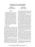
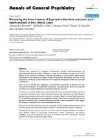
(Figure 1A). At the manufacturer’ sthresholdof100U/
ml, a sensitivity of 60.4% and a specificit y of 98.9% were
calculated for the dia gnosis of SLE. The results for eight
disease controls were false-positives. Six of these eight
non-SLE patients being positive in Anti-dsDNA-NcX
had either positive anti-dsDNA or antinucleosome
ELISA results at a specificity of 98.9% (Figure 1B). Nota-
bly, none of these eight S LE patients tested positive in
the CLIF or Farr assay.
Out of interest, the m edical records of all non-SLE
patients who tested positive in the Anti-dsDNA-NcX
ELISA were checked (Figure 1B). In five patients, causes
other than SLE with the potential to induce anti-dsDNA
antibodies were found, namely, drugs and fever indicating
an unknown infection. Combining Anti-dsDNA-NcX test
results and med ical history with consequent exclusion o f
these five patients would lea d to an increa sed spe cificity
of 99.5% at the manufacturer’s threshold.
Analysis of test criteria
To further a ssess the performance criteria of the Anti-
dsDNA-N cX ELISA with those of other assays for mea-
suring antibodies against dsDNA and/or nucleosomes,
the new ELISA was compared with the anti-dsDNA
ELISA, the antinucleosome ELISA (free of Scl-70 and
histone H1) and the Farr assay in sera from 964 indivi-
duals by using ROC curve analysis (Table 1).
To chec k whether the performance of a single te st
system was significantly better than another one, we
additionally tested the reported AUC values for signifi-
cant differences. This formal statistical comparison
revealed that the Anti-dsDNA-NcX ELISA and the anti-
nucleosome ELISA were significantly better than the
anti-dsDNA ELISA (p =0.0024andp = 0.0029, respec-
tively). The Farr assay was not significantly better than
any other ELISA, nor was the opposite the case.
The Anti-dsDNA-NcX ELISA revealed superior results
in all performance criteria. The CLIF assay was not inte-
grated into ROC curve analyses because it is a semi-
quantitative test. However, a sensitivity of 28.02% at a
specificity of 98.15% was separat ely cal culated fo r the
CLIF assay. To allow direct comparison, the sensitivities
of all o ther test systems are also shown at a specificity
of 98.15% in Table 1.
Comparison of test systems in terms of reactivity and
diagnosis determination
As considerable differences were obtai ned in ROC curve
analysis, two clinically relevant questi ons were addressed
to further elucidate the similarities (question 1) and differ-
ences (question 2) of the test systems used. First, how
often are sera positive in one test and positive in another
test system? Second, how often are sera negative in one
test but positive in another test system? Answers to the
second question would give physicians clues to how often
they might miss the correct diagnosis by determination of
disease-specific autoantibodies using only one test system.
To integrate the CLIF assay with its specificity of
98.15%, individual cutoffs of the other test systems
at the same specificity (see also Table 1) were used.
Intersections of the three ELISAs (Anti-dsDNA-NcX,
antinucle osome and a nti-dsDNA) and separat ely o f
dsDNA-NcX with the Farr and CLIF assays are illu-
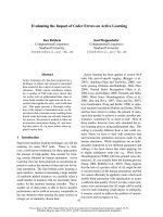
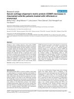
strated in a Venn diagram shown in Figure 2. Of 207
sera, 139 were positive in all of the three ELISAs, which
were compared at cutoffs read out at a specificity o f
98.15%. Of these 139 positive sera , 99.28% were positive
with the Anti-dsDNA-NcX ELISA, while 9.28% were
exclusively positive with the Anti-dsDNA-NcX ELISA.
In a comparison of the different detection techniques
(Figure 2B), 1 49 sera were positive in the Farr assay,
Anti-dsDNA-NcX ELISA or the CLIF assay. Exclusively
positive sera were found as follows: two (1.3%) in the
CLIF assay, six (4.0%) in the Farr assay and 29 (19.5%)
in the Anti-dsDNA-NcX ELISA. Among these three
tests, 138 (92.6%) of all 149 positive sera were positive
in the Anti-dsDNA-NcX ELISA.
Biesen et al. Arthritis Research & Therapy 2011, 13:R26
/>Page 3 of 9
To answer the second question and t o reveal differ-
ences between the assays, we further determined how
often serum is negative in one test but positive in another
test (Table 2). Clinically relevant for the verific ation of
diagnosis, these freque ncies indicate how often a diagno-
sis of SLE may be missed by using only one test.
Clinical associations of assays
To reveal clinical associations of inve stigated test sys-
tems, assay titers of patients with a distinct present clin-
ical finding were compared with those of patients
without that finding using the Mann-Whitney U test
(Table 3). Clinical findings were read out of medical
700
800
/
ml
A
)
400
500
600
N
cX ELISA in IU
/
100
200
300
Anti-dsDNA-
N
SLE Myositis SSc SS RA ND
0
n=88n=81n=26 n=400n=207 n=162
Diagnosis NcX dsDNA Nuc Farr CLIFT ANA Comments
SSc
132 6
88 6
50 8
38
neg
1:1000
B)
SSc
132
.
6
88
.
6
50
.
8
3
.
8
neg
.
1:1000
-
SSc 115.4 21.5 23.0 2.6 neg. 1:1000 C3Ļ; minocycline
SS 125.3 103.0 5.1 1.5 neg. 1:3200 -
RA 322.6 13.1 134.3 6.3 neg. 1:1000 C3Ļ; lymphadenopathy
RA 266.4 5.1 107.5 4.6 neg. 1:3200 Fever of unknown origin
RA
233 5
148 1
49
60
1 320
Ad li b
RA
233
.
5
148
.
1
4
.
9
6
.
0
neg.
1
:
320
Ad
a
li
muma
b
RA 104.3 206.8 5.7 4.0 neg. 1:3200 Adalimumab
RA 102.5 181.6 1.9 3.4 neg. 1:1000 Sulfasalazine
Figure 1 Right- positive and false-posi tive test results of ant i-double-stranded DNA n ucleosome-complexed enzyme-linked
immunosorbent assay (Anti-dsDNA-NcX ELISA). (A) Scatterplot showing Anti-dsDNA-NcX ELISA immunoglobulin G results in 964 different
sera. Dotted line represents the manufacturer’s threshold (100 IU/ml). Values >800 IU/ml were set to 800 IU/ml for a clearer arrangement of the
figure. SLE, systemic lupus erythematosus; SSc, systemic sclerosis; SS, Sjögren’s syndrome; RA, rheumatoid arthritis; ND, normal donors; (B) Table
showing all non-SLE patients who tested positive in the Anti-dsDNA-NcX ELISA listed with the test results of all measured assays and clinically
relevant findings. Positive test results according to a comparable specificity of 98.9% are marked in bold. Nuc, anti-dsDNA-nucleosome ELISA;
Farr, radioimmunoassay; CLIF, Crithidia luciliae immunofluorescence assay; ANA, antinuclear antibodies; C3, complement component 3.
Biesen et al. Arthritis Research & Therapy 2011, 13:R26
/>Page 4 of 9
records consisting of ACR criter ia, mSLEDAI 2000
score, laboratory parameters and immunosuppressants
and/or antimalarials.
Using the global mSLEDAI 2000 score (items used to
score for anti-dsDNA and complements were excluded
to avoid bias), o nly the Anti-dsDNA-NcX ELISA (r
s
=
0.145, p = 0.034) and anti-Nuc ELISA (r
s
= 0.143, p =
0.034) were significantly correlated on the basis of
Spearman’ s rank order correlation Spearman’ srank
order correlation, but neither the anti-dsDNA ELISA (r
s
= 0.113, p = 0.074) nor t he Farr assay (r
s
= 0.118, p =
0.065) showed a significant correlation.
All test systems were significantly associa ted with urin -
ary casts and decre ased complement component 3 (C3)
at the time of bloo d sampling. Notably, some item s were
related to only one assay. So, the mSLE DAI 2000 items
pericarditis and decreased complement component 4
(C4) were exclusively associated with elevated titers in
the anti-dsDNA ELISA. The Farr assay was exclusiv ely
connected to the presence of pleuri tis. Th e CLIF assay
was associated with prior hematological manifestation
according to an ACR c riterion. It is noteworthy that the
SLEDAI 2000 item proteinuria was solely asso ciated with
higher levels found by the Anti-dsDNA-NcX ELISA.
Of our 207 patients, 101 had histologically proven
lupus nephritis. Of those 101 p atients, 61 were not con-
sidered in further subgroup analysis because of a miss-
ing biopsy within 1 year before blood draw. Seven
patients actually had lupus nephritis class II, 13 had
class III, 12 had class IV and eight had class V according
Table 1 Performance criteria in ROC analysis
a
Criteria Anti-dsDNA-NcX Anti-dsDNA Antinucleosome Farr
Area under the curve 0.9133
b
0.8455 0.9118 0.8823
95% confidence interval 0.89 to 0.94 0.81 to 0.88 0.89 to 0.94 0.85 to 0.91
Sensitivity at specificity of 95% 72.5%
b
55.6% 67.2% 65.7%
(cutoff) (>49.7) (>58.8) (>6.8) (>5.1)
Sensitivity at specificity of 98% 66.7%
b
43.0% 58.0% 55.6%
(cutoff) (>71.5) (>99.7) (>11.1) (>6.0)
Sensitivity at specificity of 98.15%
c
66.7%
b
41.5% 55.6% 55.6
(cutoff) (>72.03) (>105.8) (>12.1) (>6.0)
Sensitivity at specificity of 99% 59.9%
b
35.8% 53.6% 54.1%
(cutoff) (>103.4) (>151.6) (>14.8) (>6.5)
Maximum sum of sensitivity + specificity 170.9
b
159.1 168.7 162.1
a
dsDNA, double-stranded DNA. Test criteria for the Anti-dsDNA-NcX, anti-dsDNA and antinucleosome enzyme-linked immunosorbent assays (ELISAs), as well as
for the anti-dsDNA radioimmunoassay (also known as a Farr assay), were calculated using receiver operating characteristic (ROC) curve analysis based on test
readings of 964 samples from 207 systemic lupus erythematosus (SLE) patients, 357 disease controls and 400 healthy donors. Outcome parameters of ROC curve
analysis were diagnosis versus no diagnosis of SLE.
b
Highest value over all four assays.
c
This specificity was calculated for the Crithidia luciliae
immunofluorescence (CLIF) assay.
Farr
CLIFT
dsDNA
Nuc
A
)B)
75
0
0
1
50
3
2
6
Farr
CLIFT
dsDNA
Nuc
40
10
13
3
56
29
dsDNA-NcXdsDNA-NcX
Figure 2 Comparison of patient sera that tested positive in the
anti-double-stranded DNA nucleosome-complexed enzyme-
linked immunosorbent assay (Anti-dsDNA-NcX ELISA) and
other investigated test systems shown in Venn diagrams. (A)
Absolute numbers of patient sera that tested positive in the Anti-
dsDNA-NcX ELISA (dsDNA-NcX), the anti-dsDNA ELISA (dsDNA) and
the anti-dsDNA nucleosome ELISA (Nuc) and their combined
intersections are shown. (B) The same analysis as in Figure 2A is
shown, including different detection techniques using Anti-dsDNA-
NcX ELISA, Farr assay (radioimmunoassay) and Crithidia luciliae
immunofluorescence (CLIF) assay is presented. Cutoffs for all
positive test results were read out of receiver operating
characteristic curve analysis at a specificity of 98.15% because this
specificity was calculated for the CLIF assay.
Table 2 Frequency of sera being positive in one test
system and negative in another test system
a
SLE patients
(N = 207)
NcX
+
(n = 138)
dsDNA
+
(n = 86)
Nuc
+
(n = 115)
Farr
+
(n = 115)
CLIF
+
(n = 58)
NcX
-
(n = 69) - 1% 0% 13% 7%
dsDNA
-
(n = 121)
44% - 33% 35% 14%
Nuc
-
(n = 92) 25% 12% - 21% 8%
Farr
-
(n = 92) 35% 14% 21% - 5%
CLIF
-
(n = 149) 57% 30% 43% 57% -
a
SLE, system ic lupus erythematosus; NcX, anti-dsDNA-nucleosome-complexed
enzyme-linked immunosorbent assay (ELISA); dsDNA, double-stranded DNA;
Nuc, anti-dsDNA-nucleosome ELISA; Farr, radioimmunoassay; CLIF, Crithidia
luciliae immunofluorescence assay. Cutoffs for all test systems were read out
of receiver operating characteristic curve analysis at a specificity of 98.15% to
include the CLIF assay in that analysis. Frequencies of sera being positive in
one selected test (column headings) and negative in another test system (left
column stub headings) were filled to demonstrate the extent of cross-
reactivity between tests. The positive sera were compared with the negative
sera to calculate percentages. For example, among all anti-dsDNA-NcX ELISA-
negative sera (n = 69), 1% were positive on the basis of the anti-dsDNA ELISA.
Biesen et al. Arthritis Research & Therapy 2011, 13:R26
/>Page 5 of 9
to the International Society of Nephrology Working
Group on the Classification of Lupus Nephritis/Renal
Pathology Society Working Group on the Classification
of Lupus Nephrit is guidelines [26]. Prior or present
renal involvement (n = 101) was significantly associated
with elevated test results in the anti-NcX ELISA (P =
0.002), anti-Nuc ELISA ( P = 0.004), anti-dsDNA ELISA
( P = 0.008) and the Farr assay (P = 0.042), but not in
the CLIF assay (P = 0.812) (all P values based on the
Mann-Whitney U test). Further analysis of defined
classes revealed an exclusive correlation of lupus nephri-
tis class IV class (n = 12) with elevated antibody levels
on the basis of Anti-Nuc, Anti-dsDNA and Anti-
dsDNA-NcX ELISA results using Spearman’s rank order
correlation (r
s
= 0.192, r
s
=0.189,r
s
= 0.183, r espec-
tively; all p < 0.01).
Association of assays with disease activity over time
Monitoring of disease activity is of prime imp ortance in
the clinical care of SLE patients. Regarding the correla-
tion of anti-dsDNA antibodies with disease activity, con-
tradicting reports have been published in the literature,
ranging from a po sitive [8] over a missing [9] to a neg a-
tive correlation [7]. In the longitud inal prospectiv e
approach over 10 months used in our present study, 20
SLE patients were monitored with an overall total of 69
patient visit s resulting in 49 different data po ints. In all
visits, no change in thera py within the past 4 weeks had
occurred before medical records and blood were taken.
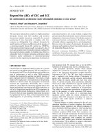
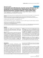
To test whether changes in distinct labo ratory para-
meters can reflect changes in disease activity, the anti-
dsDNA ELISA, the Farr assay, the antinucleosome
ELISA, the Anti-dsDNA-NcX ELISA, C3 and C4 were
compared with the changes in mSLEDAI 2000 score
over time (Figure 3). The mSLEDAI 2000 score was
defined with an exclusion of items for dsDNA and com-
plement components to avoid bias. In the follow-up
study, none of the traditional biomarkers (anti-dsDNA
ELISA, Farr assay or C3 or C4) were correlated with dis-
ease activity over time. However, Anti-dsDNA-NcX
ELISA correlated best (r = 0.4970, P = 0.0003), followed
by the antinucleosome ELISA (r = 0.4605, P = 0 .0009)
using linear regression. A subgroup analysis was not
performed because of the limited number of patients.
Discussion
In this study , the frequency of autoantibodies aga inst
dsDNA-complexed nucleosomes was investigated in
Table 3 Comparison of test assay titers in patients with versus those without a distinct present clinical finding
a
P value
Disease feature Number of patients NcX dsDNA Nuc Farr CLIF
ACR criteria (ever), N = 207
Renal 101 0.01 0.03 0.01 ns ns
Neurologic 23 0.01 0.03 0.01 0.04 ns
Hematological 134 ns ns ns ns 0.04
mSLEDAI 2000 (current), N = 165
Casts 3 0.03 0.04 0.04 0.02 0.04
Hematuria 13 <0.001 0.001 0.003 0.002 ns
Proteinuria 16 0.04 ns ns ns ns
Leukocyturia 3 0.04 0.02 ns ns ns
Pleuritis 5 ns ns ns 0.01 ns
Pericarditis 10 ns 0.02 ns ns ns
Complement 84 0.003 0.005 0.004 0.01 0.03
Fever 8 0.02 0.04 ns ns ns
Thrombocytes 5 ns 0.04 ns ns 0.03
Laboratory (current), N = 165
Decreased lymphocytes 91 0.02 0.02 ns 0.03 ns
Decreased monocytes 27 0.02 0.02 ns ns ns
Decreased C-reactive protein 62 0.02 0.04 0.04 ns ns
Decreased C3 83 0.004 0.001 0.01 0.004 ns
Decreased C4 63 ns 0.04 ns ns ns
Proteinuria >150 mg/d 39 0.01 ns 0.04 ns ns
a
Comparison was performed using the Mann-Whitney U test. This table is reduced to statistically significant findings to increase readability. Significance with
regard to any feature was always found for incr eased values of autoantibody assays. The number of patients with a positive finding (A CR and SLEDAI 2000)oran
abnormal finding (local laboratory) of all patients is given. Current values refer to values on the date of blood withdrawal. NcX, anti-dsDNA-nucleosome-
complexed enzyme-linked immunosorbent assay (ELISA); dsDNA, dsDNA, double-stranded DNA; Nuc, anti-dsDNA-nucleosome ELISA; Farr, radioimmunoassay; CLIF,
Crithidia luciliae immunofl uorescence assay; ACR, American College of Rheumatology; mSLEDAI 2000, modified Systemic Lupus Erythematosus Disease Activity
Index 2000 [25]; C3, complement component C3; C4, complement component C4; ns, not significant.
Biesen et al. Arthritis Research & Therapy 2011, 13:R26
/>Page 6 of 9
different rheumatic diseas es and healthy individuals and
compared with levels of autoantibodies against dsDNA
or n ucleosomes alone. We have demonstrated that the
use of dsDNA-complexed nucleo somes in stead o f
dsDNA or nucleosomes individually as binding sub-
strates is superior in ensuring the diagnosis of and
assessing disease activity in SLE.
To comprehensively investigate the performance of the
Anti-dsDNA-NcX ELISA, s era of 207 SLE patients, 357
disease controls and 400 healthy individuals were tested.
A sensitivity of 60.4% and a specificity of 98.9% were cal-
culated for the diagnosis of SLE at the manufacturer’ s
threshold of 100 IU/ml. Only 8 (2.2%) of 357 disease con-
trols with other rheumatic autoimmune diseases had ele-
vated anti-dsDNA-NcX res ults. Where as in s ix of these
eight patients either the anti-dsDNA ELISA or the anti-
nucleosome ELISA also revealed positive test results,
none of these sera were positive in the Farr or CLIF
assay. The Farr and CLIF assays also revealed false-posi-
tive test results, but not in t hose eight Anti-dsDNA-NcX
ELISA-positive disease controls (Table 1).
In five anti-dsDNA-NcX false-positive disease con-
trols, we found other circumstances potentially causing
elevated a utoa ntibody levels, such as minocycl ine, sulfa-
salazine, TNF inhibitors and an unknown infection with
fever. Exclusion of these assumed five false-positive
events resulted in an increased specificity of the Anti-
dsDNA-NcX ELISA from 98.9% to 99.5%.
To further compare the performance of the Anti-
dsDNA-NcX ELISA with other est ablish ed test syst ems,
a ROC curve analysis comprising 964 individuals was
conducted. In that analysis, the Anti-dsDNA-NcX
ELISA had the best perf ormance among the investigated
test systems. The superior performance criteria of the
Anti-dsDNA-NcX ELISA, especially in direct compari-
son to the anti-dsDNA and antinucleosome ELISAs,
resulted from the novel approach of utilizing dsDNA-
loaded nucleosomes instead of dsDNA or nucleosomes
alone. In addition, nearly 10% of all sera were positive in
the Anti-dsDNA-NcX ELISA, but this was not the case
in the anti-dsDNA ELISA or the antinucleosome ELISA,
indicating that dsDNA-loaded nucleosomes are more
consistent with the naturally appearing antigen.
Beyondthesefindings,comparativedatafromROC
curve analysis once more indicate that ANuA are also a
highly frequent an d very spe cific feature of SLE. There-
fore, taking into account t hat ANuA arise earlier than
anti-dsDNA antibodies [15], ANuA should be strongly
considered as a criterion for the classification and diag-
nosis of SLE with the proviso that the determination be
performed using a well-characte rized test system with
proven specificity.
By stu dying differences between assays, we also found
that all investigated test systems were able to indicate a
positive test result when another test reported a negative
test result (Table 2). The Anti-dsDNA-NcX ELISA
Figure 3 Changes in disease activity versus changes of six defined laboratory parameters over time. All results are based on a total of 69
patient visits of 20 different systemic lupus erythematosus patients. Delta values were calculated by subtracting values for a defined parameter
from an actual visit from a defined parameter from the last visit (for example, ΔC3 = C3
(visit n +1)
-C3
(visit n)
. Only changes in Anti-dsDNA-NcX
and antinucleosome ELISA results correlated significantly with changes in modified Systemic Lupus Erythematosus Disease Activity Index 2000
(mSLEDAI 2000 [25]) score over time. The mSLEDAI items for double-stranded DNA (dsDNA) and complement components C3 and C4 were
excluded to avoid bias. Linear regression analysis was used to calculate significance.
Biesen et al. Arthritis Research & Therapy 2011, 13:R26
/>Page 7 of 9
showed the potential to completely replace the anti-
dsDNA ELISA and ANuA ELISA, but not the Farr or
CLIF assay. In harmony with the superior performance
criteria in ROC curve analysis, the Anti-dsDNA-NcX
ELISA revealed the lowest percentages of sera positive
in a ssays other than the Anti-dsDNA-NcX ELISA. Sur-
prisingly, and in c onflict with current clinical practice,
thereweresomeserathatwerefoundtobepositivein
the CLIF assay but negative i n another test system.
Thus, to cancel the CLIF assay because another
upstream test (for example, anti-dsDNA ELISA) deliv-
ered a negative test r esult might circumvent a c orrect
diagnosis of SLE in some cases. To increase the l ikeli-
hood of correct diagnosis of SLE in clinically suspected
cases, it appears useful to order several assays and tech-
niques in parallel, si nce there is still no test that detects
all antibody specificities. The observed differences are
most likely caused by the differe nt techniques used in
the investigated assays.
Analysis of clinical findings with test results of investi-
gated assays in our SLE patient cohort revealed that
increased titers were differentially associated with neuro-
logical, renal and hematological involvement according
to ACR criteria. Moreover, high titers were preferentially
associated with active lupus nephritis class IV, casts,
hematuria , proteinuri a, leukocyturia, leukocytopenia,
monocytopenia and consumption of C3. The highest
number of significant associations with clinical feature s
(n = 14) was revealed by the anti-dsDNA ELISA, fol-
lowed by the Anti-dsDNA-NcX ELISA (n =13).
Remarkable differences were found in this analysis
between the anti-dsDNA ELISA, the Farr assay and the
CLIF assay, all of which target anti-dsDNA. Underlying
methodical differences (radioimmunoassay versus immu-
nofluorescence assa y versus ELISA) might contribute to
that phenomenon.
All assays were significantly correlated to global dis-
ease activity (assessed by mSLEDAI 2000 score) in the
cross-sectional survey. Using serial data of 20 patients
(69 patient visits), we further assessed whether test sys-
tems are useful for monitoring disease activity over
time. Surprisingly, changes in broadly accepted biomar-
kers were not significantly associated with changes in
disease activity over time. These findings are in harmony
with recent data derived from a much larger serial ana-
lysis o f 1,116 patient visits [9]. Strikingly, both ELISAs
containing nucleosomes as antigens were well correlated
with disease activit y over time. However, because of the
limited number o f patients and samples per patient, the
results have to be confirmed in larger studies.
Conclusions
The nonradioactive Anti-dsDN A-NcX ELISA, which is
based on dsDNA-loaded nucleosomes as antigens,
demonstrated excellent test characteristics for the assess-
ment of the diagnosis and disease activity that can be opti-
mized even if clinicians interpret deliv ered test results
within a medical context and consider the presence of
drugs and infections having the potential to induce auto-
antibodies against dsDNA and/or nucleosomes.
Additional material
Additional file 1: Supplementary Table S1. Characterization of SLE
patients. SLEDAI, Systemic Lupus Erythematosus Disease Activity Index;
1
Number of patients with fully accessible SLEDAI 2000 data [25];
2
SACQ,
serologically active clinical quiescent.
Abbreviations
ACR: American College of Rheumatology; AUC: area under the curve; CLIF:
Crithidia luciliae immunofluorescence; dsDNA: double-stranded DNA; ELISA:
enzyme-linked immunosorbent assay; mSLEDAI 2000: modified Systemic
Lupus Erythematosus Disease Activity Index; RA: rheumatoid arthritis; RIA:
radioimmunoassay; ROC: receiver operating characteristic; SLE: systemic lupus
erythematosus; SS: Sjögren’s syndrome; SSc: systemic sclerosis.
Acknowledgements
This work was supported by grants from EUROIMMUN Medizinische
Labordiagnostika AG and the German Research Foundation (Collaborative
Research Centre SFB650, TP17).
Author details
1
Department of Rheumatology and Clinical Immunology, Charité
Universitätsmedizin Berlin, Chariteplatz 1, Berlin D-10117, Germany.
2
EUROIMMUN Medizinische Labordiagnostika AG, Seekamp 31, Lübeck
D-23560, Germany.
Authors’ contributions
KE, CD, GRB, FH and WSc designed the study. FB, RB, TR, MB, AB and AR
acquired data.
RB and FH analyzed and interpreted the data. RB, GRB, WSc, WSc and FH
prepared the manuscript. OJ, RB and AR performed statistical analysis. KE,
CD, WSt, FH and WSc were responsible for overall project management. RB
had full access to all of the data in the study and takes responsibility for the
integrity of the data and the accuracy of the data analysis.
Competing interests
RB was employed from August 2006 until March 2009 by Charité University
Hospital with third-party funds paid by EUROIMMUN Medizinische
Labordiagnostika AG. CD and AR are employees of EUROIMMUN
Medizinische Labordiagnostika AG, Lübeck, Germany. WSc and WSt are
board members of EUROIMMUN AG. The other authors declare that they
have no conflict of interest.
Received: 21 August 2010 Revised: 6 January 2011
Accepted: 10 February 2011 Published: 10 February 2011
References
1. Rahman A, Isenberg DA: Systemic lupus erythematosus. N Engl J Med
2008, 358:929-939.
2. Sherer Y, Gorstein A, Fritzler MJ, Shoenfeld Y: Autoantibody explosion in
systemic lupus erythematosus: more than 100 different antibodies
found in SLE patients. Semin Arthritis Rheum 2004, 34:501-537.
3. Isenberg DA, Manson JJ, Ehrenstein MR, Rahman A: Fifty years of anti-ds
DNA antibodies: are we approaching journey’s end? Rheumatology
(Oxford) 2007, 46:1052-1056.
4. Hahn BH: Antibodies to DNA. N Engl J Med 1998, 338:1359-1368.
5. Barzilai O, Ram M, Shoenfeld Y: Viral infection can induce the production
of autoantibodies. Curr Opin Rheumatol 2007, 19:636-643.
6. Borchers AT, Keen CL, Gershwin ME: Drug-induced lupus. Ann N Y Acad Sci
2007, 1108:166-182.
Biesen et al. Arthritis Research & Therapy 2011, 13:R26
/>Page 8 of 9
7. Ho A, Magder LS, Barr SG, Petri M: Decreases in anti-double-stranded DNA
levels are associated with concurrent flares in patients with systemic
lupus erythematosus. Arthritis Rheum 2001, 44:2342-2349.
8. ter Borg EJ, Horst G, Hummel EJ, Limburg PC, Kallenberg CG: Measurement
of increases in anti-double-stranded DNA antibody levels as a predictor
of disease exacerbation in systemic lupus erythematosus: a long-term,
prospective study. Arthritis Rheum 1990, 33:634-643.
9. Bauer JW, Petri M, Batliwalla FM, Koeuth T, Wilson J, Slattery C, Panoskaltsis-
Mortari A, Gregersen PK, Behrens TW, Baechler EC: Interferon-regulated
chemokines as biomarkers of systemic lupus erythematosus disease
activity: a validation study. Arthritis Rheum 2009, 60:3098-3107.
10. Casciola-Rosen LA, Anhalt G, Rosen A: Autoantigens targeted in systemic
lupus erythematosus are clustered in two populations of surface
structures on apoptotic keratinocytes. J Exp Med 1994, 179:1317-1330.
11. Baumann I, Kolowos W, Voll RE, Manger B, Gaipl U, Neuhuber WL,
Kirchner T, Kalden JR, Herrmann M: Impaired uptake of apoptotic cells
into tingible body macrophages in germinal centers of patients with
systemic lupus erythematosus. Arthritis Rheum 2002, 46:191-201.
12. Bruns A, Blass S, Hausdorf G, Burmester GR, Hiepe F: Nucleosomes are
major T and B cell autoantigens in systemic lupus erythematosus.
Arthritis Rheum 2000, 43:2307-2315.
13. Mohan C, Adams S, Stanik V, Datta SK: Nucleosome: a major immunogen
for pathogenic autoantibody-inducing T cells of lupus. J Exp Med 1993,
177:1367-1381.
14. Chabre H, Amoura Z, Piette JC, Godeau P, Bach JF, Koutouzov S: Presence
of nucleosome-restricted antibodies in patients with systemic lupus
erythematosus. Arthritis Rheum 1995, 38:1485-1491.
15. Amoura Z, Chabre H, Koutouzov S, Lotton C, Cabrespines A, Bach JF,
Jacob L: Nucleosome-restricted antibodies are detected before anti-
dsDNA and/or antihistone antibodies in serum of MRL-Mp lpr/lpr and
+/+ mice, and are present in kidney eluates of lupus mice with
proteinuria. Arthritis Rheum 1994, 37:1684-1688.
16. Amoura Z, Piette JC, Bach JF, Koutouzov S: The key role of nucleosomes
in lupus. Arthritis Rheum 1999, 42:833-843.
17. Rahman A, Hiepe F: Anti-DNA antibodies: overview of assays and clinical
correlations. Lupus 2002, 11:770-773.
18. Suer W, Dähnrich C, Schlumberger W, Stöcker W: Autoantibodies in SLE
but not in scleroderma react with protein-stripped nucleosomes. J
Autoimmun 2004, 22:325-334.
19. Tan EM, Cohen AS, Fries JF, Masi AT, McShane DJ, Rothfield NF, Schaller JG,
Talal N, Winchester RJ: The 1982 revised criteria for the classification of
systemic lupus erythematosus. Arthritis Rheum 1982, 25:1271-1277.
20. Hochberg MC:
Updating the American College of Rheumatology revised
criteria for the classification of systemic lupus erythematosus. Arthritis
Rheum 1997, 40:1725.
21. Arnett FC, Edworthy SM, Bloch DA, McShane DJ, Fries JF, Cooper NS,
Healey LA, Kaplan SR, Liang MH, Luthra HS, Medsger TA Jr, Mitchell DM,
Neustadt DH, Pinals RS, Schaller JG, Sharp JT, Wilder RL, Hunder GG: The
American Rheumatism Association 1987 revised criteria for the
classification of rheumatoid arthritis. Arthritis Rheum 1988, 31:315-324.
22. Vitali C, Bombardieri S, Jonsson R, Moutsopoulos HM, Alexander EL,
Carsons SE, Daniels TE, Fox PC, Fox RI, Kassan SS, Pillemer SR, Talal N,
Weisman MH, European Study Group on Classification Criteria for Sjögren’s
Syndrome: Classification criteria for Sjögren’s syndrome: a revised
version of the European criteria proposed by the American-European
Consensus Group. Ann Rheum Dis 2002, 61:554-558.
23. Subcommittee for scleroderma criteria of the American Rheumatism
Association Diagnostic and Therapeutic Criteria Committee: Preliminary
criteria for the classification of systemic sclerosis (scleroderma). Arthritis
Rheum 1980, 23:581-590.
24. Tanimoto K, Nakano K, Kano S, Mori S, Ueki H, Nishitani H, Sato T, Kiuchi T,
Ohashi Y: Classification criteria for polymyositis and dermatomyositis.
J Rheumatol 1995, 22:668-674.
25. Gladman DD, Ibañez D, Urowitz MB: Systemic Lupus Erythematosus
Disease Activity Index 2000. J Rheumatol 2002, 29:288-291.
26. Weening JJ, D’Agati VD, Schwartz MM, Seshan SV, Alpers CE, Appel GB,
Balow JE, Bruijn JA, Cook T, Ferrario F, Fogo AB, Ginzler EM, Hebert L, Hill G,
Hill P, Jennette JC, Kong NC, Lesavre P, Lockshin M, Looi LM, Makino H,
Moura LA, Nagata M, International Society of Nephrology Working Group
on the Classification of Lupus Nephritis; Renal Pathology Society Working
Group on the Classification of Lupus Nephritis: The classification of
glomerulonephritis in systemic lupus erythematosus revisited. Kidney Int
2004, 65:521-530.
doi:10.1186/ar3250
Cite this article as: Biesen et al.: Anti-dsDNA-NcX ELISA: dsDNA-loaded
nucleosomes improve diagnosis and monitoring of disease activity in
systemic lupus erythematosus. Arthritis Research & Therapy 2011 13:R26.
Submit your next manuscript to BioMed Central
and take full advantage of:
• Convenient online submission
• Thorough peer review
• No space constraints or color figure charges
• Immediate publication on acceptance
• Inclusion in PubMed, CAS, Scopus and Google Scholar
• Research which is freely available for redistribution
Submit your manuscript at
www.biomedcentral.com/submit
Biesen et al. Arthritis Research & Therapy 2011, 13:R26
/>Page 9 of 9