Báo cáo y học: "Treatment of experimental adjuvant arthritis with a novel folate receptor-targeted folic acidaminopterin conjugate" doc
Bạn đang xem bản rút gọn của tài liệu. Xem và tải ngay bản đầy đủ của tài liệu tại đây (877.67 KB, 18 trang )
RESEARCH ARTIC LE Open Access
Treatment of experimental adjuvant arthritis with
a novel folate receptor-targeted folic acid-
aminopterin conjugate
Yingjuan Lu
1
, Torian W Stinnette
1
, Elaine Westrick
1
, Patrick J Klein
1
, Mark A Gehrke
1
, Vicky A Cross
1
,
Iontcho R Vlahov
1
, Philip S Low
2
and Christopher P Leamon
1*
Abstract
Introduction: Folate receptor (FR)-expressing macrophages have been shown to accumulate at sites of
inflammation, where they promote development of inflammatory symptoms. To target such a macrophage
population, we designed and evaluated the biologic activity of EC0746, a novel folic acid conjugate of the highly
potent antifolate, aminopterin.
Methods: Using a FR-positive subclone of murine macrophage-derived RAW264.7 cells and rat thioglycollate-
elicited macrophages, we studied the effect of EC0746 on dihydrofolate reducta se activity, cell proliferation, and
cellular response towards bacterial lipopolysaccharide as well as IFNg activation. The EC0746 anti-inflammatory
activity, pharmacokinetics, and toxicity were also evaluated in normal rats or in rats with adjuvant-induced arthritis;
that is, a FR-positive macrophage model that closely resembles rheumatoid arthritis in humans.
Results: EC0746 suppresses the proliferation of RAW264.7 cells and prevents the ability of nonproliferating rat
macrophages to respond to inflammatory stimuli. In the macrophage-rich rat arthritis model, brief treatment with
subcutaneously administered EC0746 is shown to mediate an FR-specific anti-inflammatory response that is more
potent than either orally administered methotrexate or subcutaneously delivered etanercept. More importantly,
EC0746 therapy is also shown to be ~40-fold less toxic than unmodified aminopterin, wi th fewer bone marrow
and gastrointestinal problems.
Conclusions: EC0746 is the first high FR-binding dihydrofolate reductase inhibitor that demonstrates FR-specific
anti-inflammatory activities both in vitro and in vivo. Our data reveal that a relatively toxic anti-inflammatory drug,
such as aminopterin, can be targeted with folic acid to inflammatory macrophages and thereby relieve
inflammatory symptoms with greatly reduced toxicity.
Introduction
A phenomenon characteristic of many autoimmune and
inflammatory disorders is persistent and unrestrained
macrophage activation [1]. This extensive build-up of
tissue-infiltrating macrophages consists of a destructive
cell population made up of both locally activated macro-
phages and inflammatory monocytes that have been
recruited from the blood in large quantities. In rheuma-
toid arthritis (RA) the synovial joints are enriched with
these activated macrop hages, where they play a primary
role in the pathophysiology of joint destructi on and dis-
ease progression [2,3]. Based on the concept that inflam-
matory diseases can be caused or worsened by activated
macrophages, many therapeutic interventions for inflam-
matory disorders have focused on suppressing or neu-
tralizing one or more proinflammat ory products
released by these macrophages. Examples of such thera-
peutics include agents that reduce TNFa (for example,
etanercept, infliximab, adalimumab), IL-1 (anakinra),
and IL-6 (tocilizumab, atlizumab) [4,5]. Other biologic
agents targeting IL-12/IL-23 (ustekinumab), B cells
(rituximab), and T cells (abatacept, alefacept) are also
available as a second-line or third-line treatment when
* Correspondence:
1
Endocyte, Inc., 3000 Kent Avenue, West Lafayette, IN 47906, USA
Full list of author information is available at the end of the article
Lu et al. Arthritis Research & Therapy 2011, 13:R56
/>© 2011 Lu et al.; licensee BioMed Central Ltd. This is an open access article distributed under the terms of the Creative Commons
Attribution License ( which permits unrestricted use, distribution, and reproduction in
any medium, provided the original work is properly cited .
anti-TNF agents fail [6]. Despite remarkable success,
biologics remain prohibitively expensive (~$16,000 per
year) [7], and a majority of them carry a black-box
warning for increased risk of serious infections [8].
Alternatively, methotrexate (MTX; molecular weight
454.4) has a long history of use in treating rheumatic
diseases, and it continues to be the most prescribed
medicine (taken orally at 7.5 to 25 mg/week) even in
the current era of the aforementioned biologic therapies
[9]. As a well-known antifolate, MTX inhibits multiple
folate-dependent enzymes involved in biosynthesis of
purines/thymidylate and several amino acids, in particu-
lar dihydrofolate reductase (DHFR) and 5-aminoimada-
zole-4-carboxamide ribonucleos ide transformylase
[10,11]. Inhibition of 5-aminoimadazole-4-carboxamide
ribonucleoside transformylase also causes the release of
adenosine, a potent endogenous anti-inflammatory
agent, at sites of inflammation [11]. Although the pre-
cise anti-inflammatory mechanism(s) by which MTX
functions remains unclea r, its therapeutic activiti es may
include suppression of proliferation of immune cells
responsible for inflammation, induction of T-cell apop-
tosis, and alterations in cell recruitment and cytokine
production [11]. Compared with most disease-modifying
anti-rheumatic drugs, MTX is generally considered to
be well tolerated, and people who are prescribed MTX
can remain on t his medication for many years [9].
Approximately one-third of RA patients discontinue
MTX therapy, however, due to drug-related toxicities
and/or poor responses [12,13], and many are put on a
combination treatment with a biological agent [9].
The molecular predecessor of MTX is aminopterin
(AMT), a compound that was initially discov ered as a
chemotherapeutic agent but was abandoned in the
1950s in favor of MTX due to high toxicity and low
therapeutic index [14]. Historically, AMT was the first
antifolate used to treat inflammatory disorders (RA and
psoriasis), and it produced a rapid improvement in dis-
ease activity, but not without toxic reactions [14]. There
is renewed interest in AMT, however, because while it
shares similar pharmacological actions to MTX, the pre-
decessor appears to be more potent when compared in
murine models of air-pouch inflammation (~40-fold)
and arthritis (~20-fold) [15,16]. The superior anti-
inflammatory action of AMT is due in part to its higher
affinity for folypolyglutamate synthetase, an enzyme
responsible for intracellular retention of folates and anti-
folates [15,17]. In preclinical studies, however, the
increased potency of AMT does n ot come without an
increase in toxicity, wherein t he reported acute 50%
lethal dose for oral exposure of AMT in mice is 3 mg/
kg, compared with 89 mg/kg for MTX [18]. A contri-
buting factor to this toxicity is that, like MTX and most
antifolates, AMT enters cells via the reduced-folate
carrier (RFC), a ubiquitously expressed anion transpor-
ter present in normal tissues [19], and probably through
a second ubiquitously expressed proton-coupled folate
transporter that is responsible for intestinal folate
resorption under low pH conditions [20].
While clinical success of antifolates for the treatment
of inflammatory diseases validates the pharmacology of
this class of agents, the issues of t oxicity and temporal
response highlight the need for further improvement.
One possible solution for decreasing the toxicity due to
nonspecificexposurewhileatthesametimeenhancing
drug uptake at the inflamed site is to target the antifo-
late more selectively to the inflammatory cells of inter-
est. A potential cellular target for incr eased selectivity in
the treatment of inflammatory diseases came with the
discovery that activa ted (but not resting) macrophages
express a functional folate receptor (FR) known as FRb
[21-23]. This finding has allowed for the rational exp loi-
tation of FR-mediated therapeutic int ervention as well
as diagnostic tools (reviewed in [24,25]). For example,
folate-targeted imaging agents have been shown to accu-
mulate in sites of active inflammation in animals [25,26]
and selectively within the inflamed joints of RA patients
[27]. Likewise, recent efforts in therapy include dsFv
anti-FRb-targeted Pseudomonas exoto xin A [28], folate-
hapten-mediated immunotherapies [29,30] and antifo-
lates designed to bind FR [23,31]. Although each of the
aforementioned approaches holds promise for yielding
new therapeutic options for patients, there have been no
reports to date on the use of folic acid (FA) for targeting
smal l molecular weight anti-inflammatory drugs to sites
of inflammation.
In our present study we investigated the biological
activities of EC0746, a FA-AMT conjugate designed to
intracellularly deliver an AMT analog specifically via the
FR. The anti-inflammatory activity of EC0746 was evalu-
ated in a series of in vitro and in vivo studies using FR-
positive macrophage cell lines and a rat adjuvant-induced
arthritis (AIA) model. Since MTX and etanercept are
part of the current standard of care for RA and other
rheumatic diseases, EC0746 was also compared against
oral MTX at eq uimol ar doses and against a limited high-
dose regimen of etanercept. Finally, we determined the
plasma pharmacokinetics and the maximum tolerated
dose (MTD) of EC0746 v ersus AMT. Our investigation
provides the first evidence that a FA-targeted small mole-
cule anti-inflammatory agent may be useful as a mac ro-
phage-specific intervention with an advantage of
improved therapeutic index over its parent drug.
Materials and methods
Reagents
EC0746 (molecular weight 2,236), shown in Figure 1a,
was synthesized as previously described [32]. [
3
H]FA
Lu et al. Arthritis Research & Therapy 2011, 13:R56
/>Page 2 of 18
was purchased from Amersham (Arlington Heights, NY,
USA). AMT, MTX, Pseudomonas aeruginosa lipopoly-
saccharide(LPS),andaDHFRcolorimetricassaykit
were purchased from Sigma-Aldrich (St Louis, MO,
USA). Murine IFNg was purchased from PeproTech
(Rocky Hill, NJ, USA). Etanercept was purchased from
CVS Pharmacy (We st Lafayette, IN, USA). The cell pro-
liferation (2,3-bis(2-methoxy-4-nitro-5-sulfo-phe nyl)-2H-
tetrazolium-5-carboxanilide (XTT)) and TNFa ELISA
kits were purchased from Roche Applied Science (India-
napolis, IN, USA) a nd eBioscience (San Diego, CA,
USA), respectively. The Proteome Profiler™ rat cytokine
array (Panel A) was purchased from R&D Systems (Min-
neapolis, MN, USA). Heat-killed Mycobacteria butyri-
cum was purchased from BD Diagnostic Systems
(Sparks, MD, USA). All other reagents were obtained
from major suppliers.
Animals, thioglycollate-elicited peritoneal macrophages,
and cell culture
All animal care and use were performed according to
National Institutes of Health guidelines and in compli-
ance with protocols approved by the Purdue Animal Use
and Care Committee. Female Lewis rats (175 to 200 g)
were purchased from Harlan Sprague Dawley (Indianapo-
lis, IN, USA) and were allowed to acclimate for 1 week.
To obtain peritoneal macrophages, rats were dosed intra-
peritoneally with an aged thioglycollate (TG) medium (20
ml/kg ) and euthanize d 3 days later. The peritoneal cavity
of the animals was lavaged with 60 ml ice-cold phos-
phate-buffered saline to collect peritoneal exudate. The
TG-elicited macrophages in the peritoneal fluids were
obtained after a red cell lysing step and a 2-hour adher-
ence in folate-free RPMI 1640 medium (Mediatech,
Manassas, VA, USA) containing 1% heat-activated fetal
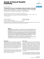
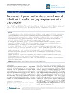
Figure 1 EC0746 folate receptor binding affinities. (a) Chemical structure of EC0746. There are four separate functional components to this
novel construct: the folate receptor (FR)-targeting moiety folic acid (FA; black), the drug moiety aminopterin (AMT; red), a saccharo-amino acid
peptide-based spacer of ((saccharo-gGlu)-gGlu)
2
-gCys (blue), and a hydrazide/disulfide-containing linker (green). (b) Relative binding affinities of
EC0746 in comparison with AMT and methotrexate (MTX) using FRa-expressing KB cells and FRb-expressing CHO-FRb cells. The assays were
performed in triplicate at 37°C using each compound as a competitor to displace [
3
H]FA from binding to FR-expressing cells. Numbers shown
next to each test article are relative affinity values with FA itself set at 1.
Lu et al. Arthritis Research & Therapy 2011, 13:R56
/>Page 3 of 18
calf serum and antibiotics. The purity of the resulting
macrophage population (herein referred to as rat TG-
macs) was d etermined to be ~90% pure ba sed on
CD11b/c expression (data not shown). The RAW264.7
macrophage cell line is a FR-expressing subclone of
ATCC TIB71 (a murine macrophage-derived tumor ce ll
line) that has been adapted to grow under folate-deficient
conditions. Previously we have reported that rat TG-
macs expre ss ~20-fold less FR than RAW264.7 cells, but
these receptors (isotope not identified due to the lack of
ant i-rat FRb antibodies) can internalize folate conjugates
at a rate consistent with their levels of FR expression
[25]. Unless otherwise specified, all cells were maintained
in the folate-free RPMI 1640 medium containing 10%
heat-activated fetal c alf serum and antibiotics (FFRPMI)
under a 5% CO
2
atmosphere.
Relative affinity assays
The relative affinities of EC0746, AMT, and MTX were
determined according to a previously established
method except that both KB cells and CHO-FRb cells
were used as the sources of FR [33]. KB cells are a
human cancer cell line known for elevated expression
of FRa.CHO-FRb cells were obtained from Manohar
Ratnam, Department of Bi ochemistry and Cancer Biol-
ogy, The University of Toledo (Toledo, OH, USA). This
cell line was originally generated by stable integration
and amplification of a human FRb cDNA expression
construct in CHO-K1 cells [34]. The relative affinity
value was defined as the inverse molar ratio of com-
pound required to displace 50% of [
3
H]FA bound to FR
on KB cells or CHO-FRb cells, and the relative affinity
of FA for the FR was set to 1; that is, values <1 reflect
weaker affinity than FA, and values >1 reflect stronger
affinity.
Dihydrofolate reductase inhibition assay
RAW264.7 cells growing in FFRPMI medium in 10 cm
cell culture dishes (BD Falcon, Lincoln Park, NJ, USA)
were treated with EC0746 (100 nM), EC0746 (100 nM)
plus 100-fold molar excess of FA (10 μM), or FA alone
(10 μM). After a 2-hour exposure, the drug-containing
media were replaced and the cells were allowed to incu-
bate further for 22 hours in fresh FF RPMI medium
(referred in the text as a 22-hour chase). Meanwhile,
AMT and MTX were kept at 100 nM fo r the entire 24-
hour incubation period. All cells were subsequently
lysed in the radioimmunoprecipitation assay lysis buffer,
and DHFR activities in the whole cell lysates were deter-
mined using a commercial DHFR assay kit (Sigma-
Aldrich). This spectrophotometric assay monitors the
enzymatic conversion of dihyd rofolic acid to tetrahydro-
folic acid by DHFR and the disappearance of the co-
factor nicotinamide adenine dinucleotide phosphate at
340 nm. The results were normalized to the values of
the untreated control cells.
XTT and TNFa assays
RAW264.7 cells in 96-well plates (3.5 × 10
4
cells/well)
were treated with 10-fold serial dilutions of EC0746 (≤1
μM) in FFRPMI medium without and with 100-fold
molar excess of FA. After a 2-hour exposure, the drug-
containing media were replaced and the cells were
allowed to incubate further for 70 hours (referred in the
text as a 70-hour chase). In comparison, the cells were
also treated continuously with AMT for 72 hours. Four
hours prior to the end of incubation, LPS was added to
the treated ce lls at a final concentration of 100 ng/ml.
Then 100 μl culture supernatants were collected for
TNFa analysis by ELISA. The cell viability was assessed
by adding XTT to the remaining media for an additional
4 hours following the manufacturer’s instructions. To
evaluation of a cytostatic effect, RAW264.7 cells seeded
at 1 × 10
6
cells/well in six-well plates were subjected to
2-hour exposure and a 70-hour chase with 0, 0.1, 10,
and 1000 nM EC0746 without and with excess FA. At
the end of the incubation (no LPS added), the surviving
cell s (that is, still viable cells) were recovered and redis-
tributed in equal number s in fresh medium for an addi-
tional 72 hours. The cell proliferation was again
assessed by the XTT assay. All results were expressed as
the percentage absorbance (minus background) relative
to the untreated control cells.
Rat cytokine array analysis
This analysis w as performed on rat TG-macs to com-
pare EC0746 against AMT and MTX for their abilities
to inhibit cytokine production after LPS/IFNg co-stimu-
lation. Using our standard condition of 2-hour pulse
plus a 70-hour chase period, rat TG-macs (harvested
the day before) were given vehicle (media only), EC0746
(100 nM), EC0746 (100 nM) plus 100-fold molar excess
of FA (10 μ M) , or FA alone (10 μM). For unconjugated
base drugs, 100 nM AMT and MTX were present con-
tinuously for the entire 72-hour incubation p eriod.
Twenty-four hours prior to the end of incubation, LPS
(5 μg/ml) and IFNg (100 ng/ml) were added to the cells
to stimulate cytokine production. The presence of cyto-
kines/chemokines in the culture media was detected
using a rat cytokine antibody array kit (R&D Systems)
capable of detecting 29 analytes in duplicate spots. The
total pixel intensity for each sp ot in the array was quan-
titated using the NIH ImageJ software [35], subtracted
from the background, and averaged for each analyte.
Adjuvant-induced arthritis
Prior to immunization with adjuvant, female Lewis rats
were fed a folate-deficient diet (Harlan Teklad,
Lu et al. Arthritis Research & Therapy 2011, 13:R56
/>Page 4 of 18
Indianapolis, IN, USA) for ~10 days to reduce serum
folate competition from high-folate-containing regular
rodent chow [36]. The rats were then inoculated intra-
dermally (at the base of tail) with 0.5 mg heat-killed
M. butyricum (BD Diagnostic Systems) in 100 μllight
mineraloil(Sigma-Aldrich,StLouis,MO,USA).Paw
edema (degree of arthritis) in rats was assessed using an
arthritis scoring system: 0 = no edema or arthritis; 1 =
swelling in one type of joint; 2 = swelling in two types of
joint; 3 = swelling in three types of joint; 4 = swelling of
the entire paw [37]. A total score for each rat is calcu-
lated by summing the scores for each of the four paws,
giving a maximum of 16 per animal. Notably, the first
appearance of the signs or symptoms of arthritis in this
model occurs around day 10 (typically between days 9
and 11) with distinctive but mild re dness and/or swelling
in small areas of the foot, but not necessary involving
joints at that point. On the first day of treatment, rats
with desired arthritis scores were distributed evenly
across the control and treatment groups (n = 5). For each
study, two or three rats from the same colony were not
induced for arthritis and were used as healthy controls.
Unless noted otherwise, all drug treatments started on
day 10 after arthritis induction and lasted for two conse-
cutive weeks with biweekly (BIW, Mondays and Thurs-
days) or once-weekly (QW, Mondays) dosing regimens.
At the completion of each study (day 24 or 4 days after
the last treatment), rats were euthanized by CO
2
asphyxiation and were processed for paw weight (cut at
the hairline) and spleen weight. The removed hind paws
were immersion-fixed in 10% buffered formalin and sub-
ject ed to radiog raphic and/or histopathological analyses.
When needed, X-ray radiographic images of the arthritic
hind paws were taken using a Kodak Imaging Station In
Vivo FX system (Carestream Molecular Imaging, New
Haven, CT, USA).
EC0746 was given subcutaneously (s.c.) in a dosing
range of 25 to 1,000 nmol/kg (QW or BIW); MTX was
given s.c. or orally at 250 nmol/kg (BI W) or 1,650 nmol/
kg (QW); and etanercept (10 mg/kg) was given s.c. once
every 3 days for a 12-day span. The QW MTX dosing
regimen was to mimic MTX administration in humans,
and this particul ar dose of 1,650 nmol/kg per week (that
is, 0.75 mg/kg per week) was reportedly active as an intra-
peritoneal agent in the AIA model [38]. To distinguish the
anti-inflammatory mechanisms of EC0746 and MTX in
vivo, a therapeutically irrelevant folate-containing competi-
tor (EC0923, molecular weight 672) was used in 500-fold
molar excess to block the activities of EC0746 and MTX
at 250 nmol/kg (BIW).
Radiographic and histopathological assessments
Formalin-fixed hind paws were examined by a board-certi-
fied veterinary radiologist who had no knowledge of the
study groups. Specific criteri a were used to establish a
numerical grade of severity for each radiographic change:
increased soft tissue volume (0 to 4), narrowing or widen-
ing of joint spaces (0 to 5), subchondral erosion (0 to 3),
periosteal reaction (0 to 4), osteolysis (0 to 4), subluxation
(0 to 3), and degenerative joint changes (0 to 3). Scores are
limited to the tarsus, and the maximum possible score per
foot was 2 6 [39]. The histopathological analysis was also
performed in a blind fashion by an independent contract
laboratory (Bolder BioPATH Inc., Boulder, CO, USA). The
arthritic ankles were scored on a scale of 0 to 5 for inflam-
mation, bone resorption, pannus formation, and cartilage
damage, with a maximal histology score of 20 per foot [40].
Pharmacokinetic studies
Female Lewis rats with jugular vein catheters (Harlan
Sprague Dawley) were used to assess the plasma pharma-
cokinetics of EC0746 and unconjugated AMT. The ani-
mals were divided into two main groups, one given a
single dose of EC0746 s.c. and the second a single dose of
AMT s.c., both at 500 nmol/kg. Whole blood samples
(300 μl) were collected from three animals per time point
at the following time points: 1 minute, 10 minutes,
30minutes,1hour,2hours,3hours,4hours,and
8 hours after injection. The blood samples were placed
into anticoagulant tubes containing 1.7 mg/ ml K
3
EDTA
and 0.35 mg/ml N-maleoyl-b-alanine (0.35 mg/ml) in a
0.15% acetic acid solution. Plasma samples were obtained
by centrifugation for 3 minutes at ~2,000 × g and sto red
at -80°C. The amounts of EC0746 and AMT in the
plasma and the two primary metabolites of EC0746
(AMT and AMT hydrazide) were determined by liquid
chromatography/mass spectrometry/mass spectrometry.
Preliminary toxicity evaluations
The short-term toxicity and MTD of EC0746 and AMT
were evaluated in healthy rats following the standard
BIW subcutaneous dosing regimen used for efficacy
studies. Further, these animals were put on a folate-
deficient diet for ~20 days before treatment to mat ch
the folate deficiency status of AIA rats used for therapy.
The folate-deficient but nonarthritic animals were thus
given increasing doses of EC0746 and AMT for two
consecutive weeks on a BIW basis. A MTD dose was
definedasthedosethathadcausedatleast13to14%
weight loss combined with clinical signs of stress, and at
least one animal in the group receiving a dose greater
than MTD needing to be euthanized. Standard hemato-
logic and blood chemistry parameters were examined as
needed along with histopathology.
Statistics
Statistical analyses were performed using the computer
program GraphPad Prism (GraphPad Software Inc., San
Lu et al. Arthritis Research & Therapy 2011, 13:R56
/>Page 5 of 18
Diego, CA, USA). Data were analyzed using Student’s t
test or the Mann-Whitney U t est (nonparametric). If
applicable, data were further analyzed across treatment
groups using one-way analysis of variance. P <0.05was
considered statistically significant in all tests.
Results
EC0746 folate receptor binding affinities
The chemical s tructure of EC0746 is shown in Figure
1a. There are four separate functional components to
this novel construct: the FR-targeting moiety FA, the
drug moiety AMT, a saccharo-amino acid peptide-based
spacer of ((saccharo-gGlu)-gGlu)
2
-gCys, and a hydrazide/
disulfide-containing linker. The sugar-modified peptide
spacer has previously been shown to reduce the liver
clearance of FA-drug conjugates [41,42]. The disulfide
bond-based linker is designed to remain largely stable in
the circulation but to fall apart quickly within the endo-
somal structures [43,44].
Like any FA-drug conjugate, the first step in the
EC0746 screening process was to make sure that it
maintains a high binding affinity towards the cell-surface
FR to allow for efficient uptake via endocytosis. As
shown in Figure 1b, EC0746 retains a relatively high
binding affinity for both KB and CHO-FRb cells with
affinity values of 0.50 and 0.27, respectively. In contrast,
AMT and MTX are both poor binders with their
respective relative affinity values of 0.004 and 0.018 on
KB cells and similar values of 0.004 and 0.005 on CHO-
FRb cells.
Target-specific antiproliferative activity against RAW264.7
cells
AMT is a potent inhibitor of DHFR, and therefore we
tested the ability of EC0746 to inhibit DHFR in a man-
ner that was dependent on its cell uptake by FR-
mediated endocytosis. For this purpose, we employed a
FR-p ositive subclone of the murine macrophage-derived
RAW264.7 cell line (see Material s and methods). The
parent RAW264.7 macrophage cell line has been widely
used for the study of immunosuppressive drugs [45],
and in our opinion c ould serve as a model for a subpo-
pulation of inflammatory monocytes and macrophages
that have proliferative capacity [46,47]. Hence, FR-posi-
tive RAW264.7 cells were given only a 2-hour pulse of
100 nM EC0746 without or with a 100-fold excess of
FA (10 μ M) followed by a 22-hour cha se. In compari-
son, the cells were also treated with 100 nM AMT and
MTX, except that these untargeted drugs were left on
cells for the entire 24-hour incubation period. As shown
in Figure 2a, EC0746 activity was similar to AMT and
MTX with regard to the extent of DHFR inhibition;
however, the inhibitory activity of EC0746 was blocked
by excess FA, indicating that the observed DHFR
inhibition was dependent on FR-mediated cellular
uptake. Notably, RAW264.7 cells were 100% viable
under these treatment conditions and whole cell lysates
were recovered for the determination of DHFR activity.
As an additional control, exposure of the cells to FA
alone was found to be benign.
Because DHFR is an enzyme that is critical for the S-
phase of c ell proliferation [48], EC0746 was evaluated
for its antiproliferative activity in comparison with
AMT. RAW264.7 cells (at ~40% confluency) were
exposed for 2 hours to 10-fold serial dilutions o f
EC0746 (0.01 nM to 1 μM) without or with 100-fold
excess FA, followed by a 70-hour chase. In addition,
LPS (100 ng/ml) was added to the culture media
4 hours before the end o f incubation to stimulate the
release of TNFa, a key proinflammatory product of acti-
vated macrophages. Meanwhile, R AW264.7 cells were
treated with AMT for 72 hours continuously over the
same concentration range. As determined by the XTT
assay (Figure 2b), EC0746 showed a dose-dependent
inhibition of cell proliferation with a relative 50% inhibi-
tory concentration value of ~0.3 nM. Importantly, the
observed antiproliferative effect was 100% competitive in
the presence of excess FA, indicating a FR-specific mode
of action. Likewise, EC0746-treated RAW264.7 cells pro-
duced less TNFa after LPS stimulation with a relative
50% inhibitory conce ntration value of ~1.6 nM, and the
observed effect was also 100% competitiv e by excess FA
(Figure 2c). Interestingly, EC0746 appeared to have a
cytostatic effect on RAW264.7 cells with a maximum
growth inhibition of ~50% at concentrations ≥1nM.In
fact, these surviving cells could no longer divide when
redispersed into fresh medium for an additional 72-hour
incubation (Figure 2d).
Taken together, these data demonstrate that EC0746
completely halted the proliferation of RAW264.7 cells in
a FR-dependent manner b ut did not kill them; instead,
these cells appeared to have experienced a prolonged
arrest. The reason for such comparison is that, following
the initial 2-hour pulse, the m ajority of EC0746 will
remain bound to the cell surface FR for subsequent
internalization during the drug-free chase period,
whereas the untargeted AMT will not bound.
Immunomodulatory effect on rat peritoneal macrophages
Unlike RAW264.7 cells, rat TG-macs display little pro-
liferative ac tivity ex vivo and therefore were used in our
studies to re present inflammatory macrophages in a low
proliferative state. Not surprisingly, neither EC0746 nor
AMT or MTX affected TG-macs viability after 72-hour
incubation at concentrations as high as 10 μM. As TG-
macs can be further activated in vitro,however,we
explored the ability of EC 0746 to block cytokine pro-
duction after exposing them to LPS and IFNg,two
Lu et al. Arthritis Research & Therapy 2011, 13:R56
/>Page 6 of 18
signals required for a full activation of macrophages
[49]. Using our standard condition of a 2-hour pulse
with the test article plus a 70-hour chase, TG-macs
were treated with 100 nM EC0746 without or with
excess FA for competition. LPS (5 μg/ml) and IFNg
(100 ng/ml) were then added to the treated cells
24 hours prior to the end of incubation to stimulate the
release of cytokines/chemokines, which were then
detected with a rat cytokine antibody array. As shown in
Figure 3a,b, LPS/IFNg co-stimulation of TG-macs
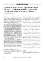
Figure 2 EC0746 is a folate-receptor-specific dihydrofolate reductase inhibitor with potent cytostatic effect on RAW264.7
macrophages. (a) RAW264.7 cells were given a 2-hour pulse of 100 nM EC0746 ± 10 μM folic acid (FA) followed by a 22-hour chase.
Aminopterin (AMT) and methotrexate (MTX) were allowed to incubate for 24 hours. The dihydrofolate reductase activities in whole cell lysates
(in duplicate) were normalized to untreated control cells (mean ± standard error of the mean). *P < 0.05. (b), (c) RAW264.7 cells were subjected
to a 2-hour pulse followed by a 70-hour chase of a 10-fold serial dilution of EC0746 ± 100-fold molar excess of FA. Free AMT was allowed to
incubate for 72 hours continuously. Four hours prior to the end of incubation, lipopolysaccharide (100 ng/ml) was added to stimulate TNFa
production. The (b) cell viability and (c) TNFa in culture media were determined by XTT and ELISA assays, respectively. Results expressed as the
percentage of control in absorbance (mean ± standard error of the mean in triplicates). (d) RAW264.7 cells were treated with indicated
concentrations of EC0746 ± excess FA (2-hour pulse plus a 70-hour chase). The surviving cells were redistributed in equal numbers in fresh
medium and allowed to incubate further for 72 hours. The cell proliferation was again determined by the XTT assay.
Lu et al. Arthritis Research & Therapy 2011, 13:R56
/>Page 7 of 18
promoted the release of ~19 cytokines/chemokines, 11
of which (Figure 3c) showed a FR-specific inhibition by
EC0746 (in at leas t three independent analyses), includ-
ing a few key proinflammatory mediators (TNFa,IL-1b,
macrophage inflammatory protein-1a, monokine
induced by IFNg, and so forth). These data collectively
indicated that the levels of FRs on TG-macs were suffi-
cient for EC0746 to remedially affect cytokine responses
associated with macrophage activation, and that the
observed anti-inflammatory action of EC0746 can be
independent of anti-macrophage proliferation.
Assessment of efficacy and dose/schedule dependency
in vivo
To establish a proof of concept for EC0746 in vivo,we
chose the macrophage-rich rat AIA model where a pre-
ferential uptake of FA-targeted imaging agents is consis-
tently seen in sites of active inflammation (arthritic
paws, liver, and spleen) [25,26]. The rat AIA model
resembles many characteristics of RA in humans and
has been widely used for the study of novel anti-inflam-
matory agents [50]. In our animal studies (n =5per
group), the onset of arthritis usually occurred around
day 10 after intradermal inoculation of M. butyricum
and was very aggressive. Multiple study endpoints were
taken to assess the effectiveness, including the arthritis
score (that is, paw edema) measured by a semiquantita-
tive visual sc oring system (see Materials and Methods),
the change in body weight (at the plateau of the disease,
untreated control animals lost ~14 to 20% of their origi-
nal weights), the paw weight, as an alternative assess-
ment of paw edema, and the spleen weight, as an
assessment of splenomegaly (an enlargement of the
spleen).
In a preliminary study (data not shown), a BIW regi-
men of EC0746 (500 nmol/kg, s.c.) was tested in AIA
rats presenting with varying degrees of arthritis (for
example, mean arthritis scores of ~0 versus 2 on the
first day of treatment). EC0746 was found to be fast act-
ing in treating AIA from the disease onset (that is,
mean starting arthritis score of ~0 on day 10); conse-
quently, these animals maintained a low arthritis score
(~1) and a steady body weight throughout the course o f
study. In rats with more established diseases (for exam-
ple, mean starting arthritis score of ~2 on days 10 to 13
post induction), EC0746 treatment also improved the
overall severity of the disease, but to a lesser extent.
Here, the maximum reduction in arthritic scores was
~50%, but the accompanying weight loss due to the
induction process was not reversed. When the percen-
tage increases in paw and splee n weights were analyzed,
EC0746 treatment yielded ~10-fold (paw edema) and
threefold (splen omega ly) improvem ents in rats with low
starting arthritis, and the corresponding improvements
were ~2.5-fold and twofold in rats bearing more estab-
lished diseases.
To fully investigate the dose-response relationship and
schedule dependency, EC0746 was administe red s.c. to
rats starting around the disease onset (days 9 to 11;
mean, day 10) with a dose range of 25 to 500 nmol/kg
BIW, or 1,000 nmol/kg given QW. As summarized in
Table 1, EC0746 treatment on days 10, 13, 17, and 20
displayed an ~10-fold linear dose response from 25 to
250 nmol/kg, with R
2
values of 1.00 (percentage inhibi-
tion in p aw edema) and 0.99 (reduction in splenome-
galy), respectively. The maximal activity of EC0746 was
achieved at 250 nmol/kg per dose, yi elding ~91% inhibi-
tion in paw edema, >3-fold to fourfold improvement in
splenomegaly, and with no apparent weight loss. There
was no statistical difference between the 250 and
500 nmol/kg dosing regimens of EC0746 in all end-
points assessed, suggesting a (FR) saturating dose
response in efficacy. When administered QW at 1,000
nmol/kg (days 10 and 17), EC0746 remained effective
with ~72% inhibition in paw edema, but this schedule
did not control the fast progressing AIA to the same
degre e as the optimal BIW dosing regimen (≥250 nm ol/
kg). Because of the schedule-dependent nature of the
response, animals receiving the Q W EC0746 treatment
also lost ~7% of their original body weights due to
arthritis progression (Table 1). In summary, EC0746 was
shown to be highly effective against AIA, more effective
when dosed BIW tha n QW, and capable of halting dis-
ease progression by controlling both local (joints) and
systemic (spleen) inflammation.
In vivo folate receptor specificity: proof of concept
To confirm in vivo target specificity of EC0746, we con-
ducted competition studies in AIA rats using a benign
folate-containing competitor (EC0923) to block t he FR
binding advantage of EC0746 (Figures 4 and 5). EC0923
(pteroyl-gGlu-d-Asp-d-Asp) is a high-affinity water-
soluble FA-peptide conjugate used in our laboratory for
in vivo comp etition studies rather than FA because high
doses of the latter can cause renal damage due to preci-
pitation in th e kidneys [51] . As described in Materials
and methods, four groups of AIA rats were given a stan-
dard BIW subcutaneous dosing regimen of either noth-
ing (that is, arthritic control), EC0746 alone (250 nmol/
kg), EC0746 (250 nmol/kg) plus a 500-fold molar excess
of EC0923 (125 μmol/kg), or EC0923 alone (125 μmol/
kg). All treatments lasted for 2 weeks, beginning 10 days
after the arthritis induction (disease onset).
As illustrated in Figure 4, EC0923 alone did not have
any impact on the development or severity of arthritis.
EC0746 alone was highly effective, as expected from
previous results (Table 1). Conversely, the activity of
EC0746 was nearly completely blocked by the presence
Lu et al. Arthritis Research & Therapy 2011, 13:R56
/>Page 8 of 18
of co-administered EC0923 in all clinical parameters
assessed: arthritis score (Figure 4a), change in body
weight (Figure 4b), and percentage increases in paw
(Figure 4c) and sp leen weights (Figure 4d). Radiographic
analysis of the a rthritic paws (Figure 4e,f) confirmed
minimal radiographic changes in EC0746-treated ani-
mals (similar to the healthy controls), whereas signifi-
cant joint erosions were seen in the untreated arthritic
controls and in the animals that had been treated with
EC0923 alone or with EC0746 plus EC0923.
Microscopically (Figure 5a,b), severe joint deteriora-
tions (that is, synovial inflammation, bone resorption,
pannus formation, and cartilage damage) were detected
in the arthritic control a nimals and in the animals trea-
ted with EC0923. In contrast, three out of five animals
treated with EC0746 had no lesions, resulting in 88 to
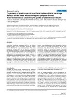
Figure 3 EC0746 has an immunomodulatory effect on folate-receptor-expressing rat TG-macs. Rat TG-macs were treated with media only,
100 nM EC0746 ± 10 μM folic acid (FA), or FA alone (10 μM) for 2 hours followed by a 70-hour chase. In comparison, the cells were also treated
with 100 nM free aminopterin (AMT) and methotrexate (MTX) for 72 hours. At 24 hours before the end of incubation, all cells were stimulated
with lipopolysaccharide (LPS) (5 μg/ml) plus IFNg (100 ng/ml). The cytokines/chemokines produced in culture supernatants were detected using
a rat cytokine array. (a) Cytokine release profiles of rat TG-macs stimulated with LPS and IFNg with or without drug treatment. (b) Cytokine array
map. (c) Mean pixel intensity (y axis) determined for each array position and plotted for the 11 products, which were detected at three times
above background levels and in at least three independent experiments. Data shown are mean ± standard error of the mean. *P < 0.05 when
compared with its corresponding cytokine level in the media only sample. CINC, cytokine-induced neutrophil chemoattractant; LIX, LPS-induced
CXC chemokine; MIG, monokine induced by IFNg; MIP-1a, macrophage inflammatory protein-1a; RANTES, regulated upon activation, normal
T-cell expressed and secreted; sICAM, soluble intracellular adhesion molecule; TIMP-1, tissue inhibitor of metalloproteinase 1; VEGF, vascular
endothelial growth factor.
Lu et al. Arthritis Research & Therapy 2011, 13:R56
/>Page 9 of 18
100% decreases in individually scored parameters
(Figure 5a), thus representing an overall decrease of
94% in the summed scores (Figure 5c). Notably, the ani-
mals treated with EC0746 plus the 500-fold excess of
EC0923 had a significa ntly decreased inflammation
score (24%; Figure 5a), but all other scored parameters
were nonsignificantly decreased (9 to 21%; Figure 5a).
Accordingly, the EC0746/EC0923-treated arthritic ani-
mals had an overall decrease of ~19% in the summed
scores, which was significantly less than the 94% reduc-
tion in animals treated with EC0746 alone (P <0.05;
Figure 5c). Likewise, the dorsal to ventral paw thickness
in the EC0746/EC0923-treated animals was decreased
by 22%, far less than the 94% reduction in the EC0746-
treated animals (P < 0.05; Figure 5d).
Overall, the res ults presented in Figures 4 and 5 show
a good correlation between macroscopic and micro-
scopic examinations of the arthritic animals, supporting
the fact that the anti-arthritic activities of EC0746 were
predominantly FR mediated.
To better understand EC0746 specificity in vivo,we
turned our attention to AMT and MTX. B oth agents
are active comp arators of EC0746, the former being the
parent drug and the latter being the most commonly
prescribed antifolate in the clinic. Given the large differ-
ences in FR affinities (Figure 1b) and the abilities of
MTX and AMT to enter other cells via RFC or protein-
coupled folate transporter, EC0746 was predicted to
affect a different population of host immune cells
than MTX and AMT, especially in a situation where
FR-positive macrophages play a big role in chronic
inflammatory responses. For years, RA patients receiving
antifolate therapy have been given folate supplementa-
tion to reduce adverse eff ects and to extend treatment
durations [12,52]. Because EC0746 contains both FA
and AMT moieties, a question arose as to whether the
anti-arthritic activity of EC0746 in AIA rats was due to
apparent folate supplementation of AMT. In an effort to
address t his question, we mixed unmodified AMT with
FA (1:1) and dosed AIA rats at a level matching the
well-tolerated BIW dose of EC0746 (500 nmol/kg s.c.).
Unfortunately, after one or two doses, all animals trea-
ted with the simple mixture had to be euthanized due
to severe an emia and gastrointestinal distress (lethargy,
bloody diarrhea, and so forth). More importantly,
whereas AMT is obviously a very toxic agent, its FA-tar-
geted form (that is, EC0746) is not.
Regarding MTX, which is a weaker FR binder than
EC0746, one might predict t hat the activity of MTX in
AIA rats would not be blocked by EC0923 under the
same competing conditions described above. To investi-
gate this hypothesis, three separate groups of AIA rats
were s.c. dosed BIW with either nothing, MTX alone
(250 nmol/kg), or MTX (250 nmol/kg) plus excess
EC0923 (125 μmol/kg). As assessed by arthritis scores
(Figure 6a), percentage increases in paw and spleen
weights (Figure 6b), and the change in body weight
(Figure 6c), the anti-arthritic activity of MTX was not
significantly blocked by the presence of the EC0923
competitor (P > 0.05; see figure legends). Taken
together, these data confirmed that EC0746 and MTX
were different from each other with regards to treating
active inflammation via FR-targeted and non-targeted
mechanisms of action, respectively.
EC0746 is more efficacious than oral methotrexate and
subcutaneous etanercept
Since MTX and etanercept are part of the current stan-
dard of care for RA, we compared EC0746 against both
drugs in the rat AIA model using clinically relevant
Table 1 EC0746 anti-arthritis activity in comparison with methotrexate and etanercept
Treatment Dose Frequency Inhibition in paw edema (%)
a
Splenomegaly
b
Body weight change (%)
c
Control - - 0 117 ± 22 -16 ± 1
EC0746 (s.c.) 25 nmol/kg Biweekly 0 ± 25
d
73 ± 8
e
-17 ± 1
100 nmol/kg Biweekly 35 ± 11
d
52 ± 13
e
-14 ± 1
250 nmol/kg Biweekly 91 ± 4
d
25 ± 6
e
-0.5 ± 3
500 nmol/kg Biweekly 91 ± 9 37 ± 7 0.4 ± 4
1,000 nmol/kg Once weekly 72 ± 12 39 ± 5 -7 ± 3
MTX (p.o.) 250 nmol/kg Biweekly 70 ± 5 42 ± 17 -14 ± 2
1,650 nmol/kg Once weekly 47 ± 10
f
- -10 ± 2
MTX (s.c.) 250 nmol/kg Biweekly 78 ± 10 24 ± 3 -2 ± 4
1,650 nmol/kg Once weekly 63 ± 13
f
7±8
Etanercept (s.c.) 10 mg/kg Every 3 days 46 ± 9
f
42 ± 7 -15 ± 2
Rats with adjuvant arthritis were treated at disease onset (10 days post arthritis induction) with EC0746, methotrexate (MTX), and etanercept at indicated doses,
dosing routes, and dosing frequencies. s.c., subcutaneously; p.o., per orally.
a
Inhibition in paw edema is calculated based on paw weight on day 24: 100 ×
(arthritic control - treated)/(arthritic control - healthy).
b
Splenomegaly is defined as the percentage increase in spleen weight relative to the spleen weights of
healthy rats.
c
On day 24 relative to body weight on the first day of treatment (day 10). Linear regression analysis:
d
R
2
= 1.00 (paw) and
e
R
2
= 0.99 (spleen).
f
Calculated based on arthritis scores on day 24 (paw weights were not obtained).
Lu et al. Arthritis Research & Therapy 2011, 13:R56
/>Page 10 of 18
dosing routes. In people, MTX is generally taken QW
by mouth, but there is a considerable variation in bioa-
vailability (28 to 94% at 15 mg/week) [ 53]. In our preli-
minary tests, we found QW oral MTX treatment (0.75
mg/kg or 1,650 nmol/kg) yielded ~50% efficacy in AIA
rats (based on arthritic scores), but the animals experi-
enced ~10% weight loss due to disease progression
(Table 1). This w as not unexpected as MTX also has a
low and variable oral bioavailability in rats due to
limited absorption and intestinal degradatio n (21% at
0.1 mg/kg) [54]. On an equimolar basis, QW MTX
(1,650 nmol/kg) s.c. was found to be slightly more
potent than QW oral MTX, yielding ~63% maximum
reduction in arthritis score and a slightly less weight
loss (~7%) (Table 1). Conversely, etanercept is a fully
humanized recombinant TNF receptor (p75)-Fc fusion
protein given as BIW injections to patients who have
failedorhavebecomelessresponsivetoMTX[5].
Figure 4 EC0746 demonstrates folate-receptor-specific anti-inflammatory activities in vivo. Starting on day 10 after arthritis induction, rats
with developing adjuvant-induced arthritis (n = 5) were given a biweekly subcutaneous dosing regimen of EC0746 (250 nmol/kg) without or
with a 500-fold molar excess of EC0923 as the folate competitor. For comparison purposes, methotrexate (MTX) (250 nmol/kg) was dosed orally
following the same schedule as EC0746, and etanercept (10 mg/kg) was given subcutaneously on days 10, 13, 16, 19, and 22. Multiple endpoints
are shown: (a) arthritis score; (b) change in body weight; (c) percentage increase in paw weight; (d) percentage increase in spleen weight; (e)
representative X-ray images of arthritic hind paws taken using a Kodak Imaging Station; and (f) radiographic score of arthritic hind paws. *P <
0.05 when compared with the arthritic control group.
Lu et al. Arthritis Research & Therapy 2011, 13:R56
/>Page 11 of 18
Returning to our study, shown in Figures 4 and 5,
EC0746 was also compared directly against oral MTX
on an equimolar BIW basis (that is, 250 nmol/kg), while
etanercept (10 mg/kg) was s.c. dosed every 3 days over a
12-day span (see Materials and methods). Although
rodents develop neutralizing antibodies against etaner-
cept, this limited high-dose regimen of etanercept was
reported to be active in rodent models of arthritis
[55,56].
As expected, both MTX and etanercept were active in
improving various sy mptoms of the experimental AIA
(Figure 4 and Table 1), but their effects were far from
optimum and neither therapy controlled arthritis quickly
enough to prevent substantial weight loss (Figure 4b and
Table 1). Histological grading of arthritic ankles (Figure 5)
showed oral MTX-treated animals had significa nt red uc-
tions (66 to 84% from the untreated arthritic control s) in
all scored parameters (Figure 5a,b), and there was a 73%
significant decrease in t he summed score (Figure 5c).
While etanercept did not appear to be as effective as oral
MTX, animals treated with etanercept also had significant
reductions in inflammation (42%) and bone resorption
(55%), which contributed to a significant 43% decrease in
the summed score (Figure 5a,c). Further, the dorsal to ven-
tral paw thicknesses in both MTX-treated and etanercept-
treat ed AIA rats were significantly decreased by 63% and
Arthritic EC0746 EC0746 + Competitor MTX Etanercept
0
1
2
3
4
5
6
7
Inflammation
Pannus
Cartilage damage
Bone resorption
Compet i torCo ntr ol
*
*
*
*
*
Histology scor es
Arthritic EC0746 EC0746 + Competitor MTX Etanercept
0
2
4
6
8
10
12
14
16
18
Competito r
0%
19 %
7%
73%
43%
94%
Control
*
*
*
Sum histology score
ArthriticArthritic
EC0746EC0746
EC0746+competitorEC0746+competitor
Competitor aloneCompetitor alone
MTXMTX
EtanerceptEtanercept
Arthritic Healthy EC0746 EC0746 +
C
ompetitor MTX Etanercept
0
1
2
3
4
5
6
7
8
9
10
11
12
Co mpe ti t or
100%
0%
94%
22 %
11%
63%
40%
Con tro l
*
*
*
*
*
Treatment
g
roups
Mean paw thi ckness
(mm)
(
a
)
(c)
(d)
(b)
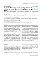
Figure 5 Histopathological assessment. The histopathological analysis was performed on formalin-fixed arthritic hind paws by Bolder BioPATH
Inc. (Boulder, CO, USA). (a) Individual histological scores of ankle joints on a scale of 0 to 5 for inflammation, bone resorption, pannus formation,
and cartilage damage with a maximal histology score of 20 per foot. (b) Representative photomicrographs (16x) of the ankle closest to the (c)
mean summed histological score. (d) Dorsal to ventral paw thickness for each group. Notably, the arthritic control animal showed very severe
inflammation (S), bone resorption (arrowhead) with mild pannus (small arrow), and cartilage damage (large arrow). *P < 0.05 when compared
with the arthritic control group. MTX, methotrexate.
Lu et al. Arthritis Research & Therapy 2011, 13:R56
/>Page 12 of 18
40%, respectively (Figure 5d). In almost all parameters
assessed (Figures 4 and 5, and Table 1), howeve r, s.c.
administered EC0746 consistently outperformed both oral
MTX and etanercept regimens - showing greater improve-
ments in arthritis scores, arthritis-related weight loss, paw
edema, radiographic changes, histological scores, and dor-
sal to ventral paw thicknesses. Notably, all three agents
decreased splenomegaly in AIA rats, but their effects were
not statistically different from each other (Table 1, calcu-
lated from Figure 4d).
Pharmacokinetics and metabolism
As shown in Table 1, EC0746 demonstrated a linear
dose-efficacy relationship in AIA ra ts, suggesting good
bioavailability after administration s.c. Because EC0746
contains a hydrazide/disulfide-based releasable linker
(Figure 1a), we anticipated that AMT and AMT hydra-
zide would be the two primary metabolites in vivo.Ona
cellular level, AMT and AMT hydrazide were found to
be equally potent in the inhibition of cell proliferation
and LPS-stimulated TNFa production in RAW264.7
macrophages (data no t shown). To study the pharmaco-
kinetics of EC0746, healthy rats were given a single sub-
cutaneous injection of EC0746 ( 500 nmol/kg) an d the
plasma concentrations of EC0746 as well as the poten-
tial metabolites, AMT, and AMT hydrazide, were moni-
tored for up to 8 hours. For comparison, unconjugated
AMT was examined at a matching dose (500 nmol/kg s.
c.) after a single administration.
EC0746 was found to reach the bloodstream within
minutes, with the maximum concentration (321 nmol/
l) occurring approximately 10 to 3 0 minutes post dose,
and maintained a plateau until 60 minutes after the
injection. EC0746-derived AMT and AMT hydrazide
were both detectable in plasma with maximum con-
centration values of 23 and 11 nmol/l, respectively
(curves nearly superimposable), but both metabolites
showed an approximate 30-minute delay from the time
at which EC0746 maximum concentration occurred.
While EC0746 itself was cleared rapidly from the
blood with an elimination half-life of ~35 minutes, the
elimination half-lives of the two metabolites were three
to five times longer at 1 17 minutes (AMT) and 187
minutes (AMT hydrazide), respectively. The corre-
sponding area under the curve values for EC0746 and
its metabolites AMT and AMT hydrazide were 32.5,
4.4, and 2.9 nmol*minute/ml. Similar to EC0746, the
maximum concentration (601 nmol/l) of unconjug ated
AMT in the plasma occurred ~30 minutes after dos-
ing; however, its elimination half-life was ~140 min-
utes, which is actually comparable with the values for
EC0746-derived AMT and AMT hydrazide. The area
under the curve value of the s.c. dosed AMT was mea-
sured at 61.3 nmol*minute/ml.
Taken together, EC0746 metabolism in vivo appeared
to re sult in a delayed release of AMT and AMT hydra-
zide, but these two metabolites behaved more like free
AMT rather than EC0746 with regards to elimination.
Based on the area under the curve responses, ~18% of
active drug exposure/release (AMT plus AMT hyd ra-
zide) was detected in the plasma over the 8-hour collec-
tion period in the EC0746-dosed animals.
Figure 6 Methotrexate displays a lack of competi tion for
folate-receptor-binding sites and a nonfolate-receptor-targeted
anti-arthritic activity. In an identical dosing fashion as was carried
out for EC0746 in Figure 4, adjuvant-induced arthritis rats (n =5)
were treated subcutaneously with methotrexate (MTX) (250 nmol/kg
biweekly) for 2 weeks without or with a 500-fold molar excess of
EC0923 as the folate competitor. Anti-arthritic activities are shown:
(a) arthritis score; (b) percentage increases in paw and spleen
weights; and (c) percentage change in body weight. ns, not
significant.
Lu et al. Arthritis Research & Therapy 2011, 13:R56
/>Page 13 of 18
Preliminary short-term toxicity assessment
Since the toxicity of antifolates can be easily masked by
rodent diets enriched with FA [57], healthy rats on a
folate-deficient diet (Harlan Teklad, Madison, WI, USA)
were used to determ ine the MTDs of EC0746 and AMT.
Following the same dosing regimen used for aforemen-
tioned anti-arthritis therapies (that is, four subcutaneous
doses given in 2 weeks), the MTDs of EC074 6 and AMT
were determined to be 2,000 nmol/kg and 50 nmol/kg,
respectively, representing a 40-fold difference in toxicity.
At supra-MTD doses, the toxicologic findings in
EC0746-treated rats were similar to those treated with
AMT, including diarrhea, swollen muzzle, leucopenia,
thrombocytopenia, and opportunistic infections. While
immunosuppression a nd gastrointestinal toxicity appear
to be dose-limiting for these compounds in folate-defi-
cient rats, EC0746 showed less of the gastrointestinal-
associated side effects than AMT at their respective
MTD doses.
Discussion
There are two natural isoforms of the membrane FR
(FRa and FRb), and both bind FA with a high affinity (K
D
<1 nM) [58]. FRa is best known for its overexpression on
human epithelial cancers, and is present on the apical
surfaces of limited normal epithelial cell types (proximal
tubules of the kidneys, choroid plexus of the brain, and
alveolar epithelial cells of the lungs) [58]. FRb is found
on myelogenous leukemias [59] and has now become a
promising biomarker for activated macrophages and
monocytes [21-23]. To date, it is not entirely clear which
subpopulation(s) of activated macrophages (and mono-
cytes) expresses FR b and what r ole these receptors play
in chronic inflammation [24]. Adding to this complexity,
activated macrophages are known to be a heterogeneous
population of extraordinarily versatile cells that are func-
tionally dynamic based on their microenvironment [49].
Because FR-positive macrophages are found to express
various macrophage activation markers (that is, CD8 0,
CD86, Ly6C/G, TNFa, and reactive oxygen species) [22],
depleting or inactivati ng this effector cell population may
have a profound effect on the immune system. In the
present study we evaluated EC0746, a novel FA-AMT
construct that utilizes the high-affinity FA ligand as a tar-
geting moiety to facilitate the specific delivery of an anti-
folate pharmacophore to FRa/FRb-expressing cells (Fig-
ure 1b) and to inhibit DHFR in a FR-dependent manner
(Figure 2a). Notably, our relative affinity values for AMT
and MTX are consistent with previously reports for these
and other antifolates that have weak affinities for both FR
isoforms because their preferred route of cellular entry is
via the RFC [19].
Since it was challenging to find an in vitro cell model
that would realistically mimic the pathophysiological
properties of activated macrophages in vivo,we
employed the high FR-expressing and highly prolifera-
tive RAW264.7 macrophages, as well as the low FR-
expressing and low-proliferating rat TG-macs, to repre-
sent macrophages in different states of activation/
inflammation. Rat TG-macs were chosen b ecause they
could be e asily obtained, and they express a functional
FR at a similar level to that of peritoneal macrophages
isolated from AIA rats at the plateau of their disease
[25]. Moreover, the rat TG-macs isolated as described
represent a heterogeneous population of ~90% CD1 1b/
c-positive cells, and ~70% of the CD11b/c-positive cell
popu lation are FR-pos itive upon staining with a fluores-
cent folate probe (data not shown). Using these two cell
models, EC0746 treatment resulted in two independent
but FR-specific mechanisms of action: an anti-prolifera-
tive effect against RAW264.7 cells (Figure 2b to 2d), and
an anti-inflammatory effect against rat TG-macs without
affecting cell viability or proliferation (Figure 3). Overall,
our in vitro results suggested that EC0746 might be
active against a heterogeneous population of FR-positive
macrophages (and monocytes) at sites of active inflam-
mation. It remains uncertain, however, whether macro-
phages from the peritonea l cavity differ from those
present in an inflamed joint, whet her rat TG-macs have
other immunoinflammatory mechanisms that make
themdifferentfrommacrophagesisolatedfromthe
arthritic animals, and whether ex vivo isolated macro-
phages lose FR expression and, cons equently, inflamma-
tory impetus in cell culture.
For in vivo efficacy assessment, the rat AIA model has
been our c hoice due to its well-documented systemic
inflammation involving activated macrophages [60,61]
and the presence of FR-positive macrophage subpop ula-
tions [25]. The obvious disadvantage of this model i s its
aggressive and acute nature (full-blown arthritis in 2 to
3 weeks), which makes it unlike human RA. Owing to
thedifficultyofobtainingrelevant clinical samples, we
have not been able to directly c ompare the level of FR
expression on activated macrophages in AIA rats with
that found on cells within the joints of RA patients. A
second, cautious limitation of this model is that the cir-
culating serum folate levels in rats are supra-physiol ogi-
cally high due to the supplementation of commercial
rodent chows [36]. Because unnaturally high folate levels
can act as a competitor for FR binding, rats used in this
study were fed a nonsupplemented diet. Although spec-
ulative, it is possible that FR levels in resident macro-
phages may have inadvertently been upregulated in the
arthritic rats during the course of study. Nonetheless,
BIW EC0746 treatment (s.c.) was found to be highly
effective in alleviating overall symptoms of AIA, espe-
cially when the treatment started at disease onset. The
EC0746 anti-arthritic activity was also dose and schedule
Lu et al. Arthritis Research & Therapy 2011, 13:R56
/>Page 14 of 18
dependent (Table 1), and appeared to be FR-specific
since a benign folate ligand (EC0923) could efficiently
block its overall effect by competing for FR-binding sites
in vivo (Figures 4 and 5). Although MTX is a weak
bicarboxylic acid structurally related to FA, its binding
affinities to FR-expressing cells (KB, CHO-FRb)were
~56-fold to 200-fold lower than that of FA (Figure 1b).
Accordingly, the anti-arthritic activity of MTX was not
blocked to a significant degree by EC0923 under the
same competing conditions (Figure 6). This finding also
supported EC0923 compromising the efficacy of EC0746
through its interaction with the FR and not through any
antifolate mechanism of action. Finally, as a structure-
matched negative control, we also tested an analog of
EC0746 that was constructed with the unnatural, and
biologically inert d-enantiomer of AMT. This com-
pound, which shares similar FR binding affinity and
identical molecule weight to EC0746, was found to be
completely inactive both in vitro and in vivo (data not
shown). These results helped confirm that an anti-
inflammatory response is not automatically triggered by
the simple binding of a ligand to the FR present on acti-
vated macrophages, and that specific activity is depen-
dent on the endocytosis of the conjugate followed by
biological cleavage of the linker and cytosolic release of
a biologically active drug.
MTX generally appears as effective as EC0746 on an
equimolar basis when both drugs are administered BIW
via a subcutaneous r oute in AIA rats (Table 1). Our in
vitro and in vivo results, however, confirmed that these
two agents were significantly different from each other
with regards to targeting FR-positive and FR-negative
inflammatory cells. In t hat regard, Matsuyama and col-
leagues - who studied MTX transport via FRb in RA
synovia l macrophages - suggested that folate antagonists
with a higher affinity towards FRb could be more useful
in RA treatmen t [21]. EC07 46 did consistently outper-
form oral MTX on an equimolar basis, and it appeared
to be more active than etanercept (Figur es 4 and 5, and
Table 1), although the latte r is a genetically engineered
human protein with unknown potency to rat TNFa.
Limited by its molecular properties (size, charge, hydro-
gen bonding potential, and so forth), EC0746 does not
meet the common criteria for oral drug delivery [62], so
this route of dosing was not initially explo red. In addi-
tion, the high toxicity of AMT in rodents precluded
further investigation into the anti-arthritic activity of
this base drug. In fac t, we found in folate-deficient rats
that EC0746 was approximately 40-fold less toxic than
AMT (all given by subcutaneous administration). Folate
conjugation and targeting therefore provided a therapeu-
tic window to AMT where none exists otherwise in AIA
rats. It is worth mentioning, however, that AMT is cur-
rently b eing studied in psoriasis patients using very low
doses (NCT00937027; Syntrix Biosystems Inc., Auburn,
WA, USA).
Pharmacokinetically, EC0746 (500 nmol/kg) adminis-
tered s.c. was absorbed quickly into the central com-
partment from the subcutaneous space, with the time
to maximum concentration occurring at approximately
10 to 30 minutes, followed by a short elimination half-
life (~35 minutes). In comparison, unconjugated AMT
at the matching subcutaneous dose exhibited a similar
time to maximum concentration of 30 minutes, but
the elimination half-life increased significantly (~140
minutes). Based on our experiences with folate-tar-
geted chemotherapeutic agents, a shorter elimination
half-life (as seen for EC0746) is favorable because it
minimizesnonspecifictissueexposurewithoutredu-
cing the FR targeting potential [42]. EC0746 was also
found to be metabolized in vivo, and the t wo major
metabolites (AMT and AMT hydrazide) exhibited
longer elimination half-lives than EC0746. Because
antifolates like AMT enter normal cells via the RFC or
protein-coupled folate transporter, we suspect that
EC0746-derived AMT/AMT hydrazide would behave
like free AMT on a cellular level (except for a 30-min-
ute delay in appearance in the plasma). A comparison
of the area under the curve values for EC0746 and
released AMT/AMT hydrazide indicates that as much
as ~18% free drug was detected in the plasma. The
question therefore arose regarding how much these
two metabolites might contribute to the overall efficacy
and toxicity of EC0746 administered to AIA rats.
While this topic deserves further study, there are three
reasons we do not believe that free drug significantly
affected the potency of EC0746 therapy. First, we have
shown that a 2-week BIW treatment of 500 nmol/kg
EC0746 s.c. is a safe and effective dosing regimen
(Table 1). Using the pharmacokinetic observations as a
guide for exposure at this dose level, and considering
the fact that roughly 18% free drug is released in circu-
lation following a 500 nmol/kg EC0746 dose, then a
90nmol/kgdoseofAMTwouldbepredictedtobe
both effective and nontoxic. Not only did our toxicol-
ogy studies show that doses of AMT above 50 nmol/kg
resulted in considerable morbidity a nd mortality, how-
ever, but 40-fold higher EC0746 doses were found to
be well tolerated. Second, the anti-arthritic activity of
EC0746 could be blocked by the presence of the folate
competitor EC0923 (Figures 4 and 5), indicating that
the mechanism of action for EC0746 was specific for
FR expression and not a nontargeted effect due to the
metabolites AMT/AMT hydrazide. Third, animal data
presented within this report suggest that the efficacy of
EC0746 was probably not driven entirely by its phar-
macokinetic properties in the plasma, but rather by its
dynamics on target cells expressing a functional FR.
Lu et al. Arthritis Research & Therapy 2011, 13:R56
/>Page 15 of 18
Targeting activated macrophages via the FR is a
rational approach to curtail macrophages’ resistance to
apoptosis and to inhibit their ability to produce tissue-
damaging products. Our investigation provides the first
evidence that anti-inflamm atorydrugs,likeAMT,can
be directly linked to FA or folate-like ligands to yield an
improved therapeutic index. Recently, a few oral ly active
small molecule drugs have shown promising results in
MTX-failure patients, including two Janus kinase inhibi-
tors, INCB028050 [50] and CP-690,550 [63,64], and the
Syk kinase inhibitor R788 [65]. These novel anti-inflam-
matory agents appear t o be better tolerated and they
can yield similar efficacy as anti-TNF biologics. As most
rheumatologists still prefer MTX as the first disease-
modifying anti-rheumatic drug for RA treatment, if
approved these new agents are likely to position them-
selves after MTX failure but before the use of bi ologics.
Owing to the lack of MTX-resistant animal models of
inflammation, the ability of EC0746 to overcome MTX
resistance was not investigated. In the clinic, studies
have shown the need to gradually increase the dose of
MTX in order to achieve an optimal therapeutic benefit,
and some people will eventually bec ome nonresponsive
to MTX. One of the reasons for the lack of response is
that RFC-mediated transport across the cell membrane
utilizes an anion exchange mechanism that requires a
higher saturating dose to work efficiently [66]. EC0746
mayovercomesuchdeficiencybydeliveringAMTto
FR-positive inflammatory macrophages and increasing
their intracellular retention of the drug.
Conclusions
The therapeutic value of EC0746 needs to be validated
in the clinic compared against other existing treatment
options. One encouraging fact may be that inflammatory
macrophages and monocytes do not turnover as fast as
their normal counterparts, and t hese cells may maintain
their FR expression in time to allow for FR-specific
intervention. For long-term therapy and managemen t,
EC0746 would probably cost less to produce than biolo-
gics, and it could potentially be self-administered at a
frequency yet to be determined in the clinic (not in ani-
mal models). Overall, the potential application of
EC0746 may include macrophage-rich inflammatory dis-
orders such as RA, psoriasis, a therosclerosis, Crohn’ s
disease, uveitis, vasculitis, and diabetes (many of which
share some common pathogenic mechanisms).
Abbreviations
AIA: adjuvant-induced arthritis; AMT: aminopterin; BIW: biweekly; DHFR:
dihydrofolate reductase; ELISA: enzyme-linked immunosorbent assay; FA:
folic acid; FFRPMI: folate-free RPMI 1640 medium; FR: folate receptor; IFN:
interferon; IL: interleukin; LPS: lipopolysaccharide; MTD: maximum tolerated
dose; MTX: methotrexate; QW: once weekly; RA: rheumatoid arthritis; RFC:
reduced-folate carrier; s.c.: subcutaneously; TG: thioglycollate; TNF: tumor
necrosis factor; XTT: 2,3-bis(2-methoxy-4-nitro-5-sulfo-phenyl)-2H-tetrazolium-
5-carboxanilide.
Acknowledgements
The authors would like to acknowledge Wilfredo Ayala-López for providing
RAW264.7 cells, Jeremy Vaughn for synthesis of EC0923, and Kristin Wollak
for assistance in animal work.
Author details
1
Endocyte, Inc., 3000 Kent Avenue, West Lafayette, IN 47906, USA.
2
Department of Chemistry, 560 Oval Drive, Purdue University, West Lafayette,
IN 47907, USA.
Authors’ contributions
YL contributed to the experimental design, data acquisition, data analysis
and interpretation, and drafted the manuscript. TWS contributed to the in
vitro experimental design, data acquisition, and data analysis and
interpretation. EW and VAC contributed to in vivo data acquisition and data
analysis. PJK and MAG contributed to design, and data analysis and
interpretation involving toxicology and pharmacokinetics. IRV contributed to
synthetic chemistry involving EC0746 and EC0923. PSL contributed to
experimental design and edited the manuscript. CPL contributed to the
experimental design, data analysis and interpretation, and edited the
manuscript. All authors read and approved the final manuscript.
Competing interests
Except for PSL, all authors are full-time employees and stockholders of
Endocyte, Inc. PSL is a full-time professor at Purdue University (West
Lafayette, IN, USA). PSL is also a founder, the Chief Science Officer, and a
compensated member of Endocyte’s Board of Directors. A patent application
has been submitted to cover the data disclosed in this manuscript. No other
financial interests apply for any authors.
Received: 18 January 2011 Revised: 23 February 2011
Accepted: 4 April 2011 Published: 4 April 2011
References
1. Hamilton JA, Tak PP: The dynamics of macrophage lineage populations
in inflammatory and autoimmune diseases. Arthritis Rheum 2009,
60:1210-1221.
2. Haringman JJ, Gerlag DM, Zwinderman AH, Smeets TJ, Kraan MC, Baeten D,
McInnes IB, Bresnihan B, Tak PP: Synovial tissue macrophages: a sensitive
biomarker for response to treatment in patients with rheumatoid
arthritis. Ann Rheum Dis 2005, 64:834-838.
3. Kinne RW, Stuhlmuller B, Burmester GR: Cells of the synovium in
rheumatoid arthritis. Macrophages. Arthritis Res Ther 2007, 9:224.
4. Tarner IH, Muller-Ladner U, Gay S: Emerging targets of biologic therapies
for rheumatoid arthritis. Nat Clin Pract Rheumatol 2007, 3:336-345.
5. Taylor PC, Feldmann M: Anti-TNF biologic agents: still the therapy of
choice for rheumatoid arthritis. Nat Rev Rheumatol 2009, 5:578-582.
6. van Vollenhoven RF: Treatment of rheumatoid arthritis: state of the art
2009. Nat Rev Rheumatol 2009, 5:531-541.
7. Wolfe F, Michaud K: The loss of health status in rheumatoid arthritis and
the effect of biologic therapy: a longitudinal observational study. Arthritis
Res Ther 2010, 12:R35.
8. Martin-Mola E, Balsa A: Infectious complications of biologic agents. Rheum
Dis Clin North Am 2009, 35:183-199.
9. Braun J, Rau R: An update on methotrexate. Curr Opin Rheumatol 2009,
21:216-223.
10. Assaraf YG: Molecular basis of antifolate resistance. Cancer Metastasis Rev
2007, 26:153-181.
11. Chan ES, Cronstein BN: Methotrexate – how does it really work? Nat Rev
Rheumatol 2010, 6:175-178.
12. van Ede AE, Laan RF, Rood MJ, Huizinga TW, van de Laar MA, van
Denderen CJ, Westgeest TA, Romme TC, de Rooij DJ, Jacobs MJ, de
Boo TM, van der Wilt GJ, Severens JL, Hartman M, Krabbe PF, Dijkmans BA,
Breedveld FC, van de Putte LB: Effect of folic or folinic acid
supplementation on the toxicity and efficacy of methotrexate in
rheumatoid arthritis: a forty-eight week, multicenter, randomized,
double-blind, placebo-controlled study. Arthritis Rheum 2001,
44:1515-1524.
Lu et al. Arthritis Research & Therapy 2011, 13:R56
/>Page 16 of 18
13. Bohanec Grabar P, Logar D, Lestan B, Dolzan V: Genetic determinants of
methotrexate toxicity in rheumatoid arthritis patients: a study of
polymorphisms affecting methotrexate transport and folate metabolism.
Eur J Clin Pharmacol 2008, 64:1057-1068.
14. Alarcon GS: Methotrexate use in rheumatoid arthritis. A clinician ’s
perspective. Immunopharmacology 2000, 47:259-271.
15. Cole PD, Zebala JA, Alcaraz MJ, Smith AK, Tan J, Kamen BA:
Pharmacodynamic properties of methotrexate and Aminotrexate during
weekly therapy. Cancer Chemother Pharmacol 2006, 57:826-834.
16. Zebala JA: Aminopterin dosage forms and methods for inflammatory
disorders. US Patent 7312217 2007.
17. Matherly LH, Voss MK, Anderson LA, Fry DW, Goldman ID: Enhanced
polyglutamylation of aminopterin relative to methotrexate in the Ehrlich
ascites tumor cell in vitro. Cancer Res 1985, 45:1073-1078.
18. Werkheiser WC: The biochemical, cellular, and pharmacological action
and effe cts of the folic a cid antagonists . Cancer Res 1963,
23:1277-128 5.
19. Westerhof GR, Schornagel JH, Kathmann I, Jackman AL, Rosowsky A,
Forsch RA, Hynes JB, Boyle FT, Peters GJ, Pinedo HM: Carrier- and receptor-
mediated transport of folate antagonists targeting folate-dependent
enzymes: correlates of molecular-structure and biological activity. Mol
Pharmacol 1995, 48:459-471.
20. Urquhart BL, Gregor JC, Chande N, Knauer MJ, Tirona RG, Kim RB: The
human proton-coupled folate transporter (hPCFT): modulation of
intestinal expression and function by drugs. Am J Physiol Gastrointest Liver
Physiol 2010, 298:G248-G254.
21. Nakashima-Matsushita N, Homma T, Yu S, Matsuda T, Sunahara N,
Nakamura T, Tsukano M, Ratnam M, Matsuyama T: Selective expression of
folate receptor beta and its possible role in methotrexate transport in
synovial macrophages from patients with rheumatoid arthritis. Arthritis
Rheum 1999, 42:1609-1616.
22. Xia W, Hilgenbrink AR, Matteson EL, Lockwood MB, Cheng JX, Low PS: A
functional folate receptor is induced during macrophage activation and
can be used to target drugs to activated macrophages. Blood 2009,
113:438-446.
23. van der Heijden JW, Oerlemans R, Dijkmans BA, Qi H, van der Laken CJ,
Lems WF, Jackman AL, Kraan MC, Tak PP, Ratnam M, Jansen G: Folate
receptor beta as a potential delivery route for novel folate antagonists
to macrophages in the synovial tissue of rheumatoid arthritis patients.
Arthritis Rheum 2009, 60:12-21.
24. Hansen M, Low P: Folate receptor positive macrophages: cellular targets
for imaging and therapy of inflammatory and autoimmune diseases. In
Targeted Drug Strategies for Cancer and Inflammation. Edited by: Jackman
AL, Leamon CP. New York: Springer; .
25. Lu Y, Leamon C: Targeting activated macrophages via a functional folate
receptor for potential treatment of autoimmune/inflammatory disorders.
In Targeted Drug Strategies for Cancer and Inflammation. Edited by: Jackman
AL, Leamon CP. New York: Springer; .
26. Turk MJ, Breur GJ, Widmer WR, Paulos CM, Xu LC, Grote LA, Low PS: Folate-
targeted imaging of activated macrophages in rats with adjuvant-
induced arthritis. Arthritis Rheum 2002,
46:1947-1955.
27.
Matteson EL, Lowe VJ, Prendergast FG, Crowson CS, Moder KG,
Morgenstern DE, Messmann RA, Low PS: Assessment of disease activity in
rheumatoid arthritis using a novel folate targeted radiopharmaceutical
Folatescan. Clin Exp Rheumatol 2009, 27:253-259.
28. Nagayoshi R, Nagai T, Matsushita K, Sato K, Sunahara N, Matsuda T,
Nakamura T, Komiya S, Onda M, Matsuyama T: Effectiveness of anti-folate
receptor beta antibody conjugated with truncated Pseudomonas
exotoxin in the targeting of rheumatoid arthritis synovial macrophages.
Arthritis Rheum 2005, 52:2666-2675.
29. Paulos CM, Varghese B, Widmer WR, Breur GJ, Vlashi E, Low PS: Folate-
targeted immunotherapy effectively treats established adjuvant and
collagen-induced arthritis. Arthritis Res Ther 2006, 8:R77.
30. Yi YS, Ayala-Lopez W, Kularatne SA, Low PS: Folate-targeted hapten
immunotherapy of adjuvant-induced arthritis: comparison of hapten
potencies. Mol Pharm 2009, 6:1228-1236.
31. Nagayoshi R, Nakamura M, Ijiri K, Yoshida H, Komiya S, Matsuyama T:
LY309887, antifolate via the folate receptor suppresses murine type II
collagen-induced arthritis. Clin Exp Rheumatol 2003, 21:719-725.
32. Vlahov IR, You F, Santhapuram HKR, Wang Y, Vaughn JF, Hahn SJ,
Kleindl PJ, Fan M, Leamon CP: Design and regioselective synthesis of a
new generation of targeted therapeutics. Part 3: Folate conjugates of
aminopterin hydrazide for the treatment of inflammation. Bioorg Med
Chem Lett 2010, 21:1202-1205.
33. Leamon CP, You F, Santhapuram HK, Fan M, Vlahov IR: Properties
influencing the relative binding affinity of pteroate derivatives and drug
conjugates thereof to the folate receptor. Pharm Res 2009, 26:1315-1323.
34. Wu M, Fan J, Gunning W, Ratnam M: Clustering of GPI-anchored folate
receptor independent of both cross-linking and association with
caveolin. J Membr Biol 1997, 159:137-147.
35. ImageJ. [ />36. Leamon CP, Reddy JA, Dorton R, Bloomfield A, Emsweller K, Parker N,
Westrick E: Impact of high and low folate diets on tissue folate receptor
levels and antitumor responses toward folate-drug conjugates. J
Pharmacol Exp Ther 2008, 327:918-925.
37. Simjee SU, Jawed H, Quadri J, Saeed SA: Quantitative gait analysis as a
method to assess mechanical hyperalgesia modulated by disease-
modifying antirheumatoid drugs in the adjuvant-induced arthritic rat.
Arthritis Res Ther 2007, 9:R91.
38. Montesinos MC, Yap JS, Desai A, Posadas I, McCrary CT, Cronstein BN:
Reversal of the antiinflammatory effects of methotrexate by the
nonselective adenosine receptor antagonists theophylline and caffeine:
evidence that the antiinflammatory effects of methotrexate are
mediated via multiple adenosine receptors in rat adjuvant arthritis.
Arthritis Rheum 2000, 43:656-663.
39. Fletcher DS, Widmer WR, Luell S, Christen A, Orevillo C, Shah S, Visco D:
Therapeutic administration of a selective inhibitor of nitric oxide
synthase does not ameliorate the chronic inflammation and tissue
damage associated with adjuvant-induced arthritis in rats. J Pharmacol
Exp Ther 1998, 284:714-721.
40. Bendele AM, Chlipala ES, Scherrer J, Frazier J, Sennello G, Rich WJ,
Edwards CK: Combination benefit of treatment with the cytokine
inhibitors interleukin-1 receptor antagonist and PEGylated soluble tumor
necrosis factor receptor type I in animal models of rheumatoid arthritis.
Arthritis Rheum 2000, 43
:2648-2659.
41.
Vlahov IR, Santhapuram HK, You F, Wang Y, Kleindl PJ, Hahn SJ, Vaughn JF,
Reno DS, Leamon CP: Carbohydrate-based synthetic approach to control
toxicity profiles of folate-drug conjugates. J Org Chem 2010, 75:3685-3691.
42. Leamon CP, Reddy JA, Klein PJ, Vlahov IR, Dorton R, Bloomfield A,
Nelson M, Westrick E, Parker N, Bruna K, Vetzel M, Gehrke M, Nicoson JS,
Messmann RA, Lorusso PM, Sausville EA: Reducing undesirable hepatic
clearance of a tumor-targeted vinca alkaloid via novel saccharopeptidic
modifications. J Pharmacol Exp Ther 2011, 336:336-343.
43. Leamon CP, Reddy JA: Folate-targeted chemotherapy. Adv Drug Deliv Rev
2004, 56:1127-1141.
44. Yang J, Chen H, Vlahov IR, Cheng JX, Low PS: Evaluation of disulfide
reduction during receptor-mediated endocytosis by using FRET imaging.
Proc Natl Acad Sci USA 2006, 103:13872-13877.
45. Attur MG, Patel R, Thakker G, Vyas P, Levartovsky D, Patel P, Naqvi S, Raza R,
Patel K, Abramson D, Bruno G, Abramson SB, Amin AR: Differential anti-
inflammatory effects of immunosuppressive drugs: cyclosporin,
rapamycin and FK-506 on inducible nitric oxide synthase, nitric oxide,
cyclooxygenase-2 and PGE2 production. Inflamm Res 2000, 49:20-26.
46. Xaus J, Comalada M, Valledor AF, Cardo M, Herrero C, Soler C, Lloberas J,
Celada A: Molecular mechanisms involved in macrophage survival,
proliferation, activation or apoptosis. Immunobiology 2001, 204:543-550.
47. Gordon S, Taylor PR: Monocyte and macrophage heterogeneity. Nat Rev
Immunol 2005, 5:953-964.
48. Ranganathan P: An update on methotrexate pharmacogenetics in
rheumatoid arthritis. Pharmacogenomics 2008, 9:439-451.
49. Mosser DM, Edwards JP: Exploring the full spectrum of macrophage
activation. Nat Rev Immunol 2008, 8:958-969.
50. Fridman JS, Scherle PA, Collins R, Burn TC, Li Y, Li J, Covington MB,
Thomas B, Collier P, Favata MF, Wen X, Shi J, McGee R, Haley PJ, Shepard S,
Rodgers JD, Yeleswaram S, Hollis G, Newton RC, Metcalf B, Friedman SM,
Vaddi K: Selective inhibition of JAK1 and JAK2 is efficacious in rodent
models of arthritis: preclinical characterization of INCB028050. J Immunol
2010, 184:5298-5307.
51. Fink M, Henry M, Tange JD: Experimental folic acid nephropathy.
Pathology 1987, 19:143-149.
52. Hoekstra M, van Ede AE, Haagsma CJ, van de Laar MA, Huizinga TW,
Kruijsen MW, Laan RF: Factors associated with toxicity, final dose, and
Lu et al. Arthritis Research & Therapy 2011, 13:R56
/>Page 17 of 18
efficacy of methotrexate in patients with rheumatoid arthritis. Ann
Rheum Dis 2003, 62:423-426.
53. Oguey D, Kolliker F, Gerber NJ, Reichen J: Effect of food on the
bioavailability of low-dose methotrexate in patients with rheumatoid
arthritis. Arthritis Rheum 1992, 35:611-614.
54. Kuroda T, Namba K, Torimaru T, Kawashima K, Hayashi M: Species
differences in oral bioavailability of methotrexate between rats and
monkeys. Biol Pharm Bull 2000, 23:334-338.
55. Inglis JJ, Nissim A, Lees DM, Hunt SP, Chernajovsky Y, Kidd BL: The
differential contribution of tumour necrosis factor to thermal and
mechanical hyperalgesia during chronic inflammation. Arthritis Res Ther
2005, 7:R807-R816.
56. Williams RO: Collagen-induced arthritis in mice: a major role for tumor
necrosis factor-alpha. Methods Mol Biol 2007, 361:265-284.
57. Mauritz R, Peters GJ, Kathmann I, Teshale H, Noordhuis P, Comijn EM,
Pinedo HM, Jansen G: Dynamics of antifolate transport via the reduced
folate carrier and the membrane folate receptor in murine leukaemia
cells in vitro and in vivo. Cancer Chemother Pharmacol 2008, 62:937-948.
58. Elnakat H, Ratnam M: Distribution, functionality and gene regulation of
folate receptor isoforms: implications in targeted therapy. Adv Drug Deliv
Rev 2004, 56:1067-1084.
59. Shen F, Ross JF, Wang X, Ratnam M: Identification of a novel folate
receptor, a truncated receptor, and receptor type beta in hematopoietic
cells: cDNA cloning, expression, immunoreactivity, and tissue specificity.
Biochemistry 1994, 33:1209-1215.
60. Smith-Oliver T, Noel LS, Stimpson SS, Yarnall DP, Connolly KM: Elevated
levels of TNF in the joints of adjuvant arthritic rats. Cytokine 1993,
5:298-304.
61. Issekutz AC, Issekutz TB: Monocyte migration to arthritis in the rat utilizes
both CD11/CD18 and very late activation antigen 4 integrin
mechanisms. J Exp Med 1995, 181:1197-1203.
62. Burton PS, Goodwin JT, Vidmar TJ, Amore BM: Predicting drug absorption:
how nature made it a difficult problem. J Pharmacol Exp Ther 2002,
303:889-895.
63. Coombs JH, Bloom BJ, Breedveld FC, Fletcher MP, Gruben D, Kremer JM,
Burgos-Vargas R, Wilkinson B, Zerbini CA, Zwillich SH: Improved pain,
physical functioning and health status in patients with rheumatoid
arthritis treated with CP-690,550, an orally active Janus kinase (JAK)
inhibitor: results from a randomised, double-blind, placebo-controlled
trial. Ann Rheum Dis 2010, 69:413-416.
64. Cohen S, Zwillich SH, Chow V, Labadie RR, Wilkinson B: Co-administration
of the JAK inhibitor CP-690,550 and methotrexate is well tolerated in
patients with rheumatoid arthritis without need for dose adjustment. Br
J Clin Pharmacol 2010, 69:143-151.
65. Weinblatt ME, Kavanaugh A, Burgos-Vargas R, Dikranian AH, Medrano-
Ramirez G, Morales-Torres JL, Murphy FT, Musser TK, Straniero N, Vicente-
Gonzales AV, Grossbard E: Treatment of rheumatoid arthritis with a Syk
kinase inhibitor: a twelve-week, randomized, placebo-controlled trial.
Arthritis Rheum 2008, 58:3309-3318.
66. van der Heijden JW, Dijkmans BA, Scheper RJ, Jansen G: Drug Insight:
resistance to methotrexate and other disease-modifying antirheumatic
drugs – from bench to bedside. Nat Clin Pract Rheumatol 2007, 3:26-34.
doi:10.1186/ar3304
Cite this article as: Lu et al.: Treatment of experimental adjuvant
arthritis with a novel folate receptor-targeted folic acid-aminopterin
conjugate. Arthritis Research & Therapy 2011 13:R56.
Submit your next manuscript to BioMed Central
and take full advantage of:
• Convenient online submission
• Thorough peer review
• No space constraints or color figure charges
• Immediate publication on acceptance
• Inclusion in PubMed, CAS, Scopus and Google Scholar
• Research which is freely available for redistribution
Submit your manuscript at
www.biomedcentral.com/submit
Lu et al. Arthritis Research & Therapy 2011, 13:R56
/>Page 18 of 18