Báo cáo y học: " A folate receptor beta-specific human monoclonal antibody recognizes activated macrophage of rheumatoid patients and mediates antibodydependent cell-mediated cytotoxicity" ppsx
Bạn đang xem bản rút gọn của tài liệu. Xem và tải ngay bản đầy đủ của tài liệu tại đây (1.31 MB, 12 trang )
RESEARC H ARTIC L E Open Access
A folate receptor beta-specific human monoclonal
antibody recognizes activated macrophage of
rheumatoid patients and mediates antibody-
dependent cell-mediated cytotoxicity
Yang Feng
1*
, Jiayin Shen
2
, Emily D Streaker
3
, Michael Lockwood
4
, Zhongyu Zhu
1
, Philip S Low
2
and
Dimiter S Dimitrov
1
Abstract
Introduction: Folate receptor beta (FRb) is only detectable in placenta and limited to some hematopoietic cells of
myeloid lineage in healthy people. Studies have indicated that FRb is over-expressed in activated macrophages in
autoimmune diseases and some cancer cells. In this study we aimed to develop an FRb-specific human
monoclonal antibody (mAb) that could be used as a therapeutic agent to treat rheumatoid arthritis and other
autoimmune diseases, as well as FRb positive cancers.
Methods: Functional recombinant FRb protein was produced in insect cells and used as antig en to isolate a mAb,
m909, from a human naïve Fab phage display library. Binding of Fab and IgG1 m909 to FRb was measured by
ELISA, surface plasmon resonance, immune fluorescence staining, and flow cytometry. Antibody-dependent cell-
mediated cytotoxicity (ADCC) was evaluated with FRb positive CHO cells as target cells and isolated peripheral
blood monocytes as effector cells in an in vitro assay.
Results: Fab m909 bound with relatively high affinity (equilibrium dissociation constant 57 nM) to FRb. The IgG1
m909 showed much higher (femtomolar) avidity as measured by ELISA, and it bound to FRb positive cells in a
dose-dependent manner, but not to parental FRb negative cells. m909 did not compete with folate for the binding
to FRb on cells. m909 was not only able to select FRb positive, activated macrophages from synovial fluid cells of
arthritis patients as efficiently as folate, but also able to mediate ADCC in FRb positive cells.
Conclusions: Unlike folate-drug conjugates, m909 selectively binds to FRb, does not recognize FRa, and has at
least one effector function. m909 alone has potential to eliminate FRb positive cells. Because m909 does not
compete with folate for receptor binding, it can be used with folate-drug conjugates in a combination therapy.
m909 can also be a valuable research reagent.
Introduction
Folate (folic acid or vitamin B9) is essential for the bio-
synthesis of nucleotide bases and for many other methy-
lation reactions. Not surprisingly, folic acid is required
in increased amounts by rapidly dividing cells, such as
cancer cells. In normal cells, folates are taken in through
the reduced folate carrier (RFC) or proton-coupled
folate transporter (PCFT), which are membrane-span-
ning proteins that f acilitate bidirectional transportation
of reduced folate across the plasma membrane and
endosome membranes [1]. RFC is ubiquitously
expressed in normal tissues and some tumors.
In addition to RFC and PCFT, a limited number o f
cells express folate receptors (FRs) that can mediate uni-
directional transportation of folates into cells. Among
the four isoforms of FRs identified (a, b, g,andδ), a
and b isoforms of FR are glycosylphosphati dylinositol
(GPI)-anchored proteins with two N-glycosylation sit es,
* Correspondence:
1
Protein Interactions Group, CCRNP, NCI-Frederick, NIH, 1050 Boyle Street,
Frederick, MD 21702, USA
Full list of author information is available at the end of the article
Feng et al. Arthritis Research & Therapy 2011, 13:R59
/>© 2011 Feng et al.; licensee BioMed Central Ltd. This is an open access article distributed under the terms of the Creative Commons
Attribution License ( s/by/2.0), which pe rmits unrestricted use, distribution, and reproduction in
any medium, provided the original work is properly cited.
and both have high affinity (K
D
of approximately 1 nM)
for folate [2]. It is conce ivable that FRs are useful when
folate supply is low or when rapid cell growth requires
elevated uptake of folate. Whereas FRa is expressed
mainly in the apical surface of some polarized epithelial
cells of normal tissues an d on many cancer cells of
epithelial origin [3], FRb is limited mostly to placenta
and some hematopoietic cells of the myelogenous line-
age [4]. FRb is also expressed on myelogenous leukemia
(for example, acute myelogenous leukemia (AML) and
chronic myelogenous leukemia) [2,5]. Although no FRb-
specific mAb has been studied in any clinical setting, a
phase 2 trial (NCT00318370) has been completed for a
humanized antibody against FRa (Farletuzumab) by
Morphotek (Exton, PA, USA) to treat relapsed ovarian
cancers after platinum chemotherapy [6]. Two more chi-
meric antibodies to FRa, MOv19 and MOv18, have
been reported [7], and treatment of a xenograft mouse
model with fusion protein of interleukin-2 and MOv19
singl e-chain variable fragment (scFv) has been shown to
reduce the tumor volume [8].
A number of reports have shown that FRb is present
on activated macrophages that accumulate at sites of
inflammation and in some tumors [9-11]. Resting
macrophages, which are abundant in normal tissues and
participate in homeostasis, have not been found to
express FRb. Resting macrophages can become activated
by stimulation with cytokines or fragments of patho-
genic microbes, resulting in the enhanced ability to kill
and damage disease-causing microorganisms [12]. How-
ever, when activated inappropriately such as in autoim-
mune diseases, macrophages can cause severe tissue
damage. Activated macrophages have been reported to
be part of, but not limited to, important mechanisms in
the following diseases: rheumatoid arthritis, lupus, ather-
osclerosis, psoriasis, diabetes, and transplantation rejec-
tion. Reports have shown that these activated
macrophages in the intimal lining and sublining layer of
synovial tissues from rheumatoid patients have receptors
for folate, which are not present on resting macrophages
[5,10]. Mouse perit oneal macrophages recruited by sub-
lethal injection of live Pseu domonas aeruginosa have
FRb expressio n, whereas other cell populations, granulo-
cytes, lymphocytes, or erythrocytes do not [5]. In rodent
arthritis models, targeting activated macrophages with
folate conjugates attenuates systemic and p eri-joint
inflammation and bone degradation [13,14]. Further-
more, the abundance of ac tivated macrophages in rheu-
matoid arthritic joints, as measured by the uptake of a
folate-linked imaging agent, could be related to the
degree of articular inflammation [15]. In addition to
infiltrating autoimmune and inflammatory diseases,
macrophages infiltrate solid tumors, promoting tumor
growth and metastasis by secreting proangiogenic
factors and growth factors and by suppressing CD8
+
T
cells. These tumor-associated macrophages have ele-
vated levels of FRb on their surface. The activated
macrophages also have cell surface marker proteins (for
example, CD86, CD80, and CD11b). It seems that, given
the critical role of activated macrophages in autoim-
mune diseases and tumors, a therapeutic agent that tar-
gets these cells will have wide applications in the clinic.
A substantial fraction of chronic myelogenous leukemia
and AML cells also express FRb [16,17].
In this study, we developed a fully human antibody,
m909, specific to human FRb (hFRb), and demonstrated
that this antibody is able to target FRb-positive cells,
including engineered cells as well as macrophages from
rheumatoid patients, and induced antibody-dependent
cell-mediat ed cytotoxicity ( ADCC) of these cells. There-
fore, m909 could be developed as a therapeutic candi-
date to treat the aforementioned autoimmune diseases
and FRb-positive tumors/leukemia.
Materials and methods
Expression of recombinant folate receptor beta
Human folate receptor beta (FRb)fragmentincluding
amino acids 22 to 236 (the numbering is based on the
sequence in NP001107007 in the National Center for
Biotechnology Information database) was cloned from
pcDNA3 to a baculovirus transfer vector pAcGP67 via
SmaI and EcoRI sites. The primers used for the subclon-
ing are 5’-cagt
cccgggcaggacaggactgat-3’ and 5’-gctggtga-
gatgcttcatcatcatcatcatcattg a
gaattcgact-3’ (restriction sites
underlined). The expression plasmid was co-transfected
with BaculoGold viral DNA into SF9 insect cells in
accordance with the instructions of the manufacturer
(BD Bioscience, San Diego, Cali fornia, USA). SF9 ce lls
were infected with the high-titer viral stock for FRb
expression. Recombinant FRb (rFRb )proteinwasiso-
lated from conditioned medium with a nickel-chelating
column and was further purified with a Superdex75 10/
300GL gel filtration column in PBS. The recombinant
product had extra resi dues of alanine, aspartic acid, pro-
line and glycine (ADPG) on the N-terminus and six his-
tidines on th e C-terminus. Purity of rFRb was examined
with 4% to 12% NuPAGE.
Functional analysis of recombinant folate receptor beta
rFRb wasallowedtobindtoNi-NTAbeadsandwas
incubated with 0.1 μ M folate-FITC (folate-fluorescein
isothiocyanate) or folate-FITC solution and 100 μM
unlabelled folate in PBS. After incubation for 1.5 hours
at 4°C, the NTA bead slurry was centrifuged a t 1,000 g
for 3 minutes, and the NTA beads were washed with 20
mM imidazole buffer. The protein on NTA beads was
released with 250 mM imidazole/PBS. The supernatant
containing the eluted rFRb was recovered and analyzed
Feng et al. Arthritis Research & Therapy 2011, 13:R59
/>Page 2 of 12
for fluorescence. Ni-NTA beads incubated with PBS
were used as negative control.
Antibody selection by phage display
Purified FRb was used for panning of a human naïve
Fab phage library in accordance with the protocol
described in [18]. Three hundred colonies were picked
from the last two rounds of panning and rescued with
helper phage for screening. Two unique clones were
selected for further aff inity improvement by light-chain
shuffling. Briefly, the heavy-chain sequence (NcoI and
SpeI fragment ) of the clone was gel-purified and ligated
with the light-chain repertoire of the Fab library. The
sub-library was further screened with rFRb for three
rounds. The clone with the best affinity, m909, was
characterized here.
Antibody expression and purification
The Fab fragment and IgG were prepared from HB2151
cells and 293Free Style cells, respectively, as described in
[18]. Purified Fab has 6xHis and FLAG tags on its C-
terminus.
ELISA binding assay
rFRb dilutedinPBSwascoatedona96-wellplateat50ng/
well at 4°C overnig ht. Wells were blocked with 100 μLof
4% nonfat dry milk/PBS (MPBS) for 1 hour at 37°C. Anti-
bodies were diluted at indicated concentrations, and each
concentration was tested with duplicate w ells at 50 μL/well.
After2-hourincubationat37°C,thewellswerewashed
four times with PBST (0.05% Tween 20 in PBS). Bound Fab
was detected with anti-FLAG-HRP mAb (1:1,000) (Sigma-
Aldrich, St. Louis, MO, USA) for 1 hour at 37°C. Wells
were washed again with PBST, the substrate ABTS was
added (50 μL/well), and the absorbance was read at 405
nm. For ELISA with IgG, a goat anti-human Fc IgG (Jack-
son ImmunoResearch Laboratories, Inc., West Grove, PA,
USA) conjugated with HRP was used at 1:1,000.
Surface plasmon resonance analysis
Binding of m909 Fab to human rFRb was assaye d by
using a Biacore X100 instrument (GE Healthcare, Piscat-
away, New Jersey, USA). Purified rFRb was diluted in 10
mM sodium acetate buffer (pH 5.0) and immobilized on
a CM5 sensor chip with an amine coupling kit. The
reference flow cell was treated with the amine coupling
reagent without exposure to rFR b . The running buffer
was HBS-EP (10 mM HEPES, pH 7.4, 150 mM NaCl, 3
mM EDTA, 0.05% surfactant P20). m909 Fab, diluted
with the running buffer, was allowed to flow through
the cells. The chip was regenerated with 10 mM glycine
(pH 2.5) and 1 M NaCl. The sensorgram was analyzed
with BIAevaluation software (GE Healthcare), and data
were fitted to a 1:1 binding model.
Flow cytometry
CHOK1 cells (FRb-negative) and CHO-hFRb (expressing
high levels of hFRb on their surface) and preB L1.2
(having low levels of hFRb surface expression) were ana-
lyzed in accordance with the procedure described in
[19]. Flow cytometry was conducted with monocytes
and macrophages isolated from pa tients: Synovial cell s
from patients with rheumatoid arthritis or monocytes
from healthy donors were isolated with Ficoll gradient
separation and were stained with the appropriate marker
antibodies (anti-CD14, -CD16, or -CD11b) for 30 min-
utes on ice. Samples were washed three times with PBS,
and this was followed by incubation with folate-Oregon
Green(100nM)for60minutesat37°Corwith50nM
m909-FITC for 60 minutes on ice. In competition stu-
dies with folate-Oregon Green, cells were c o-incubated
with 10 μM unlabelled folate to competitively block all
FR. Isotype control IgG was used as negati ve control for
m909. Flow cytometry was performed on FACSCalibur,
and CellQuest was used for data acquisition and ana-
lyses. The fluorescence gate for FR expression (x-axis)
was set so that less than 1% of macrophages were
counted as FR-positive in the presence of folate- Oregon
Green plus 100-fold excess unlabelled folate. Similarly,
the fluorescence gate for activation markers was s et so
that less than 1% of the macrophages appeared to be
positive when examined with a non specific ant ibody iso-
type control. Experiments from each group were
repeated at least three times, and representative data
from each group are shown.
Collection of synovial fluids from patients with
rheumatoid arthritis
Rheumatoid arthritic synovial fluid samples were
obtained from four patients whose rheumatoid arthritis
was diagnosed at Indiana University Health Arnett
(Lafayette, IN, USA). All procedures were approved by
the institutional review boards of Purdue University and
Lafayette Home Hospital and St. Elizabeth Medical Cen-
ter. Patients were recruited to the study after informed
consent.
Confocal microscopy
hFRb stably transfected CHO-hFRb cells, CHO-K1 cells,
and KB nasopharyngeal epidermoid cells were seeded in
chambered coverglass wells and allowed to adhere for
24 to 36 hours in a 37°C incubator. Unattached cells
were rinsed off with warm PBS, and attached cells were
incubated with 50 nM FITC-m909 IgG for 1 hour at 37°
C and then washed three times with cold PBS. KB cells
were also incubated with an FRa-specific mouse mAb
conjugated with FITC to show FRa expression on these
cells. The bind ing of antibodi es to cel ls was visualized
with an IX81 inverted microscope (Olympus America
Feng et al. Arthritis Research & Therapy 2011, 13:R59
/>Page 3 of 12
Inc., Center Valley, PA, USA) equipped with an FV10 00
confocal unit and a 60 ×/1.2 NA (numerical aperture)
oil objective. A 488-nm argon laser was used to excite
the FITC. The green color imaging was captured with
the spectral detector, and the emission spectrum of
fluorescein was monitored between 500 and 530 nm.
Images were processed using FLUOVIEW software
(Olympus America Inc.).
Cell lysis by antibody-dependent cell-mediated
cytotoxicity
Peripheral blood mononucl ear cells (PBMCs) were iso-
lated from healthy donors by means of Ficoll-Paque
Plus (GE Healthcare). Collection of blood from donors
was approved by the NCI-Frederick Research Donor
Program. The viability of isolated cells was greater than
95%. PBMCs were seeded in a 96-well plate in RPMI,
10% fetal bovine serum at 500,000 cells per well. Cells
were incubated at 37°C and allowed to attach to the
wells for 3 hours. Unattached cells were rinsed off by
two washes of warm PBS; cells attached to the wells
were used as the effector cells. Target cells, CHOK1,
CHO-hFRb, or preB L1.2 cells, were trypsinized and
resuspended into single-cell suspensions. The target
cells were incubated with 8, 40, or 200 nM IgG m909 or
control IgG at room temperature for 30 minutes and
then added to effector cells at 10,000 cells per well. The
ratio of effector to target cells was 50:1. The plate was
centrifuged at 300 g for 5 minutes and incubated at 37°
C for 24 hours. Supernatant (100 μL) was transferred to
an all-white plate, and 100 μL of CytoTox-ONE reagent
(Promega Corporation, Madison, WI, USA) was added
to each well. The lactate dehydrogenase released from
lysed cells converted the CytoTox substrate to fluores-
cent resazurin, whose signal was detected in fluorometer
(excitation wavelength of 560 nm and emission wave-
length of 590 nm). The percentage of specific lysis was
calculated as follows: (experimental tr eatment-effector
cell control)/(high control-target cell control) × 100%.
Measurement of target cells alone treated w ith 1% Tri-
ton X-100 was used as the high control. Each treatment
was carried out in six duplicate wells and each assay
plate included control wells.
Results
Expression and functional analysis of recombinant folate
receptor beta
The hFRb is a 255-amino acid membrane protein with a
short signal peptide (amino acids 1 to 22) and a C-term-
inal transmembrane tail (amino acids 237 to 255), which
forms the G PI anchorage. Thus, the part of FRb from
amino acids 23 to 236 represents its fu nctional extracel-
lular domain in the mature protein and would be ideal
for using as bait in phage library screening. This frag-
ment was designed for recombinant expression in insect
cells using a baculoviral system. The recombinant FRb
(rFRb) was purified from culture supernatant with a
nickel-chelating column followed by Superdex75 column
chromatography. The rFRb ran as a tight doublet on
reduced NuPAGE, most likely because of heterogeneo us
post-translational modifications such as glycosylation
(Figure 1a) . The doublet profile of folate-binding protein
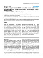
Figure 1 Recombinant rFRb is functional in binding folate. (a) Purified rFRb was resolved on 4% to 12% NuPAGE gel. The rFRb appears as a
tight doublet. Molecular weight markers (in kilodaltons) are on the left side of the gel image. (b) rFRb is able to bind to folate. Open squares
indicate NTA control with FITC-folate, filled squares indicate rFRb with FICT-folate and 100-fold excess unlabelled folate, and filled diamonds
indicate rFRb with FITC-folate. FITC emission is detected at 518 nm. FITC, fluorescein isothiocyanate; rFRb, recombinant folate receptor beta.
Feng et al. Arthritis Research & Therapy 2011, 13:R59
/>Page 4 of 12
has been reported for chicken riboflavin-binding protein
[20,21], which is closely related to FRb.Importantly,
rFRb was confirmed to bind t o its ligand, folate (Figure
1b). The purified rFRb was t hen used in the following
panning experiments.
Selection of m909 from human naïve Fab phage display
library with recombinant folate receptor beta
An Fab clone was first selected from the human naïve
Fab phage display library and had an estimated 100 nM
half maximal effective concentration (EC
50
)torFRb on
ELISA. This clone was affinity-matured using a light-
chain shuffling method an d rescreened with rFRb under
a more stringent set of conditions. A Fab clone with bet-
ter affinity, m909, was selected from the maturation pro-
cess (Figure 2a), and this is the antibody characterized in
the present study. m909 was converted into both single-
chain (scFv) and IgG1 formats for different applications.
On ELISA, Fab m909 was shown to exhibit an EC
50
of
approximately 10 to 50 nM, whereas its IgG1 showed sig-
nificant binding to rFRb on ELISA even at femtomolar
concentrations (Figure 2b), implying the importance of
the avidity effect for its binding. It should be noted that
m909 in either format did not bind to the a isoform of
the human FR (data not shown). The binding kinetics of
m909 Fab was further t ested with the surface plasmon
resonance Biacore instrument. Fab m909 has an equili-
brium dissociation constant (K
D
) equal to 57 nM (k
a
=
2.793 × 10
4
1/Ms and k
d
= 0.001584 1/s) (Figure 3),
which is in agreement with the estimation from the
ELISA binding assay.
m909 binds to native human folate receptor beta on cell
surface
To further characterize the binding ability of m909
IgG1, we tested to see whether it might recognize native
FRb on the surface of the cell. This was investigated
through flow cytometry and immunofluorescent staining
of cells. CHO-hFRb cells are engineered from parental
CHO-K1 cells (FRb-negative) and stably express rela-
tively high levels of hFRb on their surface. In flow cyto-
metry, m909 IgG1 bound to CHO-hFRb cells (Figure
4a) but not to CHO-K1 cells, indicating that the anti-
body recognizes FRb specifica lly and does not recognize
other membrane proteins on these cells. Another cell
line, preB L1.2, has been stably transfected with FRb but
showed lower levels of FRb expression (Figure 4b).
When m909 and its isotype IgG were evaluated for
binding by flow cytometry to these cells, dose-dependent
binding was observed, albeit at a much lower intensity
than that of binding to CHO-hFRb cells. m909 did not
bind to CHO-K1 cells at the highest concentration
tested (data not shown).
Figure 2 In vitro ELISA binding of anti-rFRb Fabs and affinity-
matured clone m909. (a) The parental Fab clone and light-chain
shuffled clones were tested on ELISA wells coated with rFRb. All
Fabs were purified from periplasm of Escherichia coli and tested
from 1,500 to 0.0192 nM. m909 was the clone featured in the study.
LC38 and LC41 are two other affinity-matured clones. (b) m909 IgG
was tested for binding to rFRb on ELISA. The highest IgG1
concentration tested was 1,000 nM and was serially diluted at 1:10.
m909 IgG showed binding even at 1 fM. ELISA, enzyme-linked
immunosorbent assay; Fab, antigen-binding fragment; OD, optical
density; rFRb, recombinant folate receptor beta.
Figure 3 Biacore analysis of m909 Fab. rFRb was immobilized on
a CM5 sensor chip at 210 resonance units (RU). m909 Fab at 1, 10,
100, 400, and 800 nM was tested. A 1:1 binding model gave the
equilibrium dissociation constant (K
D
) of 56.72 nM. The colored lines
are the binding curves, and the black lines are the fitted curves.
Fab, antigen-binding fragment; rFR b, recombinant folate receptor
beta.
Feng et al. Arthritis Research & Therapy 2011, 13:R59
/>Page 5 of 12
It was also of interest to explore whether m909 might
bindtothesamesiteonFRb as does folate. To answer
this question, we incubated folate-FITC with CHO-
hFRb cells in the presence of varying concentrations of
m909 IgG and found that the addition of unlabelled
IgG1 m909 did not change the folate-FITC signal
intensity (Figure 4c). Next, folate-FITC was co-incu-
bated with CHO-hFRb cells in the presence of increas-
ing concentrations of FITC-labelled m909 IgG. It was
found that the addition of m909-FITC increased the sig-
nal intensity over that of folate-FITC alone (Figure 4d),
indicating that the bindings of folate a nd m909 are not
Figure 4 Flow cytometry of m909 IgG on FRb-positive cells. hFRb-positive cells were incubated with 0.001, 0.01, 0.1, 1, 10, and 100 nM m909
IgG. An isotype control IgG1 was included in the test at 100 nM. Cells were analyzed in FACSCalibur. (a) CHO-hFRb cells. (b) PreB L1.2 cells. Black
line indicates isotype control IgG, and colored lines indicate 0.001 to 100 nM m909. (c) CHO-hFRb cells were incubated with 10 nM folate-FITC
(green line) and 10, 100, and 1,000 nM unlabelled m909 (other colored lines). (d) CHO-hFRb cells were incubated with 10 nM folate-FITC and
varying concentrations of m909-FITC. Binding of m909 and folate did not interfere with each other. Black line indicates negative control, green
line indicates FITC-folate alone, and other colored lines indicate FITC-folate with 0.1, 1, 10, or 100 nM m909-FITC. FITC, fluorescein isothiocyanate;
FRb, folate receptor beta; hFRb, human folate receptor beta.
Feng et al. Arthritis Research & Therapy 2011, 13:R59
/>Page 6 of 12
mutually exclusive and that they have at least an addi-
tive effect, if not a synergistic one.
FRs are GPI-linked membrane proteins that are readily
accessible to drugs, and this renders them potential tar-
gets for treatment of arthritis and c ancers. To confirm
that m909 can inde ed bind to FRb on intact cells, we
investigated whether m909 binding could be visualized
on cell surfaces. FITC-labelled m909 was incubated with
CHO-hFRb or CHO-K1 cells cultured in coverglass
wells, and cells were examined by confocal microscopy
to investigate the subcellular localization of the antibody
binding. As shown in Figure 5, IgG1 m909 staining was
foundpredominantlyontheplasmamembraneof
CHO-hFRb cells, and little staining was detectable inside
cells. These results agree with the flow cytometry results
and further indicate that receptor downregulation was
minor under the experimental condition (37°C for 1
hour). The isotype control IgG1-FITC did not have any
detectable staining in CHO-hFRb cells, nor did m909
stain the parental CHO-K1 cells. KB nasopharyngeal
epidermoid cells have been reported to display only the
a isoform of FR on their surface [22]. Staining with a
mousemAbspecifictoFRa showed that KB cells have
a significant amount of FRa on their surface (the last
panel in Figure 5). IgG1 m909 failed to stain KB cells,
indicating that it is specific for hFRb, in agreement with
the ELISA results.
m909 binds to human folate receptor beta selectively on
inflammatory monocytes and activated macrophages
from synovial fluid of arthritis patients
Several reports have shown that activated macrophages
and monocytes in autoimmune diseases have elevated
levels of FRb [5,11,23]. In addition, some solid tumors
are infiltrated with macrophages, among which a high
percentage are FRb-positive [9,24]. These macrophages
are capable of secreting cytokines, growth factors, and
proangiogenic factors. Eliminating activated macro-
phages from autoimmune disease tissues and tumors
could be beneficial t o these patients. Therefore, we
investigated whether m909 recognizes these diseased
cells by isolating such cells from two sources.
First, it has been observed that approximately 1 0% of
the PBMCs of apparently healthy people express mea-
surable numbers of hFRb, perhaps in response to a low
constitutive inflammatory process. To explore whether
m909 might bind these monocytes, we separated
CD14
high
PBMCs into CD16
+
and CD16
-
groups. The
latter group of cells represents the myelomonocytic line-
age cells with high levels of receptor for lipopolysacchar-
ide ( or endotoxin) but lacks FcgRIII (a hallmark of
natural killer (NK) cells). We found that, among the
PBMCs from this particular donor examined, approxi-
mately 17% of the CD14
high
CD16
-
cells have FR on their
surface, shown by incubation with 100 nM labelled
folate (Figure 6a). Importantly, a similar percentage of
these cells was found to be positive for hFRb,shown
similarlybyincubationwith5nMm909IgG(Figure
6b). These results demonstrate that m909 selects FR-
positive cells from monocytes as efficiently as folate at
tested concentrations. Furthermore, this assay provides
confirmation that it is hFRb, not the hFRa, that is upre-
gulated in the activated macrophages.
Next, we isolated synovial macrophages from four
patients with rheumatoid arthritis. Previous studies have
shown that synovial macrophages collected from
patients with arthritis have e levated FRb, and this hFRb
is able to internalize folate-conjugated drugs [5,10].
Activated macrophages were first select ed with the
CD11b marker. The subpopulation of cells was further
analyzed for hFRb expression either through folate-Ore-
gon Green or m909-FITC. It was found that fluorescent
folate can label approximately 14.5% of macrophages
(Figure 6c), whereas m909 selected ap proximately
11.17% of activated macrophages (Figure 6d). Competi-
tion with 1,000-fold excess of non-labelled folate and an
isotype control IgG1 were used in these tests to subtract
background. These results indicated that m909 and
folate are similarly effective in the selection o f activated
macrophages. Together, these data indicate that m909
specific ally recognizes FRb-positive inflammatory mono-
cytes and activated macrophages from patients.
m909 induces antibody-dependent cell-mediated
cytotoxicity with human folate receptor beta-positive
cells
During an initial attempt to examine whether m909
might affect the growth behavior of hFRb-positive cells,
we did not detect any impact of the antibody. However,
because cells decorated with human IgG are often
recognized and destroyed by NK cells, we decided to
explore whether m909 might mediate ADCC. To test
this possibility, PBMCs were isolated from healthy
donors and incubated with CHO-hFRb,CHO-K1,or
preB L1.2 cells a t a ratio of effector cells to target cells
of 50:1. The IgG1 m909 or an isotype control IgG was
incubated with cells at varying concentrations. IgG1
m909 was found to induce specific target cell lysis in an
hFRb level-dependent fashion; m909 induced signifi-
cantly more lysis in CHO-hFRb cells than preB L1.2
cells (Figure 7), which have positive but lower levels of
FRb than CHO-hFRb cells. I n the parental CHO-K1
cells, m909 did not induce any detectable cell lysis. The
control IgG did not have cytotoxicity at 200 nM. It
seems that cell lysis reached maximum at 8 nM IgG1
m909; this probably reflects t he saturation of m909
binding o n CHO-hFRb cells at this level. The notion is
supported by data in Figure 4a that 10 nM (the orange
Feng et al. Arthritis Research & Therapy 2011, 13:R59
/>Page 7 of 12
Figure 5 Confocal laser microscopy images show the specific binding of FITC-m909 to CHO-FRb cells.HumanFRb stably transfected
CHO-FRb cells (a,b,g,h), CHO-K1 cells (c,i), and KB nasopharyngeal epidermoid cells (d-f,j-l) were incubated with 50 nM m909 IgG-FITC for 1
hour at 37°C and were washed three times with cold phosphate-buffered saline. Images (a-f) were captured with transmitted light. Images (g-l)
were captured using a charge-coupled device camera with identical settings below the saturation limits. Isotype IgG1-FITC did not give any
binding signal in CHO-FRb cells (b,f). Mouse anti-human FRa mAb together with goat anti-mouse IgG-FITC secondary antibody showed the
expression of human FRa on KB cells (f,l). FITC, fluorescein isothiocyanate; FR, folate receptor.
Feng et al. Arthritis Research & Therapy 2011, 13:R59
/>Page 8 of 12
line) and 100 nM (the da rk blue line) m909 IgG had
almost the same binding. Together, these results indi-
cate that IgG1 m909 bound to the cell surface was able
to attract NK cells and thereby mediate specific cell kill-
ing by NK cells.
Discussion
FRs have been the focus of studies for decades [25].
Only in the past 10 years have their roles in cancer
treatment been actively researched. Among the studies
reported to date, a majority of them focus on targeting
FR with its ligand folate. Folate-drug conjugates have
achieved c onsiderable successes in diagnosis and treat-
ment of many diseases, especially rheumatoid arthritis.
These folate conjugates are designed to kill diseased
cells through one of two mec hanisms: direct surface tar-
geting/binding or folate-activated receptor endocytosis.
Antibody-based therapeutics have advanced signifi-
cantly in the past decade because of the recombinant
antibody technologies. S everal chimeric antibodies
Figure 6 m909 binds to FR b
+
CD14
high
CD16
-
subset (inflamm atory monocytes) of human PBMCs and activated macrophag es from
synovial fluid of rheumatoid arthritis patients. Human PBMCs were stained with PE-anti-CD14 and Tricolor-anti-CD16 antibodies and (a)
folate-Oregon Green (FOG) or (b) m909-FITC. (a) The cells were stained with 100 nM folate-FOG in the absence (solid black histogram) or
presence of an excess (10 μM) of free folic acid to competitively occupy FR (filled gray histogram). (b) The cells were stained with 5 nM m909
IgG-FITC (solid black histogram) or 5 nM control IgG1-FITC (filled gray histogram). Among the CD14
high
CD16
-
cells, m909 selected 16.83% of
cells and folate selected 17.87%. (c,d) Synovial fluid cells from four patients with rheumatoid arthritis were first labelled with anti-CD11b to stain
human macrophages and then incubated with (c) 100 nM folate-FOG in the absence (unfilled histogram) or presence of an excess (100 μM) of
free folic acid to competitively occupy FR (filled gray histogram) or (d) 50 nM m909-FITC (unfilled histogram) or 50 nM control IgG1-FITC (filled
gray histogram). A representative flow plot with the percentage of FRb-positive cells within each gate is shown. FITC, fluorescein isothiocyanate;
FR, folate receptor; PBMC, peripheral blood mononuclear cell.
Feng et al. Arthritis Research & Therapy 2011, 13:R59
/>Page 9 of 12
targeting the receptor FRa have been reported, and one
such antibody, Farletuzumab, was studied in a phase 2
clinical trial [26]. At present, no human antibody speci-
fic to FRb has been reported. A rat mAb to mouse FRb
was reported to reduce tumor-associated macrophages
when its single chain was fused with immunotoxin and
used to treat rat glioma in a nude mouse model [9].
TheuniqueexpressionprofileofFRb in activated
macrophages and AML supports the notion of t argeting
FRb for treatment of autoimmune diseases and myelo-
genous leukemias. In AML cells, the level of FRb can be
upregulated by treatment with all-trans retinoic acid
[27], thereby increasing the specificity of FRb-targeted
therapy.
In this study, we have produced the first functional
recombinant hFRb reported in the literature. m909
was selected from a human Fab library with rFRb and
was found to specifically recognize hFRb but not the a
isoform of FR, giving it an advantage when the binding
of FRa is to be avoided. m909 is also capable of selec-
tively binding to inflammatory monocytes and acti-
vated macrophages from the synovial fluid of patients
with rheumatoid arthritis. Whereas the activated
macrophages have elevated levels of FRb,thenormal
residential macrophages do not express FRb.There-
fore, m909 is a good candidate for diagnosis and treat-
ment of both autoimmune diseases that involve
activated macrophages and tumors that are infiltrated
with activated macrophages.
Many imaging and therapeutic agents using folate have
been reported, as folate has advantages as a small mole-
cule, being easy to produce and to conjugate to drugs, as
well as having quick clearance from the circulation when
used as an imaging agent [28]. However, in some cases, a
receptor-targeted method is preferred. For example, some
cells have FRs, but owing to changes in post-translational
modifications, these receptors do not bind to folate [ 4].
Folate and its conju gates do not distinguish between the
two isoforms of FR. When only FRb, not the a isoform of
FR, is required for targeting, m909 allows the specificity.
Antibodies by nature are stable proteins, and m909 should
have a relatively long half life in circulation, providing an
option when prolonged treatment targeting FRb is desired.
m909 mediates ADCC in FRb-positive cells, suggesting
tha t it could be used to eliminate activated macrophages
or AML cells as a monotherapy without the need to cou-
ple it to drugs. Our experiments showed that there is a sig-
nificant amount of m909-bound FRb on CHO-FRb cell
surfaces after incubation at 37°C for 1 hour. Many surface
receptors undergo downmodulation upon antibody bind-
ing. The fact that the intensity of m909-bound receptor
remains strong after 37°C incubation implies that the
receptor internalization is slow or represents a small frac-
tion of the receptor or that recycling and rebinding over-
whelm the internalization. In any case, the presence of
m909-decorated receptor at high levels allows time for NK
cells and macrophages to kill these disease cells.
Because m909 and folate do not interfere with each
other’sbindingonFRb (Figure 4c, d), m909 and folate-
drug conjugates may be used in combination to increase
efficacy. This feature of m909 is also important for
m909 monotherapy to work in a clinical setting because,
in the serum of healthy people, there is a significant
level of free folate (18.2 μg/L or 42 nM on average) [29].
Also, binding of m9 09, unlike folate-drug conjuga tes,
will not be affected by the folate levels in the system.
m909 was selected from a naïve human antibody
library and its sequence does not deviate significantly
from the germline sequences. The V gene of m909
heavy chain ha s 98.61% identity to its closest germline
gene IGHV1-3*01, and the V gene of the light chain has
96.77% identity to IGLV3-19*01. The close homology of
m909 to germline genes supports a possibility that it
will be well to lerated by t he immune system. Finally,
because no high-affinity FRb-specific antibody is com-
mercially available, m909 can b e used as a research
reagent to study the function of FRb.
Conclusions
m909 has approximately 57 nM affinity in Fab form and
femtomolar avidity in IgG1 form. Unlike folate-drug
Figure 7 m909 induces ADCC in F Rb -positive cells but not in
FRb-negative cells. Freshly isolated PBMCs were incubated with
target cells (CHO-hFRb, PreB L1.2, or CHO-K1) at a ratio of 50:1 in
the presence of m909 IgG1 at three different concentrations (8, 40,
or 200 nM) or an isotype control IgG1 at 200 nM (first set of
columns). ADCC was detected with CytoTox-ONE reagent, allowing
measurement of the lactate dehydrogenase released by lysed target
cells. The percentage of specific lysis was calculated as described in
Materials and methods. Dark bar indicates preB L1.2 cells, gray bar
indicates CHO-hFRb cells, and clear bar indicates CHO-K1 cells.
ADCC, antibody-dependent cell-mediated cytotoxicity; FRb, folate
receptor beta; PBMC, peripheral blood mononuclear cell.
Feng et al. Arthritis Research & Therapy 2011, 13:R59
/>Page 10 of 12
conjugates, m909 selectively binds to FRb,doesnot
recognize FRa, and has at least one effector function.
m909 alone has the potential to eliminate FRb-positive
cells. Because m909 does not compete with folate for
rec eptor binding, it can be used with folate-drug conju-
gates in a combination therapy. m909 can also be a
valuable research reagent.
Abbreviations
ADCC: antibody-dependent cell-mediated cytotoxicity; AML: acute
myelogenous leukemia; EC
50
: half maximal effective concentration; ELISA:
enzyme-linked immunosorbent assay; Fab: antigen-binding fragment; FITC:
fluorescein isothiocyanate; FR: folate receptor; FRβ: folate receptor beta; GPI:
glycosylphosphatidylinositol; hFRβ: human folate receptor beta; K
D
:
equilibrium dissociation constant; mAb: monoclonal antibody; NK: natural
killer; PBMC: peripheral blood mononuclear cell; PBS: phosphate-buffered
saline; PBST: 0.05% Tween 20 in phosphate-buffered saline; PCFT: proton-
coupled folate transporter; RFC: reduced folate carrier; rFRβ: recombinant
folate receptor beta; scFv: single-chain variable fragment.
Acknowledgements
We thank members of our groups for helpful discussions. This project was
supported by the Intramural Research Program of the National Institutes of
Health (NIH), National Cancer Institute, Center for Cancer Research, and by
federal funds from the National Cancer Institute, NIH, under contract N01-
CO-12400. The content of this publication does not necessarily reflect the
views or policies of the Department of Health and Human Services, nor
does mention of trade names, commercial products, or organizations imply
endorsement by the US Government.
Author details
1
Protein Interactions Group, CCRNP, NCI-Frederick, NIH, 1050 Boyle Street,
Frederick, MD 21702, USA.
2
Department of Chemistry, Purdue University, 560
Oval Drive, West Lafayette, IN 47906, USA.
3
BRP, SAIC-Frederick, Inc., NCI-
Frederick, 1050 Boyle Street, Frederick, MD 21702, USA.
4
Indiana University
Health Arnett Physicians, 2600 Greenbush Street, Lafayette, IN 47904, USA.
Authors’ contributions
YF helped to initiate the study, carry out experiments in different areas,
analyze data, and write the manuscript. JS helped to carry out experiments
in different areas, analyze data, and write the manuscript. EDS helped to
carry out experiments in different areas. ML provided patient samples. ZZ
provided the library. PSL and DSD helped to initiate the study, analyze data,
and write the manuscript. All authors read and approved the final
manuscript.
Competing interests
PSL has received fees and stocks from Endocyte Inc. (West Lafayette, IN,
USA), a company that he founded in 1995 to develop treatments for cancer.
Because Endocyte plans to develop drugs (but not antibodies) for treatment
of autoimmune and inflammatory diseases, this relationship could constitute
a conflict of interest. The National Institutes of Health and Purdue University
have applied for a patent claiming m909; however, the authors receive no
benefits from the patent application. The authors declare that they have no
other competing interests.
Received: 10 January 2011 Revised: 4 March 2011
Accepted: 8 April 2011 Published: 8 April 2011
References
1. Matherly LH, Goldman DI: Membrane transport of folates. Vitam Horm
2003, 66:403-456.
2. Elnakat H, Ratnam M: Distribution, functionality and gene regulation of
folate receptor isoforms: implications in targeted therapy. Adv Drug Deliv
Rev 2004, 56:1067-1084.
3. Basal E, Eghbali-Fatourechi GZ, Kalli KR, Hartmann LC, Goodman KM,
Goode EL, Kamen BA, Low PS, Knutson KL: Functional folate receptor
alpha is elevated in the blood of ovarian cancer patients. PLoS One 2009,
4:e6292.
4. Reddy JA, Haneline LS, Srour EF, Antony AC, Clapp DW, Low PS: Expression
and functional characterization of the beta-isoform of the folate
receptor on CD34(+) cells. Blood 1999, 93:3940-3948.
5. Xia W, Hilgenbrink AR, Matteson EL, Lockwood MB, Cheng JX, Low PS: A
functional folate receptor is induced during macrophage activation and
can be used to target drugs to activated macrophages. Blood 2009,
113:438-446.
6. Ebel W, Routhier EL, Foley B, Jacob S, McDonough JM, Patel RK, Turchin HA,
Chao Q, Kline JB, Old LJ, Phillips MD, Nicolaides NC, Sass PM, Grasso L:
Preclinical evaluation of MORAb-003, a humanized monoclonal antibody
antagonizing folate receptor-alpha. Cancer Immun 2007, 7:6.
7. Coney LR, Mezzanzanica D, Sanborn D, Casalini P, Colnaghi MI, Zurawski VR
Jr: Chimeric murine-human antibodies directed against folate binding
receptor are efficient mediators of ovarian carcinoma cell killing. Cancer
Res 1994, 54:2448-2455.
8. Melani C, Figini M, Nicosia D, Luison E, Ramakrishna V, Casorati G,
Parmiani G, Eshhar Z, Canevari S, Colombo MP: Targeting of interleukin 2
to human ovarian carcinoma by fusion with a single-chain Fv of
antifolate receptor antibody. Cancer Res 1998, 58:4146-4154.
9. Nagai T, Tanaka M, Tsuneyoshi Y, Xu B, Michie SA, Hasui K, Hirano H, Arita K,
Matsuyama T: Targeting tumor-associated macrophages in an
experimental glioma model with a recombinant immunotoxin to folate
receptor beta. Cancer Immunol Immunother 2009, 58:1577-1586.
10. van der Heijden JW, Oerlemans R, Dijkmans BA, Qi H, van der Laken CJ,
Lems WF, Jackman AL, Kraan MC, Tak PP, Ratnam M, Jansen G: Folate
receptor beta as a potential delivery route for novel folate antagonists
to macrophages in the synovial tissue of rheumatoid arthritis patients.
Arthritis Rheum 2009, 60:12-21.
11. Varghese B, Haase N, Low PS: Depletion of folate-receptor-positive
macrophages leads to alleviation of symptoms and prolonged survival
in two murine models of systemic lupus erythematosus. Mol Pharm 2007,
4:679-685.
12. Mosser DM: The many faces of macrophage activation. J Leukoc Biol 2003,
73:209-212.
13. Nagayoshi R, Nakamura M, Ijiri K, Yoshida H, Komiya S, Matsuyama T:
LY309887, antifolate via the folate receptor suppresses murine type II
collagen-induced arthritis. Clin Exp Rheumatol 2003, 21:719-725.
14. Paulos CM, Varghese B, Widmer WR, Breur GJ, Vlashi E, Low PS: Folate-
targeted immunotherapy effectively treats established adjuvant and
collagen-induced arthritis. Arthritis Res Ther
2006, 8:R77.
15.
Paulos CM, Turk MJ, Breur GJ, Low PS: Folate receptor-mediated targeting
of therapeutic and imaging agents to activated macrophages in
rheumatoid arthritis. Adv Drug Deliv Rev 2004, 56:1205-1217.
16. Blaser BW, Gonit M, Qi H, Shatnawi A, Guimond M, Lee RJ, Ratnam M:
Induction of folate receptor type beta in a bone marrow
engraftment model of acute myelogenous leukemi a. Leukemia 2007,
21:2233-2235.
17. Puig-Kroger A, Sierra-Filardi E, Dominguez-Soto A, Samaniego R,
Corcuera MT, Gomez-Aguado F, Ratnam M, Sanchez-Mateos P, Corbi AL:
Folate receptor beta is expressed by tumor-associated macrophages
and constitutes a marker for M2 anti-inflammatory/regulatory
macrophages. Cancer Res 2009, 69:9395-9403.
18. Feng Y, Zhu Z, Xiao X, Choudhry V, Barrett JC, Dimitrov DS: Novel human
monoclonal antibodies to insulin-like growth factor (IGF)-II that potently
inhibit the IGF receptor type I signal transduction function. Mol Cancer
Ther 2006, 5:114-120.
19. Feng Y, Xiao X, Zhu Z, Streaker E, Ho M, Pastan I, Dimitrov DS: A novel
human monoclonal antibody that binds with high affinity to
mesothelin-expressing cells and kills them by antibody-dependent cell-
mediated cytotoxicity. Mol Cancer Ther 2009, 8:1113-1118.
20. Zanette D, Monaco HL, Zanotti G, Spadon P: Crystallization of hen
eggwhite riboflavin-binding protein. J Mol Biol 1984, 180:1185-1187.
21. Shen F, Wang H, Zheng X, Ratnam M: Expression levels of functional
folate receptors alpha and beta are related to the number of N-
glycosylated sites. Biochem J 1997, 327(Pt 3):759-764.
22. Schultz RM, Andis SL, Shackelford KA, Gates SB, Ratnam M, Mendelsohn LG,
Shih C, Grindey GB: Role of membrane-associated folate binding protein
in the cytotoxicity of antifolates in KB, IGROV1, and L1210A cells. Oncol
Res 1995, 7:97-102.
Feng et al. Arthritis Research & Therapy 2011, 13:R59
/>Page 11 of 12
23. Matteson EL, Lowe VJ, Prendergast FG, Crowson CS, Moder KG,
Morgenstern DE, Messmann RA, Low PS: Assessment of disease activity in
rheumatoid arthritis using a novel folate targeted radiopharmaceutical
Folatescan. Clin Exp Rheumatol 2009, 27:253-259.
24. Qi H, Ratnam M: Synergistic induction of folate receptor beta by all-trans
retinoic acid and histone deacetylase inhibitors in acute myelogenous
leukemia cells: mechanism and utility in enhancing selective growth
inhibition by antifolates. Cancer Res 2006, 66:5875-5882.
25. Wagner C: Cellular folate binding proteins; function and significance.
Annu Rev Nutr 1982, 2:229-248.
26. ClinicalTrials.gov homepage. [].
27. Pan XQ, Zheng X, Shi G, Wang H, Ratnam M, Lee RJ: Strategy for the
treatment of acute myelogenous leukemia based on folate receptor
beta-targeted liposomal doxorubicin combined with receptor induction
using all-trans retinoic acid. Blood 2002, 100:594-602.
28. Low PS, Henne WA, Doorneweerd DD: Discovery and development of
folic-acid-based receptor targeting for imaging and therapy of cancer
and inflammatory diseases. Acc Chem Res 2008, 41:120-129.
29. Lawrence JM, Petitti DB, Watkins M, Umekubo MA: Trends in serum folate
after food fortification. Lancet 1999, 354:915-916.
doi:10.1186/ar3312
Cite this article as: Feng et al.: A folate receptor beta-specific human
monoclonal antibody recognizes activated macrophage of rheumatoid
patients and mediates antibody-dependent cell-mediated cytotoxicity.
Arthritis Research & Therapy 2011 13:R59.
Submit your next manuscript to BioMed Central
and take full advantage of:
• Convenient online submission
• Thorough peer review
• No space constraints or color figure charges
• Immediate publication on acceptance
• Inclusion in PubMed, CAS, Scopus and Google Scholar
• Research which is freely available for redistribution
Submit your manuscript at
www.biomedcentral.com/submit
Feng et al. Arthritis Research & Therapy 2011, 13:R59
/>Page 12 of 12