Báo cáo Y học: A model for recognition of polychlorinated dibenzo-p-dioxins by the aryl hydrocarbon receptor docx
Bạn đang xem bản rút gọn của tài liệu. Xem và tải ngay bản đầy đủ của tài liệu tại đây (399.76 KB, 6 trang )
A model for recognition of polychlorinated dibenzo-
p
-dioxins
by the aryl hydrocarbon receptor
M. Procopio
1
, A. Lahm
2
, A. Tramontano
3
, L. Bonati
1
and D. Pitea
1
1
Dipartimento di Scienze dellÕAmbiente e del Territorio, Universita
Á
degli Studi di Milano-Bicocca, Milano, Italy;
2
Istituto di Ricerche di Biologia Molecolare P. Angeletti, Pomezia (Roma), Italy;
3
Dipartimento di Scienze Biochimiche ÔRossi FanelliÕ, Universita'di Roma ÔLa SapienzaÕ, Roma, Italy
Ligand binding by the aryl hydrocarbon receptor (AhR), a
member of the bHLH-PAS family of transcriptional reg-
ulatory proteins, has been mapped to a region within the
second ÔPASÕ domain, a conserved sequence motif ®rst
discovered in the P er-ARNT-Sim family of pr oteins. In
addition to the bacterial photoactive yellow protein (PYP),
which had been proposed as a structural prototype for the
three dimensional fold of PAS domains, two crystal
structures of the P AS domain have recently been deter-
mined: the human potassium channel HERG and the
heme binding domain of the bacterial O
2
sensing FixL
protein. The three structures reveal a highly conserved
structural framework in e volutionary rather distant PAS
domains, p rovide a more g eneral view of how these
domains can recognize their ligands and suggest a
structure±function relationship that we exploited to build a
three-dimensional model of the ligand binding domain
(LBD) of the mouse aryl hydrocarbon receptor (mAhR).
The model allowed us to putatively i dentify the residues
responsible for the recognition of polychlorinated dibenzo-
p-dioxins (PCDDs) by AhR receptors and to formulate an
hypothesis on the signal transduction mechanism.
Keywords: aryl h ydrocarbon receptor; polychlorinated
dibenzo-p-dioxins; structure prediction; protein modelling;
molecular recognition.
Studies on the biological mechanism of action of polychlo-
rinated dibenzo-p-dioxins (PCDDs) indicate that their
biological effects are mediated by binding to a speci®c
cytoplasmic protein, the aryl hydrocarbon receptor (AhR)
[1].
Ligand-induced activation of AhR initiates a process
whereby t he receptor i s transformed into a nuclear
transcription factor by forming a complex with the protein
ARNT (Ah receptor nuclear translocator). Speci®c recog-
nition of XRE DNA sequences (xenobiotic responsive
elements) by the ligand-activated AhR/ARNT heterodimer
then induces transcription of genes encoding xenobiotic
metabolizing enzymes [2]. Understanding the PCDD±AhR
binding process at a molecular level is therefore a key step
for gaining insight into the biological mechanism of action
of these compounds.
The structure±activity relationship (SAR) of the PCDD±
AhR interaction has been studied with th e aim of correlat-
ing physico-chemical properties o f the ligand s and their
biological activities [3±5]. In particular, we analysed a series
of PCDDs with varying binding af®nities [4,5] on t he basis
of their molecular electrostatic potential (MEP) and molec-
ular polarizability and concluded that the requirements f or
high af®nities are the concentration o f negative M EP values
at the e xtremes of t he ligand's long axis and a d epleted
charge above and below the aromatic rings. This led to the
hypothesis that there are favorable interactions with a
receptor nucleophilic site in the central part of the ligand
and with electrophilic sites at both sides of the principal
molecular axis. A necessary next step to understand the
PCDD±AhR interaction and t o identify the amino-acid
residues directly inte racting w ith P CDDs is the construction
of a three-dimensional model for the AhR ligand binding
domain (LBD).
AhR and ARNT belong to the Per-ARNT-Sim (PAS)
family of proteins [6,7], whose members act as transcrip-
tional activators, sensor modules of two-component regu-
latory sy stems o r a s ion channels [8]. PAS domains are
found predominantly in p roteins that are involved, directly
or indirectly, in signal transduction. Their known functions
are in some cases to mediate protein±protein interactions
and, in other cases, s uch as f or AhR, ligand a nd/or cofactor
binding [8].
In AhR, two PAS domains are present in a 270-residue
region encompassing two imperfect repeats of 110 amino
acids ( PAS-A and PAS-B) separated by a sequence of 50
amino acids. A minimal LBD was mapped in the mouse
AhR (mAhR) between amino acids 230 and 397, the region
that encompasses the PAS-B repeat [9]. While deletion of
the PAS-A repeat (amino acids 121±182) reduced ligand
binding only to 30%, deletion of the PAS-B repeat (amino
acids 259± 374) completely abolished binding, as did deletion
Correspondence to A.Tramontano,DepartmentofBiochemical
Sciences ÔRossi FanelliÕ, University of Rome ÔLa SapienzaP.le Aldo
Moro, 500185 Rome. Fax: + 39 06 91093482,
Tel.: + 39 06 91093207, E-m ail:
Abbreviations: AhR, aryl hydrocarbon receptor; PYP, photoactive
yellow protein; LBD, ligand binding domai n; mAhR, mouse aryl
hydrocarbon receptor; PCDD, polychlorinated dibenzo-p-dioxin;
ARNT, Ah rec e ptor nu clear t r anslocator; XRE, xenobiotic responsive
elements; SAR, structure±activity relationship; MEP, molecular
electrostaticpotential;PAS,Per-ARNT-Simproteinfamily;TCDD,
tetrachlorodibenzo-p-dioxin.
(Received 10 August 2001, revised 20 September 2001, accepted 16
October 2001)
Eur. J. Biochem. 269, 13±18 (2002) Ó FEBS 2002
of the complete PAS region [9]. In the same study it was
already shown that modi®cations outside the PAS domain
had no effect on ligand binding.
A s tructural prototype for the three-dimensional fold of
the PAS domain superfamily has been proposed to be the
structure of the photoactive yellow protein (PYP), a
bacterial light-sensing protein [10]. However, the crystal
structures of two other PAS domains [11,12] have been
recently determined and their analysis a llowed us t o build a
three-dimensional model of the mAhR LBD and to
investigate its ligand binding site at the molecular level.
RESULTS AND DISCUSSION
Structure prediction
Application of a re cursive
PSI
-
BLAST
[13] search (default
parameters) against the nonredundant protein sequence
database revealed a high number of matches between the
mAhR LBD and many other PAS proteins, including
hypoxia-inducible factor 1a, several histidine kinases, light
receptors, regulatory proteins, clock proteins (such as the
period clock protein PER), sensor proteins (oxygen/redox
sensors) and ion channels. T he crystal structures of t he PAS
domains of two of these proteins were recently solved: the
human potassium channel HERG [11] and the heme
binding domain of the bacterial O
2
sensing FixL protein
[12]. Both structures were detected after four (HERG) or
eight (FixL)
PSI
-
BLAST
iteration cycles, as was the PYP
protein (iteration 6). Although E values w ere initially rather
high (> 0.1) for all three structures, E values for HERG
and FixL became highly signi®cant (< 10
)4
)asthesearch
progressed. A search including only the database of known
protein structures neither found any of these structures, nor
highlighted any other statistically signi®cant homologies.
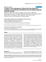
The structures of the HERG and FixL P AS domains are
showninFig.1togetherwiththePYPstructure.Giventhe
low level of sequence identity [15], the high structural
conservation is quite unexpected: all the structures are
formed by a ®ve-stranded antiparallel b sheet with a helices
on one side. Although all three domains belong to proteins
involved in signalling p rocesses a nd are expected t o transmit
a signal through protein±protein i nteractions, they have
developed quite different mechanisms to perform their
function. While the HERG PAS domain does not bind a
ligand [11], both the FixL PAS domain and PYP are
activated by ligands; in FixL, oxygen binding at the heme
binding PAS domain controls the activity of a h istidine
kinase domain [12]; and in PYP, a local conformational
change occurs once the p-hydroxycinnamoyl cromophore is
bound [10].
The largest conformational difference between the FixL
and the HERG and PYP structures occurs in the so-called
helical connector, the long central helix, which sh ows a
translational displacement of 7A
Ê
that allows the accom-
modation of the heme cofactor (Fig. 1C) [12]. The hydro-
phobic core of the three domains is generally well conserved,
but two buried residues in FixL differ signi®cantly in size
from the structurally equivalent residues in PYP and
HERG, again favoring the heme binding. These are g lycines
224 and 251 that substitute Phe96 a nd Val120 in PYP, and
Phe98 and Leu127 in HERG [16].
For both FixL and PYP, structures are known for the
inactive and active signaling states. In the case of PYP,
conformational changes occur in the neighborhood of the
p-hydroxycinnamoyl cromophore and are transmitted to
the s urface of the protein primarily through t he cromophore
and Arg52 [10]. In FixL, the heme propionate groups are
suggested to relay the spin transition signal by transducing
the increased planarity of the pu ckered porphyrin ring into
backbone and side-chain conformational changes within a
loop (residues 211±215) immediately following the helical
connector [12]. The suggested signal transducing regions of
PYP and FixL are thus located at the opposite ends of the
Fig. 1. Schematic representation of the HERG (A), PYP (B), and FixL (C) PAS domains displaying the high degree of structural similarity. The
largest shift amongst the conserved s econdary element position o ccurs i n FixL due t o the presence of the large heme c ofactor. Secondary structure
elements are colored blue (strands ) and red (helices), cofactor ligands gr een. (A), (B) and (C) were generat ed using
RIBBONS
[28]. Coordinate sets
used correspond to entries 1BV5(FixL) [12], 2PYP(PYP) [ 10] and 1BYW(HERG) [11] of the PDB protein data bank [14].
14 M. Procopio et al. (Eur. J. Biochem. 269) Ó FEBS 2002
long central he lix, h ighlighting th e imp ortance of this region
and t he ¯anking loops as the critical regulatory region of the
PAS domain family [16], with the remainder of the PAS fold
serving as a structural scaffold.
When sequence s imilarity between the t arget a nd
potential template sequences is low, as in our case, t he
correctness of the alignment plays a crucial role both in the
selection of the correct template and in the expected ®nal
quality of the model. Other information, such as predicted
and observed secondary structures of the target and
template proteins, and sequence and structure conservation
in their families, should therefore be used to re®ne the
alignment.
The consensus secondary structure for residues 230±397
of th e mAhR LBD a s predicted by the
JPRED
server [17,18] is
reported i n Fig. 2 together with the o bserved s econdary
structure of the three template candidates. The ®nal
sequence alignment used for modelling is reported in the
same ®gure. This sequence alignment differs somehow from
PAS domain alignment recently p roposed [8], as it was
produced manually taking into account th e predicted
secondary structure of AhR LBD, the observed secondary
structure and FSSP structural alignment for FixL, HERG
and PYP, and the c onservation pattern in a m ultiple
alignment of AhR sequences. For clarity, in Fig. 2 we only
show some of these latter sequences and, for comparison, a
subset of sequences from the related ARNT proteins.
The AhR sequences in Fig. 2 w ere selected for their
differences in the response to PCDDs: the human Ah
receptor that has an af®nity fo r 2,3,7,8-TCDD sixfold lower
than mAhR [19]; the AhR-1 ortholog of Caenorhabditis
elegans (AhR-1C.E.), neither photoaf®nity labeled by a
dioxin analog, nor activated by b-naphto¯avone in a yeast
system [20]; the rainbow trout AhRa that binds TCDD [21]
and t he Microgadus Tomcod AhR also activated by TCDD
[22].
All alignments were manipulated using the interactive
display program
SEAVIEW
[23].
Fig. 2. Alignment of Ah receptors and their predict ed secondary structure against the three structural templates a ligned a ccording to FSSP. a Helices
and b strands are represented as white and black b ars, respectively. Secondary structure assignment for FixL, PYP a nd HERG is derived from the
PDB entries. Colouring sc heme for resid ues: red: acidic ; blue: basic; purple : polar; yellow: Cys; brown : aromatic; gree n: hydrophob ic; orange:
Ser,Thr; gr ey: Pro; wh ite: Gly.
Ó FEBS 2002 A model for PCDD ± AhR recognition (Eur. J. Biochem. 269)15
Modelling
Because of the closer functional homology (noncovalent
interaction with a ligand) we used FixL as a template for
modelling. This choice was also motivated by the observa-
tion that the helical connector in FixL is translated away
from the b sheet with respect to HERG and PYP (Fig. 1)
thus allowing binding of the heme-ligand, a situation
expected to be present in a similar fashion also in AhR.
The sequence corresponding to mAhR residues 275±380
was therefore inscribed onto the structural template pro-
vided by FixL a ccording to the alignmen t shown in F ig. 2 ,
and, subsequently, the necessary i nsertions and d eletions
were modeled (Fig. 3A). AhR residues 381±397 were not
modeled b ecause the corresponding helix in FixL is pointing
away from the barrel and should not be involved in ligand
binding. AhR residues 286±288 c ould be modeled using the
corresponding loop of equal length from the HERG
structure, while all other insertions and deletions were
constructed u sing a fragment database s earch procedure
[24]. The one-residue insertion at position 314 c orrelates well
with the presence of a hydrophilic residue at the spatially
close position 282 replacing the buried hydrophobic Ile
present in FixL.
Fig. 3. Comparison between the mAhR model (A,C) and the parental FixL PAS d omain structure (B,D). In (A) a nd (B) r esidu es tha t in¯ue nce the size
of the ligand p ocket a re h ighligh ted. The arrow indicates t he shift of the he lical co nn ector ( orange) i n Ah R with resp ect t o F ixL. Lo op region s w here
insertions or deletions had to be accomodated in the AhR model are coloured magenta. In (C) and (D) a close-up of the AhR and FixL ligand
binding po ckets is sh own. T he k ey e lements in the proposed signal t ransduct ion m echanism fo r FixL, a change in side-chain conformation of
Arg206, Thr210 and Asp212, are conserved in AhR with Arg333, Thr337 and Glu339 equivalently positioned, r eady to sense and transduce the
presence of the PCDD ligand . Addition al AhR residues involved i n ligand reco gnition and discrimin ation a re Arg282 and Gln377 close to t he polar
end of the ligand inside the pocket. TCDD and heme cofactor atoms are colored green (carbon), red (oxygen), blue (nitrogen), yellow ( iron) and
magenta (chlorine).
16 M. Procopio et al. (Eur. J. Biochem. 269) Ó FEBS 2002
The postulated signaling loop of FixL (amino acids 211±
215) following the helical connector had to be substituted by
a s horther frag ment in mAhR (Fig . 2, a mino acids 335±338)
that could only be achieved in our model by manually
shifting the helix towards the b sheet.
A l ast insertion occurred at G ly368, in a loop region
unlikely to be involved in interactions with t he ligand.
The backbone geometry of the resulting model was
regularized with
WHAT IF
[25], the side-chain rotamers of
substituted residues optim ized using
SCWRL
[26] and the
model analyzed without any further modi®cations.
A model for recognition of PCDDs by Ah receptors
The most noticeable conformational difference between the
mAhR model a nd the FixL template is the relative position
of the helical connector that moves closer to the b sheet, thus
reducing t he size of the binding cavity entrance (Fig. 3). The
helix position, intermediate between that observed in HERG
and in FixL, correlates well with the functional role of the
hydrophobic core in the three proteins, while HERG lacks
any binding activity; the modeled mAhR binds PCDDs and
FixL has to accomodate the larger heme cofactor.
The AhR residues at positions important for heme
binding in FixL support our model. Gly224 and Gly251 in
the hydrophobic core of FixL correspond to Leu347 and
Ala375 in mAhR thus reducing the size of the cavity. This is
also consistent with site-directed mutagenesis r esults that
identi®ed Ala375 as critical for the ligand binding activity
[19]. Interestingly, there is also a good correlation between
the size of the side-chain at this position and the size of
the ligand. While the latter decreases from FixL to AhR
to HERG, the side-chain volume increases (from Gly251
to Ala375 to Leu127). Moreover, human AhR and
AhR-1C.E., both with reduced af®nity for PCDDs, have
bigger side-chains at this position (Val and Leu, respectively)
partially ®lling t he binding cavity. The residue coordinating
the f erric heme ion in FixL, His200, is substituted by Cys327
in all AhR receptors, except for AhR-1C.E. where methi-
onine is present.
At the entrance of the FixL ligand cavity, Arg220, that
binds a heme propionate group, is replaced by Thr in all
AhR (Thr343 in mAhR), except for human AhR and
AhR-1C.E. that have isoleucine and leucine, respectively.
While the CG2 methyl group of Thr could m ediate hydro-
phobic interactions with the ligand, both i soleucine and
leucine will partially block the entrance and reduce af®nity.
None of these residues, characteristic of the AhR
proteins, are conserved in the homologous ARNT proteins
(Fig. 2) which have no ligan d binding activity.
Additional information ab out the PCDD±AhR binding
can be deduced by analyzing the proposed mechanisms for
signal transduction of FixL [12,16,27]. A ccording to Perutz
et al. [27], the pathway s tarts at Ile215, Leu236, Ile238,
which form a hydrophobic triad around the heme ligand.
The movements of these residues are transmitted to, and
ampli®ed by, a loop that includes Pro213, and then
transmitted to other atoms including the h eme p ropio-
nates. A different key residue has been proposed by Gong
et al. [12] who indicated the interaction between heme
propionates and His (or Arg) 214 as the starting event of
the protein conformational change. It has also been
observed that, going from the unbound to the bound
state, Arg206 affects the position of Asp212, which in turn
undergoes the largest c onformational change o f all the
sidechains [12].
Interestingly, although the conformation of the signaling
loop had to be altered in the mAhR model, Arg206 and
Thr210 of FixL are in e quivalent structural positions as
Arg333 and Thr337 in mAhR and Asp212 of FixL is
replaced by the very s imilar Glu339. These three r esidues are
conserved in all Ah receptors and are not present in the
other PAS proteins analyzed. Therefore, by analogy with
the FixL mechanism, it is conceivable that, once PCDD is
bound, Arg333 in mAhR is involved in the interaction with
one of the chlorine atoms and breaks the hydrogen bond
with Glu339 that changes conformation.
The ligand with t he highest af®nity for the AhR is 2,3,7,8-
TCDD and our model can be used to investigate its mode of
binding, under t he assumption that the molecular plane of
TCDD is in a similar position as that o f the heme group in
FixL. We highlight in Figs 3C,D, the residues predicted to
mediate k ey ligand interactions in the proposed binding
cavity. The size of Ala375 is important for ligand accom-
modation, Cys327 co uld interact with the electrophilic
central region of TCDD [4], Thr343 possibly stabilizes the
complex by hydrophobic interactions, Arg333, at the
entrance of the cavity, may guide TCDD t owards its
binding site by long-range electrostatic interactions and, by
interacting with chlorine atoms of TCDD, may promote a
signal transduction mechanism through Glu339, similar to
that of FixL. Two additional residues, Arg282 and Phe345,
are shown in the Fig. 3. While Arg282, replaced by Gln in
some Ah receptors and pointing t o the TCDD chlorinated
side, may contribute to the binding by electrostatic interac-
tions or hydrogen bond, Phe345, lining one side of the
ligand pocket, could interact with the aromatic ringsystem
of TCDD. Ultimately, Gln377, characteristic of all Ah
receptors and not present i n o ther PAS proteins, could f orm
hydrogen bonds w ith chlorine atoms in the predicted
binding cavity for TCDD.
Most of the proposed interactions ®t well with the
electrostatic characteristics we highlighted in previous
QSAR studies on ligand properties [4,5]. The requirements
of a nucleophilic site in the central part of the ligand and of
electrophilic sites at the sides of the principal molecular axis
are both explained by our model.
CONCLUSIONS
Given the limitations in today's modelling and prediction
techniques, the model presented here has to be considered
only an a pproximate and probably i ncomplete picture of the
ligand binding domain of AhR and of its interactions with
PCDDs. It should also be emphasized that the LBD is part
of a much larger protein and some features of the Ah
receptor system might not be explainable in terms of the
isolated domain.
PYP has been previously proposed as an app ropriate
structural template for AhR, but our analysis of the recently
determined structures of the FixL and the HERG P AS
domains strongly suggests that a model based on FixL is
more likely to be correct. On one hand, the availability of
the t hree structures indicates that the position of the helical
connector can differ. On the other, the closer functional
homology between FixL and AhR, the secondary structure
Ó FEBS 2002 A model for PCDD ± AhR recognition (Eur. J. Biochem. 269)17
prediction and the size of the ligand all point to FixL as a
more suitable candidate.
Our model, although based on low sequence similarity, is
capable of explaining all known experimental and theoret-
ical data and therefore we believe it to be accurate enough t o
serve as a framework for furt her experiment s such as site
directed mutagenesis of residues proposed to mediate the
AhR±PCDD interaction and docking calculations to more
accurately de®ne the orientation of the ligand i n the binding
cavity.
ACKNOWLEDGEMENTS
The ®nancial support by the Italian CNR (grant no. 98.03245.ST74)
and the Fondazione Lomb ardia per l'Ambiente is gratefully acknowl-
edged.
REFERENCES
1. Landers, J.P. & Bunce, N.J. (1991) The Ah receptor and the
mechanism of dioxin t oxicity. Biochem. J. 276, 273±287.
2. Sogawa, K. & Fujii-Kuriyama, Y. (1997) Ah receptor, a novel
ligand-activated transcription factor. J. Biochem. 122, 1075±1079.
3. Waller, C.L. & Mckinney, J.D. (1995) Three-dimensional quan-
titative structure-activity relationships of dioxins and dioxin-like
compounds: model validation and Ah receptor characterization.
Chem. Res. Toxicol. 8, 847±858.
4. Bonati, L., Fraschini, E., Lasagni, M., Modoni, E.P. & Pitea, D.
(1995) A h ypothesis o n t he me chanism o f P CDD b iological
activity based on molecular electro static potential modeling.
Part 2. J. Mol. Struct. (THEOCHEM) 340, 83±95.
5. Fraschini, E., Bonati, L. & Pitea, D. (1996) Molecular polariz-
ability as a tool for u nderstan ding the b in ding properties of
polychlorinated dibenzo-p-dioxins: de®nition of a reliable com-
putational procedure. J. Phys. C hem. 100, 10564±10569.
6. Hahn, M.E., (1991) The a ryl hydrocarbon re ceptor: a comparative
perspective. Comp. Biochem. Physiol. Part C 121, 23±53.
7. Gu, Y.Z., Hogenesch, J.B. & Brad®eld, C.A. (2000) The PAS
superfamily: sensors of environmental and developmental signals.
Annu. Rev. Pharmacol. Toxicol. 40, 519±561.
8. Taylor, B.L. & Zhulin, I.B. (1999) PAS domains: internal sensors
of oxygen, redox potential, and light. Microbiol. Mol. Biol. Rev.
63, 479±506.
9. Fuk unaga, B.N., Probst, M.R., Reisz-Porszasz, S. & Hankinson,
O. (1995) Identi®cation of functional domains of the aryl hydro-
carbon receptor. J. Biol. Chem. 27 0, 29270±29278.
10. Pellequer, J.L., Wager-Smith, K.A., Kay, S.A. & Getzo, E.D.
(1998) Photoactive yellow protein: a structural prototype for the
three-dimensional fold o f the PAS domain superfamily. Proc. Natl
Acad. Sci. USA 95, 5884±5890.
11. Cabral, J.H.M., Lee, A., Cohen, S.L., Chait, B.T., Li, M. &
Mackinnon, R. ( 1998) Crystal structure an d functional analysis of
the HERG potassium channel N terminus: a eukaryotic PAS
domain. Cell 95, 649±655.
12. Gong, W., Hao, B., Mansy, S.S., Gonzalez, G., Gilles-Gonzalez,
M.A. & Chan, M.K. (1998) Structure of a biological oxygen
sensor: a new mechanism for heme-driven signal transduction.
Proc. Natl A cad. Sci. USA 95, 15177±15182.
13. Altsc hul, S .F., Madden, T .L., S cha
È
er, A.A., Z hang, J ., Zhang, Z.,
Miller, W. & Lipman, D.J. (1997) Gapped BLAST and PSI-
BLAST: a ne w generation o f p rotein database search programs.
Nucleic Acids Res. 25, 3 389±3402.
14. Berman, H.M., Westbrook, J., Feng, Z., Gilliland, G., B hat, T.N.,
Weissig, H., Shindyalov, I.N. & Bourne, P.E. (2000) The p rotein
data bank. Nucleic Acids Res. 28, 235±242.
15. Holm, L. & Sander, C. (1996) Mapping the protein universe.
Science 273, 595±602.
16. Pellequer, J.L., Brudler, R. & Getzo, E.D. (1999) Biological
sensors: more than one way to sense oxygen. Current Biol. 9,
R416±R418.
17. Cu, J .A., Clamp, M.E., Siddiqui, A.S., Finlay, M. & Barton, G .J.
(1998) JPred: a consensus secondary structure prediction server.
Bioinformatics 14 , 892±893.
18. Cu, J.A. & Barton, G.J. (1999) Evaluation and improvement of
multiple sequence methods for protein secondary s tructure
prediction. Proteins 34, 508±519.
19. Ema,M.,Ohe,N.,Suzuki,M.,Mimura,J.,Sogawa,K.,Ikawa,S.
& Fujii-Kuriyama, Y. (1994) Dioxin binding activities of poly-
morphic forms of mouse and human arylhydrocarbon receptors.
J. Biol. C hem. 269, 27337±27343.
20. Powe ll-Coman, J.A., Brad®eld, C.A. & W oo d, W.B. (1998)
Caenorhabditis elegans orthologs of the aryl hydrocarbon recep-
tor and its heterodimerization partner the aryl hydrocarbon
receptor nu clear translocator. Proc. Natl Acad. Sci. USA 95,
2844±2849.
21. Abnet, C.C., Tanguay, R.L., Hahn, M.E. & W.Peterson, R.E.
(1999) Two forms of aryl hydrocarbon receptor type 2 in rainbow
trout (Oncorhync hus mykiss). Evidence for dierential expression
and enhancer speci®city. J. Biol. Chem. 274, 15159±15166.
22. Roy, N.K. & Wirgin, I. (1997) Characterization of the aromatic
hydrocarbon r ecepto r gene and i ts expression in Atlantic tomcod.
Arch. Biochem. Biophys. 344, 373±386.
23. Galtier, N., Gouy, M. & Gautier, C . (1996) SEAVIEW and
PHYLO_WIN: two graphic tools for sequence alignment and
molecular phylogeny. Comput. Appl. Biosc. 12, 543±548.
24. Molecular Simulation Inc. (1998) INSIGHT II Molecular Simu-
lation Inc. San D iego, CA, USA.
25. Vriend, G. (1990) WHAT IF: a molecular modeling and drug
design program. J. Mol. Graph. 8, 52±56.
26. Bower, M.J., Cohen, F.E. & Dunbrack, R.L. Jr (1997) Prediction
of protein side-chain rotamers from a backbone-dependent rot-
amer library: a new homo logy modeling t ool. J. Mol. Biol. 267,
1268±1282.
27. Perutz, M.F., Paoli, M. & Lesk, A.M. (1999) Fix L, a haemo-
globin that acts as an oxygen sensor: signalling mechanism and
structural basis of its homology with PAS domains. Chem. Biol. 6,
R291±R297.
28. Carson, M. (1991) RIBBONS 2.0. J. Appl. Cryst. 24, 958±961.
18 M. Procopio et al. (Eur. J. Biochem. 269) Ó FEBS 2002