Báo cáo y học: "Notochordal conditioned media from tissue increases proteoglycan accumulation and promotes a healthy nucleus pulposus phenotype in human mesenchymal stem cells" pot
Bạn đang xem bản rút gọn của tài liệu. Xem và tải ngay bản đầy đủ của tài liệu tại đây (474.86 KB, 13 trang )
RESEARCH ARTICLE Open Access
Notochordal conditioned media from tissue
increases proteoglycan accumulation and
promotes a healthy nucleus pulposus phenotype
in human mesenchymal stem cells
Devina Purmessur
1
, Rachel M Schek
2
, Rosalyn D Abbott
2
, Bryan A Ballif
3
, Karolyn E Godburn
2
and
James C Iatridis
1*
Abstract
Introduction: Notochordal cells (NCs) are influential in development of the intervertebral disc (IVD) and species
that retain NCs do not degenerate. IVD repair using bone marrow derived mesenchymal stem cells (MSCs) is an
attractive approach and the harsh microenvironment of the IVD suggests pre-differentiation is a necessary first step.
The goal of this study was to use soluble factors from NCs in alginate and NCs in their native tissue to differentiate
human MSCs to a young nucleus pulposus (NP) phenotype.
Methods: Human MSCs (cultured under micromass conditions for 21 days in hypoxia) were differentiated with
conditioned medium derived from porcine notochordal cells in native tissue (NCT) or in alginate beads (NCA), and
compared with chondrogenic (TGFb-3) or basal medium. A PCR array of 42 genes was utilized to screen a large
number of genes known to be associated with the healthy NP phenotype and pellet cultures were also evaluated
for glycosaminoglycan content, histology and viability. Proteomic analysis was used to assess candidate soluble
factors in NCA and NCT.
Results: Notochordal cell conditioned media had diverse effects on MSC phenotype. NCT resulted in the highest
levels of glycosaminoglycan (GAG), as well as up-regulation of SOX9 and Collagen II gene expression. NCA
demonstrated effects that were catabolic yet also anti-fibrotic and minimally hypertrophic with down-regulation of
Collagens I and III and low levels of Collagen X, respectively. Micromass culture and hypoxic conditions were
sufficient to promote chondrogenesis demonstrating that both basal and chondrogenic media produced similar
phenotypes. Candidate matricellular proteins, clusterin and tenascin were identified by proteomics in the NCA
group.
Conclusions: NCs secreted important soluble factors capable of differentiating MSCs to a NP phenotype
synthesizing high levels of proteoglycan while also resisting collagen fiber expression and hypertrophy, yet results
were sen sitive to the conditions in which media was generated (cells in alginate versus cells in their native tissue)
so that further mechanistic studies optimizing culture conditions and defining important NC secreted factors are
required. Matricellular proteins, such as clusterin and tenascin, are likely to be important to optimize differ entiation
of MSCs for maximum GAG production and reduced collagen fiber expression.
* Correspondence:
1
Leni and Peter W. May Department of Orthopaedics, Mount Sinai School of
Medicine, One Gustave L. Levy Place, Box 1188, New York, NY 10029-6574,
USA
Full list of author information is available at the end of the article
Purmessur et al. Arthritis Research & Therapy 2011, 13:R81
/>© 2011 Purmessur et al.; licensee BioMed Central Ltd. This is an open access article distributed under the terms of the Creative
Commons Attribut ion License (http://creativecommon s.org/licenses/by/2.0), whi ch permits unrestricted use, distribution, and
reproduction in any medium, provided the original work is properly cited.
Introduction
Current surgical therapies to treat intervertebral disc
(IVD) degeneration i nclude spinal fusion and arthro-
plasty; these methods are highly invasive and are often
associated with reduced patient mobility [1]. Cell based
therapies are an attractive alternative since they may be
applied in a minimally invasive manner with the ability
to address an underlying cause of degeneration. I VD
degeneration is associated with increased cell apoptosis
and senescence, an up-regulation of pro-inflammatory
and pain-related proteins, and ultimately, a breakdown
of the disc matrix [2-5]. Cell-based therapies aim to
restore metabolic homeostasis within the IVD and
reduce inflammation by replacing or augmenting the
disc cells at an early stage of degeneration. Such thera-
pies can adapt and integrate with the native tissue
microe nvironment restoring structure and function with
limited long term side effects. One promising cell choice
is mesenchymal stem cells (MSCs). MSCs are multipo-
tent cells predominantly found in bone marrow that
have the plasticity to differentiate into cells of the chon-
drocytic, adipogenic and osteogenic lineages [6]. How-
ever, there is evidence to suggest that MSCs may not be
well suited to the hostile anaerobic environment of the
diseased IVD [7,8] so that long term survival and inte-
gration within the disc may require pre-differentiation
of the MSCs in culture towards a phenotype more
representative of native IVD cells.
There are at least two cell populations in the disc, the
fibroc hondrocytes that populate and maintain the annu-
lus fibrosus (AF) and the more chondrocytic cells in the
nucleus pulposus (NP). The NP cells are often described
as being “ chondrocyte-like” as a consequence of their
morphology and the extracellular matrix proteins they
synthesize (such as collagen type II and aggrecan). The
glycosaminoglycan (GAG) to hydroxyproline ratio is an
important distinguishing characteristic between NP cells
with ratios as high as 27:1 and hyaline chondrocytes
with ratios as low as 2:1 [9].
MSCs are a promising potential cell source for IVD
repair, as described by a number of in vitro and in vivo
studies [10-19]. The interaction between MSCs and cells
ofthenativeIVD,includingtheadaptationofMSCsto
the IVD microenvironment , enhanced MSC metabolism
and biosynthesis; however, the magnitude of effects
appears to be d ependent on cell ratio and whether the
cell cont act is in direct or direct [12,1 8-20]. Studies sug-
gest that a ratio of 75% NP:25% MSC with direct cell-
cell contact provides the optimal culture conditions for
MSC differentiation and matrix expression toward a
chondrocyte-like phenotype [18]. This interaction
appears to be independent on MSC source, as both
autologous an d allogenic MSCs interact favorably with
NP cells [16,19]. In vivo, the ability of MSCs to improve
biosynthesis and restore homeostasi s within degenerated
IVD is likely to be dependent on their long term survi-
val in the native IVD microenvironment. Injection of
undifferentiated MSCs into the IVDs of small animal
models such as degenerated rabbit IVDs depleted of NP
tissue demonstrated survival of MSCs for up to 48
weeks [14]. However, the tissue composition (NP
matrix) and cell populations (predominantly notochordal
NP cells) in these animal models differ radically from
those present clinically in human degenerated IVDs.
Differentiation of MSCs toward an NP phenotype is
more complex than differentiation towards a hyaline
chondrocyte lineage [21]. Differentiation toward an NP
phenotype is likely to depend on diverse biological para-
meters such as an appropriate choice or c ombination of
growth factors, 3D matrix, cell-cell contact and environ-
mental conditions mimicking the IVD such as hypoxia.
Further, only very recently, has the phenotype of NP
cells become more clearly defined. While no single defi-
nitive NP marker exists, many laboratories have exam-
ined potential markers associated with a “healthy NP
phenotype” in a diverse range of animal species includ-
ing human IVDs and these studies are ongoing [22-26].
The proteoglycan-rich matrix and high proteoglycan to
collagen ratio of the human NP is considered an impor-
tant marker when determining a healthy N P phenotype
[9]. Based on literature a healthy NP like phenotype can
be considered as high proteoglycan biosynthesis,
increases in the matrix proteins SOX9, collagen II,
aggrecan, phenotypic markers such as keratin-19 and
transforming growth factors 1 and 3 (TGFb-1 and -3)
[10,22,27,28]. This is coupled with decreases in collagens
I, III and X, the cat abolic enzymes matrix metalloprotei-
nases ( MMPs) and A distintegrin and metalloproteinase
with thrombospondin motifs (ADAMTSs) and inflam-
matory cytokines interleukin-1b (IL-1 b) and tumour
necrosis factor alpha (TNFa) [2,4,29,30],
The notochord plays an influential role in early devel-
opment of the IVD [31] and exposing MSCs to noto-
chordal cells (NCs) has been proposed as a powerful
method for differentiation to an NP phenotype [32]. In
a number of species, including humans, during growth
and aging, the NCs populating the NP disappear and are
replaced by chondrocytic NP cells [33]. The NP of some
species retain notochord cells into maturity, for exam-
ple, the pig, rabbit, non-chondrodystrophoid dogs and
rodents, and the IVDs of these species do not experi-
ence d egeneration of the IVD [33], suggesting an asso-
ciation between NCs with IVD development and
maintenance of the healthy NP phenotype. It has pre-
viously been shown that NCs, including conditioned
medium derived from NCs (NCCM) has enhanced IVD
cell and articular chondrocyte metabolism [34,35]. More
recent studies by Korecki et al. have shown that NCCM
Purmessur et al. Arthritis Research & Therapy 2011, 13:R81
/>Page 2 of 13
from porcine NCs seeded in alginate increased GAG
production and up-regulated Laminin B1 and collagen
type III in human MSCs after seven days in culture [32].
While NCCM demonstrates strong promise for NP dif-
ferentiation, generation of NCCM was not optimized.
For example, Korecki et al. employed NCs isolated from
their native tissue environment in order to highlight the
relevance of NCs alone. Yet, the cell matrix interactions
will influence the production of soluble factors from
NCs which maintain the healthy IVD, and it is specu-
lated that generation of N CCM within the native tissue
environment has anabolic soluble factors that may
improve differentiating potential of MSCs cells to an NP
phenotype.
We hypothesize that NCCM generated from NCs in
their native tissue environment will trigger differentia-
tion of MSCs toward an NP phenotype to a greater
extent than both notochordal media generated from NC
cells in alginate and chondrogenic media (TGFb-3)
alone. The first aim of this study was to pre-differentiate
MSCs into cells with a healthy NP-phenotype based on
custom PCR array analysis and GAG production as
defined above. A custom PCR array was designed to
evaluate expression of 42 genes chosen from recent lit-
erature in order to characterize NP cell phenotype,
matrix protein, catabolic/anti-catabolic pr otein, growth
factor and pain/inflammatory protein expression. The
second aim was to identify the optimal conditions for
generating conditioned media by comparing the effects
of CM derived from NCs seeded in alginate or derived
from notochordal tissue, as compared with chondro-
genic media with TGFb-3. The last aim consisted of a
pilot st udy of proteomic analysis of secreted protein fac-
tors from the NCT and NCA conditioned media that
may provide instructive cues and create unique extracel-
lular environments that would contribute to o ur under-
standing of how NCs influence development of a
healthy NP phenotype.
Materials and methods
Generation of conditioned media from porcine IVD cells
and tissue
The average ratio of notochordal to NP cells isolated
from the IVDs of each pig spine was 88%:12%, similar
to that found by Chen et al. [36]; therefore, the whole
pool of NP cells were taken to be predominantly noto-
chordal in nature. NP t issue was carefully isolated asep-
tically from IVDs of two- to eight-month-old female
porcine spines (n = 8) obtained within 24 hours of
death (Animal Facility Research 87 Inc., Boylston, MA,
USA). To gener ate conditioned media (CM) from noto-
chordal cells see ded in alginate beads (NCA), NP tissue
was first digested as described by Urban et al. [37].
Briefly, tissue was digested with 0.2% protease (from
Streptomyces griseus - Type XIV: P5147, Sigma-Aldrich,
ST Louis, MO USA) for one hour followed by 0.025%
collagenase (from Clostridium histolyticum type 1A:
C2674, Sigma-Aldrich) for 18 hours at room tempera-
ture. To remove remaining cell clusters, additional
digestion with Cell dissociation solution, non-enzymatic
1 × (C1419, Sigma-Aldrich) was performed for two
hours. Cells were then rinsed in 0.15 M NaCl and
encap sulated in beads at a density of 2 × 10
6
cells/ml of
1.2% low viscosity alginate (Sigma-Aldrich). Beads were
cultured in 12-well plates at a density of 10 beads/well
with 2 ml of media (low glucose DMEM, 1% Pen/Strep,
0.5% Fungizone and 1% insulin-transferrin-selenium
(ITS) (I2771, Sigma Aldrich)) for four days in hypoxia
(5% O
2
,5%CO
2
, 37°C).
For generation of CM from notochordal cells in tissue
(NCT), the NP of three porcine discs ( wet weight
approximately 0.9 to 1.3 g) were soaked per 30 mls o f
low glucose DMEM, 0.5% Fungizone and 1% Pen/Strep
without ITS for four days in hypoxia. Media was
retained and filtered through a 70 um cell strainer
(Thermo Fisher Scientific, Pittsburg, PA USA) to
remove any remaining tissue.
NCA and NCT were both filtered through MW 3000
Amicon Ultra-15 (Millipore Bedford, MA USA) and re-
suspended in 15 ml Basal media (B) (low glucose
DMEM + 1% Pen/Strep + 1% ITS) in order to remove
small metabolites and waste products. 15 ml of either
NCT or NCA was added to each Amicon Ultra-15 filter
and material on top (the concentrate) was resuspended
in 15 ml Basal media with a final concentration of 1 ×.
To verify the conditioned media used was the same
from each notochordal culture all media was pooled for
NCT and NCA respectively.
Pelleting of MSCs
Human bone marrow derived MSCs samples (age range
22 to 37 years, n = 3) were purchased from Texas A&M
(Temple, TX, USA) with the appropriate Material
Transfer Agreement and expanded in monolayer culture
in alpha MEM medium supplemented with 10% fetal
bovine serum. At passage 4, cells were pelleted at a den-
sity of 250,000 cells in 15 ml polypropylene tubes by
centrifugation at 600 g for five minutes. They were then
cultured in 500 μlofChondrogenicmedia(C)(Basal
media with 50 μg/ml ascorbate, 0.1 μM dexamethasone,
40 μg L-proline and 10 ng/ml TGFb-3) in hypoxic con-
ditions for 24 hours.
Treatment of MSCs with 4 CM types: B, NCT, NCA, and C
After 24 hours, spent media was removed and 500 μLof
either B, NCA, NCT or C were added to respective
tubes containing pellets (Table 1: study design) and
changed every three to four days, for a total culture
Purmessur et al. Arthritis Research & Therapy 2011, 13:R81
/>Page 3 of 13
period of 21 days. Media was retained to assess GAG
released to media during the 21-day culture.
Dependent variables
PCR array
Each pellet was lysed with 300 μl RNeasy Lysis RLT buffer
(Qiagen: 79216 Valencia, CA USA) and the lysates from
five pellets pooled stored at -80°C. RNA was extracted,
cDNA synthesized and custom RT
2
Profiler’ SYBR green
PCR arrays (SABiosciences : CAPH-0817A (Qiagen), Fre-
derick, MA USA) were run by the Vermont Cancer Centre
DNA analysis facility. The custom array included 42 genes
associated with NP cell function: Ph enotypic marker/
Matrix-associated protein genes, growth factor genes, cata-
bolic/anti-catabolic genes and inflammatory/pain genes
(Additional file 1 Table S1 and Additional file 2 Table S2).
Relative gene expression was calculated using the compara-
tive Ct method normalized to undifferentiated MSCs from
the same patients (Day 0) and three housekeeping genes
(18s, GAPDH and b-actin). For normalization purposes,
undetermined values for Day 0 were given an arbitrary
value of 40 as undifferentiated MSCs did not express all
the genes (leading to some catabolic genes with artificially
high fold increase s). Error ba rs were plotted as SE M s.
Glycosaminoglycan (GAG) and DNA content
To examine GAG and DNA in the cell pellet, spent media
was removed and 200 μl of lysis buffer (Sigma Aldrich: L-
8285; RNT70) was added t o each c ell pellet. This lysis buffer
is routinely used to lyse cell membranes for the release of
RNA/DNA and was also used to d issociate GAG associated
with the cell pellet. The lysate was then assessed using the
Di-methyl m ethylene Blue (DMMB) assay and the standard
curve w as generated in the lysis buffer used t o dissociate t he
cell pellets. DMMB was then normalized to DNA content
using the picogreen assay (Invitrogen, Carlsbad, CA, USA).
To quantify the GAG released to media, media samples
from the pellets of each m edia group retained over 21 days
were assesse d and each GAG m easurement subtracted from
the respective Day 0 control media (NCA. NCT, C and B
before addition to pellets) averaged f or the total 21 days and
then normalized to DNA content [38].
Cell viability
Viability was analysed with the Live/Dead Kit (Invitro-
gen). Briefly, media was removed and the pellets were
washed with PBS. Each pellet was resuspended in 100 μl
of a 2 μM Calcein AM/1 μM Ethidium Homodimer-2
(ETH-2) staining solution and the cell suspension placed
onto a microscope slide. Cells were incubated in the dark
at 37°C for 30 minutes. After incubatio n a cov er-slip was
placed on top of the suspension and cells were visualized
at 20 ×. Excitation and emission for Calcein and ETH-2
were 494/517 nm and 528/617 nm respectively with Cal-
cein staining the cytoplasm of live cells green and ETH-2
staining the nuclear envelope of dead cells red.
Histology of pellets
To visualize the intact pellet, pellets were first fixed in
formalin and then incubated with 1% Alcian Blue in
HCl f or 30 minutes, followed by final f ixation in 100%
ethanol. Pellets were embedded in freezing medium and
20 μm sections cut using a cryotome. Sections were re-
stained with 1% Alcian Blue in HCL for 30 minutes in
order to ensure full penetration of the dye to assess pro-
teoglycan quantity and location, and also 4’,6-diamidino-
2-phenylindole (DAPI, Roche Diagnostics, Mannheim,
Germany) which stains the nuclei of cells (Ultraviolet fil-
ter), followed by a wash with PBS. Images were captured
on an Olympus BX50 light microscope (Center Valley,
PA USA) at 20 × magnification.
Mass spectrometry and data analysis
While the presence of significant amounts of albumin
(present in the ITS solution) greatly reduced the signal
to noise and may have masked several important pro-
teins, distinct bands running at approximately 37 kDa
and140kDafortheNCTandNCAgroupswere
observed (Additional file 3 Figure S3). Gel regions corre-
sponding to these molecular weights were excised from
each of the three lanes and subjected to in-gel tryptic
digestion. Coomasie-stained regions of the SDS -PAGE
gel were diced into 1 mm cubes. Gel pieces were
reduced, alkylated with iodoacetamide and subjected to
in-gel digestion as described previ ously [39]. Dried pep-
tides were subjected to liquid chromatography tandem
mass spectrometry in a linear ion trap (LTQ) mass spec-
trometer (Thermo Electron Corporation Waltham, MA
USA). Data were searched against t he International Pro-
tein Index (IPI) non-redundant protein database using
Sequest; requiring tryptic specificit y; allowing
precursor m/z toleran ces of 2 Da; allowing methionine
residues to be oxidized (+15.99 Da); and requiring
Table 1 Study design for treatment of MSCs with B, C, NCA and NCT groups
Media group Number of human samples Time in culture Number of pellets per sample
PCR GAG Cell viability Alcian blue stain
B 3 21 days 5 3 1 2
C 3 21 days 5 3 1 2
NCA 3 21 days 5 3 1 2
NCT 3 21 days 5 3 1 2
Purmessur et al. Arthritis Research & Therapy 2011, 13:R81
/>Page 4 of 13
cysteine residues to be carbamidomethylated (+57.02 Da).
Peptides were filtered initially by requiring a XCorr value
> 2 for doubly-charged peptides and > 2.5 for triply-
charged peptides. Proteins having more than three pep-
tides meeting these criteria were retained and XCorr
values were then relaxed fo r peptides from these proteins
to > 1.7 for doubly-charged peptides and > 2 for triply-
charged peptides. Proteins that were common to uncon-
ditioned and conditioned media were discarded as well as
proteins that did not have at least two porcine-specific
peptides or had peptide that were no t consistent with
porcine origin as determined by the SEQUEST search
analysis and manual BLAST analysis of each remaining
peptide. To estimate t he peptide false-discovery rate for
the peptides identified in this study, we employed a sta-
tistical method using a target-decoy strategy as described
in detail previously [40] As the complete porcine pro-
teome is not available, and as the IPI indexed non-redun-
dant database is not formatted for the generation of a
decoy database, we searched all MS data against a conca-
tenated forward (target) and reverse (decoy) IPI human
protein database containing the sequences of the proteins
harboring the porcine-specific proteins identified in this
study. Using the same filtering criteria used for the non-
redundant database search, we filtered the data to a false
discovery rate of less than 0.01% at the peptide level. The
proteins harboring porcine-specific peptides were again
identified and remained in this dataset after filtering.
Thus, the odds that any one of the identified peptides
from these proteins is a false positive are less than 0.01%.
Statistics
For qRT-PCR, statist ical analysis was perform ed first by
testing fo r normality using a Ryan-joiners test. For sam-
ples that were either parametric or non-parametric, a
onesamplet-testoronesamplesignranktestofthe
ΔΔCt valu es with hypothesized value of 0 were car ried
out respectively (ΔΔCtforB,C,NCAandNCTgroups
and ΔΔCt = 0 for undifferentiated MSCs at Day 0/or B).
GAG was assessed for normality and a one-way
ANOVA with a Fisher’s P LSD was done in order to
check for significance between all media treatment
groups (with P < 0.05 significant) . Similar analysis using
aone-wayANOVAwithaFisher’s PLSD was carried
out for DNA content. For a descripti on of the statistical
approach used in the proteomic analysis see the mass
spectrometry and data analysis section above.
Results
Gene expression data (significance > 2-fold normalized to
Day 0 and B)
Gene exp ression data was normalized to Day 0 undiffer-
entiated MSCs and also to B conditioned MSCs because
of similarities between B and C groups. Only gene
expression data with signif icance greater than two fold
was discussed.
Phenotypic marker genes
Treatment of MSCs with B, C, NCA and NCT for 21
days in pellet culture had diverse effects on the gene
expression of phenotypic markers with few differences
observed compared to the basal group for these genes
(Table 2 ). C demonstrated no significant changes com-
pared to Day 0 or B for any IVD markers. B demon-
strated a significant increase in the gene expression of
the IVD marker GPC1 compared to Day 0. NCA
showed a significant down-regula tion of BGN relative to
Day 0 and B. Only NCT demonstrated significant up-
regulation of SOX9 relative to Day 0 and B (Figure 1A),
although a significant down-regulation of KRT19 relative
to Day 0. A trend of up-regulation of adipogenic
(PPARG) and osteogenic (BGLAP) markers was observed
for all treatment conditions.
Matrix-associated protein genes
B showed a significant increase in COL3A1 gene expres-
sion and decreases in ACAN and HAS1 expression rela-
tive to Day 0 (Table 2; Figure 1A). C demonstrated
significant increases COL10A1 and COL3A1 relative to
Day 0. However NCA showed significant decreases in
COL1A1 and COL3A1 ge ne expression relative to Day 0
and B. NCT significantly increased COL2A1 and
COL10A1 gene expression relative to Day 0 and B (Fig-
ure 1A).
Catabolic/anti-catabolic genes
A general up-regulation in the expression of catabolic
enzymes was observed for all media groups post culture.
BshowedsignificantincreasesinADAMTS4, MMP1,
-13, -14 and -2 r elativ e to Day 0 (Table 2; Figure 1B). C
demonstrated significant increases in MMP13, -14,and
-9 relative to Day 0. NCA showed significant increases
in MMP 1 4 and -9 relative to Day 0, and MMP1,-2,and
-3 relative to Day 0 and B. NCT demonstr ated signifi-
cant increases in MMP14, -3 and -9 relative to Day 0
and ADAMTS4, MMP1 and -13 relative t o Day 0 and B.
Of the anti-catabolic proteins assessed, only TIMP1
showed significant increases i n gene expression for B,
NCA, and NCT relative to Day 0 (Table 2; Figure 1B).
Growth factor genes
In B, relative to Day 0, gene expression of TGFB3 and IGF
was significantly up regulated and CTGF and EGF were
significant down regulated (Table 2 Figure 1C). A signifi-
cant decrease in CTGF expression relative to Day 0 and B
was observed for C. NCA showed a significant increase in
TGFB1 expression relative to Day 0 and decreases in
TGFB2 and CTGF expression relative to Day 0 and B.
NCT demonstrated significant increases in a number of
growth fact ors; TGFB1 relative to Day 0 and B, and
TGFB3 relative to Day 0 only including a significant down
regulation in CTGF expression relative to Day 0.
Purmessur et al. Arthritis Research & Therapy 2011, 13:R81
/>Page 5 of 13
Table 2 Fold changes in gene expression data for media conditions B, C, NCA and NCT Significance greater than two-
fold relative to Day 0 MSCs or Basal in bold and underlined; + = fold increase & - = fold decrease (P <0)
ALL GENES Normalized to Day 0 Normalized to Basal
B C NCA NCT C NCA NCT
BGN -1.0 +1.1 -6.1 +1.6 +1.1 -6.2 +1.6
GPC1 +2.5 +2.1 +1.5 +1.9 -1.2 -1.7 -1.2
IVD KRT19 -7.3 -1.0 -1.4 -12.7 -1.0 +1.1 -1.6
Phenotypic LAMB1 +1.6 +1.3 -1.2 +1.3 -1.3 -2.0 -1.2
SOX9 +2.59 +1.48 -1.32 +6.79 -1.65 -3.49 +2.74
MSC BGLAP +2.02 -1.13 +1.95 +3.51 -2.37 -1.12 +1.58
PPARG +27.62 +9.90 +9.53 +19.86 -2.79 +1.24 -1.41
ACAN -14.1 -6.3 -2.5 -16.2 +2.1 -1.2 -1.2
COL1A1 -1.1 +1.2 -29.7 -3.4 +1.3 -27.3 -2.9
COL10A1 +1.1 +60.4 +2.2 +61.9 +52.1 +1.7 +51.8
Matrix proteins COL2A1 -1.0 +49.2 -1.0 +15.4 +32.2 -1.0 +8.1
COL3A1 +3.6 +3.9 -11.7 -2.2 +1.1 -42.9 -7.4
eln -1.4 +12.9 -3.0 -2.4 +17.4 -20.8 -1.8
HAS1 -7.3 +1.3 +1.0 +1.1 11.4 +7.5 +9.3
Aggrecanases ADAMTS4 +6.4 +4.9 +2.2 +26.4 -1.3 -2.9 +4.2
ADAMTS5 +1.1 -2.5 +3.2 +5.1 -2.6 +2.9 +5.2
Matrix enzyme MMP1 +369.3 +9.7 +86039.1 +28040.4 -3.2 +231.6 +78.2
MMPs MMP13 +2637.8 +3056.0 +1085.0 +15582.2 +1.1 -2.5 +6.0
MMP14 +4.5 +2.2 +6.2 +6.8 -2.1 +1.3 +1.5
MMP2 +3.8 +1.8 +4.7 +4.0 -2.1 +1.2 +1.0
MMP3 +36.1 +184.4 +203663.7 +1973.3 +11.7 +12436.5
+120.9
MMP9
+22.2 +5.9 +19.1 +3.2 -3.8 -1.1 -7.3
TIMP1 +4.99 +1.77 +5.90 +9.12 -2.60 +1.26 +1.79
Anti-catabolic TIMP2 +1.46 -1.10 +1.26 +1.66 -1.64 -1.09 +1.14
TIMP3 -1.00 -1.00 -1.00 +3.68 -1.00 -1.00 +3.08
TGFB1 +1.82 +1.92 +2.01 +3.96 +1.06 +1.07 +2.11
TGFB2 +1.43 -3.34 -4.91 -1.80 -4.86 -6.78 -2.49
Growth factors TGFB TGFB3 +8.31 +1.36 +3.08 +5.27 -6.23 -2.97 -1.58
TGFBR1 -4.52 -4.61 -1.03 +1.05 -1.00 +4.19 +4.83
TGFBR2 +1.03 +1.65 +1.25 +1.17 1.59 +1.18 +1.11
CTGF -8.9 -3.9 -76.5 -9.6 +2.4 -8.2 -1.0
EGF -3.3 -7.1 -1.1 -1.3 -2.1 +1.5 +2.4
FGF1 -1.6 -1.5 +1.6 +9.2 +1.1 +2.4 +14.1
General IGF1 +169.7 +104.0 -1.0 +87.4 -1.6 -1.0 -1.9
PDGFA -1.63 -1.57 -1.39 -1.06 +1.02 +1.15 +1.46
WISP1 +3.27 +2.62 -2.44 +1.90 -1.35 -8.23 -1.75
IL1B +22.7 +15.1 +9911.6 +444.5 -2.1 +310.6 +14.7
TNF +1.01 +1.66 +90.93 +5.33 +1.56 +72.86 +3.32
Inflammatory CASP3 +1.3 -1.0 -1.2 +1.2 -1.3 -1.8 -1.1
BDNF -12.9 -4.7 -1.3 -6.3 -1.2 +3.1 +2.0
NGF -4.0 -2.7 +1.4 -4.4 -1.6 +1.6 -1.1
TAC4 -1.08 +1.03 +4.22 +1.28 +1.09 +4.33 +1.36
Purmessur et al. Arthritis Research & Therapy 2011, 13:R81
/>Page 6 of 13
Inflammatory/pain genes
B showed a significant decrease in BDNF expression
relative to Day 0 (Table 2). C demonstrated no signifi-
cant changes relative to Day 0 or B. In NCA a signifi-
cant up-regulation of IL-1B relative to Day 0 was
observed and TNFa was increased relative to both Day
0 and B. NCT also demonstrated a significant up-regula-
tion of IL1B, however unlike NCA it down-regulated
NGF relative to Day 0.
Cell viability
The majority of cells appeared alive and healthy (stained
green) with very few dead cells (stained red) after three
weeks culture (data not shown). There are no differences
in cell viability between media groups. This suggests that
despite all media conditions demonstrating some cata-
bolic/inflammatory effects they did not induce cell death.
GAG and DNA content
GAG in the pellet (normalized to DNA content): A
measurable amount of GAG was observed in the cells
pellets o f all media groups; however a sig nificantly
greater amount of GAG was demonstrated in the NCT
relative to B, C and NCA (10.99 μg +/- 0.76 compared
to 5.50 μg +/- 0.23, 6.28 μg +/- 0.43 and 6.64 μg+/-
0.46 ( μ gGAG/μg DNA) respectively) (Figure 2). GAG
released to media (normalized to DNA content): No sig-
nificant differences in GAG released to me dia were
observed between media groups (data not shown). No
significant differences in DNA content were observed
between B, C and NCA compared to the NCT group
(0.162 μg +/- 0.009, 0.149 μg +/- 0.009, 0.172 μg+/-
0.008 compared to 0.134 μg +/- 0.009 respectively)
(Additional file 4 Figure S4).
Histology of pellets
Alcian blue staining showed a general trend of light
staining in the middle with stronger staining along the
periphery of the pellet for B, C and NCA m edia groups;
however, NCA demonstrated darker staining along the
periphery (Figur e 3). The NCT media group showed the
most dramatic staining throughout, indicating that this
0.01
0.1
1
10
100
1000
Fold Change
B
C
NCA
NCT
1
10
100
1000
10000
100000
1000000
1
0000000
Fold Change
0.01
0.1
1
10
100
1000
TGFB1
TGFB3
CTGF
FGF1
IGF1
Fold Change
0.1
1
10
100
1000
10000
100000
IL1B
TNF
NGF
Fold Change
A
B
C
D
*
*
*
*
*
*
*
*
*
*
*
*
*
*
*
*
*
*
*
*
*
*
*
*
*
*
*
*
*
*
*
*
*
*
*
*
*
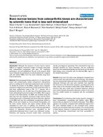
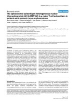
Figure 1 Gene expression data in MSCs 3 weeks post treatment. qRT-PCR results for human mesenchymal stem cells (MSCs) (n =3)
pelleted in 3D culture and treated with conditioned media from four groups: B = Basal, C = Chondrogenic, NCA = notochordal NP cells in
alginate; NCT = notochordal NP cells in tissue for 21 days. Fold change in mRNA levels were calculated with the ΔΔC
T
method relative to three
housekeeping genes and undifferentiated (Day 0) MSCs from the same patient. * indicates significantly different from Day 0 and bar indicates
significantly different from Basal, P < 0.05; Error bars are expressed as SEMs. 1A: Phenotypic marker/Matrix-associated protein genes. The most
prominent results were the significant increase in Sox9 and Col2A1 for NCT media compared to basal and the significant decrease in Col 1A1
and Col3A1 for NCA media; B: Catabolic/anti-catabolic genes. All media conditions had high expression levels for catabolic genes. Very low
levels of mRNA for catabolic proteins resulted in very high relative expression levels relative to Day 0 controls. Most relevant comparisons,
therefore, are with other groups; C: Growth factor genes. A general up-regulation in expression was observed in particular for NCT, with the
exception of CTGF with all media groups; D: Inflammatory/pain genes. Up-regulation of pro-inflammtory cytokines was noted for NCA and NCT
however significant down-regulation of NGF was observed with NCT.
Purmessur et al. Arthritis Research & Therapy 2011, 13:R81
/>Page 7 of 13
0.000
2.000
4.000
6.000
8.000
10.000
12.000
14.000
B
C
NCA
NCT
ug GAG/ug DNA
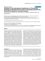
Figure 2 Quantification of proteoglycan in MSC pellets. Di-methyl methylene Blue (DMMB) analysis was used to assess the amount of
Glycosaminoglycan (GAG) associated with the human mesenchymal stem cell (MSC) pellet 21 days post culture with four media groups; B =
Basal, C = Chondrogenic, NCA = notochordal NP cells in alginate; NCT = notochordal NP cells in tissue normalized to DNA content using the
Picogreen assay (μgGAG/μgDNA). Bar indicates significance, P < 0.05. Significantly more GAG was associated with the pellets of the NCT group
in comparison to all other media groups.
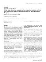
Figure 3 Histology of MSC pellets 3 weeks post culture. Human mesenchymal stem cell (MSC) pellets were sectioned at 20 μm and stained
with Alcian Blue (for GAG) and 4’,6-diamidino-2-phenylindole (DAPI) (cell nuclei) after 21 days culture with four media groups; either B = Basal, C
= Chondrogenic, NCA = notochordal NP cells in alginate; NCT = notochordal NP cells in tissue. (scale bar = 50 μm). GAG was observed for all
media groups however NCT demonstrated the greater abundance of GAG throughout the whole pellet with fewer stained nuclei compared to
other groups.
Purmessur et al. Arthritis Research & Therapy 2011, 13:R81
/>Page 8 of 13
media type produced the most proteoglycans. DAPI
staining showed uniform staining throughout the pellet
with fewer cells appearing along the periphery. C and
NCA media groups were the most cellular with NCT
being t he least. This is consistent with picogreen assay
data which demonstrated a greater DNA content for C
and NCA compared to NCT (data not shown).
Proteomics of media groups
To d etermine if the NCT and NCA conditioned media
showed major differences in protein profiles compared
to the unconditioned control m edia, and as a first
approach to determine if porcine-specific protein factors
could even be identified in the condition ed media, equal
volumes of each medium were subjected to denaturing
SDS-PAGE and the gel was stained with coomassie blue
to v isualize proteins. Seve ral proteins were identified in
both regions of the conditioned media samples that
were not identified in the control sample. However, only
three proteins were identified by more than one peptide
whose amino acid sequences were unambiguously por-
cine in origin (Table 3). The “.” in each peptide
sequence indicates the site of tryptic cleavage and is pre-
ceded (at the amino-terminus) or followed (at the car-
boxyl-terminus) by the amino acid in the porcine
sequence. Also indicated are the pre-processed predi-
cated molecular masses of the protein precursors.
BLAST searches were performed on each peptide and if
the bovine protein ortholog had an ident ical sequence
(allowing for isobaric exchanges between leucine and
isoleucine) it is so note d. Values of multiple parameters
of the proteomics SEQUEST search for each peptide are
listed and include the cross correlation score (XCorr),
the charge state (z), the delta correlation score o f the
next unique peptide sequence (Unique ΔCn), and the
measured mass difference of the precursor compared to
its theoretical mass in parts per million m/z (Δppm).
Also noted are the number of times each peptide was
identified and the number of entries in the non-redun-
dant protein database reported by SEQUEST with the
exact amino acid sequence of the peptide. § indicates a
redundancy was u ncovered in bovine by BLAST se arch-
ing. “M*” denotes an oxidized methionine residue. All
three of the porcine-specific identified proteins origi-
nated uniquely from the NCA sample: clusterin found
in the 37 kDa range and tenascin and alpha-2-macroglo-
bulin found in the 140 kDa range. Table 3 lists the pep-
tides identified from these three proteins in each sample
and details their relevant proteomic metrics.
Discussion
Human MSCs are a potential cell source for regenera-
tion of the degenerated human IVD yet the appropriate
method for pre-differentiation remains unclear. This
Table 3 Porcine-specific proteins/peptides identified via proteomic analysis of NCA
NCA
region
Porcine Protein and Peptides
Identified
Identical in Bovine
Ortholog
XCorr z Unique
ΔCn
Times
Identified
Redundancy in
Database
140 kDa alpha-2-macroglobulin precursor (163
kDa)
K.IKEEGTEVELTGK.G No 4.311 2 0.359 2 0
R.SSGSLLNNAIK.G No 3.07 2 0.343 2 5
R.TPQIITILEK.A No 2.95 2 0.324 2 0
R.KYSNPSTCFGGESQAICEK.F No 2.646 3 0.129 1 0
R.QEFEM*KLEVEAK.I Yes 1.823 3 0.079 1 1§
K.YGAATFTR.T No 1.716 2 0.17 1 10
140 kDa tenascin precursor (191 kDa)
K.ATLTGLRPGTEYGIGVSAVK.G No 4.872 3 0.564 1 0
R.LNYGLPSGQPVEVQLPR.N No 4.721 3 0.51 2 0
R.GLEPGQEYTILLTAEK.G No 3.424 2 0.388 2 0
R.VATYLPTPEGLK.F Yes 2.706 2 0.34 2 1§
K.ESSLTLLWR.T No 2.582 2 0.308 2 0
R.VPGDQTSTTIR.E No 2.418 2 0.301 2 4
37 kDa clusterin precursor (52 kDa)
R.ASNIM*DELFQDR.F No 4.341 2 0.449 1 0
R.KSLLSSLEEAKK.K No 3.739 3 0.231 2 0
K.TLIEQSNEERK.S No 3.135 2 0.232 2 0
R.QQSHVM*DIM*EDSFNR.A No 3.08 3 0.235 2 0
K.AISDKELQEM*STEGSK.Y Yes 3.051 3 0.333 2 1§
K.TLIEQSNEER.K No 2.692 2 0.326 3 0
Purmessur et al. Arthritis Research & Therapy 2011, 13:R81
/>Page 9 of 13
study differentiated human MSCs for 21 days, using two
notochordal cell media conditions as well as chondro-
genic and basal media groups. The lack of clear NP phe-
notypic markers led to examination of a number of
outcomemeasuresincludinguseofgeneprofilingwith
a custom PCR array of 42 genes associated with a
healthy NP phenotype, an important innovation of this
study, as well as assessments of GAG, histology, and cell
viability. Culture of human MSCs in NCT stimulated
anabolic changes most similar to a healthy NP pheno-
type rather than a chondrogenic phenotype with
increased proteoglycan, while NCA conditioning
resulted in significant down-regulation of fibrotic genes
and minimal effects on the hypertrophic gene COLX.
Chondrogenic and basal groups demonstrated many
similarities in gene expression compared to Day 0 (pre-
culture) conditions, suggesting that micromass culture
under hypoxic conditions produces a similar gene pro-
file as MSCs cultured with Chondrogenic media.
NCT conditio ning of MSCs resulted in up-regulation
of SOX9, COL2,andTGFB3 that could be associated
with a healthy NP phenotype [10,27,28]. This was corro-
borated at the protein level with a significant increase in
GAG associated with the cell pellet as shown by the
DMMB assay and Alcian blue staining despite a
decrease in ACAN at the gene level. The increase in
GAGB observed for NCT relative to other media groups
suggests that the cell phenotype induce d with NCT was
more closely an NP than chondrocyt ic phenotype. N CT
also demonstrated an increase in matrix enzymes and
IL-1B. However, be cause significant incr eases in most
anabolic genes and GAG were also observed, it is possi-
ble tha t the catabolic effects induced by NCT are asso-
ciated with remodeling during differentiation rather
than a catabolic cell phenotype [41].
NCA had an anti-fibrotic effect on MSC differentia-
tion with significant down-regulation of COL1A1 and
COL3A1. Whilst significant increases were observed in
COLX for both NCT and C, for NCA minimal changes
in the hypertrophic marker COLX were noted. A com-
mon problem of in vitro induced chondrogenesis is
hypertrophic differentiation of MSCS with increased
expression of Collagen × [29,42]. Hypertrophic differen-
tiation and calcification has also been shown during
intervertebral disc degeneration [43]. Minimal changes
in COLX expression suggests that NC cells in alginate
alone may produce soluble factors cap able of limiting or
preventing hypertrophy and inhibiting synthesis of cer-
tain fibrous proteins. Unlike NCT, NCA had little
impact on anabolic gene expression however accumula-
tion of GAG was observed in MSCs after 21 days. These
results are in contrast to a previous study that used con-
ditioned media from NCs in alginate constructs to treat
humanMSCsforsevendaysandfoundincreasesin
expression of matrix proteins [32]. We can speculate
that differences in gene expression may be due to the
different time courses of the studies (7 days versus 21
days) or the differences in methods of CM generation.
A preliminary proteomics study demonstrated that
NCA media conditions contained porcine alpha-2-
macroglobulin, clusterin and tenascin. Intriguingly, these
proteins have been implicated as cytoactive proteins that
could be involved in reducing fibrous collagens, limiting
matrix degradation or hypertrophy. Alpha-2-macroglo-
bulin is an endoproteinase inhibitor present in blood
and joint fluid which functions as a substrate for matrix
enzymes such as ADAMTS-4 and -5 and inhibits their
activity [44]. Clusterin is known to be a multifunctional,
secreted glycoprotein expressed in diverse locations,
implicated in regulating complement activation an d cell
death in injured and degenerati ng tissues, and may have
a cytoprotective effect on chondrocytes including NP
cells [45,46]. Tenascin is an extracellular matrix glyco-
protein known to be abundant in the annulus of young
IVDs and localized pericellularly in degenerated IVDs,
and possibly could have a role in fibronectin - disc cell
interactions [47]. The biological roles of these proteins
were not tested in this study, therefore, their effects are
speculative and require further validation to confirm
such roles.
Effects observed for C were consistent with the chon-
drocyte cell phenotype (a trend of increa sing SOX9 and
COL2 expression) at the gene and also at the protein
level with GAG detected in the cell pellet. Results for B
showed many similarities to that of C including the pre-
sence of GAG in the cell pellet. The only principal dif-
ferences were a lack of CO L2 expression and up-
regulation of the phenotypic marker GPC1, growth fac-
tor TGFB3, and anti-catabolic protein TIMP1.These
changes were unexpected as B was a control group.
This suggests that the initial dose of TGFb-3 for 24
hours followed by 3D culture/hypoxia for three weeks
was sufficient to differentiate MSCs toward a chondro-
genic phenotype. As a consequence of these unexpected
findings, relative gene expression was normalized to Day
0, undifferentiated human MSCs, rather than B when
examining the effects of NCA and NCT. Consequently,
certain genes (that is, catabolic) were expressed at extre-
mely low levels at Day 0 and relative expression levels
are at very high orders of magnitude for all groups.
Until very recently no definitive markers of the IVD or
NP cell phenotype were available, therefore markers o f
the chondrocyte phenotype were often used to assess
MSC differentiation (for example, SOX9) [9,27]. Micro-
array analysis of rat, bovine and canine IVD tissue has
identified several candidate phenotypic markers such as
Glypican, Biglycan, Keratin 19 and Laminin B1 [22,23].
However, studies have also shown that species
Purmessur et al. Arthritis Research & Therapy 2011, 13:R81
/>Page 10 of 13
differences and degree of degeneration can influence the
magnitude of expression of these genes, questioning
their suitability as IVD/NP phenotypic m arkers [24,25].
In this study, little change at the gene expression level
was observed for these markers. Optimal NP phenotypic
markers are a moving target as research contin ues to
advance, and recent studies identified up to 12 NP posi-
tive and 36 negative marker genes using microarray ana-
lysis of human IVD cells, a subset of which were then
examined in differentiated human MSCs [26]. Future
studies, therefore, require investi gation of such markers
to accurately assess differentiation of MSCs toward an
NP phenotype.
The enha nced production of GAG in the NCT group
suggests that NCs in their native tissue environment
were able to differentiate MSCs toward an NP pheno-
type. At the gene level a decrease in aggrecan expression
was noted however as protein data (GAG in pellets and
histology) confirmed the presence of proteoglycan it is
speculated that this effect may correspond to a negative
feedback loop; as aggrecan has been made the cells
down-regulate its gene expression, or alternately to the
accumulation of GAG without production of aggrecan
core protein. The increased GAG content is likely asso-
ciated with greater total accumulation of GAG (as
observed histologically wi th no differences in cell viabi-
lity per group) as well as increased GAG per cell. The
notochordal rich ECM is likely to influence the proteins
present in the notochordal conditioned media from tis-
sue and could be responsible for the observed effects on
MSCs for this media group. Indeed the observed effects
are likely a consequence of either soluble factors pro-
duced by NCs only when they are situated in their
native matrix or factors derived from the matrix itself
(for example, matricellular proteins). Although the SDS-
PAGE and proteomic analysis of the NCT conditioned
did not reveal observable differences in specific growth
and differentiation factors, due largely to the masking
effect of the bovine serum albumin in the samples, pre-
vious studies suggest many candidates. For example, the
matricellular protein CTGF (connective tissue growth
factor) has been implicated as an anabolic factor respon-
sible for the effects mediated by NCCM on IVD bio-
synthesis [48]. In this study, we observed significant
down-regulation of CTGF at the gene level for all media
groups. We suggest that this decrease in CTGF may
represent a negative feed-back mechanism in which
CTGF may have been synthesized at the beginning of
culture or been present at sufficient levels in the CM.
This study used an in vitro micromass culture system
with human MSCs and porcine notochordal derived
conditioned media for d ifferentiation toward a healthy
NP phenotype. This cross-species comparison was justi-
fied as human BM-MSCs a re currently the most
clinically relevant cell source for disc repair and porcine
notochordal cells unlike human notochordal cells are
readily available; however, it cannot be ruled out that
species differences may have had an impact on the
results obtained and is, therefore, a limitation of the
study. Our goal is to use the NC conditioned media to
identify proteins as therapeutic agents and not to ulti-
mately use porcine tissue to generate the therapeutic
agent. Thus, this is solely an experimental model and
there would not be a cross-species concern if this
approach were ultimately used clinically. Proteoglycan
measure ments using both the DMMB assay and histolo-
gical analyses using Alcian blue demonstrated similar
trends adding confidence to both measurements; how-
ever, the presence of guanidine thiocyanate in the cell
pellet lysis solution may inter fere with the DMMB assay
and could have an effect on total GAG content [49].
The relatively small sample size can also be considered
a limitation however trends were consistent between
specimens and only significant changes were discussed
here. NCA and NCT demonstrated differing e ffects on
MSC differentiation at the gene and protein level and
differences may be accounted for by the native cell-
matrix interactions present in NCT compared to NCA
with cells cultured alone. Alternatively cell extraction
for the NCA group may have affected notochordal cell
phenotype therefore gene profiling pre and post condi-
tioning would be a necessary next step to determine
this. NCA and NCT also demonstrated difference s with
regard to proteomic analysis with proteins identified in
NCA only. This could be explained by the presence of
BSA in the media masking larger matricellular proteins
derivedfromnativenotochordaltissueandalsothedif-
ferent cell-matrix environment as mentioned above.
These lines of inquiry as well as proteomic analysis of
albumin-free conditioned media will be the subj ect of
future studies.
Conclusions
Using a custom PCR array of 42 genes associated with
the healthy NP cell phenotype we have shown that CM
derived from NCs had diverse effects o n MSC differen-
tiation tow ard a NP phe notype and this w as dependent
on the conditions in which the CM was generated. In
their native IVD matrix NCs enhanced MSC differentia-
tion toward an NP phenotype with increased production
of GAG whilst CM derived from NCs alone cultured in
alginate inhibited fib rotic genes and induced minimal
effects on hypertrophic gene expression compared to
standard chondrogenic media containing TGFb-3. This
was confirmed by histology and analysis of GAG in pel-
lets. Likely candidates for the observed effects include
anabolic matricellular proteins derived from the NC
matrix itsel f. However, CM from NCs alone in alginate
Purmessur et al. Arthritis Research & Therapy 2011, 13:R81
/>Page 11 of 13
warrants further investigation du e to the inhibitory
effects observed on fibrotic genes and minimal effects
on hypertrophic matrix proteins, of which clusterin and
tenascin are possible candidate proteins identified in this
study which require further validation. The development
of an optimal method to pre-condition MSCs for injec-
tion into a degenerated IVD depends on our ability to
successfully combine multiple factors. In addition to
correctly formulated media, appropriate culture condi-
tions will include proper MSC microenvironment (cell-
cell/cell-matrix), and oxygen tension and mechanical sti-
mulus. Once this has been realized, a therapy in which
MSCs can restore the health of a degenerated IVD may
be possible.
Additional material
Additional file 1: Table S1. The 42 genes associated with NP
phenotype assessed in human MSCs treated with Basal, Chondrogenic,
media from Notochordal NP cells in alginate and Notochordal NP cells in
tissue using custom qRT-PCR array (SYBR green).
Additional file 2: Table S2. The complete gene names of the 42 genes
associated with NP phenotype assessed.
Additional file 3: Figure S3. Coomassie-stained SDS-PAGE gel of equal
volumes of control (or Basal medium prior to conditioning), NCA and
NCT medias. Molecular weight standards are in the first lane and their
values are in kDa. Asterisks denote the approximately 140 kDa and
approximately 37 kDa regions that were cut from each lane and
subjected to proteomic analysis.
Additional file 4: Figure S4. DNA content in MSC cell pellets 21 days
after treatment with Basal, Chondrogenic, media from Notochordal NP
cells in alginate and Notochordal NP cells in tissue assessed using the
Picogreen Assay (μg DNA per pellet).
Abbreviations
ADAMTSs: A distintegrin and metalloproteinase with thrombospondin
motifs; AF: Annulus fibrosus; B: basal; C: Chondrogenic; CTGF: connective
tissue growth factor; CM: conditioned media; DAPI: 4’,6-diamidino-2-
phenylindole; DMB: Di-methyl methylene Blue; ETH-2: ethidium Homodimer-
2; GAG: glycosaminoglycan; IL-1 β: interleukin-1 beta; ITS: Insulin-Transferrin-
Selenium; IVD: intervertebral disc; LC-MS/MS: Liquid Chromatography
tandem mass spectrometry; MSCs: mesenchymal stem cells; MMPs: matrix
metalloproteinases; NCA: notochordal cells in alginate; NCCM: conditioned
medium derived from NCs; NCs: notochordal cells; NCT: notochordal cells in
tissue; NP: nucleus pulposus; SDS-PAGE: Sodium Dodecyl Sulfate-
Polyacrylamide Gel Electrophoresis; TGFβ-3: transforming growth factor beta
3; TNFα: tumor necrosis factor alpha.
Acknowledgements
This work supported by grants from the NIH (R21AR056037), the AO
Research Fund (project F-09-10I) of the AO Foundation, and the Vermont
Genetics Network through NIH grant P20 RR16462 from the INBRE Program
of the NCRR (B.A.B.). We gratefully acknowledge technical assistance of Tim
Hunter and Mary Lou Shane at the Vermont Cancer Center DNA Analysis
Facility and Bin Deng at the Vermont Genetics Network Proteomics Core
facility.
Author details
1
Leni and Peter W. May Department of Orthopaedics, Mount Sinai School of
Medicine, One Gustave L. Levy Place, Box 1188, New York, NY 10029-6574,
USA.
2
The University of Vermont, 33 Colchester Avenue, Burlington, VT
05401, USA.
3
Department of Biology and Vermont Genetics Network
Proteomics Facility, The University of Vermont, 109 Carrigan Drive,
Burlington, VT 05405, USA.
Authors’ contributions
DP was involved in the study design, performed all experimental work, data
analysis and interpretation, and drafted the manuscript. RMS participated in
the study design, data interpretation, and helped to draft the manuscript.
RDA contributed to the study design, experimental work, data analysis and
interpretation. BB performed the SDS-PAGE and proteomic assessment of
media groups, data analysis and write-up and helped with data
interpretation. KEG participated in the study design, experimental work and
data analysis. JCI secured funding, contributed to the study design,
organized and executed the study and helped with data analysis and
interpretation including drafting the manuscript. All authors read and
approved the manuscript.
Competing interests
The authors declare that they have no competing interests.
Received: 14 December 2010 Revised: 2 May 2011
Accepted: 31 May 2011 Published: 31 May 2011
References
1. Sakai D: Future perspectives of cell-based therapy for intervertebral disc
disease. Eur Spine J 2008, 17(Suppl 4):452-458.
2. Le Maitre CL, Pockert A, Buttle DJ, Freemont AJ, Hoyland JA: Matrix
synthesis and degradation in human intervertebral disc degeneration.
Biochem Soc Trans 2007, 35:652-655.
3. Le Maitre CL, Freemont AJ, Hoyland JA: Accelerated cellular senescence in
degenerate intervertebral discs: a possible role in the pathogenesis of
intervertebral disc degeneration. Arthritis Res Ther 2007, 9:R45.
4. Le Maitre CL, Freemont AJ, Hoyland JA: The role of interleukin-1 in the
pathogenesis of human intervertebral disc degeneration. Arthritis Res
Ther 2005, 7:R732-745.
5. Purmessur D, Freemont AJ, Hoyland JA: Expression and regulation of
neurotrophins in the non-degenerate and degenerate human
intervertebral disc. Arthritis Res Ther 2008, 10:R99.
6. Pittenger MF, Mackay AM, Beck SC, Jaiswal RK, Douglas R, Mosca JD,
Moorman MA, Simonetti DW, Craig S, Marshak DR: Multilineage potential
of adult human mesenchymal stem cells. Science 1999, 284:143-147.
7. Wuertz K, Godburn K, Neidlinger-Wilke C, Urban J, Iatridis JC: Behavior of
mesenchymal stem cells in the chemical microenvironment of the
intervertebral disc. Spine (Phila Pa 1976) 2008, 33:1843-1849.
8. Wuertz K, Godburn K, Iatridis JC: MSC response to pH levels found in
degenerating intervertebral discs. Biochem Biophys Res Commun 2009,
379:824-829.
9. Mwale F, Roughley P, Antoniou J: Distinction between the extracellular
matrix of the nucleus pulposus and hyaline cartilage: a requisite for
tissue engineering of intervertebral disc. Eur Cell Mater 2004, 8:58-63,
discussion 63-54.
10. Steck E, Bertram H, Abel R, Chen B, Winter A, Richter W: Induction of
intervertebral disc-like cells from adult mesenchymal stem cells. Stem
Cells 2005, 23:403-411.
11. Yoshikawa T, Ueda Y, Miyazaki K, Koizumi M, Takakura Y: Disc regeneration
therapy using marrow mesenchymal cell transplantation: a report of two
case studies. Spine (Phila Pa 1976) 2010, 35:E475-480.
12. Watanabe T, Sakai D, Yamamoto Y, Iwashina T, Serigano K, Tamura F,
Mochida J: Human nucleus pulposus cells significantly enhanced
biological properties in a coculture system with direct cell-to-cell
contact with autologous mesenchymal stem cells. J Orthop Res 2010,
28:623-630.
13. Sakai D, Mochida J, Yamamoto Y, Nomura T, Okuma M, Nishimura K,
Nakai T, Ando K, Hotta T: Transplantation of mesenchymal stem cells
embedded in Atelocollagen gel to the intervertebral disc: a potential
therapeutic model for disc degeneration. Biomaterials 2003, 24:3531-3541.
14. Sakai D, Mochida J, Iwashina T, Watanabe T, Nakai T, Ando K, Hotta T:
Differentiation of mesenchymal stem cells transplanted to a rabbit
degenerative disc model: potential and limitations for stem cell therapy
in
disc regeneration. Spine (Phila Pa 1976) 2005, 30:2379-2387.
15. Risbud MV, Albert TJ, Guttapalli A, Vresilovic EJ, Hillibrand AS, Vaccaro AR,
Shapiro IM: Differentiation of mesenchymal stem cells towards a nucleus
Purmessur et al. Arthritis Research & Therapy 2011, 13:R81
/>Page 12 of 13
pulposus-like phenotype in vitro: implications for cell-based
transplantation therapy. Spine 2004, 29:2627-2632.
16. Sobajima S, Vadala G, Shimer A, Kim JS, Gilbertson LG, Kang JD: Feasibility
of a stem cell therapy for intervertebral disc degeneration. Spine J 2008,
8:888-896.
17. Le Maitre CL, Baird P, Freemont AJ, Hoyland JA: An in vitro study
investigating the survival and phenotype of mesenchymal stem cells
following injection into nucleus pulposus tissue. Arthritis Res Ther 2009,
11:R20.
18. Richardson SM, Walker RV, Parker S, Rhodes NP, Hunt JA, Freemont AJ,
Hoyland JA: Intervertebral disc cell-mediated mesenchymal stem cell
differentiation. Stem Cells 2006, 24:707-716.
19. Le Visage C, Kim SW, Tateno K, Sieber AN, Kostuik JP, Leong KW:
Interaction of human mesenchymal stem cells with disc cells: changes
in extracellular matrix biosynthesis. Spine 2006, 31:2036-2042.
20. Yang SH, Wu CC, Shih TT, Sun YH, Lin FH: In vitro study on interaction
between human nucleus pulposus cells and mesenchymal stem cells
through paracrine stimulation. Spine (Phila Pa 1976) 2008, 33:1951-1957.
21. Richardson SM, Hoyland JA, Mobasheri R, Csaki C, Shakibaei M,
Mobasheri A: Mesenchymal stem cells in regenerative medicine:
opportunities and challenges for articular cartilage and intervertebral
disc tissue engineering. J Cell Physiol 2010, 222:23-32.
22. Lee CR, Sakai D, Nakai T, Toyama K, Mochida J, Alini M, Grad S: A
phenotypic comparison of intervertebral disc and articular cartilage cells
in the rat. Eur Spine J 2007, 16:2174-2185.
23. Rutges J, Creemers LB, Dhert W, Milz S, Sakai D, Mochida J, Alini M, Grad S:
Variations in gene and protein expression in human nucleus pulposus in
comparison with annulus fibrosus and cartilage cells: potential
associations with aging and degeneration. Osteoarthritis Cartilage
18:416-423.
24. Gilson A, Dreger M, Urban JP: Differential expression level of cytokeratin
8 in cells of the bovine nucleus pulposus complicates the search for
specific intervertebral disc cell markers. Arthritis Res Ther 2010, 12:R24.
25. Minogue BM, Richardson SM, Zeef LA, Freemont AJ, Hoyland JA:
Transcriptional profiling of bovine intervertebral disc cells: implications
for identification of normal and degenerate human intervertebral disc
cell phenotypes. Arthritis Res Ther 2010, 12:R22.
26. Minogue BM, Richardson SM, Zeef LA, Freemont AJ, Hoyland JA:
Characteriation of the human nucleus pulposus cell phenotype and
evaluation of novel marker gene expression to define adult stem cell
differentiation. Arthritis Rheum 2010, 62:3695-3705.
27. Sive JI, Baird P, Jeziorsk M, Watkins A, Hoyland JA, Freemont AJ: Expression
of chondrocyte markers by cells of normal and degenerate
intervertebral discs. Mol Pathol 2002, 55:91-97.
28. Risbud MV, Di Martino A, Guttapalli A, Seghatoleslami R, Denaro V,
Vaccaro AR, Albert TJ, Shapiro IM: Toward an optimum system for
intervertebral disc organ culture: TGF-beta 3 enhances nucleus pulposus
and anulus fibrosus survival and function through modulation of TGF-
beta-R expression and ERK signaling. Spine 2006,
31:884-890.
29. Mwale F, Girard-Lauriault PL, Wang HT, Lerouge S, Antoniou J,
Wertheimer MR: Suppression of genes related to hypertrophy and
osteogenesis in committed human mesenchymal stem cells cultured on
novel nitrogen-rich plasma polymer coatings. Tissue Eng 2006,
12:2639-2647.
30. Seguin CA, Pilliar RM, Roughley PJ, Kandel RA: Tumor necrosis factor-alpha
modulates matrix production and catabolism in nucleus pulposus tissue.
Spine 2005, 30:1940-1948.
31. Walmsley R: The development and growth of the intervertebral disc.
Edinb Med J 1953, 60:341-364.
32. Korecki CL, Taboas JM, Tuan RS, Iatridis JC: Notochordal cell conditioned
medium stimulates mesenchymal stem cell differentiation toward a
young nucleus pulposus phenotype. Stem Cell Res Ther 2010, 1:18.
33. Roughley PJ: Biology of intervertebral disc aging and degeneration:
involvement of the extracellular matrix. Spine 2004, 29:2691-2699.
34. Erwin WM, Inman RD: Notochord cells regulate intervertebral disc
chondrocyte proteoglycan production and cell proliferation. Spine 2006,
31:1094-1099.
35. Aguiar DJ, Johnson SL, Oegema TR: Notochordal cells interact with
nucleus pulposus cells: regulation of proteoglycan synthesis. Exp Cell Res
1999, 246:129-137.
36. Chen J, Yan W, Setton LA: Molecular phenotypes of notochordal cells
purified from immature nucleus pulposus. Eur Spine J 2006, 15 Suppl
15:303-311.
37. Guehring T, Wilde G, Sumner M, Grunhagen T, Karney GB, Tirlapur UK,
Urban JP: Notochordal intervertebral disc cells: sensitivity to nutrient
deprivation. Arthritis Rheum 2009, 60:1026-1034.
38. Farndale RW, Buttle DJ, Barrett AJ: Improved quantitation and
discrimination of sulphated glycosaminoglycans by use of
dimethylmethylene blue. Biochim Biophys Acta 1986, 883:173-177.
39. Zappaterra MD, Lisgo SN, Lindsay S, Gygi SP, Walsh CA, Ballif BA: A
comparative proteomic analysis of human and rat embryonic
cerebrospinal fluid. J Proteome Res 2007, 6:3537-3548.
40. Elias JE, Gygi SP: Target-decoy search strategy for increased confidence
in large-scale protein identifications by mass spectrometry. Nat Methods
2007, 4:207-214.
41. Bertram H, Boeuf S, Wachters J, Boehmer S, Heisel C, Hofmann MW,
Piecha D, Richter W: Matrix metalloprotease inhibitors suppress initiation
and progression of chondrogenic differentiation of mesenchymal
stromal cells in vitro. Stem Cells Dev 2009, 18:881-892.
42. Weiss S, Hennig T, Bock R, Steck E, Richter W:
Impact of growth factors
and PTHrP on early and late chondrogenic differentiation of human
mesenchymal stem cells. J Cell Physiol 2010, 223:84-93.
43. Rutges JP, Duit RA, Kummer JA, Oner FC, van Rijen MH, Verbout AJ,
Castelein RM, Dhert WJ, Creemers LB: Hypertrophic differentiation and
calcification during intervertebral disc degeneration. Osteoarthritis
Cartilage 2010, 18:1487-1495.
44. Tortorella MD, Arner EC, Hills R, Easton A, Korte-Sarfaty J, Fok K, Wittwer AJ,
Liu RQ, Malfait AM: Alpha2-macroglobulin is a novel substrate for
ADAMTS-4 and ADAMTS-5 and represents an endogenous inhibitor of
these enzymes. J Biol Chem 2004, 279:17554-17561.
45. Connor JR, Kumar S, Sathe G, Mooney J, O’Brien SP, Mui P, Murdock PR,
Gowen M, Lark MW: Clusterin expression in adult human normal and
osteoarthritic articular cartilage. Osteoarthritis Cartilage 2001, 9:727-737.
46. Khan IM, Salter DM, Bayliss MT, Thomson BM, Archer CW: Expression of
clusterin in the superficial zone of bovine articular cartilage. Arthritis
Rheum 2001, 44:1795-1799.
47. Gruber HE, Ingram JA, Hanley EN Jr: Tenascin in the human intervertebral
disc: alterations with aging and disc degeneration. Biotech Histochem
2002, 77:37-41.
48. Erwin WM, Ashman K, O’Donnel P, Inman RD: Nucleus pulposus
notochord cells secrete connective tissue growth factor and Up-regulate
proteoglycan expression by intervertebral disc chondrocytes. Arthritis
Rheum 2006, 54:3859-3867.
49. Hoemann CD, Sun J, Chrzanowski V, Buschmann MD: A multivalent assay
to detect glycosaminoglycan, protein, collagen, RNA, and DNA content
in milligram samples of cartilage or hydrogel-based repair cartilage. Anal
Biochem 2002, 300:1-10.
doi:10.1186/ar3344
Cite this article as: Purmessur et al.: Notochordal conditioned media
from tissue increases proteoglycan accumulation and promotes a
healthy nucleus pulposus phenotype in human mesenchymal stem
cells. Arthritis Research & Therapy 2011 13:R81.
Submit your next manuscript to BioMed Central
and take full advantage of:
• Convenient online submission
• Thorough peer review
• No space constraints or color figure charges
• Immediate publication on acceptance
• Inclusion in PubMed, CAS, Scopus and Google Scholar
• Research which is freely available for redistribution
Submit your manuscript at
www.biomedcentral.com/submit
Purmessur et al. Arthritis Research & Therapy 2011, 13:R81
/>Page 13 of 13