Báo cáo y học: " Functional and phenotypical comparison of myofibroblasts derived from biopsies and bronchoalveolar lavage in mild asthma and scleroderma" ppsx
Bạn đang xem bản rút gọn của tài liệu. Xem và tải ngay bản đầy đủ của tài liệu tại đây (784.21 KB, 10 trang )
BioMed Central
Page 1 of 10
(page number not for citation purposes)
Respiratory Research
Open Access
Research
Functional and phenotypical comparison of myofibroblasts derived
from biopsies and bronchoalveolar lavage in mild asthma and
scleroderma
Kristoffer Larsen
1
, Johan Malmström
1,2
, Marie Wildt
1,3
, Camilla Dahlqvist
1
,
Lennart Hansson
4
, György Marko-Varga
5
, Leif Bjermer
4
, Agneta Scheja
3
and
Gunilla Westergren-Thorsson*
1
Address:
1
Experimental Medical Science, Division of Vascular and Airway Research, Lund University, S-221 84 Lund, Sweden,
2
Institute for
Molecular Systems Biology, ETH Hönggerberg, CH-8093 Zürich, Switzerland,
3
Department of Rheumatology, Lund University Hospital, S-221 85
Lund, Sweden,
4
Department of Respiratory Medicine and Allergology, Lund University Hospital, S-221 85 Lund, Sweden and
5
Analytical
Chemistry, Lund University, S-221 00 Lund, Sweden
Email: Kristoffer Larsen - ; Johan Malmström - ;
Marie Wildt - ; Camilla Dahlqvist - ; Lennart Hansson - ;
György Marko-Varga - ; Leif Bjermer - ; Agneta Scheja - ;
Gunilla Westergren-Thorsson* -
* Corresponding author
Abstract
Background: Activated fibroblasts, which have previously been obtained from bronchoalveolar lavage fluid
(BALF), are proposed to be important cells in the fibrotic processes of asthma and scleroderma (SSc). We have
studied the motility for BALF derived fibroblasts in patients with SSc that may explain the presence of these cells
in the airway lumen. Furthermore, we have compared phenotypic alterations in activated fibroblasts from BALF
and bronchial biopsies from patients with mild asthma and SSc that may account for the distinct fibrotic responses.
Methods: Fibroblasts were cultured from BALF and bronchial biopsies from patients with mild asthma and SSc.
The motility was studied using a cell migration assay. Western Blotting was used to study the expression of alpha-
smooth muscle actin (α-SMA), ED-A fibronectin, and serine arginine splicing factor 20 (SRp20). The protein
expression pattern was analyzed to reveal potential biomarkers using two-dimensional electrophoresis (2-DE)
and sequencing dual matrix-assisted laser desorption ionization time-of-flight mass spectrometry (MALDI-TOF-
TOF). The Mann-Whitney method was used to calculate statistical significance.
Results: Increased migration and levels of ED-A fibronectin were observed in BALF fibroblasts from both groups
of patients, supported by increased expression of RhoA, Rac1, and the splicing factor SRp20. However, these
observations were exclusively accompanied by increased expression of α-SMA in patients with mild asthma.
Compared to BALF fibroblasts in mild asthma, fibroblasts in SSc displayed a differential protein expression pattern
of cytoskeletal- and scavenger proteins. These identified proteins facilitate cell migration, oxidative stress, and the
excessive deposition of extracellular matrix observed in patients with SSc.
Conclusion: This study demonstrates a possible origin for fibroblasts in the airway lumen in patients with SSc
and important differences between fibroblast phenotypes in mild asthma and SSc. The findings may explain the
distinct fibrotic processes and highlight the motile BALF fibroblast as a potential target cell in these disorders.
Published: 23 January 2006
Respiratory Research 2006, 7:11 doi:10.1186/1465-9921-7-11
Received: 23 August 2005
Accepted: 23 January 2006
This article is available from: />© 2006 Larsen et al; licensee BioMed Central Ltd.
This is an Open Access article distributed under the terms of the Creative Commons Attribution License ( />),
which permits unrestricted use, distribution, and reproduction in any medium, provided the original work is properly cited.
Respiratory Research 2006, 7:11 />Page 2 of 10
(page number not for citation purposes)
Background
Excessive extracellular matrix (ECM) deposition in skin
and internal organs such as the lungs is one of the features
of SSc [1]. A similar process occurs in patients with mild
asthma where the fibrosis is limited to the peribronchial
areas of the lung [2]. Due to the ability of fibroblasts to
regulate the normal ECM turnover, these cells are consid-
ered to be important in fibrosis [3,4]. Since fibrosis is far
more dominant in SSc as opposed to that in mild asthma,
one would anticipate finding differences in characterizing
the respective fibroblasts. The actual tissue sources of
fibroblasts in fibrosis are not completely understood, but
the residing fibroblast pool has been suggested to contain
different clones that may account for the disease pathol-
ogy [5]. In addition, the recruitment of circulating fibrob-
last progenitor cells such as fibrocytes, and the
involvement of epithelial mesenchymal transition have
been proposed as complementary sources to the residing
tissue fibroblast pool in fibrotic disorders [6-9].
In the early phase of airway remodeling, fibroblasts
migrate into the tissue, an event that is facilitated by pro-
teins associated with the actin cytoskeleton and intracellu-
lar signaling pathways involving small GTPases such as
RhoA and Rac1, which induce formation of stress fibers
and focal adhesions [10]. Once activated, fibroblasts
acquire a myofibroblast phenotype that is characterized
by an increased expression of α-SMA and an increased
secretion of ECM molecules [4]. This differentiation proc-
ess can be induced by factors such as transforming growth
factor-beta (TGF-β) and alternatively spliced fibronectin
that contains the type III extra domain A (ED-A fibronec-
tin) [11,12]. The splicing factor SRp20 has been suggested
to be important in determination of site selection on the
pre-mRNA in exon inclusion of ED-A fibronectin [11,13].
Activated fibroblasts have previously been cultured from
bronchial biopsies from patients with mild asthma and
SSc, which has led to new insights into these disorders
[14,15]. Furthermore, fibroblasts have been obtained
from BALF from patients with SSc, and recently also from
patients with mild asthma where increased motility and
deposition of ECM components were important features
for these cells [16,17]. The BALF fibroblasts are likely to
play an important role in the early stages of airway remod-
eling due to the specific ECM production observed,
including increased levels of the pro-fibrotic proteogly-
cans biglycan and versican. In patients with mild asthma,
the increased motility observed in BALF fibroblasts was
suggested to account for the presence of these cells in the
airway lumen, however, this possible linkage has not been
studied in SSc-derived BALF fibroblasts.
In this study, we hypothesized that BALF fibroblasts in SSc
display alterations in cell motility which may account for
the presence of these cells in the airway lumen. Further-
more, we hypothesized that there are phenotypic distinc-
tions between BALF fibroblasts from patients with mild
asthma and SSc, which may account for the different
fibrotic processes observed in these disorders. Differences
in fibroblast migration, splicing of ECM, and protein
expression pattern may reveal new biomarkers and mech-
anisms involved in the severe disease pathology of SSc.
Methods
Subjects, bronchoalveolar lavage, and sampling of lung
tissue
Patients suffering from SSc and alveolitis (n = 10, 4 male/
6 female) aged 29–69 diagnosed by HRCT were included
in the study. All patients met the standards for the Ameri-
can College of Rheumatology criteria for SSc. Four
patients had diffuse cutaneous SSc and six had limited
cutaneous SSc. The patients were not treated with any
putative disease modifying drugs.
Patients with mild asthma with BALF fibroblasts (n = 5, 4
female/1 male) fulfilled the criteria of the American Tho-
racic Society. These patients had a positive phadiotope
staining, PD
20
< 2 mg/ml of methacholine stimulation,
stable asthmatic conditions, free of infections 6 weeks
before bronchoscopy, and no corticosteroid treatment 6
months before the study. Informed consent was given
from all subjects in the study. A more thoroughly descrip-
tion of these patients with mild asthma have been pre-
sented earlier [16]. BAL was performed by flushing the
airways with up to 140 ml of 0.9% NaCl, and the recov-
ered fluid was used for analysis. Bronchial biopsies were
collected as previously described [14]. This study was fully
approved by the local ethical committee (LU 193-01 and
LU 339-00).
Cell cultures
Fibroblasts were cultured from the BALF and bronchial
biopsies from patients with mild asthma and SSc as previ-
ously described [14]. Fibroblasts were used in passage 5–
7. For western blots, the cells were harvested in lysis-buffer
containing 10% glycerol, 1% Nonidet 40, 50 mM Tris,
100 mM NaCl, 2 mM MgCl
2
, 2 mM Na orthovanadate, 1
µg/ml PMSF, 1 µg/ml aprotinin and 20 µg/ml leupeptin.
Western blot
The protein content of the lysed cells was determined
using a Bradford protein reagent kit (Pierce, Rockford, IL).
Equal amounts of protein were loaded on 4–12% Bis-Tris
gels (Invitrogen, Uppsala, Sweden) with MOPS running-
buffer. Western Blotting was performed as previously
described [18]. The separated proteins were incubated
with primary antibodies against human α-SMA (DAKO,
Glostrup, Denmark), human RhoA (Santa Cruz Biotech,
Santa Cruz, CA), human Rac1 (Transduction Labs, Lexing-
Respiratory Research 2006, 7:11 />Page 3 of 10
(page number not for citation purposes)
ton, KY), human ED-A/Fibronectin (Abcam Ltd, Cam-
bridge, Cambridgeshire, UK), human TGF-β (R&D
Systems, Abingdon, UK), or human SRp20 (Zymed Labs
Inc, South San Francisco, CA). A secondary HRP-labelled
rabbit-antimouse (DAKO, Glostrup, Denmark) antibody
was used and the intensity of the bands on the membrane
were analysed using the Gel-Pro™ Analyser software
(Media Cybernetics, Silver Spring, MD).
Cell migration assay
The migration of the cultured fibroblasts was analyzed as
previously described [18]. Briefly, fibroblasts (30,000
cells) were cultured for 6 hours within a cloning cylinder.
The cylinder was removed and the fibroblasts were
allowed to migrate for 48 h. The cells were fixed in 1% glu-
taraldehyde and stained for 2 hours in 0.5% crystal violet
prior to the distance measurements. The migration was
measured as distance traveled for 200 cells from the bor-
der of the removed cylinder.
Stress fiber analysis
For stress fiber analysis, cells were seeded (5000 cells/
well) under the conditions described above. Thereafter,
cells were fixed in 4% paraformaldehyde in PBS for 15
minutes. After permeabilization in 0.5% Triton X-100 in
PBS for 5 minutes, and blocking with 1% BSA in PBS for
30 minutes, the cells were incubated for 30 minutes with
Alexa Fluor™ 488 phalloidin probe (Molecular Probes,
The Netherlands) diluted in blocking buffer. Cells were
rinsed carefully between each step. A Nikon Microphot-
FXA fluorescent microscope (Nikon, Japan) was used to
study the cells. Monoclonal mouse antibody against pax-
illin was used, followed by Alexa Fluor™ 584 goat-anti-
mouse IgG (Molecular Probes, The Netherlands).
Proteome expression
Two-dimensional 2-DE was performed as previously
described [16]. Briefly, cells were harvested in solubiliza-
tion solution (7 M urea, 2 M thiourea, 2% ((chloamido-
propyl)-dimethylammonio)- propanesulfonate
(CHAPS). 10 mM dithiotreitol (DTT) and 0.33% immobi-
lized pH gradient (4–7) buffer (IPG) (Amersham Bio-
sciences, Uppsala, Sweden) were added to the samples,
which were rehydrated with Immobiline DryStrips (180
mm, pH 4–7, Amersham Biosciences, Uppsala, Sweden).
The isoelectric focusing step was performed using a Multi-
phor
®
II (Amersham Biosciences, Uppsala, Sweden)
according to the following schedule: 300 V 1 min, 3500 V
25 h until approximately 85 kVh were reached. The strips
were applied on 14% homogeneous duracryl gels and
electrophoresis was performed at 100 V for 18 h using a
Hoefer™ DALT gel apparatus (Amersham, San Francisco,
CA).
Gels were stained by silvernitrate according to
Shevchenko et al. [19] and scanned using a Bio-Rad GS-
710 gel scanner (Bio-Rad, Hercules, CA). Preparative gels
were stained using Brilliant Blue G-Colloidal (Sigma-
Aldrich, Saint-Louis, MO) according to the instructions
from the manufacturer. Image analysis was performed
using PDQuest 7.01 2-D gel analysis software (Bio-Rad,
Hercules, CA). Each spot on the gel was given an inte-
grated optical density (IOD) value by the software that
was compared to the total amounts of spots and is there-
fore referred to as ppm of the total IOD of all valid spots.
The statistical evaluation of the differential protein expres-
sion pattern was performed using Ludesi Interpreter soft-
ware (Ludesi AB, Lund, Sweden). Protein spots that
displayed a two-fold or larger differential expression pat-
tern were considered as spots of interest. These spots were
excised from the gels, washed with 50 mM ammonium
bicarbonate buffer, followed by three rounds of ace-
tonitrile, treated overnight with 10 ng/ml trypsin
(Promega, Madison WI) and acidified with 0.5% trifluoric
acid.
The samples were desalted and concentrated by Ziptip
(Millipore, Bedford, MA) according to the manufacturer's
instructions and thereafter placed on polished stainless
steel target plates together with 7.5 mg/mL a-cyano-4-
hydroxycinnamic acid dissolved in 60:40 acetonitrile-
water. The MALDI plates were analyzed in automated
mode on the AB4700 Proteomics Analyzer (Applied Bio-
systems, Framingham, MA) with 1000 laser shots in MS
mode and with internal two-point calibration on trypsin
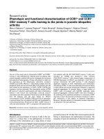
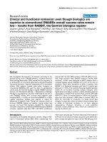
Migration of fibroblasts from BALF and bronchial biopsies from patients with SScFigure 1
Migration of fibroblasts from BALF and bronchial
biopsies from patients with SSc. Fibroblasts were cul-
tured from BALF and bronchial biopsies in a "clone-cylinder",
which was removed after 24 h. The distance from the border
of the removed cylinder covered by the cells was measured
after an additional 48 h. Values are presented as means ±
SEM for n = 5 patients/group. *Significant difference when
comparing the migration between BALF fibroblasts and
biopsy fibroblasts from patients with SSc.
0
50
100
150
200
250
300
350
400
450
500
SSc Biopsy SSc BALF
Distance (µm)
*
Respiratory Research 2006, 7:11 />Page 4 of 10
(page number not for citation purposes)
peptides. MS/MS spectra were acquired using up to 3000
laser shots/precursor unless the pre-defined signal-to-
noise level in the MS/MS acquisition was achieved sooner.
The MS/MS data were submitted for database to Mascot
(Matrix Science Inc. Boston, MA) with a parent mass error
tolerance of 25 ppm and mass fragments with an error tol-
erance of 0.2 Da.
Statistical methods
Mean values ± standard error of the mean (SEM) were cal-
culated and the Mann-Whitney method was used for anal-
yses of statistical significance. All values of p < 0.05 (*)
were considered significant.
Results
BALF fibroblasts in SSc display increased migration and
expression of small GTPases
BALF fibroblasts were established from 5 out of 10
patients with SSc. This is similar to previous findings in
patients with mild asthma where these cells could be
established from 5 out of 12 patients [16]. To study if
BALF fibroblasts in SSc could originate from the submu-
cosa, differences in cell migration were studied in fibrob-
lasts from BALF and bronchial biopsies from patients with
SSc. A significant 1.2-fold (p < 0.05) increase in cell migra-
tion was reported for the BALF fibroblasts from patients
with SSc when compared to fibroblasts from bronchial
biopsies (Fig 1). These observations are in accordance
with previous findings where BALF fibroblasts from
patients with mild asthma displayed a significant increase
in cell migration [16].
To study if the increased migration of BALF fibroblasts
was linked to the expression of the small GTPases RhoA
and Rac1, Western Blotting was performed on cultured
fibroblasts. Several studies have demonstrated that these
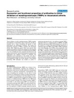
proteins are of importance in cell migration [10]. A signif-
icant increase of RhoA (1.5-fold increase, p < 0.05) and
Rac1 (1.3-fold increase, p < 0.05) was observed in the
BALF fibroblast cultures (Figs 2A–B). These increases are
also in accordance with previous results from BALF
fibroblasts from patients with mild asthma [16].
Increased levels of ED-A fibronectin and SRp20 in BALF
fibroblasts
Next we examined the production of the alternatively
spliced form of cellular fibronectin, ED-A fibronectin, in
BALF fibroblasts, which was compared to that of fibrob-
lasts from bronchial biopsies in patients with SSc and
mild asthma. A significant 2-fold increase (p < 0.05) was
Expression of RhoA and Rac1 in fibroblasts from BALF and bronchial biopsies from patients with SSc and mild asthmaFigure 2
Expression of RhoA and Rac1 in fibroblasts from BALF and bronchial biopsies from patients with SSc and mild
asthma. Fibroblasts were harvested in lysis buffer as described in the Method section. Equal amounts of protein were loaded
on 4–12% Bis-Tris gels. Western Blotting was performed to study the expression of RhoA (A) and Rac1 (B) where the optical
density of the bands was measured to determine the expression. Values are presented as means ± SEM for n = 5 patients/
group. *Significant difference when comparing the expression of RhoA and Rac1 between BALF fibroblasts and biopsy fibrob-
lasts from patients withSSc.
0
2
4
6
8
10
12
14
16
18
20
SSc biopsy
SSc BALF
0
20
40
60
80
100
120
SSc Biopsy SSc BALF
*
Intensity/µg protein
Intensity/µg protein
SSc biopsy SSc BALF
22 kDa 21 kDa
SSc biopsy
SSc BALF
*
A
B
Respiratory Research 2006, 7:11 />Page 5 of 10
(page number not for citation purposes)
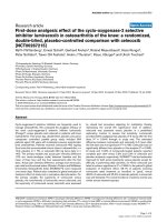
seen in the BALF fibroblast cultures from patients with SSc
and a 5-fold increase (p < 0.05) was observed in BALF
fibroblasts from patients with mild asthma when com-
pared to biopsy fibroblasts (Fig 3A). The antibody used
reacts with an epitope located in the ED-A sequence of cel-
lular fibronectin. Analysis of the immunological determi-
nant recognized by the antibody shows three fragments of
47, 44, and 52 kDa. Interestingly, the three fragments
were visible on the Western Blot membrane in BALF
fibroblasts from both disorders, but only one fragment
was seen in the biopsy cultures from patients with mild
asthma and SSc. No difference in total production of
fibronectin was seen between the two fibroblast pheno-
types in patients with SSc or mild asthma (Fig 3B). The
proportion of ED-A fibronectin in patients with SSc and
asthma from the BALF fibroblasts was 60% ED-A, whereas
in the biopsy cultures 25% of the total fibronectin produc-
tion was ED-A fibronectin.
Next, we studied if the serine-arginine (SR) splicing factor
SRp20 was regulated, since it has been proposed to be
involved in the increased expression of alternative splicing
of fibronectin. A 1.4-fold increase of SRp20 expression
was seen in BALF fibroblasts from patients with asthma (p
< 0.05) and a 1.3-fold increase was observed in SSc when
compared to fibroblasts from bronchial biopsies (p <
0.05) (Fig 4).
Cultured fibroblasts from BALF and biopsies in mild
asthma and SSc display myofibroblast phenotype and
unaltered TGF-
β
production
It has been shown that increased ED-A fibronectin levels
are associated with increased α-SMA levels [12]. There-
fore, the cells were analyzed for expression of the myofi-
broblast marker α-SMA to study the phenotype of the
cultured fibroblasts. The fibroblasts expressed α-SMA in
both groups of patients where a significant 8-fold (p <
0.05) increase was observed in BALF fibroblasts from
patients with mild asthma when compared to fibroblasts
from bronchial biopsies (Fig 5A). When compared to
BALF fibroblasts in SSc, the BALF fibroblasts from patients
with mild asthma displayed a 1.3-fold increase in α-SMA
expression (p < 0.05). No difference in α-SMA expression
was observed between the two groups of SSc fibroblasts,
but the expression was larger (5-fold, p < 0.05) in SSc
biopsy fibroblasts than in biopsy fibroblasts from patients
with mild asthma. The actin filaments in all cells were
arranged into stress fibers (Fig 6). These findings suggest
similar phenotypes in BALF- and biopsy fibroblasts in SSc
but a larger difference in α-SMA expression in BALF
Expression of fibronectin isoforms in fibroblasts from BALF and bronchial biopsies from patients with mild asthma and SScFigure 3
Expression of fibronectin isoforms in fibroblasts from BALF and bronchial biopsies from patients with mild
asthma and SSc. Fibroblasts were cultured from bronchial biopsies and BALF from patients with SSc and mild asthma and
harvested in lysis buffer as described in the Method section. Equal amounts of protein were loaded on 4–12% Bis-Tris gels. The
production of the alternatively spliced isoform ED-A fibronectin (A) and cellular fibronectin (B) was measured using Western
Blot with human ED-A fibronectin and cellular fibronectin antibodies. Quantification of fibronectin expression was performed
by measuring the optical density of the bands. Values are presented as means ± SEM for n = 5 patients/group.
A
B
50 kDa
0
10
20
30
40
50
60
70
Asthma
Bio ps y
Asthma
BAL F
SSc Biops y SSc BALF
0
20
40
60
80
100
120
Asthma
Bio ps y
Asthma
BAL F
SSc Biops y SSc BALF
Intensity/µg protein
Intensity/µg protein
240 kDa
Asthma
Bio ps y
Asthma
BAL F
SSc Biops y SSc BALF
Asthma
Bio ps y
Asthma
BAL F
SSc Biops y SSc BALF
p<0.05 p<0.05
Respiratory Research 2006, 7:11 />Page 6 of 10
(page number not for citation purposes)
fibroblasts from patients with mild asthma when com-
pared to corresponding biopsy fibroblasts.
The production of TGF-β, which has been shown to
induce a myofibroblast phenotype with elevated levels of
α-SMA expression in cultured fibroblast, was studied in
the fibroblasts from BALF and bronchial biopsies in both
patient groups. A tendency towards reduced TGF-β pro-
duction was seen for the BALF fibroblast cultures from
patients with SSc when compared to asthma, however,
this decrease was not statistically significant (data not
shown). No alterations were seen in production between
the remaining cultured BALF- and biopsy fibroblasts in
patients with SSc or mild asthma (data not shown).
BALF fibroblasts from patients with SSc and mild asthma
display a differential proteome
The next set of experiments was addressed to explore dif-
ferences between SSc and mild asthma on a molecular
level by using two-dimensional gel electrophoresis (2-DE)
in the range of pH 4–7 and MALDI-TOF-TOF, in the hope
of revealing important markers for the different fibrotic
processes. A series of triplicate gels were studied where the
master gel for each patient group comprised of approxi-
mately 500 unique protein spots. The differential protein
expression pattern between BALF fibroblasts from
patients with SSc and mild asthma displayed 24 differen-
tially expressed spots of statistical significance (p < 0.05).
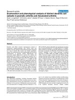
Of these differentially expressed spots, 13 protein spots
displayed a statistical significant 2-fold or larger difference
in expression and these were matched in all gels (Fig 7A).
A protein score >57 with more than two matched peptides
were considered to be a significant identification. We were
able to identify 6–29 peptides that yielded a protein score
ranging from 163–585, thus indicating a high probability
for the identified proteins. These proteins were divided
into different groups depending on their functional role
(Fig 7B); cytoskeletal-associated, cell cycle regulating-,
scavenger- and metabolic proteins. The proteins that dis-
played the largest differences in protein expression were
cytoskeletal associated proteins and scavenger proteins.
The proteome in fibroblasts from BALF and bronchial
biopsies from patients with mild asthma have been previ-
ously shown to include differentially expressed proteins
involved in cell migration [16]. However, when compar-
ing the fibroblast proteome between BALF and bronchial
biopsies from patients with SSc, only three proteins dis-
played a significant differential expression pattern (data
not shown). Again, this indicates that BALF- and biopsy
fibroblast cultures of patients with SSc are more similar in
phenotype, which correlates to the similar levels of α-SMA
expression (Fig 5). In addition, a comparison between
Production of SRp20 in fibroblasts from BALF and bronchial biopsies from patients with SSc and mild asthmaFigure 4
Production of SRp20 in fibroblasts from BALF and
bronchial biopsies from patients with SSc and mild
asthma. Fibroblasts were harvested in lysis buffer as
described in the Method section. Equal amounts of protein
were loaded on 4–12% Bis-Tris gels. The level of SRp20
expression was determined by Western Blotting and further
quantified by measuring the optical density of the bands. Val-
ues are presented as means ± SEM for n = 5 patients/group.
Asthma Biopsy Asthma BALF SSc Biopsy SSc BALF
20 kDa
0
20
40
60
80
100
120
Asthma
Biopsy
Asthma
BALF
SSc Biopsy SSc BALF
p<0.05 p<0.05
Expression of α-SMA in fibroblasts from BALF and bronchial biopsies from patients with SSc and mild asthmaFigure 5
Expression of α-SMA in fibroblasts from BALF and
bronchial biopsies from patients with SSc and mild
asthma. Fibroblasts were cultured from bronchial biopsies
and BALF from patients with SSc and mild asthma and har-
vested in lysis buffer as described in the Method section.
Equal amounts of protein were loaded on 4–12% Bis-Tris
gels. The expression of α-SMA was detected using Western
Blot with human α-SMA antibodies and further quantified by
measuring the optical density of the bands. Values are pre-
sented as means ± SEM for n = 5 patients/group.
0
10
20
30
40
50
60
70
80
90
100
Intensity/µg protein
Asthma Biopsy Asthma BALF SSc Biopsy SSc BALF
Asthma
Biopsy
Asthma
BAL F
SSc Biops y SSc BALF
45 kDa
p<0.05
p<0.05
p<0.05
Respiratory Research 2006, 7:11 />Page 7 of 10
(page number not for citation purposes)
asthma biopsy fibroblasts and SSc biopsy fibroblasts were
performed, however no significant regulated proteins
could be observed using this approach.
Discussion
In this study, we have reported increased motility in BALF
fibroblasts from patients with SSc when compared to
fibroblasts from corresponding bronchial biopsies, which
proposes a possible mesenchymal origin for these cells.
The increased migration is in accordance with previous
studies on BALF fibroblasts from patients with mild
asthma [16]. This finding was accompanied by an ele-
vated expression of the small GTPases RhoA and Rac1.
These observations are important since RhoA and Rac1
have been suggested to be involved in cell migration by
formation of stress fibers as well as the formation and
maintenance of focal adhesions [20]. Differences in phe-
notype between fibroblasts cultured from bronchial biop-
sies and BALF from patients with SSc and mild asthma
were characterized which may be key factors in the dis-
tinct fibrotic responses of these disorders. The production
of ED-A fibronectin was elevated in BALF fibroblasts from
patients with SSc and mild asthma when compared to
fibroblasts cultured from corresponding bronchial biop-
sies. This alternatively spliced form of cellular fibronectin
that contains the ED-A domain is associated with wound
healing and fibrosis in diseases such as SSc [11]. The
expression of this matrix molecule serves as a marker of
extracellular matrix that is closely linked to intracellular α-
SMA expression myofibroblasts [21]. Furthermore, the
elevated levels of ED-A fibronectin in BALF fibroblasts
may also explain the increased migration observed in
these cells [22]. The elevated levels of the splicing factor
SRp20 in the BALF fibroblast cultures may explain the
induced expression of ED-A fibronectin. TGF-β specifi-
cally induces the expression of SRp20, which when over-
expressed promotes the alternative splicing of fibronectin
[13]. The TGF-β-induced expression of ED-A fibronectin is
required for TGF-β-triggered increase of α-SMA [12]. The
cultured fibroblasts from BALF and bronchial biopsies
from SSc and mild asthma expressed α-SMA, which sug-
gests that these cells display a myofibroblast phenotype.
The increased expression of α-SMA in BALF fibroblasts
from patients with mild asthma and SSc proposes distinct
BALF fibroblast phenotypes in these disorders. Although
the levels of α-SMA expression in fibroblasts from BALF
and bronchial biopsies in SSc did not display a distinct
pattern, the levels were elevated when compared to biopsy
fibroblast in mild asthma. These observations are impor-
tant since they may reflect the differentiated stage the cells
are cultured from, which in turn may reflect the degree of
fibrosis in the tissue.
Since BALF fibroblasts in SSc and mild asthma display
increased migration and express important myofibroblast
markers such as α-SMA and ED-A fibronectin, the differ-
ential protein expression profile between the two BALF
fibroblast groups were studied by using 2-DE and MALDI-
TOF-TOF to reveal factors that may account for the dis-
tinct fibrotic processes in these disorders. This approach is
an excellent tool when identifying high abundant pro-
teins but less efficient when studying membrane associ-
ated- and low abundant proteins. Nevertheless, many of
the high abundant proteins within range for the 2-DE are
involved in important cellular mechanisms, including
fibrosis. Cytoskeletal proteins, such as vimentin, tropo-
myosin, and actin associated proteins were identified in
elevated levels in the SSc BALF fibroblasts, which may all
account for the motile phenotype that characterizes the
BALF fibroblast. Moreover, these proteins are elevated in
activated myofibroblasts since they have been suggested
to be involved in the increased intracellular trafficking
and secretion of ECM molecules, which is an important
feature of the myofibroblast in fibrotic tissue. Ran-bind-
ing protein 1 (RanBP1) has been shown to be induce
migration [23]. This protein was expressed in BALF
fibroblasts from patients with mild asthma and SSc, but
was significantly increased in the latter group. This obser-
vation may therefore explain the increased migration
characteristic for the BALF fibroblasts from patients with
Actin expression in fibroblasts from BALF and bronchial biopsies from patients with SSc and mild asthma are arranged into stress fibersFigure 6
Actin expression in fibroblasts from BALF and bron-
chial biopsies from patients with SSc and mild
asthma are arranged into stress fibers. Fibroblasts were
cultured from bronchial biopsies and BALF from patients
with SSc and mild asthma. Cells were seeded on four-well
chamber slides (5000 cells/well), stained with Alexa Fluor™
488 phalloidin showing stress fibers and analyzed using a fluo-
rescence microscope.
Respiratory Research 2006, 7:11 />Page 8 of 10
(page number not for citation purposes)
SSc. Several scavenger proteins involved in oxidative stress
and redox processes such as disulfide isomerase (ERp60)
and glutathione S-transferase P (GSTP1-1), displayed ele-
vated levels in BALF fibroblasts from patients with SSc.
Interestingly, oxidative stress is considered an important
factor in patients with SSc in contributing to vascular
damage leading to an activation of fibroblasts and inflam-
matory cells [24]. Therefore, the increased levels of scav-
enger proteins in BALF fibroblasts from patients with SSc
may reflect a response to the elevated levels of oxidative
stress, mediated by free radicals observed in patients with
SSc.
The small number of differentially expressed proteins
between fibroblasts from BALF and bronchial biopsies
from patients with SSc suggests that these two fibroblast
Protein expression pattern in BALF fibroblast cultures from patients with SSc and mild asthmaFigure 7
Protein expression pattern in BALF fibroblast cultures from patients with SSc and mild asthma. Cells were cul-
tured in six-well plates and harvested as described in the Method section. The lysed cells were separated by 2-DE. A repre-
sentative 2-D gel from asthma and SSc BALF fibroblasts are presented and the significant differentially expressed spots when
are marked with arrows (A). The identified differentially expressed proteins in fibroblasts from BALF in patients with mild
asthma and SSc was identified using sequencing MALDI-TOF-TOF (B). IOD (ppm) is the optical density of the spots correlated
to the total optical density for all spots present in the gel. Abbreviations: Acc No. = Swissprot accession number; MW = Molec-
ular weight. IOD (ppm) = Optical density of the spots correlated to the total optical density for all spots present in the gel.
Peptide count = Number of identified peptides that could be matched to the suggested database protein. Protein Score =
Probability that the peptide counts are derived from the suggested database protein.
GSTP1-1
Keratin 10
Tropomyosin
Vimenetin
Actin-related protein 3
p16 ARC
RanBP1
Stathmin
pI 4-7
Mw 90-5 kDa
ERp60
A
Protein Name Group Acc No. Peptide count Protein score Protein MW Protein pI SSc IOD (ppm) Asthma IOD (ppm)
Vimentin Cytoskeletal P08670 29 479 53.6 5.06 3722 499
Actin-related protein 3 " P32391 15 260 47.8 5.06 7840 3784
Actin-related protein 2/3 16kDa subunit (p16-ARC) " O15511 6 250 16.2 5.47 1151 0
Tropomyosin isoform " Q15657 21 373 28.5 4.89 9775 1885
Ran-specific GTPase-activating protein (RanBP1) " P43487 10 233 23.4 5.19 8376 2528
Stathmin Cell-Cycle P16949 15 427 17.2 5.77 1915 1057
Glutathione S-trasferase P (GSTP1-1) Scavenger P09211 8 394 23.4 5.44 15124 4014
Ubiquitin carboxyl-terminal hydrolase isozyme (UCH-L3) " P15374 9 378 26.3 4.84 1000 3647
Thioredoxin-dependent peroxidase reductase precursor " P30048 7 320 28.0 7.67 3714 5847
Disulfide isomerase ER-60 (ERp60) " P03101 24 585 57.1 5.98 17483 6733
6-phosphogluconolactonase (6PGL) Other O95336 11 309 27.8 5.70 1146 3427
Apolipoprotein A-I precursor " P15497 27 566 30.2 5.71 959 4433
Keratin 10 " Q8N175 13 163 59.0 5.01 3988 1844
B
SSc Asthma
Thioredoxin peroxidase
reductase precursor
UCH-L3
Apolipoprotein
A-1 precursor
6PGL
Respiratory Research 2006, 7:11 />Page 9 of 10
(page number not for citation purposes)
phenotypes are relatively similar, an observation that was
further supported by the small differences in α-SMA
expression. In contrast to this observation, BALF fibrob-
lasts from SSc and mild asthma display important distinc-
tions in α-SMA and protein expression pattern. These
observations emphasize the complex diversity of myofi-
broblast phenotypes present in the human fibrotic lung,
which have been shown in previous studies to exhibit dif-
ferent affinity and activation from cytokines and growth
factors such as TGF-β [25]. Fibroblasts have the ability to
produce TGF-β by themselves through an autocrine mech-
anism that has been suggested to be of importance in
maintaining the myofibroblast phenotype by inducing
increased levels of α-SMA [26]. We did not observe, how-
ever, any differences in the production of TGF-β from the
fibroblasts alone in this study. ECM components such as
heparin, biglycan and decorin that are produced by
myofibroblasts can affect the differentiation process in an
autocrine manner [5,27] and may thus represent a possi-
ble TGF-β independent pathway for the observed differ-
ences in α-SMA expression. In addition, a contribution of
other TGF-β-producing cells in the early passages such as
eosinophils and macrophages may also affect levels of
ED-A fibronectin reported in the analyzed BALF fibrob-
lasts in later passages [28]. Increased levels of BALF eosi-
nophils have been reported in patients with mild asthma
with BALF fibroblasts when compared to patients with
asthma and control subjects without the presence of these
cells, however if this linkage is present in SSc remains to
be elucidated in future studies [16]. The origin of the BALF
fibroblasts is not known but since fibroblasts reside in
areas beneath the basement membrane, it is tempting to
speculate that fibroblasts with increased motility would
migrate to the airway lumen upon possible stimuli or
damages to the airway epithelium. Another possible ori-
gin for the BALF fibroblasts includes the recruitment of
fibroblast progenitor cells, termed fibrocytes, from the cir-
culation. In SSc, the endothelial cells are damaged by
mediators such as free radicals, which may facilitate traf-
ficking of cells from the circulation through the endothe-
lium to interact with fibroblasts [24].
Conclusion
The characterization of BALF fibroblasts from patients
with SSc and the comparison with patients with mild
asthma emphasize the importance of activated fibroblasts
in these disorders. The increased motility in fibroblasts
derived from BALF when compared with those derived
from bronchial biopsies suggests a potential submucosal
origin for these cells. Moreover, the findings in this study
highlight important distinctions in fibroblast phenotype
between the two disorders which may reflect the different
disease pathology in SSc. This makes the BALF fibroblast
an interesting target cell for future therapies of lung fibro-
sis observed in SSc.
Competing interests
The author(s) declare that they have no competing inter-
ests.
Authors' contributions
KL: drafted the manuscript, participated in the organiza-
tion of the manuscript, performed all two-dimensional 2-
DE experiments and sample preparation prior to the mass
MALDI-TOF-TOF analysis, and performed some of the
cell migration assays and cell culture experiments.
JM: performed all mass MALDI-TOF-TOF analyses and
peptide database searches. He also participated in the
organization of the manuscript.
MW: handled a majority of the cell culture experiments, as
well as participated in the planning of the manuscript.
CD: Technician who performed many of the Western Blot
experiments and was also involved in the organization of
the manuscript.
LH: Clinician who performed many of the bronchoscopy
sessions when collecting SSc biopsies and BALF and was
involved in organization of the manuscript.
GMV: One of the initiators of this study who has collabo-
rated in the organization of the manuscript.
AS: One of the initiators of this study who has collabo-
rated in the organization of the manuscript.
LB: Clinician who performed many of the bronchoscopy
sessions when collecting asthma biopsies and BALF and
was involved in organization of the manuscript.
GWT: Group leader who (in collaboration with AS) initi-
ated the study and organized the manuscript.
Acknowledgements
The authors would like to thank Dr Ellen Tufvesson, Annika Andersson-
Sjöland and Anna Lindström for laboratory and technical skills. This work
was supported by grants from the Swedish Medical Research Council
(11,550), Heart-Lung Foundation, CFN-Centrala Försöksdjursnämden,
Greta and John Kock, Alfred Österlund, Anna-Greta Crafoord Founda-
tions, Riksföreningen mot Rheumatism, Gustaf V:s 80 Årsfond, and the
Medical Faculty, Lund University.
J.M. was supported by a Wennergren foundation postdoctoral fellowship.
References
1. Atamas SP, White B: Cytokine regulation of pulmonary fibrosis
in scleroderma. Cytokine Growth Factor Rev 2003, 14:537-550.
2. Davies DE, Wicks J, Powell RM, Puddicombe SM, Holgate ST: Airway
remodeling in asthma: new insights. J Allergy Clin Immunol 2003,
111:215-225.
3. Selman M, Pardo A: Idiopathic pulmonary fibrosis: an epithelial/
fibroblastic cross-talk disorder. Respir Res 2002, 3:3.
Publish with BioMed Central and every
scientist can read your work free of charge
"BioMed Central will be the most significant development for
disseminating the results of biomedical research in our lifetime."
Sir Paul Nurse, Cancer Research UK
Your research papers will be:
available free of charge to the entire biomedical community
peer reviewed and published immediately upon acceptance
cited in PubMed and archived on PubMed Central
yours — you keep the copyright
Submit your manuscript here:
/>BioMedcentral
Respiratory Research 2006, 7:11 />Page 10 of 10
(page number not for citation purposes)
4. Tomasek JJ, Gabbiani G, Hinz B, Chaponnier C, Brown RA: Myofi-
broblasts and mechano-regulation of connective tissue
remodelling. Nat Rev Mol Cell Biol 2002, 3:349-363.
5. Westergren-Thorsson G, Sime P, Jordana M, Gauldie J, Sarnstrand B,
Malmstrom A: Lung fibroblast clones from normal and fibrotic
subjects differ in hyaluronan and decorin production and
rate of proliferation. Int J Biochem Cell Biol 2004, 36:1573-1584.
6. Kasai H, Allen JT, Mason RM, Kamimura T, Zhang Z: TGF-beta1
induces human alveolar epithelial to mesenchymal cell tran-
sition (EMT). Respir Res 2005, 6:56.
7. Postlethwaite AE, Shigemitsu H, Kanangat S: Cellular origins of
fibroblasts: possible implications for organ fibrosis in sys-
temic sclerosis. Curr Opin Rheumatol 2004, 16:733-738.
8. Schmidt M, Sun G, Stacey MA, Mori L, Mattoli S: Identification of
circulating fibrocytes as precursors of bronchial myofibrob-
lasts in asthma. J Immunol 2003, 171:380-389.
9. Willis BC, Liebler JM, Luby-Phelps K, Nicholson AG, Crandall ED, du
Bois RM, Borok Z: Induction of epithelial-mesenchymal transi-
tion in alveolar epithelial cells by transforming growth fac-
tor-beta1: potential role in idiopathic pulmonary fibrosis. Am
J Pathol 2005, 166:1321-1332.
10. Fukata M, Nakagawa M, Kaibuchi K: Roles of Rho-family GTPases
in cell polarisation and directional migration. Curr Opin Cell Biol
2003, 15:590-597.
11. Leask A, Abraham DJ: TGF-beta signaling and the fibrotic
response. FASEB J 2004, 18:816-827.
12. Serini G, Bochaton-Piallat ML, Ropraz P, Geinoz A, Borsi L, Zardi L,
Gabbiani G: The fibronectin domain ED-A is crucial for myofi-
broblastic phenotype induction by transforming growth fac-
tor-beta1. J Cell Biol 1998, 142:873-881.
13. Lim LP, Sharp PA: Alternative splicing of the fibronectin EIIIB
exon depends on specific TGCATG repeats. Mol Cell Biol 1998,
18:3900-3906.
14. Malmstrom J, Larsen K, Hansson L, Lofdahl CG, Norregard-Jensen O,
Marko-Varga G, Westergren-Thorsson G: Proteoglycan and pro-
teome profiling of central human pulmonary fibrotic tissue
utilizing miniaturized sample preparation: a feasibility study.
Proteomics 2002, 2:394-404.
15. Shi-Wen X, Chen Y, Denton CP, Eastwood M, Renzoni EA, Bou-
Gharios G, Pearson JD, Dashwood M, du Bois RM, Black CM, Leask
A, Abraham DJ: Endothelin-1 promotes myofibroblast induc-
tion through the ETA receptor via a rac/phosphoinositide 3-
kinase/Akt-dependent pathway and is essential for the
enhanced contractile phenotype of fibrotic fibroblasts. Mol
Biol Cell 2004, 15:2707-2719.
16. Larsen K, Tufvesson E, Malmstrom J, Morgelin M, Wildt M, Andersson
A, Lindstrom A, Malmstrom A, Lofdahl CG, Marko-Varga G, Bjermer
L, Westergren-Thorsson G: Presence of activated mobile
fibroblasts in bronchoalveolar lavage from patients with mild
asthma. Am J Respir Crit Care Med 2004, 170:1049-1056.
17. Ludwicka A, Trojanowska M, Smith EA, Baumann M, Strange C, Korn
JH, Smith T, LeRoy EC, Silver RM: Growth and characterization
of fibroblasts obtained from bronchoalveolar lavage of
patients with scleroderma. J Rheumatol 1992, 19:1716-1723.
18. Tufvesson E, Westergren-Thorsson G: Biglycan and decorin
induce morphological and cytoskeletal changes involving sig-
nalling by the small GTPases RhoA and Rac1 resulting in lung
fibroblast migration. J Cell Sci 2003, 116:4857-4864.
19. Shevchenko A, Wilm M, Vorm O, Mann M: Mass spectrometric
sequencing of proteins silver-stained polyacrylamide gels.
Anal Chem 1996, 68:850-858.
20. Wittmann T, Waterman-Storer CM: Cell motility: can Rho
GTPases and microtubules point the way? J Cell Sci 2001,
114:3795-3803.
21. Dugina V, Fontao L, Chaponnier C, Vasiliev J, Gabbiani G: Focal
adhesion features during myofibroblastic differentiation are
controlled by intracellular and extracellular factors. J Cell Sci
2001, 114:3285-3296.
22. Inoue T, Nabeshima K, Shimao Y, Meng JY, Koono M: Regulation of
fibronectin expression and splicing in migrating epithelial
cells: migrating MDCK cells produce a lesser amount of, but
more active, fibronectin. Biochem Biophys Res Commun 2001,
280:1262-1268.
23. Wang D, Li Z, Messing EM, Wu G: Activation of Ras/Erk pathway
by a novel MET-interacting protein RanBPM. J Biol Chem 2002,
277:36216-36222.
24. Herrick AL, Matucci CM: The emerging problem of oxidative
stress and the role of antioxidants in systemic sclerosis. Clin
Exp Rheumatol 2001, 19:4-8.
25. Phan SH: Fibroblast phenotypes in pulmonary fibrosis. Am J
Respir Cell Mol Biol 2003, 29:S87-S92.
26. Schmid P, Itin P, Cherry G, Bi C, Cox DA: Enhanced expression of
transforming growth factor-beta type I and type II receptors
in wound granulation tissue and hypertrophic scar. Am J Pathol
1998, 152:485-493.
27. Desmouliere A, Rubbia-Brandt L, Grau G, Gabbiani G: Heparin
induces alpha-smooth muscle actin expression in cultured
fibroblasts and in granulation tissue myofibroblasts. Lab Invest
1992, 67:716-726.
28. Phipps S, Ying S, Wangoo A, Ong YE, Levi-Schaffer F, Kay AB: The
relationship between allergen-induced tissue eosinophilia
and markers of repair and remodeling in human atopic skin.
J Immunol 2002, 169:4604-4612.