Báo cáo y học: "Neonatal immune responses to TLR2 stimulation: Influence of maternal atopy on Foxp3 and IL-10 expression" pot
Bạn đang xem bản rút gọn của tài liệu. Xem và tải ngay bản đầy đủ của tài liệu tại đây (333.31 KB, 9 trang )
BioMed Central
Page 1 of 9
(page number not for citation purposes)
Respiratory Research
Open Access
Research
Neonatal immune responses to TLR2 stimulation: Influence of
maternal atopy on Foxp3 and IL-10 expression
Bianca Schaub*
1,6
, Monica Campo
2
, Hongzhen He
2
, David Perkins
4
,
Matthew W Gillman
3
, Diane R Gold
5
, Scott Weiss
5
, Ellice Lieberman
6
and
Patricia W Finn
2
Address:
1
University Children's Hospital Munich, Department of Pulmonary, LMU, Munich, Germany,
2
Pulmonary and Critical Care Division,
Department of Medicine, Brigham and Women's Hospital, Harvard Medical School, Boston, MA, USA,
3
Department of Ambulatory Care and
Prevention, Harvard Medical School and Harvard Pilgrim Health Care, Boston, MA, USA,
4
Immunogenetics and Transplantation, Department of
Medicine, Brigham and Women's Hospital, Harvard Medical School, Boston, MA, USA,
5
Channing Laboratory, Brigham and Women's Hospital,
Harvard Medical School, Boston, MA, USA and
6
Harvard Medical School, Boston, MA, USA
Email: Bianca Schaub* - ; Monica Campo - ;
Hongzhen He - ; David Perkins - ;
Matthew W Gillman - ; Diane R Gold - ;
Scott Weiss - ; Ellice Lieberman - ; Patricia W Finn -
* Corresponding author
Abstract
Background: Maternal atopic background and stimulation of the adaptive immune system with allergen
interact in the development of allergic disease. Stimulation of the innate immune system through microbial
exposure, such as activation of the innate Toll-like-receptor 2 (TLR2), may reduce the development of
allergy in childhood. However, little is known about the immunological effects of microbial stimulation on
early immune responses and in association with maternal atopy.
Methods: We analyzed immune responses of cord blood mononuclear cells (CBMC) from 50 healthy
neonates (31 non-atopic and 19 atopic mothers). Cells were stimulated with the TLR2 agonist
peptidoglycan (Ppg) or the allergen house dust mite Dermatophagoides farinae (Derf1), and results
compared to unstimulated cells. We analyzed lymphocyte proliferation and cytokine secretion of CBMC.
In addition, we assessed gene expression associated with T regulatory cells including the transcription
factor Foxp3, the glucocorticoid-induced TNF receptor (GITR), and the cytotoxic lymphocyte antigen 4
(CTLA4). Lymphocyte proliferation was measured by
3
H-Thymidine uptake, cytokine concentrations
determined by ELISA, mRNA expression of T cell markers by real-time RT-PCR.
Results: Ppg stimulation induced primarily IL-10 cytokine production, in addition to IFN-γ, IL-13 and TNF-
α secretion. GITR was increased following Ppg stimulation (p = 0.07). Ppg-induced IL-10 production and
induction of Foxp3 were higher in CBMC without, than with maternal atopy (p = 0.04, p = 0.049). IL-10
production was highly correlated with increased expression of Foxp3 (r = 0.53, p = 0.001), GITR (r = 0.47,
p = 0.004) and CTLA4 (r = 0.49, p = 0.003), independent of maternal atopy.
Conclusion: TLR2 stimulation with Ppg induces IL-10 and genes associated with T regulatory cells,
influenced by maternal atopy. Increased IL-10 and Foxp3 induction in CBMC of non-atopic compared to
atopic mothers, may indicate an increased capacity to respond to microbial stimuli.
Published: 21 March 2006
Respiratory Research2006, 7:40 doi:10.1186/1465-9921-7-40
Received: 28 November 2005
Accepted: 21 March 2006
This article is available from: />© 2006Schaub et al; licensee BioMed Central Ltd.
This is an Open Access article distributed under the terms of the Creative Commons Attribution License ( />),
which permits unrestricted use, distribution, and reproduction in any medium, provided the original work is properly cited.
Respiratory Research 2006, 7:40 />Page 2 of 9
(page number not for citation purposes)
Background
The early immunological mechanisms that predispose to
the development of allergic immune responses are the
focus of recent studies [1]. Prior investigations have exam-
ined the development of allergen-specific T cell memory
cells that is dominated by T helper 2 (Th2) cytokines [2].
The immunological events that drive T helper memory
development are initiated early in infancy [3], possibly
even in utero [4,5]. Already during the perinatal period
there are immunological differences in neonates at high
risk of allergy, namely relatively reduced capacity for type
1 (Th1) interferon gamma (IFN-γ) responses, compared
with low risk neonates with no family history of allergy
[6-8].
Th1 responses such as production of IFN-γ and IL-12 can
be influenced by innate, non-antigen-dependent immune
stimulation. Innate immune stimulation is in part medi-
ated via mammalian toll-like receptors (TLR). Conserved
throughout evolution, TLRs participate in innate immune
responses to a variety of microbial pathogens, for example
the cell wall component of gram-positive bacteria, pepti-
doglycan (Ppg), i.e. predominantly recognized by TLR2
[9-13], but specific cellular responses in the early immune
system have just started to be a focus of research [14].
While murine models as well as epidemiological studies
suggest an involvement of TLR4 agonists in modulating
asthma or allergic diseases [15,16], TLR2 agonists can also
decrease allergic immune responses in murine models
[17]. Recent human data demonstrated increased levels of
TLR2 in children of farmers exposed to high microbial
burden, where a very low prevalence of atopy occurs [18].
Also, genetic variation in TLR2 was described to be a
major determinant of the susceptibility to asthma and
allergies in children of farmers [19].
We hypothesized that innate stimulation of cord blood
mononuclear cells (CBMC) with a TLR2 agonist might
influence T cell responses in cord blood mononuclear
cells depending on a maternal background of atopy. Spe-
cifically, we tested whether the innate TLR 2 agonist Pep-
tidoglycan (Ppg) influences immune factors in addition
to the secretion of Th1 (IFN-γ, IL-12), Th2 (IL-13)
cytokines and TNF-α. We examined T cell subsets such as
T regulatory cells, which are characterized by secretion of
the cytokines IL-10, TGF-β, and expression of e.g. the tran-
scription factor Foxp3 and GITR. As maternal atopy is
known to increase the risk for atopic diseases in children,
we hypothesized that regulatory factors of T cells may be
diminished in CBMC of mothers with atopy.
Methods
Human study populations
Fetal cord blood samples (n = 50) were obtained from
Boston area pregnancies for laboratory-based analysis.
The subjects for the study were recruited during the prena-
tal period to participate in one of two ongoing pregnancy
studies [20,21]. Umbilical cord blood was obtained at the
time of delivery from healthy neonates born at term after
uncomplicated pregnancies. The laboratory investigators
were blinded to clinical information, and samples were
analyzed based on sample availability to perform the lab-
oratory studies.
At the time of enrollment all mothers completed a ques-
tionnaire regarding atopic status. Maternal atopy was
determined by detailed interview or questionnaire during
pregnancy and was defined as a history of doctor's diagno-
sis of asthma and/or hay fever, and/or eczema. Of the 50
cord blood samples analyzed, unblinding revealed that 31
of the mothers had no maternal history of atopy, and 19
mothers had maternal atopy. Of these 19, 2 mothers had
a doctor's diagnosis of asthma, 2 of them had also asthma
and hay fever; 7 mothers had only hay fever, 4 had only
eczema, and 4 mothers had a doctor's diagnosis of hay
fever and eczema. Demographic data regarding maternal
age and smoking, delivery type, offspring gender, birth
weight and ethnicity were not significantly different
between the two groups of atopic and non-atopic moth-
ers. Specifically, there were no smokers in any of the
groups. Exclusion criteria included multiple gestation
(twins, triplets), and inability to answer questions in Eng-
lish. Informed consent was obtained from mothers for
their participation in the study, including cord blood col-
lection. Approval was obtained from the human research
committee of the Brigham and Women's Hospital and
Harvard Pilgrim Health Care, Boston, MA.
Isolation of CBMC and lymphocyte proliferation
Cord blood samples were collected from the umbilical
vein after delivery and processed fresh, non cryo-pre-
served as previously described [22,23]. Samples were
placed in heparinized tubes and processed within 24
hours. Cord blood mononuclear cells (CBMC) were iso-
lated by density-gradient centrifugation with Ficoll-
Hypaque Plus (Pharmacia, Uppsala, Sweden) after dilu-
tion in phosphate buffer saline (PBS, Sigma Aldrich,
St.Louis, MO). Cells were washed in RPMI 1640 and
diluted in 10% human serum (Biowhittaker, Walkersville,
MD) to a concentration of 5 × 10
6
cells/ml. For the lym-
phocyte proliferation assay 0.5 × 10
6
cells/well were cul-
tured in triplicates in 96-well round-bottom tissue-culture
plates (Corning, NY, NY) for 3 days, stimulated with pep-
tidoglycan (Ppg, 10 µg/ml, Staph. Aureus, Sigma Aldrich,
St. Louis, MO), Dermatophagoides farinae (Derf1, 30 µg/
ml, Indoor Biotechnologies, Charlottesville, VA), or phy-
tohemagglutinin (PHA, 5 µg/ml, Sigma Aldrich, St.Louis,
MO) as positive control and compared to unstimulated
samples. The positive control PHA induced CBMC prolif-
eration with a stimulation index (SI) of 33 ± SEM 8. The
Respiratory Research 2006, 7:40 />Page 3 of 9
(page number not for citation purposes)
doses for anti-MHC II and anti-CD4 (each 10 µg/ml, BD
Bioscience Pharmingen, San Jose, CA) and corresponding
isotype controls and for the previous stimuli were estab-
lished in prior dose and time-course experiments. Specifi-
city of Ppg for TLR2 was determined in prior experiments
using TLR2 -/- mice demonstrating lack of spleen cell pro-
liferation after Ppg stimulation. As control, Ppg stimula-
tion in TLR4 -/- mice demonstrated increased lymphocyte
proliferation of spleen cells. Endotoxin concentrations in
Ppg, Derf1, and PHA preparations, measured by Limulus
assay, were very low (<0.01 EU/ml = 0.002 ng/ml), and
did not significantly change lymphocyte proliferation or
cytokine secretion in CBMC. By testing the functional
ability of CBMC with different doses of LPS and active
components such as Lipid A, LpA (starting at 0.01 ng to
100 ng/ml), we detected increased lymphocyte prolifera-
tion with doses of LpA above 1 ng/ml; therefore levels
below 0.01 ng/ml had no influence on the analysis.
After incubation, samples were pulsed with 1 µCi
3
H-Thy-
midine for an additional 8 hours. Cultures were per-
formed at 37°C in a humidified 5% CO
2
incubation
chamber. Cells were harvested with a Tomcat Mach II har-
vester (Wallac, Turku, Finland) onto filter plates, which
were read using a β-Counter. Proliferation was either
assessed by counts per minute (cpm) or quantified by
stimulation index (SI), which is calculated as the ratio of
mean counts per minute (cpm) of stimulated over
unstimulated replicates.
Cytokine measurements
Cells cultured in media were harvested immediately and
cell cultures, stimulated with Ppg, Derf1 or PHA as
described above, were harvested after 3 days of stimula-
tion. Supernatants were aliquoted in duplicate into 96-
well plates (50 µl/well), which are precoated with
cytokine specific antibody. Optical density was measured
at 450 nm. The cytokines IL-13, IFN-γ, IL-12, TNF-α and
IL-10 were measured by ELISA (Endogen, Rockford, IL)
according to the manufacturer's instructions. The sensitiv-
ity of the assay was 7 pg/ml for IL-13, 2 pg/ml for IFN-γ, 3
pg/ml for IL-12 (p70), 5 pg/ml for TNF-α and 3 pg/ml for
IL-10.
Real-time quantitative RT-PCR
For RNA, CBMC were stimulated with the described stim-
uli for 3 days at 5 × 10
6
cells/ml in 6-well plates. Total RNA
was isolated from CBMC with TRI Reagent (Sigma-
Aldrich, St. Louis, MO). Isolated RNA was reverse tran-
scribed with SuperScript II RNAse reverse transcriptase
(Life Technologies, Carlsbad, CA). Specific primer pairs
for GAPDH and β-actin (housekeeping genes), TLR2,
Foxp3, GITR, CTLA4 and TGF-β were designed with the
Primer Express software (Applied Biosystems, Foster City,
CA). The sequences of the forward (FW) and reverse (RE)
primer pairs used in the experiments were as follows:
GAPDH: TTGTGGAAGGGCTCATGACC (FW), TCTTCT-
GGGTGGCAGTGATG (RE), β-actin: CTATTGGCAAC-
GAGCGGTTC (FW), AGGAAGGCTGGAAAAGAGCCT
(RE), TLR2: CATTCCCTCAGGGCTCACAG (FW), TTGTT-
GGACAGGTCAAGGCTT (RE), Foxp3: GAGAAGCTGAGT-
GCCATGCA (FW), GGTCAGTGCCATTTTCCCAG (RE),
GITR: CGAGGAGTGCTGTTCCGAGT (FW), TGGAAT-
TCAGGCTGGACACAC (RE), CTLA4: ATC GCC AGC TTT
GTG TGT GA (FW), GACCTCAGTGGCTTTGCCTG (RE);
TGF-β: TTCAACACATCAGAGCTCCGA (FW), GGAGAG-
CAACACGGGTTCAG (RE). Direct detection of the PCR
product was monitored by measuring the increase in flu-
orescence caused by the binding of SYBR Green to dsDNA.
Using 5 µl of cDNA, 5 µl of primer, and 10 µl of SYBR
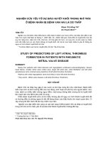
A+B. Lymphocyte proliferation following addition of anti-MHC II or anti-CD4 ab is unchanged in unstimulated CBMC and following stimulation with the innate stimulus PpgFigure 1
A+B. Lymphocyte proliferation following addition of anti-
MHC II or anti-CD4 ab is unchanged in unstimulated CBMC
and following stimulation with the innate stimulus Ppg. Fol-
lowing addition of anti-MHC II or anti-CD4 ab, lymphocyte
proliferation is decreased after stimulation with the allergen
Derf1 (p < 0.05). A+B. Lymphocyte proliferation is shown in
counts per minute (cpm) and was determined after stimula-
tion with the indicated dose of Ppg and Derf1 (30 µg/ml) for
72 h by
3
H-Thymidine uptake as described in Methods (n =
50). Anti-MHC II or anti-CD4 ab was applied in a dose of 10
µg/ml each.
0
1000
2000
3000
4000
5000
6000
7000
UPpg
cpm
No ab
anti-MHCII
anti-CD4
0
100
200
300
400
500
600
700
800
UDer f1
cpm
No ab
anti-MHC II
anti-CD4
*
†
†
*
0
1000
2000
3000
4000
5000
6000
7000
UPpg
cpm
No ab
anti-MHCII
anti-CD4
0
100
200
300
400
500
600
700
800
UDer f1
cpm
No ab
anti-MHC II
anti-CD4
*
†
†
*
*
†
†
*
Respiratory Research 2006, 7:40 />Page 4 of 9
(page number not for citation purposes)
Green Master Mix (Applied Biosystems) per well, the
gene-specific PCR products were measured continuously
by means of GeneAmp 5700 Sequence Detection System
(Applied Biosystems) during 40 cycles. All experiments
were run in duplicate, and the same thermal cycling
parameters were used. Non-template controls and dissoci-
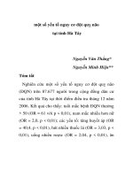
A. Lymphocyte proliferation following stimulation with Ppg and Derf1 was increased in CBMC (p < 0.001)Figure 2
A. Lymphocyte proliferation following stimulation with Ppg and Derf1 was increased in CBMC (p < 0.001). B. IFN-γ secretion
was increased following stimulation with Ppg as compared to unstimulated CBMC (U) (p < 0.001). C. IL-13 secretion was
increased following Ppg stimulation as compared to unstimulated cells (U)(p = 0.001). D. TNF-α production was increased fol-
lowing stimulation with either Ppg or Der f 1 compared to U (p < 0.001). E. IL-10 production was increased following stimula-
tion with Ppg as compared to U (p < 0.001). A-E. Lymphocyte proliferation and cytokine concentrations from supernatants of
CBMC were determined following stimulation with the indicated doses of Ppg and Derf1. Lymphocyte proliferation shown as
SI (stimulation index, ratio of mean counts per minute of stimulated over unstimulated replicates) was measured by
3
H-Thymi-
dine uptake, cytokine concentrations were measured with ELISA (Methods)(n = 50). Data are shown as Box- and whiskers-
plots (Median, whiskers: 5% and 95%-quantile) with outliers.
0
50
100
150
200
250
UPpg
Der f1
*
IL-13 (pg/ml)
0
50
100
150
200
250
UPpg
Der f1
*
0
50
100
150
200
250
UPpg
Der f1
*
IL-13 (pg/ml)
0
500
1000
1500
2000
2500
3000
*
IFN-J(pg/ml)
U Ppg Der f1
0
500
1000
1500
2000
2500
3000
*
IFN-J(pg/ml)
U Ppg Der f1
0
200
400
600
800
1000
UPpgDer f1
*
IL-10 (pg/ml)
*
0
200
400
600
800
1000
UPpgDer f1
*
IL-10 (pg/ml)
*
0
2
4
6
8
10
12
14
*
*
SI
Ppg Der f1
0
2
4
6
8
10
12
14
*
*
SI
Ppg Der f1
0
200
400
600
800
1000
1200
1400
1600
1800
2000
UPpgDer f1
*
*
TNFD (pg/ml)
0
200
400
600
800
1000
1200
1400
1600
1800
2000
UPpgDer f1
0
200
400
600
800
1000
1200
1400
1600
1800
2000
UPpgDer f1
*
*
TNFD (pg/ml)
Respiratory Research 2006, 7:40 />Page 5 of 9
(page number not for citation purposes)
ation curves were used to detect primer-dimer conforma-
tion and non-specific amplification. The threshold cycle
(C
T
) of each target product was determined and set in rela-
tion to the amplification plot of GAPDH. The C
T
is the
number of PCR cycles required for the fluorescence signal
to exceed the detection threshold value. The detection
threshold was set to the log linear range of the amplifica-
tion curve and kept constant (0.3) for all data analysis.
The difference in C
T
values of two genes was used to calcu-
late the fold difference. The level of mRNA of the individ-
ual gene is described as gene expression. The relative
quantitative results were used to determine changes in
gene expression in stimulated as compared to unstimu-
lated samples [24,25].
Statistical analysis
Data analysis was performed with SigmaStat software.
Data for lymphocyte proliferation, cytokine concentra-
tions and gene expression were not normally distributed
and could regularly not be transformed to normality.
Non-detectable cytokine concentrations were assigned to
a value of 0.01 for inclusion into the analysis. Non-para-
metric tests (Kruskal-Wallis, Mann-Whitney) were used to
compare the median of cytokine levels, proliferation val-
ues or gene expression between different groups. Statisti-
cally significant differences for the comparison of several
groups were determined by one-way ANOVA analysis fol-
lowed by a comparison of groups with the Tukey-Kramer
analysis. Data are either reported as mean ± SEM or
median ± CI depending on the distribution and presented
as box- and whiskers- plots (Median, whiskers: 5% and
95%-quantile) with outliers. We used either Pearson's or
Spearman's correlation to assess the association between
cytokine secretion and gene expression. Statistical signifi-
cance was defined by p < 0.05.
Results
Stimulation of CBMC with innate and adaptive stimuli
In CBMC of healthy neonates, we detected constitutive
expression of TLR2. TLR2 expression assessed by real time
RT-PCR was increased 3.46 fold (± 1.5) following stimu-
lation with the TLR2 agonist Ppg as compared to unstim-
ulated cells. TLR2 was also expressed on the cell surface of
mononuclear cells following Ppg stimulation detected by
flow cytometry (not shown).
Allergic (house dust mite Der f1) and innate, non-allergic
stimulation (Ppg) of CBMC led to significantly increased
proliferation following stimulation (Fig. 1A, B, black bar,
no antibody) as compared to unstimulated cells (U).
Allergen-induced lymphoproliferation was shown to be
specific for the allergen Der f1 through blockade of lym-
phoproliferation by either anti-MHCII or anti-CD4 anti-
bodies (Fig. 1A). Also, we present data demonstrating, as
expected, that the innate stimuli Ppg is not inhibited by
addition of anti-MHCII or anti-CD4 antibodies (Fig. 1B).
Regulation of cytokine secretion through innate
stimulation
To analyze the effect of innate stimuli on effector cell
responses in CBMC, we determined lymphoproliferative
responses and Th1 (IFN-γ, IL-12 (p70)) and Th2 (IL-13)
Table 1: Association of maternal atopy* with decreased IL-10 production following innate stimulation (Ppg) in CBMC.
Parameter Stimulus No maternal atopy
(Median, 25/75%)
Maternal atopy
(Median, 25/75%)
P value (Mann-Whitney
rank)
SI Ppg 1.67 (0.99/2.41) 1.45 (1.13/2.75) 0.97
Der f1 1.43 (0.98/2.35) 1.38 (0.93/1.95) 0.93
IFN-γ U 2.49 (1.05/5.83) 2.89 (1.86/4.37) 0.57
Ppg 7.53 (4.70/136.20) 6.07 (2.71/19.19) 0.22
D 3.70 (1.25/22.38) 9.48 (2.46/18.60) 0.73
IL-13 U 0.01 (0.01/3.39) 0.01 (0.01/3.69) 0.58
Ppg 8.04 (0.01/46.04) 6.50 (0.01/61.04) 0.91
D 0.01 (0.01/16.78) 4.10 (0.01/16.96) 0.64
TNF-α U 2.25 (0.01/176.85) 0.27 (0.01/7.60) 0.17
Ppg 1474.00 (1295.50/1645.00) 1529.00 (1069.12/1694.25) 0.87
D 758.30 (205.25/1340.25) 782.50 (168.30/1470.00) 0.96
IL-10 U 1.01 (0.52/2.24) 0.38 (0.01/0.77) 0.007
Ppg 69.54 (25.92/239.78) 13.47 (9.12/86.22) 0.03
D 10.28 (4.15/14.92) 6.51 (4.25/13.80) 0.68
* Maternal atopy was defined as history of doctors diagnosis of one or more of the diagnoses asthma, hay fever or eczema. N = 31 mothers without
and n = 19 mothers with atopy (differs slightly in groups depending on availability of data).
Lymphocyte proliferation is shown as SI (stimulation index, ratio of mean counts per minute of stimulated over unstimulated replicates). Cytokine
concentrations were measured with ELISA and are presented in pg/ml.
Respiratory Research 2006, 7:40 />Page 6 of 9
(page number not for citation purposes)
cytokine production as well as production of the pro-
inflammatory cytokine TNF-α and the immunoregulatory
cytokine IL-10. Lymphoproliferation was increased fol-
lowing Ppg stimulation compared to unstimulated cells
(p < 0.05), and higher as compared to Der f1-induced pro-
liferation (Fig. 2A). IFN-γ secretion was increased follow-
ing stimulation with Ppg as compared to unstimulated
cells (p < 0.001) (Fig. 2B). There was mildly increased IL-
12 (p70) secretion, though at low levels and not signifi-
cant (p = 0.18, data not shown). IL-13 was significantly
elevated following Ppg stimulation as compared to
unstimulated cells (p = 0.001) and also increased, though
not significantly, after Der f1 stimulation (p = 0.18). TNF-
α production was significantly increased following innate
(Ppg) and allergic stimulation (both p < 0.001). IL-10
secretion was significantly increased following stimula-
tion with Ppg as compared to unstimulated cells (p <
0.001), and higher than following Derf1 stimulation.
Influence of maternal atopy on cytokine secretion
We have previously shown that allergen-induced (OVA)
proliferation in CBMC from mothers with a diagnosis of
asthma was increased as compared to mothers without
asthma [26]. Here, we determined whether maternal
atopy has an influence on lymphoproliferation and
cytokine responses to innate and allergic stimulation.
Lymphoproliferative responses and Th1 (IFN-γ, IL-12) as
well as Th2 (IL-13) cytokine responses to innate and aller-
gic stimuli were comparable in CBMC with and without
maternal atopy (Table 1, data not shown). For all
cytokines, median concentrations in unstimulated CBMC
were low. TNF-α secretion as a representative pro-inflam-
matory cytokine was very high following innate stimula-
tion and similar in mothers with and without atopy.
Interestingly, IL-10 secretion was significantly higher fol-
lowing Ppg stimulation in CBMC without maternal atopy
as compared to CBMC with maternal atopy (p = 0.03,
Table 1).
T cell subpopulations
IL-10 is secreted from several cell types including macro-
phages and characteristically produced from a subpopula-
tion of T cells with regulatory capacity (T regs). As T cells
express TLR2 and Ppg stimulates proliferation, we deter-
mined important markers of these T cell subsets by real-
time RT-PCR such as expression of the transcription factor
Foxp3, the glucocorticoid-induced TNF receptor GITR, the
cytotoxic lymphocyte antigen 4 CTLA4 and the cytokine
TGF-β on CBMC. GITR expression was increased follow-
ing stimulation with Ppg as compared to unstimulated
cells, though not significantly (p = 0.07)(Fig. 3). Foxp3
and CTLA4 were both constitutively expressed at low lev-
els (data not shown), and increased following stimulation
with Ppg, however not significantly (Fig. 3). TGF-β was
expressed at low levels at baseline and decreased after
stimulation with Ppg as compared to unstimulated cells
(p = 0.03)(data not shown). Stimulation with the allergen
Der f1 resulted in non-significant changes in expression of
Foxp3, CTLA4 and GITR (not shown). To investigate this
population of T cells further, we assessed the percentage of
CD4
+
CD25
+
cells, one predominant phenotype of T regu-
latory cells following stimulation with Ppg. We found a
mild, non-significant increase in the percentage of
CD4
+
CD25
+
cells after stimulation with Ppg (data not
shown).
Effect of maternal atopy on markers of T cell
subpopulations
To determine the importance of maternal atopy on
parameters of subsets of T cells in addition to IL-10, we
assessed the expression of Foxp3, GITR and CTLA4
depending on maternal atopy (Table 2). Following stimu-
lation with Ppg, differences in T cell markers in CBMC
from children of mothers without as compared to those
with maternal atopy became apparent. Foxp3 and CTLA4
were both increased in CBMC of children of mothers
without as compared to those with maternal atopy; the
differences were marginally significant for Foxp3 (p =
0.049)(p = 0.17 for CTLA4). As IL-10 and Foxp3 were sig-
nificantly higher in CBMC from children of mothers with-
out atopy, we further assessed the correlation between IL-
10 and Foxp3. Foxp3 was positively correlated with IL-10
secretion in CBMC following stimulation with Ppg (r =
0.53, p = 0.001, Table 3). These positive correlations were
seen in CBMC from both children of mothers without and
with maternal atopy (r = 0.52, p = 0.01 and r = 0.56, p =
Table 2: Association of maternal atopy* with decreased Foxp3 expression following Ppg stimulation in CBMC.
Parameter No maternal atopy (Median,
25/75)
Maternal atopy (Median, 25/
75)
P value (Mann-Whitney rank)
Foxp3 1.59 (-0.4/3.26) -0.6 (-0.78/1.06) 0.049
GITR 1.22 (-0.17/4.28) 2.74 (-0.40/7.21) 0.43
CTLA4 1.19 (-0.22/2.02) - 0.13 (-0.77/0.79) 0.17
* Maternal atopy was defined as history of doctors diagnosis of one or more of the diagnoses asthma, hay fever or eczema. N = 31 mothers without
and n = 19 mothers with atopy (differs slightly in groups depending on availability of data).
The mRNA level of the genes is shown as fold difference in gene expression in stimulated as compared to unstimulated samples and compared to
the housekeeping gene GAPDH. Quantitative gene expression was assessed with real-time RT-PCR.
Respiratory Research 2006, 7:40 />Page 7 of 9
(page number not for citation purposes)
0.06, data not shown). Also, positive correlations were
demonstrated for increased Ppg-induced IL-10 secretion
with GITR (r = 0.47, p = 0.004) and CTLA4 (r = 0.49, p =
0.003), independent of maternal atopy.
Discussion
This study demonstrates that microbial stimulation with
the TLR2 agonist peptidoglycan in vitro modulates func-
tional immune capacities of cord blood mononuclear
cells (CBMC) from children of mothers with as compared
to without a doctors diagnosis of maternal atopy. In
CBMC from children of mothers without a doctors diag-
nosis of atopy, an increase of Ppg-induced IL-10 secretion
was paralleled by an increase of two markers of T regula-
tory cells (significantly for Foxp3 and mildly for CTLA4).
In addition, Ppg stimulation was associated with a posi-
tive correlation between IL-10 and genes associated with T
regulatory cells (Foxp3, GITR and CTLA4), suggesting
innate modulation of T regulatory cells in CBMC. These
data support the hypothesis that microbial stimulation of
CBMC leads to immune modulation in association with
the maternal atopic background.
Of note, the phenotype of T regulatory cells is not clearly
defined to date. We acknowledge the limitation of a
mixed CBMC population in this study. While this study
did not address cell type, prior studies indicate that TLRs
are present not just on monocytes and B cells but also on
T cells, underscoring a putative link between innate and
adaptive immunity [32]. The induction of both IL-10 and
IFN-γ following stimulation with Ppg in this study could
indicate a role of a specific population of T cells in human
CBMC. For example, it has been proposed that IL-10 and
IFN-γ producing CD4
+
T cells may be one of the human
equivalents of the CD4
+
CD25
+
T regulatory cells origi-
nally described in the mouse [33]. In addition, in this
study not only IL-10 but also GITR, another marker char-
acteristic for T regulatory cells, was increased following
Ppg stimulation. We present an increase of TLR2-stimu-
lated IL-10 as well as a correlation between IL-10 and
other markers of T regulatory cells. These data may indi-
cate that microbial stimulation such as Ppg can impact T
cells in the fetal immune system, potentially capable of
regulating several immune processes including cytokine
secretion. This is intriguing in the context that Ppg stimu-
lation in our murine model of asthma could decrease
allergic stimulation [17].
IL-10 secretion may be crucial in modulating the develop-
ment of the fetal immune system, and in contributing to
Th2 maturation via inhibition of IL-12 production [27].
On the other hand, regarding allergic diseases, IL-10 was
demonstrated in several studies to be associated with
lower risk for atopy or sensitization to egg protein in later
life [28,29]. The Ppg-induced increase of IL-10 in our
study could indicate a role for innate stimuli in early
immunomodulation. Furthermore, IL-10 was induced in
chronic schistosomiasis in African children, who have a
low prevalence of atopic disease [30]. Additionally, suc-
cessful allergen-desensitization therapy has been postu-
lated to work through the induction of IL-10 secreting T
regulatory cells. In support of this concept, IL-10 secreting
T regulatory cells were shown to be induced by glucocor-
ticoids and β 2-agonists, the hallmark of anti-allergic ther-
apy [31].
Furthermore, the forkhead-winged-helix family transcrip-
tion factor Foxp3 may control genes encoding T regulatory
cell-associated molecules (such as CD25, CTLA4 and
GITR). Mutations in Foxp3 lead to the X-linked immuno-
deficiency syndrome IPEX in humans (immune dysregu-
lation, polyendocrinopathy, enteropathy, X-linked
syndrome). Clinical features are autoimmune disease,
inflammatory bowel disease, severe allergy including
atopic dermatitis, food allergy, and fatal infection [34].
Foxp3 is stably expressed in mature natural T regulatory
cells; the role of Foxp3 in the development of the neonatal
immune system remains to be determined. It is intriguing
that both IL-10 and Foxp3 levels are decreased in cord
blood of neonates of mothers with atopy in our study.
Maternal atopy is known to be an important influential
factor in a child's allergic predisposition [35]. In this
study, maternal atopy is defined as doctors diagnosis of
asthma, hay fever and/or eczema. Unfortunately, data on
maternal sensitization were not available, which we
acknowledge as a potential limitation of the study. From
the literature, the prevalence of a positive skin prick test to
at least one allergen is reported in up to 60% in the 20–29
year old age range in the American NHANES population
Table 3: Correlation between IL-10 production and specific markers of T regulatory cells in the whole population (n = 50, differs
slightly in groups depending on availability of data).
Correlation Coefficient r p †
TGF-β 0.02 0.95
Foxp3 0.53 0.001
GITR 0.47 0.004
CTLA4 0.49 0.003
† Spearman rank test
Respiratory Research 2006, 7:40 />Page 8 of 9
(page number not for citation purposes)
not stratified as high or low risk for atopy [36], which
most closely represents the population in our study. The
percentage of sensitization can therefore be much higher
without having ever any atopic symptoms. Also, some
studies suggest that a history of atopic symptoms may be
more indicative of allergic disease than skin test positivity
to allergens.
Our analysis was performed in a group of 50 mothers
including 19 with maternal atopy as defined by the doc-
tor's diagnoses asthma and/or hay fever and/or atopic
eczema. Further separate analysis in the subgroups were
not statistically feasible. In addition, the diagnosis of
maternal atopy comprises a common immunological
basis for all three diseases. Regardless, the specific immu-
nological mechanisms by which maternal atopy may
influence the development of atopy in the child remain
undefined. Thus, differences in T cell regulation, possibly
T regulatory cells, depending on the maternal atopic back-
ground, may be biologically important. The study of
Amoudruz et al. in CBMC of 9 mothers with and 10 with-
out allergy is consistent with this concept [14]. In this
study, cytokine secretion of IL-6 is lower after Ppg stimu-
lation in CBMC of mothers with as compared to mothers
without allergy. Importantly, Pasare et al have shown that
the suppressive effects of CD25
+
regulatory cells can be
blocked by the presence of IL-6, produced by DC and acti-
vated through stimulation of the TLR pathways [37]. Our
study suggests that in maternal atopy, T regulatory cells
may be potentially less effective as demonstrated by
reduced secretion of IL-10 and by diminished expression
of Foxp3.
Conclusion
In conclusion, our study provides evidence that exposure
to microbial stimuli may induce the neonatal immune
system to increase IL-10 secretion. Gene expression
related to regulatory T cell subpopulations appears to be
influenced by innate stimuli, which may potentially result
in an altered phenotype or function of T cell subpopula-
tions. Our findings that IL-10 and Foxp3 expression were
reduced in mothers with atopy raise the possibility that
CBMC from their neonates may have a diminished capac-
ity to respond to microbial stimuli. Whether these pat-
terns in the context of additional genetic and
environmental factors are associated with an increased
risk of atopy in the child remains to be investigated.
Abbreviations
CBMC, cord blood mononuclear cells; LpA, Lipid A; Ppg,
Peptidoglycan; TLR, Toll-like receptor.
Competing interests
The author(s) declare that they have no competing inter-
ests.
Authors' contributions
BS designed the experiments, carried them out, analyzed
the results and drafted the manuscript. MC and HH car-
ried out part of the experiments. DP participated in study
design and data analysis. MWG, DG, SW and EL contrib-
uted to study design, and draft of the manuscript. PWF
participated in study design, experimental design, analysis
and draft of the manuscript. All authors read and
approved the final manuscript.
Acknowledgements
The authors thank Sheryl Rifas for thoughtful data review. This work was
supported by: DFG 997/1-1 (BS), NIH grants HL 56723, HL 67684, IA
45007, AI045007 (all PWF), HL 64925, HL 68041, HD34568 (all MWG), AI/
EHS 35786 (DG).
References
1. Upham JW, Holt PG, Taylor A, Thornton CA, Prescott SL: HLA-DR
expression on neonatal monocytes is associated with aller-
gen-specific immune responses. Journal of Allergy and Clinical
Immunology 2004, 114:1202-8.
2. Upham JW, Lee PT, Holt BJ, Heaton T, Prescott SL, Sharp MJ, et al.:
Development of Interleukin-12-Producing Capacity
throughout Childhood. Infect Immun 2002, 70:6583-8.
3. Prescott SL, Macaubas C, Smallacombe T, Holt BJ, Sly PD, Holt PG:
Development of allergen-specific T-cell memory in atopic
and normal children. Lancet 1999, 353:196-200.
4. Prescott SL, Macaubas C, Holt BJ, Smallacombe TB, Loh R, Sly PD, et
al.: Transplacental Priming of the Human Immune System to
Environmental Allergens: Universal Skewing of Initial T Cell
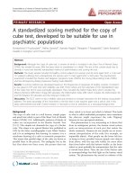
Gene expression of GITR following stimulation with Ppg was increased as compared to unstimulated cells (p = 0.07)Figure 3
Gene expression of GITR following stimulation with Ppg was
increased as compared to unstimulated cells (p = 0.07). The
mRNA level of the individual gene is shown as fold difference
in gene expression in Ppg (10 µg/ml) stimulated as compared
to unstimulated samples and compared to the housekeeping
gene GAPDH. RNA was prepared as described in Methods
(n = 50). Quantitative gene expression was assessed with
real-time RT-PCR. Data are shown as Box- and whiskers-
plots (Median, whiskers: 5% and 95%-quantile) with outliers.
-10
0
10
20
30
40
50
60
Foxp3 GITR CTLA4
Fold difference
-10
0
10
20
30
40
50
60
Foxp3 GITR CTLA4
Fold difference
Publish with BioMed Central and every
scientist can read your work free of charge
"BioMed Central will be the most significant development for
disseminating the results of biomedical research in our lifetime."
Sir Paul Nurse, Cancer Research UK
Your research papers will be:
available free of charge to the entire biomedical community
peer reviewed and published immediately upon acceptance
cited in PubMed and archived on PubMed Central
yours — you keep the copyright
Submit your manuscript here:
/>BioMedcentral
Respiratory Research 2006, 7:40 />Page 9 of 9
(page number not for citation purposes)
Responses Toward the Th2 Cytokine Profile. J Immunol 1998,
160:4730-7.
5. Warner JA, Jones AC, Miles EA, Warner JO: Prenatal sensitisa-
tion. Pediatr Allergy Immunol 1996, 7:98-101.
6. Tang ML, Kemp AS, Thorburn J, Hill DJ: Reduced interferon-
gamma secretion in neonates and subsequent atopy. Lancet
1994, 344:983-5.
7. Kondo N, Kobayashi Y, Shinoda S, Takenaka R, Teramoto T, Kaneko
H, et al.: Reduced interferon gamma production by antigen-
stimulated cord blood mononuclear cells is a risk factor of
allergic disorders – 6-year follow-up study. Clin Exp Allergy 1998,
28:1340-4.
8. Warner JA, Miles EA, Jones AC, Quint DJ, Colwell BM, Warner JO:
Is deficiency of interferon gamma production by allergen
triggered cord blood cells a predictor of atopic eczema? Clin
Exp Allergy 1994, 24:423-30.
9. Yoshimura A, Lien E, Ingalls RR, Tuomanen E, Dziarski R, Golenbock
D: Cutting edge: recognition of Gram-positive bacterial cell
wall components by the innate immune system occurs via
Toll-like receptor 2. J Immunol 1999, 163:1-5.
10. Takeuchi O, Hoshino K, Kawai T, Sanjo H, Takada H, Ogawa T, et al.:
Differential roles of TLR2 and TLR4 in recognition of gram-
negative and gram-positive bacterial cell wall components.
Immunity 1999, 11:443-51.
11. Schnare M, Barton GM, Holt AC, Takeda K, Akira S, Medzhitov R:
Toll-like receptors control activation of adaptive immune
responses. Nat Immunol 2001, 2:947-50.
12. Sabroe I, Jones EC, Usher LR, Whyte MK, Dower SK: Toll-like
receptor (TLR)2 and TLR4 in human peripheral blood gran-
ulocytes: a critical role for monocytes in leukocyte lipopoly-
saccharide responses. J Immunol 2002, 168:4701-10.
13. McCurdy JD, Olynych TJ, Maher LH, Marshall JS: Cutting edge: dis-
tinct Toll-like receptor 2 activators selectively induce differ-
ent classes of mediator production from human mast cells. J
Immunol 2003, 170:1625-9.
14. Amoudruz P, Holmlund U, Malmstrom V, Trollmo C, Bremme K,
Scheynius A, et al.: Neonatal immune responses to microbial
stimuli: Is there an influence of maternal allergy? J Allergy Clin
Immunol 2005, 115:1304-10.
15. Braun-Fahrlander C, Riedler J, Herz U, Eder W, Waser M, Grize L, et
al.: Environmental exposure to endotoxin and its relation to
asthma in school-age children. N Engl J Med 2002, 347:869-77.
16. Eisenbarth SC, Piggott DA, Huleatt JW, Visintin I, Herrick CA, Bot-
tomly K: Lipopolysaccharide-enhanced, toll-like receptor 4-
dependent T helper cell type 2 responses to inhaled antigen.
J Exp Med 2002, 196:1645-51.
17. Velasco G, Campo M, Manrique OJ, Bellou A, He H, Arestides RSS, et
al.: Toll-like Receptor 4 or 2 Agonists Decrease Allergic
Inflammation. Am J Respir Cell Mol Biol 2004, 32:218-224.
18. Lauener RP, Birchler T, Adamski J, Braun-Fahrlander C, Bufe A, Herz
U, et al.: Expression of CD14 and Toll-like receptor 2 in farm-
ers' and non-farmers' children. Lancet 2002, 360:465-6.
19. Eder W, Klimecki W, Yu L, von Mutius E, Riedler J, Braun-Fahrlander
C, et al.: Toll-like receptor 2 as a major gene for asthma in
children of European farmers. J Allergy Clin Immunol 2004,
113:482-8.
20. Schaub B, Bellou A, Gibbons FK, Velasco G, Campo M, He H, et al.:
TLR2 and TLR4 stimulation differentially induce cytokine
secretion in human neonatal, adult and murine mononuclear
cells. Journal of Interferon and Cytokine Research 2004, 24:543-52.
21. Gillman MW, Rich-Edwards J, Rifas-Shiman SL, Lieberman ES, Klein-
man KP, Lipshultz SE: Maternal age and other predictors of
newborn blood pressure. Journal of Pediatrics 2004, 144:240-5.
22. Schaub B, Tantisira KG, Gibbons FK, He H, Litonjua AA, Gillman MW,
et al.: Fetal Cord Blood: Aspects of Heightened Immune
Responses. J Clin Immunol 2005, 25:329-37.
23. Schroeter C, Schaub B, Gold DR, Contreras P, Manrique O, Gillman
MW, et al.: Nuklear factor kappa B activation in human cord
blood mononuclear cells. Pediatric Research 2004, 56:1-7.
24. Heid CA, Stevens J, Livak KJ, Williams PM: Real time quantitative
PCR. Genome Res 1996, 6:986-94.
25. Gibson UE, Heid CA, Williams PM: A novel method for real time
quantitative RT-PCR. Genome Res 1996, 6:995-1001.
26. Willwerth BM, Schaub B, Gold DR, Tantisira KG, Palmer LJ, AA L, et
al.: Prenatal, Perinatal and Heritable Influences on Cord
Blood Immune Responses. Annals of Allergy, Asthma and Immunol-
ogy 2005 in press.
27. Trinchieri G: Interleukin-12: a proinflammatory cytokine with
immunoregulatory functions that bridge innate resistance
and antigen-specific adaptive immunity. Annu Rev Immunol
1995, 13:251-76.
28. Tiemessen MM, Van Ieperen-Van Dijk AG, Bruijnzeel-Koomen CA,
Garssen J, Knol EF, Van Hoffen E: Cow's milk-specific T-cell reac-
tivity of children with and without persistent cow's milk
allergy: key role for IL-10. J Allergy Clin Immunol 2004, 113:932-9.
29. Neaville WA, Tisler C, Bhattacharya A, Anklam K, Gilbertson-White
S, Hamilton R, et al.: Developmental cytokine response profiles
and the clinical and immunologic expression of atopy during
the first year of life. J Allergy Clin Immunol 2003, 112:740-6.
30. van den Biggelaar AH, van Ree R, Rodrigues LC, Lell B, Deelder AM,
Kremsner PG, et al.: Decreased atopy in children infected with
Schistosoma haematobium: a role for parasite-induced
interleukin-10. Lancet 2000, 356:1723-7.
31. Peek EJ, Richards DF, Faith A, Lavender P, Lee TH, Corrigan CJ, et al.:
Interleukin 10 Secreting 'Regulatory' T Cells Induced by Glu-
cocorticoids and Beta2-Agonists. Am J Respir Cell Mol Biol 2005,
33:105-11.
32. Caramalho I, Lopes-Carvalho T, Ostler D, Zelenay S, Haury M,
Demengeot J: Regulatory T Cells Selectively Express Toll-like
Receptors and Are Activated by Lipopolysaccharide. J Exp
Med 2003, 197:403-11.
33. Gerosa F, Nisii C, Righetti S, Micciolo R, Marchesini M, Cazzadori A,
et al.: CD4+ T Cell Clones Producing both Interferon-
[gamma] and Interleukin-10 Predominate in Bronchoalveo-
lar Lavages of Active Pulmonary Tuberculosis Patients. Clin-
ical Immunology 1999, 92:224-34.
34. Gambineri E, Torgerson TR, Ochs HD: Immune dysregulation,
polyendocrinopathy, enteropathy, and X-linked inheritance
(IPEX), a syndrome of systemic autoimmunity caused by
mutations of FOXP3, a critical regulator of T-cell homeosta-
sis. Curr Opin Rheumatol 2003, 15:430-5.
35. Litonjua AA, Carey VJ, Burge HA, Weiss ST, Gold DR: Parental his-
tory and the risk for childhood asthma. Does mother confer
more risk than father? Am J Respir Crit Care Med 1998, 158:176-81.
36. Matricardi PM, Rosmini F, Panetta V, Ferrigno L, Bonini S: Hay fever
and asthma in relation to markers of infection in the United
States. J Allergy Clin Immunol 2002, 110:381-7.
37. Pasare C, Medzhitov R: Toll pathway-dependent blockade of
CD4+CD25+ T cell-mediated suppression by dendritic cells.
Science 2003, 299:1033-6.