Báo cáo y học: " Increased proinflammatory responses from asthmatic human airway smooth muscle cells in response to rhinovirus infection" pptx
Bạn đang xem bản rút gọn của tài liệu. Xem và tải ngay bản đầy đủ của tài liệu tại đây (1010.31 KB, 11 trang )
BioMed Central
Page 1 of 11
(page number not for citation purposes)
Respiratory Research
Open Access
Research
Increased proinflammatory responses from asthmatic human
airway smooth muscle cells in response to rhinovirus infection
Brian GG Oliver*
1
, Sebastian L Johnston
2
, Melissa Baraket
1
,
Janette K Burgess
1,3
, Nicholas JC King
4
, Michael Roth
3,5
, Sam Lim
6
and
Judith L Black
1,3
Address:
1
Department of Pharmacology, University of Sydney, NSW, 2006, Australia,
2
Department of Respiratory Medicine, National Heart and
Lung Institute, Imperial College London, UK,
3
Woolcock Institute for Medical Research, NSW 2006, Australia,
4
Department of Pathology,
University of Sydney, NSW, 2006, Australia,
5
Pulmonary Cell Research, Dept. Research, University Hospital Basel, CH-4031 Basel, Switzerland and
6
ANZAC Research Institute, University of Sydney, Sydney, Australia
Email: Brian GG Oliver* - ; Sebastian L Johnston - ;
Melissa Baraket - ; Janette K Burgess - ; Nicholas JC King - ;
Michael Roth - ; Sam Lim - ; Judith L Black -
* Corresponding author
Abstract
Background: Exacerbations of asthma are associated with viral respiratory tract infections, of
which rhinoviruses (RV) are the predominant virus type. Airway smooth muscle is important in
asthma pathogenesis, however little is known about the potential interaction of RV and human
airway smooth muscle cells (HASM). We hypothesised that rhinovirus induction of inflammatory
cytokine release from airway smooth muscle is augmented and differentially regulated in asthmatic
compared to normal HASM cells.
Methods: HASM cells, isolated from either asthmatic or non-asthmatic subjects, were infected
with rhinovirus. Cytokine production was assayed by ELISA, ICAM-1 cell surface expression was
assessed by FACS, and the transcription regulation of IL-6 was measured by luciferase activity.
Results: RV-induced IL-6 release was significantly greater in HASM cells derived from asthmatic
subjects compared to non-asthmatic subjects. This response was RV specific, as 5% serum- induced
IL-6 release was not different in the two cell types. Whilst serum stimulated IL-8 production in cells
from both subject groups, RV induced IL-8 production in only asthmatic derived HASM cells. The
transcriptional induction of IL-6 was differentially regulated via C/EBP in the asthmatic and NF-κB
+ AP-1 in the non-asthmatic HASM cells.
Conclusion: This study demonstrates augmentation and differential transcriptional regulation of
RV specific innate immune response in HASM cells derived from asthmatic and non-asthmatics, and
may give valuable insight into the mechanisms of RV-induced asthma exacerbations.
Background
Asthma exacerbation is the major contributor to morbid-
ity, mortality and health care costs associated with this
highly prevalent disease. Approximately 80% of asthma
Published: 03 May 2006
Respiratory Research 2006, 7:71 doi:10.1186/1465-9921-7-71
Received: 31 January 2006
Accepted: 03 May 2006
This article is available from: />© 2006 Oliver et al; licensee BioMed Central Ltd.
This is an Open Access article distributed under the terms of the Creative Commons Attribution License ( />),
which permits unrestricted use, distribution, and reproduction in any medium, provided the original work is properly cited.
Respiratory Research 2006, 7:71 />Page 2 of 11
(page number not for citation purposes)
exacerbations in children [1-4] and about 70% in adults
[5,6] are associated with respiratory viral infections. Rhi-
novirus (RV) is by far the most common virus type associ-
ated with asthma exacerbations [3,7,8]. RV can infect the
lower respiratory tract, as demonstrated by Papadopoulos
and co-workers (2000), who used in-situ hybridization to
detect RV infection of both bronchial epithelial and
underlying submucosal cells in biopsies obtained from
the lower airways [9]. Although the authors did not iden-
tify the infected submucosal cells it is likely that they
would have been mesenchymal in origin, eg fibroblasts
and/or smooth muscle cells, as identified by the histolog-
ical representation of positive signal for infection demon-
strated in this paper.
Normally the bronchial epithelium forms a barrier
between the airway lumen and the underlying cells. How-
ever, epithelial cells from asthmatic subjects have
impaired RV-induced apoptosis and increased RV replica-
tion and cell necrosis in comparison to cells derived from
non-asthmatic subjects [10]. Furthermore, in the asth-
matic airway there may be desquamation of the epithelial
cell layer [11], and increased smooth muscle mass [12].
These asthma specific structural changes, in combination
with RV-induced necrosis of bronchial epithelial cells,
increase the likelihood of RV infecting the underlying
smooth muscle during a naturally acquired RV infection
in asthmatic subjects.
Human airway smooth muscle (HASM) cells are actively
involved in maintaining the local immune environment,
through the production of a wide variety of immunomod-
ulatory factors [13], and modulation of their cell surface
receptors [14-16]. The host-mediated immune response
to RV is important in viral clearance from the lower respi-
ratory tract. Following RV infection, lower airway neu-
trophilia occurs [17], which is likely to be as a result of RV-
induced chemokine release. IL-8 is a potent chemotactic
agent for neutrophils [18], in addition to activating sev-
eral cell types found in the lungs. IL-6 is a complex and
pleiotropic cytokine which has many functions and may
contribute to the progression of asthma, since it polarizes
T-helper cells towards a T-helper 2 phenotype [19]. Fur-
thermore, IL-6 induces differentiation of T cells, B cells
and macrophages, in addition to contributing to the
recruitment of mononuclear cells and neutrophils
[20,21]. Previous experiments, mainly carried out using
rabbit airway smooth muscle cells and two HASM cell
lines derived from non-asthmatic healthy lung donors,
have shown that RV infection induces the production of
interleukin (IL)-1β and IL-5 [22,23].
Since it has been previously suggested that RV infection of
HASM cells mediates cytokine production [22], and since
major differences in innate responses of bronchial epithe-
lial cells from asthmatic and normal subjects to RV infec-
tion have been recently demonstrated [10], we
hypothesised that RV induction of inflammatory cytokine
release is augmented in primary HASM cells from asth-
matic compared with normal subjects. Having found this
to be the case, and with the knowledge that a transcription
factor which can bind to the IL-6 promoter region, C/EBP-
α, is absent from asthmatic but not normal HASM cells
[24] we then investigated transcriptional regulation of IL-
6 to determine whether its expression is differentially reg-
ulated in asthmatic compared to normal HASM cells.
Methods
Patient demographics
HASM cells were isolated from airway muscle bundles
obtained from 22 asthmatic subjects [mean age 23 years,
range 18–33]), and 29 non-asthmatic subjects [mean age
55 years, range 16–74] of which 17 were undergoing lung
resection for a tumour, 7 were undergoing transplantation
(cystic fibrosis [n = 1], pulmonary fibrosis [n = 2], emphy-
sema [n = 2], Eisenmenger's syndrome [n = 1], and Tetral-
ogy of Fallot [n = 1]) and 5 were healthy. Four of the non-
asthmatic subjects were females, and 23 were male, the
age and sex of two of the non-asthmatic subjects were not
available. Four of the asthmatic subjects were females, and
18 were males.
RV propagation and titration
Stocks of human RV-16 were amplified by growth in Ohio
HeLa cells as previously described [25]. In some experi-
ments RV was UV-inactivated in 24 well plates containing
200 μl of viral stock per well, at a distance of 5 cm from a
30 W UV light source (germicidal lamp G30T8, Sankyo
Denki, Japan), for 15 minutes. UV inactivation (UVi) of
RV was shown to be effective by RV titration assay. HASM
were infected with RV at a multiplicity of infection (MOI)
of 4, 0.4 and 0.04. Following exposure to RV, virus release
was assessed by titration assay [25]. Breifely, Rhinovirus
levels were estimated by titrating serially log diluted con-
centrations of the cell free supernatant in quadruplicate
upon Ohio HeLa cells. Ohio HeLa cells were seeded at a
concentration of 1 × 10
5
cells/ml in 96 well plates. To this
50 μL of viral suspension or control medium were added.
The plate was shaken for ten minutes at room tempera-
ture, and cultured for 4–6 days. After 3 days of culture,
cells were assessed for cytopathic effect (CPE). The cells
were then assessed for CPE every 24 hours, to ensure that
cell death of control wells was not occurring and therefore
biasing viral induced CPE. Viral concentration was deter-
mined as the lowest viral concentration which caused
cytopathic effect in 50% of the wells (tissue culture infec-
tive dose 50 [TCID
50
]).
Respiratory Research 2006, 7:71 />Page 3 of 11
(page number not for citation purposes)
Isolation and culture of human airway smooth muscle cells
HASM cells were isolated from bronchial tissue obtained
from either bronchoscopy, lung tissue obtained from lung
transplants, or lung tissue resected at thoracotomy, by
microdissection. Ethical approval for the use of the lung
tissue for in vitro experimentation was granted by the
Human Ethics Committee of the University of Sydney,
and the Central Sydney Area Health Service, and informed
consent was received from all subjects. This isolation and
culture of HASM cells was carried out according to a
method described by Johnson et al 1995 [26]. HASM cells
were used between passage 4–7. HASM cells were identi-
fied by morphology, and positive immunofluorescent
staining with a specific α-smooth muscle actin antibody
[26] and a calponin antibody [27]. HASM cells were
seeded in 12 well plates at a density of 3.2 × 10
4
cells / ml
in 5% FBS in DMEM (without the addition of antibiotics).
For experiments using subconfluent cells, experimenta-
tion was carried out using HASM cells 24 hours post seed-
ing, and confluent cells were obtained following 7 days of
growth.
RV infection
The medium bathing the HASM cells was removed and
replaced with medium containing RV. The cells were incu-
bated at 37°C with shaking every fifteen minutes, for one
hour. The medium was removed and the cells were
washed 3 times in sterile PBS, and 1 ml of DMEM (either
0.1% FBS or 5% FBS) added. Cells exposed to UV irradi-
ated RV and cells with no virus exposure underwent the
same infection procedure (with the absence of RV) as
infected cells. Medium was harvested at 24 hours post
infection, following centrifugation to remove non-adher-
ent HASM cells, and stored at -80°C, for analysis by ELISA
and RV titration assays. HASM cell viability was deter-
mined using manual cell counting and trypan blue exclu-
sion assays.
ELISA
ELISAs for eotaxin, tumour necrosis factor (TNF)-α, IL-6,
IL-8, and interferon (IFN)-γ were purchased from R&D
Systems Europe, Abingdon, UK. ELISAs were carried out
according to the manufacturer's instructions. The detec-
tion limits of these assays were: 15.6 pg/ml for all except
eotaxin (25 pg/ml).
Flow cytometry
Flow cytometric analysis was performed to assess the cell
surface expression of ICAM-1 (BD, North Ryde, Australia)
in comparison to an isotype control antibody (BD).
IL-6 promoter constructs – amplification and isolation
A plasmid containing a 651 bp fragment of the human IL-
6 gene was kindly provided by Shigeru Katamine (Naga-
saki University, Nagasaki, Japan). The IL-6 promoter con-
structs were designed as previously reported by Eickelberg
et al (1999) [28]. Briefly, site-directed mutagenesis was
used to inactivate various transcription factor binding
sites. The AP-1 consensus sequence (positions 283 to 276,
5'-TGAGTCAC-3') was changed to 5'-TGCAGCAC-3'; the
C/EBP consensus sequence (positions 154 to 146, 5'-
TTGCACAAT-3') was changed to 5'-CCGTTCAAT-3'; and
the NF-κB consensus sequence (positions 72 to 63, 5'-
GGGATTTTCC-3') was changed to 5'-CTCATTTTCC-3'.
Transient transfection of HASM cells
A commercially available kit (Effectene
®
transfection rea-
gent, Qiagen, Australia) was used. The use of a second
plasmid to control for transfection efficiency is used by
some researchers, however preliminary experiments indi-
cated that transfection of HASM cells with the control
Renilla luciferase reporter vector (Promega, Australia) in
addition to the IL-6 promoter construct plasmids resulted
in interference between the two plasmids, and therefore
this practice was not continued. This effect has been dem-
onstrated in other reports [29,30], and it is recommended
that two plasmids should not be used in some systems.
The manufacturer's transfection protocol was followed,
with a ratio of DNA (0.5 μg/well) to Effectene
®
transfec-
tion reagent of 1:4. Following incubation with the trans-
fection mixture for 24 hours (in 5%FBS + DMEM), cells
were washed twice with PBS, and 1 ml of 0.1% FBS in
DMEM was added to each well, and then exposed to UVi
RV (initial MOI of 4), or platelet derived growth factor
(PDGF 10 μg/well), or infected with RV at MOI of 0.04.
Control cells were cultured in the presence of 0.1% FBS in
DMEM alone. Stimulation or infection was maintained
for 24 hours, at which time cells and supernatant were
harvested for determination of luciferase activity.
Measurement of luciferase activity
Luciferase activity was determined using a commercially
available kit (Dual-Luciferase
®
reporter assay system,
Promega, Australia) according to the manufacturer's
instructions, using a Turner Biosystems luminometer,
(Promega, Australia)
Statistical methods and analysis of results
Data are presented as mean ± SEM. Data were subjected to
two tailed Student's paired T test or repeated measures
ANOVA with Dunnett's multiple comparison post test.
Data derived from different donor populations were sub-
jected to unpaired two-tailed Student's t-test. Data were
analysed using GraphPad Prism version 4.00 for Windows
(GraphPad Software, San Diego, California, USA). A
probability level of 95% (p ≤ 0.05) was considered to be
the threshold for statistical significance.
Respiratory Research 2006, 7:71 />Page 4 of 11
(page number not for citation purposes)
Results
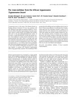
Infection of HASM cells with RV induces cell death
Firstly, we wished to confirm that RV is able to produc-
tively infect HASM cells. We compared the RV-induced
rate of cell death in asthmatic and non-asthmatic HASM
cells. Infection with RV-16 (MOI of 4) induced significant
cell death in HASM cells derived from both asthmatic and
non-asthmatic subjects, the viability of asthmatic cells was
reduced to 45 ± 13% and non-asthmatic cells to 52 ± 11%
by 24 hours post infection (p < 0.05 RV versus control, n
= 3 for both). No difference in the RV-induced cell death
between the cells derived from the two subject groups was
observed (Figure 1A). There was no significant cell death
in comparison to non-infected control cells with infection
at lower virus concentrations (0.4 and 0.04 MOI). In con-
trast, no cell death was observed following exposure to
UVi RV-16. (Figure 1B).
Infection was verified by titration assay of the tissue cul-
ture medium, with maximum RV-16 release occurring at
24 hours, decreasing at both 48 and 72 hours. When cor-
rected for cell number, greater RV-16 production occurred
in proliferating HASM cells compared to confluent cells.
However, no difference in RV production was observed,
with initial infection at a MOI of 4, between cells derived
from asthmatics and non-asthmatics in both proliferating
cells (asthmatic 46350 ± 15070 TCID
50
/ml and non-asth-
matics 44220 ± 8820 TCID
50
/ml, p > 0.05 n = 6) and con-
fluent cells (asthmatic 30010 ± 9187 TCID
50
/ml and non-
asthmatics 37860 ± 19200 TCID
50
/ml, p > 0.05 n = 5).
Infection was also verified using RT-PCR (data not
shown).
Distinct pro-inflammatory cytokine response to RV-16
infection in HASM cells derived from asthmatic and non-
asthmatic subjects
To investigate whether RV infection of HASM cells
induced augmented pro-inflammatory cytokine responses
in asthmatic compared with non-asthmatic subjects, we
next investigated IL-6, IL-8, eotaxin, TNF-α and IFN-γ
secretion into the tissue culture medium in response to RV
infection.
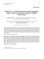
As shown in Figure 2A, infection with RV-16 (MOI of 4)
significantly increased IL-6 secretion in both non-asth-
matic (p < 0.001, n = 11) and asthmatic (p < 0.01, n = 8)
-derived HASM cells in comparison to non-infected con-
trol cells 24 hours post infection. Furthermore, the RV
induced release of IL-6 was 2.5 fold and significantly
greater from cells obtained from asthmatic than non-asth-
matic patients (asthmatic 1177 ± 281.5 n = 8 and non-
asthmatic 501.0 ± 78.7 n = 11 p = 0.017). 5% FBS stimu-
lated secretion of IL-6 in both asthmatic and non-asth-
matic cells, and in contrast to RV-induced IL-6, no
differences were found between the two subject groups
(Figure 2B). Infection with RV-16 (MOI 4) and concurrent
stimulation with 5% FBS significantly induced the release
of IL-6 in comparison to 5% FBS alone in HASM cells
derived from only non-asthmatic donors, whilst a non-
significant trend towards increased production occurred
in the asthmatic-derived cells (Figure 2B).
As shown in Figure 2C, in non-asthmatic HASM cells RV
infection caused an increase of IL-8 secretion, however,
due to variation this difference did not become significant
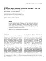
Rhinovirus infection of HASM cells induces necrotic cell deathFigure 1
Rhinovirus infection of HASM cells induces necrotic cell death. Photomicrographs of HASM cells 24 hours post: A)
Infection with rhinovirus at an MOI of 4, and B) exposure to UVi rhinovirus. The photomicrographs are of HASM cell derived
from a single asthmatic patient, and are representative of the response observed in HASM cells derived from all asthmatic and
non-asthmatic donors tested.
Respiratory Research 2006, 7:71 />Page 5 of 11
(page number not for citation purposes)
(n = 11). In contrast, RV-16 significantly increased the
secretion of IL-8 significantly above non-infected asth-
matic-derived HASM cells 24 hours post infection (p <
0.05, n = 8, Figure 3A). In contrast, stimulation with 5%
FBS, a commonly used HASM cell stimulus [24,31],
increased the production of IL-8 in both asthmatic and
non-asthmatic derived HASM cells in comparison to con-
stitutive release, and infection with RV-16 (MOI 4) and
concurrent stimulation with 5% FBS significantly induced
the release of IL-8 in comparison to 5% FBS alone in
HASM cells derived from both asthmatic and non-asth-
matic subjects. (Figure 2D).
The production of eotaxin was not induced following
infection with RV-16 at an MOI of 4, when measured 24
hours post infection. Tumour necrosis factor α and IFN-γ
were either not produced or below the limit of detection
in HASM cells derived from both asthmatic and non-asth-
matic subjects.
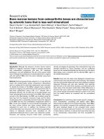
UVi RV-16-induced secretion of IL-6 and IL-8
In a separate series of experiments we compared the secre-
tion of IL-6 and IL-8 from HASM cells infected with RV-16
or exposed to UVi RV-16 for 24 hours. UVi RV stimulated
the production of IL-6 from both asthmatic (n = 6, p <
0.05) and non-asthmatic (n = 8, p < 0.05) HASM cells. Of
interest, UVi RV-16 induced similar levels of IL-6 when
compared to infectious RV-16 (Figure 3A). The secretion
of IL-8 protein was significantly increased in only the asth-
matic-derived HASM cells (n = 9, p < 0.01) in response to
UVi RV, as was found for infection with RV-16 (Figure
3B).
To ensure that IL-6 production in response to UVi RV-16
was not due to another factor in the viral inoculum, in a
separate series of experiments using the same viral stocks
and the same infection and UV irradiation protocols, IL-6
release was induced by only RV-16 and not UVi RV-16 in
alveolar macrophages (data not shown). This suggests
that the HASM response to UVi RV-16 is both a cell spe-
cific response, and due to ICAM-1 virus interactions.
Cytokine release from infected HASM cellsFigure 2
Cytokine release from infected HASM cells. A and C cytokine release in response to rhinovirus infection of HASM cells
derived from non-asthmatic (open bars) and asthmatic (black bars) subjects, measured in the cell free tissue culture superna-
tant 24 hours post infection. B and D Stimulation of HASM cells with 5% FBS and concurrent RV infection. No significant differ-
ence was observed between IL-6 and IL-8 output in response to FBS stimulation between HASM cells derived from asthmatic
and non-asthmatic volunteers. IL-6 and IL-8 were induced in both cell types in response to stimulation with 5% FBS, and con-
current infection with RV in the presence of 5% FBS induced IL-6 and IL-8 release in the non-asthmatic cells, and IL-8 in the
non-asthmatic cells. # p = 0.05 0.1% FBS versus 5%FBS, * p < 0.05 0.1% FBS versus 5%FBS, *** p < 0.001 0.1% FBS versus
5%FBS, the P value indicates the level of significance between 5%FBS and 5%FBS + RV.
Respiratory Research 2006, 7:71 />Page 6 of 11
(page number not for citation purposes)
No difference was observed between the constitutive
secretion of eotaxin and that found following exposure to
UVi RV-16 in HASM cells derived from asthmatic (UVi
RV-16 582.6 ± 247.8, compared to constitutive release
627.2 ± 272.4 pg/ml, p = 0.5, n = 5) and non-asthmatic
(UVi RV-16 795.1 ± 281.2 compared to 690.5 ± 231.3
constitutive release pg/ml, p = 0.7, n = 6) subjects.
IL-6 levels in response to RV are increased in sub-confluent
asthmatic HASM cells
HASM cells derived from asthmatic subjects have been
shown to proliferate faster than cells derived from non-
asthmatic subjects [31], however in cells grown to conflu-
ence, as used in the above studies, no difference in cell
number is found. To exclude the possibility that the differ-
ence observed in RV-16 induced IL-6 secretion between
cells derived from asthmatic and non-asthmatic subjects
was due to differences in cell number, HASM cells were
from both groups were seeded at the same density (10
4
UVi rhinovirus induces cytokine release from infected HASM cellsFigure 3
UVi rhinovirus induces cytokine release from infected HASM cells. Cytokine release in response to rhinovirus infec-
tion, and exposure to UVi rhinovirus (UVi), of HASM cells derived from non-asthmatic (open bars) and asthmatic (black bars)
donors, measured in the cell free tissue culture supernatant 24 hours post infection. a) IL-6 release n = 8 non-asthmatic and n
= 6 asthmatic derived HASM cells, b) IL-8 release n = 8 non-asthmatic and n = 9 asthmatic derived HASM cells For comparison
infectious rhinovirus is shown upon each graph where the data originate from HASM cells derived from the same donor.
Repeated measures ANOVA with Dunnett's Multiple Comparison Test post test.
Respiratory Research 2006, 7:71 />Page 7 of 11
(page number not for citation purposes)
cells/ml) and infected with RV-16 (MOI 4) 1 day post sub-
culture. As was observed in HASM cells grown to conflu-
ence, IL-6 secretion was induced 24 hours post RV-16
infection of both sub-confluent asthmatic, and sub-con-
fluent non-asthmatic-derived HASM cells. Furthermore,
as in confluent cells, the RV-16-induced IL-6 was signifi-
cantly greater in HASM cells derived from asthmatic than
non-asthmatic derived HASM cells (Figure 4).
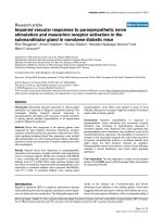
Similar cell surface expression of ICAM-1 on asthmatic
and non-asthmatic HASM cells
Since IL-6 was induced following exposure to UVi RV-16
it is likely that this resulted from interaction between
ICAM-1 (RV cellular receptor) and UVi RV-16. To ensure
that the increased production of IL-6 observed in the asth-
matic derived cells was not due to increased expression of
ICAM-1 upon these cells, we measured the cell surface
expression of ICAM-1 on asthmatic and non-asthmatic
HASM cells. As shown in Figure 5, no significant differ-
ence in the constitutive cell surface expression of ICAM-1
was found between asthmatic and non-asthmatic derived
HASM cells (asthmatic 230.5 ± 87.57 mean fluorescence
± SEM, n = 4 and non-asthmatic 254.4 ± 46.57 mean flu-
orescence ± SEM, n = 7 p > 0.05).
Transcriptional control of IL-6 production in asthmatic,
compared to normal HASM cells
We have previously demonstrated that the transcription
factor C/EBP-α is absent from asthmatic but not non-asth-
matic HASM cells [24]. Having shown that induction of
IL-6 is augmented in RV-infected HASM cells from asth-
matic donors and in the knowledge that the IL-6 promoter
region contains a C/EBP binding site we next investigated
transcriptional regulation of IL-6 induction to determine
if differences exist between asthmatic and non-asthmatic-
derived cells. In HASM cells transfected with the intact
human IL-6 promoter construct (NF-κB, C/EBP and AP1
binding sites), significant up regulation of luciferase activ-
ity was found in only asthmatic derived HASM cells in
response to infectious RV-16 (MOI 0.04). Similarly, at
higher MOIs (0.4 and 4) up regulation was observed in
only the asthmatic-derived cells (2.5 ± 0.8 and 2.2 ± 0.5 -
fold increase respectively, n = 8 for both). In comparison
with the positive control (PDGF BB, 10 ng/ml), there was
significantly increased luciferase activity of the intact IL-6
promoter construct in both HASM cells derived from asth-
matic (p < 0.01, n = 6) and non-asthmatic patients (p <
0.05, n = 7), in comparison to basal luciferase activity
(data not shown).
Since UVi RV-16 induced similar IL-6 secretion to live RV-
16 without the induction of HASM cell necrosis, and since
no up regulation of luciferase activity was observed in
HASM cells derived from non-asthmatic subjects in
response to infectious RV-16, the transcriptional control
of IL-6 production was examined using UVi RV-16. In
HASM cells derived from non-asthmatic but not asth-
matic donors, significant up-regulation of the luciferase
activity of the IL-6 promoters containing C/EBP, and C/
EBP and NF-κB deletions was observed. In contrast, luci-
ferase activity was not increased when either NF-κB or AP-
1 binding sites were deleted (Figure 6A). In summary
these observations indicate that IL-6 production in these
cells, in response to exposure to UVi RV-16, is mediated
by NF-κB and AP-1 binding sites.
In comparison, significant up-regulation of the IL-6 pro-
moter luciferase activity containing NF-κB/AP-1 deletions
and the plasmid containing an AP-1 deletion were
observed in HASM cells derived from asthmatic but not
Similar expression of ICAM-1 on asthmatic and non-asth-matic HASM cellsFigure 5
Similar expression of ICAM-1 on asthmatic and non-
asthmatic HASM cells. A typical histogram of the expres-
sion of cell surface ICAM-1 on HASM cells derived from
asthmatic (black line) and non-asthmatic (blue line) donors.
Isotype controls are represented by the red (solid filled) and
green lines for the HASM cells derived from non-asthmatic
and asthmatic donors respectively.
Cytokine release from infected subconfluent HASM cellsFigure 4
Cytokine release from infected subconfluent HASM
cells. IL-6 release in response to rhinovirus infection of sub-
confluent HASM cells, derived from non-asthmatic (open
bars) and asthmatic (black bars) donors, measured in the cell
free tissue culture supernatant 24 hours post infection. n = 6
both asthmatic and non-asthmatic derived cells.
Respiratory Research 2006, 7:71 />Page 8 of 11
(page number not for citation purposes)
non-asthmatic donors (Figure 6B). Therefore, IL-6 pro-
duction in asthmatic derived HASM cells, in response to
exposure to UVi RV, is mediated mainly by transcription
factors binding to the C/EBP binding site.
Discussion
In this study, significant differences were found in the
innate responses to RV infection between primary HASM
cells derived from asthmatic and non-asthmatic subjects.
RV-induced IL-6 release was significantly greater in the
asthmatic-derived HASM cells in comparison to cells
derived from non-asthmatic subjects. The increased
expression of IL-6 in HASM cells obtained from asthma
patients was due to differences in the activation pattern of
several transcription factors.
The present study investigated pro-inflammatory cytokine
production from HASM cells, and represents the first
reported experiments to examine RV infection of asth-
matic-derived HASM cells. Important differences in the
innate responses to RV in asthmatic compared to normal
subjects were observed. Hakonarson et al (1988) previ-
ously examined RV infection of airway smooth muscle
cells [32]. In their studies, they were able to demonstrate
that rabbit airway smooth muscle cells exposed to RV
exhibited increased constrictor responsiveness to acetyl-
choline and decreased relaxation to β-adrenoceptor stim-
ulation with isoprenaline. The mechanism by which this
occurred was reported to be RV-induced IL-1β, as blocking
the IL-1 receptor reversed the changes in contraction and
relaxation induced by exposure to RV [22]. However as
the evidence for HASM cell production of IL-1β is incon-
sistent [32], the decision was made in the present study
not to measure RV induced IL-1β.
Whilst the purpose of this study was not to determine the
kinetics of RV replication within HASM cells, the titer of
RV within the cell culture media was measured at 24 hours
post-infection. No difference was seen in the level of RV
within the cell culture medium derived from HASM cells
from asthmatic and non-asthmatic patients. RV has previ-
ously been shown to replicate to a higher titre in epithelial
cells derived from asthmatic patients [10]. This response
appears cell type specific, as it was not observed in HASM
cells in the present study.
Greater RV-induced IL-6 secretion occurred in the HASM
cells derived from asthmatic patients. This increased
release of IL-6 was RV-specific, as the mitogenic stimulus,
FBS (in the absence of RV), stimulated similar IL-6 release
in the two cell types. Furthermore, the increased IL-6
release was not due to differences in cell cycle status or cell
number, as greater RV induced IL-6 release was also
observed in sub-confluent HASM cells obtained from
asthmatic subjects. The increased IL-6 was also not due to
intrinsic differences in ICAM-1 expression between the
two cell types. The greater production of IL-6 which
occurred in HASM cells derived from asthmatic subjects in
comparison to non-asthmatic subjects is most like due to
differences in the transcriptional control of IL-6. In com-
mon with IL-6 production in response to 5% FBS, UVi RV-
induced IL-6 release in both cell types. As occurred with
5% FBS, there was no difference in the amount of IL-6
produced between the two diagnostic groups, further sug-
gesting that whilst IL-6 production is induced by virus
receptor interactions, the HASM cells derived from asth-
matic patients respond differently to infectious RV result-
ing in a greater release of IL-6. Whilst it is likely that the
majority of IL-6 induction is due to activation of ICAM-1,
up-regulation of the luciferase activity of the intact IL-6
promoter construct occurred in only asthmatic derived
Transcriptional control of IL-6Figure 6
Transcriptional control of IL-6. Luciferase activity fol-
lowing exposure to UVi rhinovirus (UVi), of the IL-6 luci-
ferase promoter construct plasmids in a) non-asthmatic
derived HASM cells and b) HASM cells derived from asth-
matic donors. The IL-6 promoter construct plasmid with no
transcription factor deletions is referred to as full length and
the various deletions are indicated by a minus sign in front of
the transcription factor binding site. All data sets from each
donor are normalised to the constitutive expression of the
full-length plasmid, which has been assigned the arbitrary
value of 100. * indicates p < 0.05 in the luciferase constructs
which contained transcription factor binding site deletions in
response to UVi RV-16. n = 6 non-asthmatic and n = 5 asth-
matic derived HASM cells.
Respiratory Research 2006, 7:71 />Page 9 of 11
(page number not for citation purposes)
HASM cells. In parallel experiments, carried out at the
same time, stimulation with PDGF BB resulted in up-reg-
ulation of the luciferase activity of the full-length IL-6 pro-
moter construct plasmid in both cell types. Therefore, it
can be assumed that the increased RV-induced IL-6 in the
asthmatic derived HASM cells is as a result of a greater
induction of IL-6-specific transcription factors. The
increased IL-6 secretion in response to RV infection from
asthmatic-derived HASM cell was also observed in sub-
confluent cells. This finding confirmed the fact that the
increased IL-6 protein production in asthmatic HASM
cells is not due to differences in cell number at the time of
infection, and moreover, is not dependent on differences
in cell cycle status.
IL-6 transcriptional regulation involves at least 4 different
transcription factor binding sites [33]: cAMP response ele-
ment binding protein (CREB), CCAAT/enhancer-binding
protein (C/EBP) binding site, activator protein (AP)-1,
and nuclear factor (NF)-κB. The transcriptional control of
IL-6 production in the asthmatic and non-asthmatic
HASM cells following exposure to UVi RV was found to
differ. IL-6 protein production in the non-asthmatic-
derived HASM cells was primarily mediated by the tran-
scription factor binding sites, NF-κB and AP-1, whilst in
the asthmatic derived HASM cells IL-6 production was pri-
marily driven by transcription factors binding to the C/
EBP binding site. It has recently been shown that asth-
matic HASM cells have a dysfunctional expression of C/
EPB α [24], which may account for the greater rate of pro-
liferation observed in asthmatic HASM cells [31]. In gen-
eral, C/EBP-α is considered to be a negative regulator of
protein transcription, therefore the absence in the asth-
matic HASM cells may disrupt the balance between excita-
tory and inhibitory C/EBPs, such that there are more pro-
transcriptional members. Thus the role of the C/EBP pro-
teins in these cells upon specific stimulation may be
skewed towards pro-transcription. In a report by Ammit
and coworkers (2002), HASM cells from non-asthmatic
donors in response to stimulation with TNF-α produce IL-
6 through transcription factors binding to the NF-κB
binding site, whilst AP-1 was shown not to be involved
[34]. In a different cell type, IL-6 production in response
to RV infection of the epithelial cell line A549 is also
mediated via NF-κB [35]. Our findings, in the non-asth-
matic HASM cells, support the role of NF-κB in RV-
induced IL-6 release.
Further differences between asthmatic and non-asthmatic
HASM cells were found upon infection with RV. Whilst
5% FBS stimulated IL-8 secretion, with this being
increased by concurrent RV infection in both asthmatic
and non-asthmatic cells, under basal conditions, both
infectious RV and UVi RV induced release of IL-8 in HASM
cells derived from asthmatic donors only.
Several reports have shown that the interaction of RV cap-
sid protein and ICAM-1 can induce a cell-mediated
inflammatory response, independent of RV replication.
This response has been found in a variety of cell types such
as epithelial cells [36], T cells [37], neutrophils [38],
monocytes [39] and importantly in the context of this
study airway smooth muscle cells [40]. In response to UVi
RV, increased IL-8 protein production was observed in the
HASM cells derived only from asthmatic donors. This is
not surprising since RV-induced IL-8 protein production
occurred only in asthmatic HASM cells. Since ICAM-1 is
stimulated by both infectious and UVi RV [40] it is likely
that IL-8 production in the HASM cells is mediated by RV
ICAM-1 interactions. Eotaxin was not found to be induced
by either infectious RV or UVi RV, in contrast to epithelial
cells, which produce eotaxin in response to RV infection
[41]. However, eotaxin is also not induced by RV infection
of lung fibroblasts (P. Bardin personal communication).
Viral replication is dependant upon evading detection by
the host's immune system. In this study we have shown
that both IL-6 and IL-8 are induced by RV infection, and
furthermore, the production of IL-6 is increased in HASM
cells derived from asthmatic patients. This indicates the
important role of the HASM cell as a modulator of the
local immune response. Whilst IL-6 has not been shown
to be directly antiviral, IL-6 can stimulate endothelial cells
to release IL-8, and therefore can contribute to the neu-
trophilic infiltration observed in viral respiratory tract
infections.
The association between asthma exacerbations and viral
infection has been known for at least the last 30 years [42-
44], and with the recent advances in molecular biology
there is now an overwhelming body of evidence in sup-
port of virus infections being the major trigger for asthma
exacerbations. The exact mechanisms behind viral infec-
tion and asthma exacerbations are not understood,
although the recent report of increased RV replication and
epithelial cell death following RV infection of bronchial
epithelial cells from asthmatic subjects [10] suggests
increased likelihood of RV infection of ASM in asthmatic
subjects. The augmented pro-inflammatory cytokine
release we have observed from asthmatic HASM cells is
therefore likely an important contribution to increased
airway inflammation associated with asthma exacerba-
tions.
Independently of productive infection, both IL-6 and IL-8
protein release were observed in the HASM cells derived
from asthmatic donors. These important mediators are
not only found to be elevated in the asthmatic airway
[45,46], but are also induced upon in vivo RV infections
[17,47], suggesting they contribute to the process of exac-
erbation, through recruitment and activation of neu-
Respiratory Research 2006, 7:71 />Page 10 of 11
(page number not for citation purposes)
trophils, as well as perhaps other cells such as eosinophils
and mast cells.
Conclusion
This study is the first to demonstrate differential regula-
tion of virus induced pro-inflammatory cytokines in
HASM cells from asthmatic and normal volunteers. The
observations reported herein have important implications
for the development of new therapies for virus induced
asthma exacerbations.
Competing interests
The author(s) declare that they have no competing inter-
ests.
Authors' contributions
BGGO: carried out the bench work, and drafted the man-
uscript
SLJ: conceived of the study, and participated in its design
and coordination and helped to draft the manuscript. MB:
carried out bronchoscopic biopsies and subject recruit-
ment and helped to draft the manuscript. JKB, NJCK, SL
participated in the study design and coordination and
helped to draft the manuscript. MR: helped with initial
luciferase assays, participated in the study design and
coordination and helped to draft the manuscript. JLB con-
ceived of the study, and participated in its design and
coordination and helped to draft the manuscript. All
authors read and approved the final manuscript.
Acknowledgements
This work was supported by the National Health and Medical Research
Council, Australia.
References
1. Chauhan AJ, Inskip HM, Linaker CH, Smith S, Schreiber J, Johnston SL,
Holgate ST: Personal exposure to nitrogen dioxide (NO2) and
the severity of virus-induced asthma in children. Lancet 2003,
361:1939-1944.
2. Rakes GP, Arruda E, Ingram JM, Hoover GE, Zambrano JC, Hayden
FG, Platts-Mills TA, Heymann PW: Rhinovirus and respiratory
syncytial virus in wheezing children requiring emergency
care. IgE and eosinophilanalyses. Am J Respir Crit Care Med 1999,
159:785-790.
3. Johnston SL, Pattemore PK, Sanderson G, Smith S, Lampe F, Josephs
L, Symington P, O'Toole S, Myint SH, Tyrrell DA, et al.: Community
study of role of viral infections in exacerbations of asthma in
9–11 year old children. BMJ 1995, 310:1225-1229.
4. Corne JM, Marshall C, Smith S, Schreiber J, Sanderson G, Holgate ST,
Johnston SL: Frequency, severity, and duration of rhinovirus
infections in asthmatic and non-asthmatic individuals: a lon-
gitudinal cohortstudy. Lancet 2002, 359:831-834.
5. Grissell TV, Powell H, Shafren DR, Boyle MJ, Hensley MJ, Jones PD,
Whitehead BF, Gibson PG: Interleukin-10 Gene Expression in
Acute Virus-induced Asthma. Am J Respir Crit Care Med 2005,
172:433-439.
6. Wark PA, Johnston SL, Moric I, Simpson JL, Hensley MJ, Gibson PG:
Neutrophil degranulation and cell lysis is associated with
clinical severity in virus-induced asthma. Eur Respir J 2002,
19:68-75.
7. Nicholson KG, Kent J, Ireland DC: Respiratory viruses and exac-
erbations of asthma in adults. BMJ 1993, 307:982-986.
8. Atmar RL, Guy E, Guntupalli KK, Zimmerman JL, Bandi VD, Baxter
BD, Greenberg SB: Respiratory tract viral infections in inner-
city asthmatic adults. Arch Intern Med 1998, 158:2453-2459.
9. Papadopoulos NG, Bates PJ, Bardin PG, Papi A, Leir SH, Fraenkel DJ,
Meyer J, Lackie PM, Sanderson G, Holgate ST, et al.: Rhinoviruses
infect the lower airways. J Infect Dis 2000, 181:1875-1884.
10. Wark PA, Johnston SL, Bucchieri F, Powell R, Puddicombe S, Laza-
Stanca V, Holgate ST, Davies DE:
Asthmatic bronchial epithelial
cells have a deficient innate immune response to infection
with rhinovirus. J Exp Med 2005, 201:937-947.
11. Braunstahl GJ, Fokkens WJ, Overbeek SE, KleinJan A, Hoogsteden
HC, Prins JB: Mucosal and systemic inflammatory changes in
allergic rhinitis and asthma: a comparison between upper
and lower airways. Clinical & Experimental Allergy 2003, 33:579-587.
12. Seow CY, Schellenberg RR, Pare PD: Structural and functional
changes in the airway smooth muscle of asthmatic subjects
[Review] [57 refs]. American Journal of Respiratory & Critical Care
Medicine 1998, 158:S179-S186.
13. Pascual RM, Awsare BK, Farber SA, Panettieri RA, Peters SP, Penn RB:
Regulation of phospholipase A2 by interleulin-1 in human
airway smooth muscle. Chest 2003, 123:433S-434S.
14. Lazaar AL, Albelda SM, Pilewski JM, Brennan B, Pure E, Panettieri RA:
T lymphocytes adhere to airway smooth muscle cells via
integrins and CD44 and induce smooth muscle cell DNA syn-
thesis. J Exp Med 1994, 180:807-816.
15. Lazaar AL, Amrani Y, Hsu J, Panettieri RA Jr, Fanslow WC, Albelda
SM, Pure E: CD40-mediated signal transduction in human air-
waysmooth muscle. J Immunol 1998, 161:3120-3127.
16. Burgess JK, Carlin S, Pack RA, Arndt GM, Au WW, Johnson PR, Black
JL, Hunt NH: Detection and characterization of OX40 ligand
expression in human airway smooth muscle cells: a possible
role in asthma? J Allergy Clin Immunol 2004, 113:683-689.
17. Gern JE, Vrtis R, Grindle KA, Swenson C, Busse WW: Relationship
of upper and lower airway cytokines to outcome of experi-
mental rhinovirus infection. Am J Respir Crit Care Med 2000,
162:2226-2231.
18. Geiser T, Dewald B, Ehrengruber MU, Clark-Lewis I, Baggiolini M:
The interleukin-8-related chemotactic cytokines GRO alpha,
GRO beta, and GRO gamma activate human neutrophil and
basophil leukocytes. J Biol Chem 1993, 268:15419-15424.
19. Diehl S, Rincon M: The two faces of IL-6 on Th1/Th2 differenti-
ation. [Review] [45 refs]. Molecular Immunology 2002,
39:531-536.
20. DiCosmo BF, Geba GP, Picarella D, Elias JA, Rankin JA, Stripp BR,
Whitsett JA, Flavell RA: Airway epithelial cell expression of
interleukin-6 in transgenic mice. Uncoupling of airway
inflammation and bronchial hyperreactivity. J Clin Invest 1994,
94:2028-2035.
21. Mul FP, Zuurbier AE, Janssen H, Calafat J, van WS, Hiemstra PS, Roos
D, Hordijk PL: Sequential migration of neutrophils across
monolayers of endothelial and epithelial cells. J Leukoc Biol
2000, 68:529-537.
22. Hakonarson H, Carter C, Maskeri N, Hodinka R, Grunstein MM: Rhi-
novirus-mediated changes in airway smooth muscle respon-
siveness: induced autocrine role of interleukin-1beta. Am J
Physiol 1999, 277:L13-L21.
23. Grunstein MM, Hakonarson H, Maskeri N, Chuang S: Autocrine
cytokine signaling mediates effects of rhinovirus on airway
responsiveness. Am J Physiol Lung Cell Mol Physiol 2000,
278:L1146-L1153.
24. Roth M, Johnson PR, Borger P, Bihl MP, Rudiger JJ, King GG, Ge Q,
Hostettler K, Burgess JK, Black JL, et al.: Dysfunctional interaction
of C/EBPalpha and the glucocorticoid receptor in asthmatic
bronchial smooth-muscle cells. N Engl J Med 2004, 351:560-574.
25. Papi A, Johnston SL: Rhinovirus infection induces expression of
its own receptor intercellular adhesion molecule 1 (ICAM-1)
via increased NF-kappaB- mediated transcription. J Biol Chem
1999, 274:9707-9720.
26. Johnson PR, Armour CL, Carey D, Black JL: Heparin and PGE2
inhibit DNA synthesis in human airway smooth muscle cells
in culture. Am J Physiol 1995, 269:L514-L519.
27. Durand-Arczynska W, Marmy N, Durand J: Caldesmon, calponin
and alpha-smooth muscle actin expression in subcultured
smooth muscle cells from human airways. Histochemistry 1993,
100:465-471.
Publish with BioMed Central and every
scientist can read your work free of charge
"BioMed Central will be the most significant development for
disseminating the results of biomedical research in our lifetime."
Sir Paul Nurse, Cancer Research UK
Your research papers will be:
available free of charge to the entire biomedical community
peer reviewed and published immediately upon acceptance
cited in PubMed and archived on PubMed Central
yours — you keep the copyright
Submit your manuscript here:
/>BioMedcentral
Respiratory Research 2006, 7:71 />Page 11 of 11
(page number not for citation purposes)
28. Eickelberg O, Pansky A, Mussmann R, Bihl M, Tamm M, Hildebrand P,
Perruchoud AP, Roth M: Transforming growth factor-beta1
induces interleukin-6 expression via activating protein-1
consisting of JunD homodimers in primary human lung
fibroblasts. J Biol Chem 1999, 274:12933-12938.
29. Farr A, Roman A: A pitfall of using a second plasmid to deter-
mine transfection efficiency. Nucleic Acids Res 1992, 20:920.
30. Huszar T, Mucsi I, Terebessy T, Masszi A, Adamko S, Jeney C, Rosivall
L: The use of a second reporter plasmid as an internal stand-
ard to normalize luciferase activity in transient transfection
experiments may lead to a systematic error. J Biotechnol 2001,
88:251-258.
31. Johnson PR, Roth M, Tamm M, Hughes M, Ge Q, King G, Burgess JK,
Black JL: Airway smooth muscle cell proliferation is increased
inasthma. American Journal of Respiratory & Critical Care Medicine
2001, 164:474-477.
32. Hakonarson H, Maskeri N, Carter C, Hodinka RL, Campbell D,
Grunstein MM: Mechanism of rhinovirus-induced changes in
airway smooth muscle responsiveness. J Clin Invest 1998,
102:1732-1741.
33. Ray A, Tatter SB, May LT, Sehgal PB: Activation of the human
"beta 2-interferon/hepatocyte-stimulating factor/interleukin
6" promoter by cytokines, viruses, and second messenger
agonists. Proceedings of the National Academy of Sciences of the United
States of America 1988, 85:6701-6705.
34. Ammit AJ, Lazaar AL, Irani C, Neill GM, Gordon ND, Amrani Y, Penn
RB, Panettieri RA: Tumor necrosis factor-alpha-induced secre-
tion of RANTES and interleukin-6 from human airway
smooth muscle cells: modulation by glucocorticoids and
beta-agonists. Am J Respir Cell Mol Biol 2002, 26:465-474.
35. Zhu Z, Tang W, Ray A, Wu Y, Einarsson O, Landry ML, Gwaltney J
Jr, Elias JA: Rhinovirus stimulation of interleukin-6 in vivo and
in vitro. Evidence for nuclear factor kappa B-dependent
transcriptional activation. J Clin Invest 1996, 97:421-430.
36. Johnston SL, Papi A, Bates PJ, Mastronarde JG, Monick MM, Hunning-
hake GW: Low grade rhinovirus infection induces a prolonged
release of IL-8 in pulmonary epithelium. J Immunol 1998,
160:6172-6181.
37. Gern JE, Vrtis R, Kelly EA, Dick EC, Busse WW: Rhinovirus pro-
duces nonspecific activation of lymphocytes through a
monocyte-dependent mechanism. J Immunol 1996,
157:1605-1612.
38. Dagher H, Ghildyal R, Freezer N, Bardin P: Chemokine Responses
In Primary Airway Fibroblasts Following Rhinovirus Infec-
tion. Respirology 2004, 9:A76-A77.
39. Johnston SL, Papi A, Monick MM, Hunninghake GW: Rhinoviruses
induce interleukin-8 mRNA and protein production in
human monocytes. J Infect Dis 1997, 175:323-329.
40. Grunstein MM, Hakonarson H, Whelan R, Yu Z, Grunstein JS, Chuang
S: Rhinovirus elicits proasthmatic changes in airway respon-
siveness independently of viral infection. J Allergy Clin Immunol
2001, 108:997-1004.
41. Papadopoulos NG, Papi A, Meyer J, Stanciu LA, Salvi S, Holgate ST,
Johnston SL: Rhinovirus infection up-regulates eotaxin and
eotaxin-2 expression in bronchial epithelial cells. Clin Exp
Allergy 2001, 31:1060-1066.
42. Minor TE, Dick EC, DeMeo AN, Ouellette JJ, Cohen M, Reed CE:
Viruses as precipitants of asthmatic attacks in children. JAMA
1974, 227:292-298.
43. Gregg I: Viral infection and asthma. Br Med J 1972, 3:824-825.
44. Lambert HP, Stern H: Infective factors in exacerbations of bron-
chitis and asthma. British Medical Journal 1972, 3:323-327.
45. Broide DH, Lotz M, Cuomo AJ, Coburn DA, Federman EC, Wasser-
man SI: Cytokines in symptomatic asthma airways. Journal of
Allergy & Clinical Immunology 1992, 89:958-967.
46. Marini M, Vittori E, Hollemborg J, Mattoli S: Expression of the
potent inflammatory cytokines, granulocyte-macrophage-
colony-stimulating factor and interleukin-6 and interleukin-
8, in bronchial epithelial cells of patients with asthma. J Allergy
Clin Immunol 1992, 89:1001-1009.
47. Fleming HE, Little FF, Schnurr D, Avila PC, Wong H, Liu J, Yagi S,
Boushey HA: Rhinovirus-16 colds in healthy and in asthmatic
subjects: similar changes in upper and lower airways.
Am J
Respir Crit Care Med 1999, 160:100-108.