Báo cáo y học: " Peroxisome Proliferator-Activated Receptor α (PPARα) down-regulation in cystic fibrosis lymphocytes" ppsx
Bạn đang xem bản rút gọn của tài liệu. Xem và tải ngay bản đầy đủ của tài liệu tại đây (520.37 KB, 15 trang )
Respiratory Research
BioMed Central
Open Access
Research
Peroxisome Proliferator-Activated Receptor α (PPARα)
down-regulation in cystic fibrosis lymphocytes
Veerle Reynders*1, Stefan Loitsch1, Constanze Steinhauer1, Thomas Wagner1,
Dieter Steinhilber2 and Joachim Bargon1
Address: 1Dept. of Internal Medicine, Division of Pneumology, University Hospital Frankfurt, Germany and 2Institute of Pharmaceutical
Chemistry, University of Frankfurt, Frankfurt am Main, Germany
Email: Veerle Reynders* - ; Stefan Loitsch - ; Constanze Steinhauer - ;
Thomas Wagner - ; Dieter Steinhilber - ; Joachim Bargon -
* Corresponding author
Published: 30 July 2006
Respiratory Research 2006, 7:104
doi:10.1186/1465-9921-7-104
Received: 16 February 2006
Accepted: 30 July 2006
This article is available from: />© 2006 Reynders et al; licensee BioMed Central Ltd.
This is an Open Access article distributed under the terms of the Creative Commons Attribution License ( />which permits unrestricted use, distribution, and reproduction in any medium, provided the original work is properly cited.
Abstract
Background: PPARs exhibit anti-inflammatory capacities and are potential modulators of the
inflammatory response. We hypothesized that their expression and/or function may be altered in
cystic fibrosis (CF), a disorder characterized by an excessive host inflammatory response.
Methods: PPARα, β and γ mRNA levels were measured in peripheral blood cells of CF patients
and healthy subjects via RT-PCR. PPARα protein expression and subcellular localization was
determined via western blot and immunofluorescence, respectively. The activity of PPARα was
analyzed by gel shift assay.
Results: In lymphocytes, the expression of PPARα mRNA, but not of PPARβ, was reduced (-37%;
p < 0.002) in CF patients compared with healthy persons and was therefore further analyzed. A
similar reduction of PPARα was observed at protein level (-26%; p < 0.05). The transcription factor
was mainly expressed in the cytosol of lymphocytes, with low expression in the nucleus. Moreover,
DNA binding activity of the transcription factor was 36% less in lymphocytes of patients (p < 0.01).
For PPARα and PPARβ mRNA expression in monocytes and neutrophils, no significant differences
were observed between CF patients and healthy persons. In all cells, PPARγ mRNA levels were
below the detection limit.
Conclusion: Lymphocytes are important regulators of the inflammatory response by releasing
cytokines and antibodies. The diminished lymphocytic expression and activity of PPARα may
therefore contribute to the inflammatory processes that are observed in CF.
Background
Cystic fibrosis (CF) is a common inherited disease caused
by mutations in the gene encoding the cystic fibrosis
transmembrane conductance regulator (CFTR), which is
an epithelial chloride channel. The disorder affects multi-
ple organs and the phenotype is extremely heterogeneous.
However, CF morbidity and mortality are mainly due to
lung disease, which is characterized by an excessive host
inflammatory response. Although CF lung disease is generally considered to be a neutrophil-mediated disorder,
Page 1 of 15
(page number not for citation purposes)
Respiratory Research 2006, 7:104
recent studies suggest a potent role for lymphocytes in the
pathogenesis of the disease [1,2]. In addition, inflammatory markers such as cytokines and eicosanoids are elevated, not only locally, in the airways, but also
systemically, thus indicating a more generalized state of
inflammation in CF [3-5].
/>
response through the production and release of cytokines,
chemokines, and/or antibodies. We noticed differences
for PPARα levels in lymphocytes. Along the same line, an
altered PPARα activity was observed in lymphocytes,
which confirmed our hypothesis.
Materials and methods
The nuclear factor-κB (NF-κB) and activated protein-1
(AP-1) transcription factors are key players in the inflammatory response by inducing the expression of cytokines,
chemokines, cell adhesion molecules and growth factors.
The actions of NF-κB and AP-1 can, however, be inhibited
by the Peroxisome Proliferator-Activated Receptors α and
γ (PPARs), which thereby exert anti-inflammatory properties [6-8]. PPARs are ligand-activated transcription factors
belonging to the nuclear hormone receptor super-family.
Fatty acids and eicosanoids are natural occurring PPAR
ligands [9,10]; fibrates and glitazones are more specific
synthetic activators for PPARα and γ, respectively. PPARs
regulate gene expression by heterodimerization with the
retinoid × receptor (RXR) and subsequent binding to specific DNA sequence elements, termed PPAR response elements (PPRE), in the promoter regions of their target
genes [11]. In addition, they can repress gene transcription in a DNA-binding independent manner through
inhibition of other signaling pathways by protein-protein
interactions and cofactor competition [6,7,12]. At present,
three distinct PPAR isoforms have been identified, called
α, β and γ. PPARα and γ agonists decrease plasma concentrations of cytokines and acute phase proteins [13-15] and
induce anti-atherosclerotic effects [16,17] and are therefore able to influence the immune response. They also
seem to play a role in airway inflammation. Similarly,
PPARα and γ agonists have been reported to inhibit airway inflammation in a murine model of asthma [18] and
a model of airway infection [19] by inhibiting eosinophil,
lymphocyte and neutrophil influx into the lung.
Moreover, CF is associated with abnormalities in fatty acid
and eicosanoid metabolism. In addition to deficiencies in
essential fatty acids in plasma, increased release of arachidonic acid (AA) from the cell membrane and elevated levels of pro-inflammatory eicosanoids in urine, blood and
airways have been reported [3,20-24]. Even cell membrane compositions seem to be disturbed with increased
levels of AA and decreased levels of docosahexaenoic acid
(DHA) [25]. Fatty acids and derivatives can regulate the
actions of PPARs and an imbalance may therefore cause
inappropriate activation of PPARs.
In conclusion, we hypothesized that the expression of
PPARs, transcription factors with anti-inflammatory
capacities, is altered in CF. To check our hypothesis, we
measured PPARα, β and γ expression in peripheral blood
cells, which are important mediators of the inflammatory
Patients
This study was approved by the Ethics Committee of the
Frankfurt University Hospital. Patients with cystic fibrosis
were between 22 and 43 years old and were all affected by
lung disease. They had a stable condition and came for
routine check-up. The clinical characteristics of our
patients are represented in Table 1. An age-matched, gender-mixed healthy control group was established for all
the experiments. Only healthy feeling volunteers, which
had not been ill for the past weeks, and which were free
from any detectable inflammation, infection or allergic
disease were selected for sampling. Due to time, technical
and sampling constraints, sample sizes vary between the
different experiments.
Measurement of IL-8 in plasma by ELISA
A commercial ELISA kit was used to measure IL-8 concentrations in plasma (R&D Systems, Germany). The instructions of the manufacturer were followed.
Measurement of sIL-2R in plasma by ELISA
A commercial ELISA kit was applied to measure soluble
IL-2 Receptor levels (R&D Systems, Germany). Prior to
use, plasma was diluted 1 to 4. The instructions of the
manufacturer were followed.
Isolation of peripheral lymphocytes and monocytes
To avoid circadian fluctuations of PPARs, blood samples
were always taken in the morning. Mononuclear cells
were isolated from whole blood by density gradient centrifugation using Lymphoprep (Axis-Shield). After washing with PBS, monocytes were separated from
lymphocytes by magnetic sorting (Miltenyi Biotec, Bergisch Gladbach, Germany). Cells were incubated with saturating concentrations of anti-CD14+ monoclonal
antibodies conjugated with super paramagnetic particles
for 20 min. by 4°C. Subsequently, cells were resolved in
PBS (containing 5 mM EDTA and 0.5% BSA) and added
on top of a separation column. Unlabeled cells, i.e. lymphocytes, were collected through elution from the column. In order to isolate the monocytes, the separation
column was detached from the strong magnet and monocytes were eluted. Purity was checked with May-Grünwald
Giemsa staining and was ≥ 97%.
Isolation of peripheral neutrophils
Density centrifugation using Polymorphprep™ solution
(Axis Shield, Heidelberg, Germany) enabled us to isolate
Page 2 of 15
(page number not for citation purposes)
Respiratory Research 2006, 7:104
/>
Table 1: Clinical characteristics of cystic fibrosis patients.
Patient
Age (years)
Gender
Genotype
P.a.1
CRP
FEV1 % pred2
FVC % pred2
1
2
3
4
5
6
7
8
9
10
11
12
13
14
15
16
17
18
19
20
33
25
30
34
37
32
26
28
37
32
23
37
34
39
25
22
43
23
39
40
F
M
M
M
M
F
F
M
M
F
M
M
F
M
M
F
M
M
M
M
dF508/R553x
dF508/dF508
dF508/dF508
dF508/?
dF508/dF508
dF508/dF508
dF508/dF508
dF508/?
dF508/dF508
dF508/dF508
dF508/?
dF508/dF508
dF508/dF508
dF508/dF508
dF508/R553x
dF508/N1303
dF508/dF508
dF508/dF508
dF508/G542x
dF508/?
+
+
+
+
+
+
+
+
+
+
+
+
+
+
+
+
+
+
0,9
0,5
0,7
0,3
0,6
2
1,32
0,91
< 0,3
< 0,3
< 0,3
< 0,3
0,94
< 0,3
1,03
0,9
< 0,3
0,8
0,4
0,4
58
74
42
59
52
60
86
31
30
76
103
74
23
60
85,9
53,6
98,8
85,3
61
64
91
90
72
80
84
75
87
48
43
92
99
101
59
83
79,7
64,6
95,5
84,5
79
80
1 Pseudomonas
2 Normal:
aeruginosa infection
80–120% of predicted
neutrophils from whole blood. The mononuclear and
polymorphonuclear leucocytes were separated into 2 distinct bands, free from red blood cells. Neutrophils were
collected, washed with PBS and checked for purity via
May-Grünwald-Giemsa staining and had to be > 95%.
Reverse transcriptase – competitive multiplex PCR/realtime PCR
Total RNA from monocytes, lymphocytes and neutrophils
was extracted with RNAzol B™ (Wak-Chemie, Germany)
and subjected to oligo(deoxythymidine)-primed firststrand cDNA synthesis using the Superscript II Preamplification System (Invitrogen, Karlsruhe, Germany). The
instructions of the manufacturers were followed.
Multiplex PCR (see Loitsch et al., 1999)[26]
Construction of internal standards
The cDNA derived from monocytes and lymphocytes was
amplified in the presence of a range of known concentrations of internal standards (competitors). Internal standards for the PPARs and GAPDH were constructed as wildtype fragments containing a deletion of nucleotides:
PPARα, β and γ cDNA with a 44, 41 and 106 bp deletion,
respectively and GAPDH cDNA with a 55 bp deletion. The
shortened fragments were obtained via PCR and the use of
following antisense primers: 5'-ATC ACA GAA GAC AGC
ATG GCC GTT CAG GTC CAA GTT TGC G-3' for PPARα,
5'-CTG CCA CAA TGT CTC GAT GTA GGA TGC TGC
GGG CCT TCT T-3' for PPARβ and 5'-TCA GCG GGA AGG
ACT TTA TGC ACT GGA GAT CTC CGC CAA C-3' for
PPARγ. The sense primers were the same as those used for
the multiplex PCR (see next paragraph). The fragments
were ligated in T-vectors (Promega) and the copy number
was calculated after spectrophotometric quantification.
Then, dilution series (1:3) of the internal standards were
established. The internal standards share identical primer
recognition sites with the wild-type target.
Competitive multiplex Polymerase Chain Reaction
Oligonucleotide primers for PCR were designed according
to published sequences: PPARα [GenBank Accession no.
Y07619]: sense 5'-TGCAGATCTCAAATCTCTGG-3', antisense 5'-ATCACAGAAGACAGCATGGC-3', amplifying a
374 bp wild-type product; PPARβ [GenBank Accession
no. L07592]: sense 5'-TTCCAGAAGTGCCTGGCACT-3',
antisense 5'-CTGCCACAATGTCTCGATGT-3'; amplifying
a 275 bp wild-type product; PPARγ [GenBank Accession
no. D83136]: sense 5'-TCTCTCCGTAATGGAAGACC-3',
antisense 5'-TCTTTCCTGTCAAGATCGCC-3', amplifying
a 660 bp wild-type product and, GAPDH [GenBank Accession no. M33197]: sense 5'-ATCTTCCAGGAGCGAGATCC-3', antisense 5'-ACCACTGACACGTTGGCAGT-3',
amplifying a 502 bp wild-type product.
2–10 μl cDNA was added to a PCR master-mix, which
contained all the primers mentioned above. Next, the mix
was divided over a series of reaction tubes into which
known concentrations of internal standards were spiked.
Cycling conditions for PCR were as follows: 94°C for 3
minutes (1 cycle), followed by 40 cycles of 94°C, 58°C,
72°C, each for 45 seconds and a final extension phase at
72°C for 10 minutes (Trio-Thermoblock, Biometra).
Page 3 of 15
(page number not for citation purposes)
Respiratory Research 2006, 7:104
The amplification products were separated by agarose gel
electrophoresis, stained with ethidium bromide and analyzed by densitometry. Densitometric data were plotted
on a log/log scale as a function of internal-standardderived PCR products and corrected for molar equivalence.
Real-time PCR
Neutrophils exhibit low levels of mRNA in general. The
classic competitive PCR was not sensitive enough and we
had to establish real-time PCR. Real-time PCR was performed by using the ABI prism 7700 sequence detector
(Perkin Elmer/Applied Biosystems). Primers and probes
were designed using the software program Primer Express
(Perkin Elmer/Applied Biosystems). For the measurement
of β-actin, a published primers/probe set was applied
[27]. The fluorogenic probes contained a reporter dye
(FAM) covalently attached at the 5'end and a quencher
dye (TAMRA) covalently attached at the 3'end. PPARα
[Genbank: NM005036]: sense 5'-CTT CAA CAT GAA CAA
GGT CAA AGC-3', antisense 5'-AGC CAT ACA CAG TGT
CTC CAT ATC A-3', probe 5'-CGG GTC ATC CTC TCA
GGA AAG GCC-3', amplicon length 99 bp; PPARβ [Genbank: L07592]: sense 5'-GGG CAT GTC ACA CAA CGC
TAT-3', antisense 5'-GCA TTG TAG ATG TGC TTG GAG
AA-3', probe 5'-CTT CTC AGC CTC CGG CAT CCG A-3',
amplicon length 147 bp; PPARγ [Genbank: D83233]:
sense 5'-GAA ACT TCA AGA GTA CCA AAG TGC AA-3',
antisense 5'-AGG CTT ATT GTA GAG CTG AGT CTT CTC3', probe 5'-CAA AGT GGA GCC TGC ATC TCC ACC TTA
TT-3', amplicon length 87 bp; β-actin [Genbank: D28354
and X00351]: sense 5'-AGC CTC GCC TTT GCC GA-3',
antisense 5'-CTG GTG CCT GGG GCG-3', probe 5'-CCG
CCG CCC GTC CAC ACC CGC C-3', amplicon length 174
bp.
Specific external controls were constructed for all target
genes by cloning a partial cDNA fragment (the amplicon
of interest obtained by classic PCR amplification) into a
pCR®2.1 vector (Invitrogen). A standard curve was generated: in each PCR run, 10-fold serial dilutions of the corresponding plasmid clone were included, with known
amounts of input copy number. In order to normalize for
inefficiencies in cDNA synthesis and RNA input amounts,
the mRNA expression of the housekeeping gene β-actin
was quantified for each sample. cDNA samples were
diluted 10 times prior to PCR amplification. PCR amplifications were performed in a total volume of 25 μl, containing 5 μl cDNA sample, 12.5 μl Taqman Universal PCR
Master Mix (Perkin Elmer/Applied Biosystems), 200–800
nM of each primer and 200 nM detection probe (Eurogentec). Each PCR amplification was performed in triplicate,
using the following conditions: 2 min. at 50°C and 10
min. at 95°C, followed by a total of 45 two-temperature
cycles: 15 s at 94°C and 1 min. at 60°C for PPARs and
/>
67°C for β-actin. PCR data were analyzed through the
application of the software 'Sequence Detector 7.1' (Perkin Elmer/Applied Biosystems).
Western blot analysis for lymphocytes
Equal amounts (80 μg) of total cell proteins were resolved
by 10% SDS-PAGE and transferred to a PVDF membrane
(Millipore Corporation, Bedford) at 80 V for 1 hour.
Membranes were incubated with mouse PPARα monoclonal antibodies (1:2000 dilution) (clone B11.80A, generous gift from Dr. Winegar, Glaxo Smith Kline) at 4°C
overnight. Protein levels were normalized using a mouse
monoclonal antibody against β-actin (1:10.000 dilution)
(Sigma). Proteins were subsequently detected through the
use of horseradish peroxidase-conjugated secondary antibodies and the chemiluminescence system ECL (Amersham Pharmacia Biotech, Buckinghamshire, UK). After
scanning (DocuGel V-System, Scananalytics), band intensities were analyzed using the software package Zero-DScan™ (Scananalytics).
Immunofluorescence assay
Cytospin glass slides were prepared by centrifugation of
105 lymphocytes using a cytospin centrifuge (Cytospin 4,
Thermo Shandon). After cells were fixed in ice-cold methanol and blocked with a solution of 2% BSA in PBS overnight at 4°C, they were permeabilized with Perm/Wash
buffer (BD Biosciences Pharmingen) and then incubated
for 2 hours with monoclonal PPARα antibody, diluted
1:10 in Perm/Wash/2%BSA buffer (clone Pα B32.51
kindly provided by Dr. Winegar, Glaxo Smith Kline [28]).
After washing, the second antibody (cy3 labeled goat-antimouse, Caltag) was added in a 1:250 dilution in Perm/
Wash/2%BSA buffer for 30 min. Following washing and
air-drying, the cells were embedded in Aquatex (Merck)
and evaluated by immunofluorescence microscopy.
Gel shift assay
Nuclear proteins from lymphocytes were prepared as
described by Dignam and coworkers [29]. A gel shift kit
for PPARα was obtained from Panomics, Inc. and the
instructions of the manufacturer were followed. Equal
amounts of nuclear protein extracts (10 μg as determined
by Bradford assay) were incubated for 30 min. with
biotin-labeled oligonucleotide probe, which corresponds
to the PPAR binding site, and then subjected to non-denaturing PAGE. Afterwards, proteins were blotted on a PallBiodyneB® (PALL Corporation) membrane and bands
were visualized after exposure to Hyperfilm™ECL (Amersham Biosciences, UK). Subsequently, equal loading was
checked via Coomassie Blue staining of the membrane.
Band intensities were analyzed using the software package
Zero-D-Scan™ (Scananalytics).
Page 4 of 15
(page number not for citation purposes)
Respiratory Research 2006, 7:104
/>
PPARα
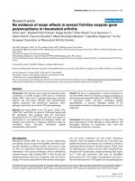
PPARα mRNA levels were significantly lower (-37%, p <
0.002) in lymphocytes of CF patients compared with control persons (Fig. 3). In monocytes, no differences were
observed in the expression of PPARα between the healthy
subjects and the CF patients (Fig. 4).
IL-8 in plasma (pg/ ml)
*
10.0
7.5
PPARβ
For both lymphocytes and monocytes, no statistical differences in the mRNA expression of PPARβ were detected
between CF patients and healthy persons (Fig. 3 and 4).
5.0
2.5
0.0
C
CF
Figure 1
measured in ELISA
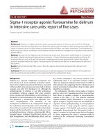
IL-8 levels by plasma of CF patients and healthy persons
IL-8 levels in plasma of CF patients and healthy persons
measured by ELISA. IL-8 levels are significantly higher in CF
patients (n = 15) than in control persons (n = 11). Results are
shown as mean ± standard error. * Significantly different (p <
0.03).
Statistical analysis
Results are expressed as mean ± SE. Statistical comparisons were made using the unpaired Student's t-test
(Sigma-plot). A value of p < 0.05 was considered significant.
Results
Interleukin-8 levels in plasma
IL-8 in plasma was measured via ELISA to demonstrate
that the patients in this study exhibit the typical elevated
systemic cytokine levels [30]. As expected, IL-8 levels were
significantly higher in CF patients compared with control
persons (7.3 pg/ml vs 2.9 pg/ml, respectively; p < 0.03)
(Fig. 1). We can therefore assume that the inflammation
cascade is not restricted to the airways, but is also found
systemically.
PPAR mRNA expression in peripheral blood cells
In order to check for differences in the expression of
PPARs between CF patients and healthy persons, we
started screening at mRNA level. All data were normalized
to the expression levels of the housekeeping genes
GAPDH or β-actin, which were equally expressed in samples of CF patients and control persons.
Monocytes and lymphocytes
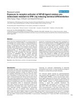
Competitive multiplex PCR products were loaded on an
agarose gel, electrophorised and stained with ethidium
bromide (see fig. 2). Bands were scanned and analyzed
with the software package Zero-D-Scan™ (Scananalytics).
PPARγ
PPARγ mRNA was detected in a few samples of monocytes
and lymphocytes, but was not quantifiable due to the
extremely low expression levels.
Neutrophils
Neutrophils are considered end-cells as DNA and most,
but not all, mRNA and protein synthesis, cease once the
myeloid cells are mature enough to enter the blood. For
that reason, mRNA levels were rather low in neutrophils
and PPAR mRNA was difficult to quantify via the classic
competitive multiplex PCR. We therefore developed realtime PCR, a highly sensitive and accurate method.
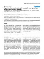
PPARα
PPARα mRNA levels were equal in neutrophils of CF
patients and healthy persons (Fig. 5A)
PPARβ
Idem, PPARβ mRNA levels were similar in both groups
(Fig. 5B).
PPARγ
PPARγ mRNA was detectable, but the low expression levels did not allow quantification.
PPARα protein levels in peripheral blood lymphocytes
measured via western blotting
mRNA analysis revealed less expression of PPARα in lymphocytes of CF patients compared with healthy persons.
On the basis of this finding we further examined the
expression of the receptor at protein level via western blotting. A single band for PPARα was observed around 60
kDA (Fig. 6A). Analysis of the band intensities demonstrated that protein levels of PPARα are significantly lower
(-26%, p < 0.05) in lymphocytes of CF patients compared
with control subjects (Fig. 6B). β-actin was measured for
normalization.
Localization of PPARα in human peripheral blood
lymphocytes
In order to identify the subcellular localization of PPARα
within peripheral blood lymphocytes, an immunofluores-
Page 5 of 15
(page number not for citation purposes)
Respiratory Research 2006, 7:104
/>
Internal standard serial dilution 1:3
Figure 2
RT-competitive multiplex PCR for PPARα, β, and γ and GAPDH in human peripheral blood lymphocytes
RT-competitive multiplex PCR for PPARα, β, and γ and GAPDH in human peripheral blood lymphocytes. Picture of ethidium
bromid stained agarose gel after electrophoresis of the amplified products. wt = wild-type amplicon, ist = internal standard
amplicon.
cence assay was developed. As shown in Fig. 7A and 7B,
the highest concentration of the protein is observed in the
cytosol, whereas the nucleus contains only trace amounts
of the transcription factor. In the context of our study, the
technique was not found appropriate for quantifying
PPARα protein levels by means of measuring the fluorescence intensity. Activity was therefore measured via gel
shift assay.
Activity of the PPARα transcription factor
Since PPARα expression is lower in lymphocytes of CF
persons, it was deemed useful to check for the activity of
the transcription factor, which was determined via gel
shift assay (Fig. 8). To this end, a commercially available
kit for PPARα was used (Panomics). The DNA-binding
element (PPRE) was not radioactive-, but biotin-labeled.
Equal amounts of nuclear extracts were loaded. The measurement of band intensities showed that PPARα DNA
binding activity was 36% less in lymphocytes of CF
patients, compared with control subjects (p < 0.01) (Fig.
8B). In order to evaluate the binding specificity, competition analysis was performed by adding 60-fold cold specific (PPRE) and unspecific oligonucleotide (see fig. 8A:
lane 2 and 3, respectively). The upper band fainted
strongly by adding cold PPRE, but remained unaltered
after adding cold unspecific oligonucleotide. Equal loading of nuclear extracts was verified via Coomassie Blue
staining of the membrane.
sIL-2 R levels in plasma
Soluble IL-2 receptor (sIL-2R), a well-known marker for Tlymphocyte activation, was measured in plasma of stable
CF patients and control persons via ELISA (Fig. 9). Normal values for sIL-2R levels in plasma are around 1020 pg/
ml. Statistical analysis revealed that CF patients exhibit
significantly higher levels of sIL-2R in plasma than
healthy persons (CF: 1521 ± 84.15 pg/ml vs C: 970 ±
56.44 pg/ml). These data indicate that peripheral T-lymphocytes of CF patients are more activated than lymphocytes of healthy subjects.
Discussion
The mechanisms behind the disturbed immune response
in CF are still largely unknown and require further
research. The aim of our study was to measure the expression of the PPAR transcription factors in patients with
cystic fibrosis and healthy subjects. Because of their
known regulatory functions in inflammatory processes,
Page 6 of 15
(page number not for citation purposes)
Respiratory Research 2006, 7:104
/>
Monocytes
**
200
PPAR mRNA levels (AU)
PPAR mRNA levels (AU)
Lymphocytes
100
0
C
PPAR
CF
C
CF
PPAR
Figure 3
mRNA peripheral levels
and 6/5) were subjected to measure PPARα patients (CF,
M/F:densitometry blood lymphocytes from multiplex = 11;
n = 15; expressionin order toand healthy subjects (C, nPCR
Human M(ale)/F(emale): 9/6) RT-competitiveCF and PPARβ
Human peripheral blood lymphocytes from CF patients (CF,
n = 15; M(ale)/F(emale): 9/6) and healthy subjects (C, n = 11;
M/F: 6/5) were subjected to RT-competitive multiplex PCR
and densitometry in order to measure PPARα and PPARβ
mRNA expression levels. Data are normalized to GAPDH
expression levels. Values are represented as means ± standard error. PPARα expression levels were 37% lower in CF
patients compared to control persons. ** Significantly different (p < 0.002).
we hypothesized that their expression and/or function
may be altered in cystic fibrosis, a disorder characterized
by an excessive host inflammatory response.
Our study confirmed that systemic inflammation was
present in our CF patients on the basis of the observed
increased levels of plasma IL-8. Our data revealed that
both PPARα mRNA and protein levels were significantly
lower in peripheral lymphocytes of CF patients than in
healthy control persons. Immunofluorescence experiments demonstrated that just a small fraction of PPARα
resides in the nucleus, whereas the cytosol contains the
larger part of the transcription factor. This was observed
for both groups. Differences in activity were demonstrated
via gel shift assay, i.e. a significant reduction of PPARα
DNA binding activity in lymphocytes of CF persons compared with healthy subjects. Finally, increased levels of
soluble IL-2 Receptor in plasma suggest that peripheral
lymphocytes are activated in cystic fibrosis.
Most CF patients become chronically infected with specific bacterial pathogens, such as Pseudomonas aeruginosa,
which cause a destructive inflammatory response in the
lung. However, several studies provide evidence that
100
75
50
25
0
C
PPAR
CF
C
CF
PPAR
Figure and 13/6) blood expression (C, = patients (CF,
via 19; M/F:PPARβ mRNA monocytes densitometry
n human peripheralmultiplex PCR and levels were M/F: 6/4)
in =RT-competitiveand healthy subjectsfromnCF 10; measured
PPARα 4
PPARα and PPARβ mRNA expression levels were measured
in human peripheral blood monocytes from CF patients (CF,
n = 19; M/F: 13/6) and healthy subjects (C, n = 10; M/F: 6/4)
via RT-competitive multiplex PCR and densitometry. Data
are normalized to GAPDH mRNA expression levels. Values
are means and standard error. Both PPAR levels were similar
in the two groups.
inflammation can occur prior to infection and that CF
lungs are primed for inflammation [30-32]. Nevertheless,
the inflammatory processes are not restricted to the respiratory tract as shown by the elevated levels of pro-inflammatory markers in the blood circuit of CF patients
[4,30,33]. Our study also demonstrated elevated levels of
IL-8 in plasma of CF patients. Therefore, monocytes, lymphocytes and neutrophils were studied, as they are important mediators of the inflammatory response, i.a. through
the release of cytokines, chemokines, and through the
production of antibodies.
Our study revealed that PPARα and PPARβ are abundantly expressed in freshly isolated monocytes and lymphocytes at mRNA level, whereas little or no PPARγ was
detected. Both PPARα and PPARβ mRNA could be measured via real-time PCR in neutrophils; PPARγ mRNA on
the other hand was not quantifiable. Statistical analysis
showed that PPARα mRNA, but not PPARβ mRNA, is significantly less expressed (-37%) in lymphocytes of CF
patients compared with control persons. The same difference could be detected at protein level via western blotting. The expression of PPARα and β mRNA in monocytes
and neutrophils was not significantly different in patients
and healthy persons. These data are supported by several
studies. First, there is evidence that PPARα mRNA and
Page 7 of 15
(page number not for citation purposes)
Respiratory Research 2006, 7:104
/>
Neutrophils
B
750
PPAR /actin (copies x 10e6)
PPAR /actin (copies x 10e6)
A
500
250
0
7500
5000
2500
0
C
CF
C
CF
Figure control persons in freshlyM/F: 6/6) human neutrophils was determined by real-time PCR for CF patients (n = 12; M/F:
7/5) and5
PPAR mRNA expression (n = 12; isolated
PPAR mRNA expression in freshly isolated human neutrophils was determined by real-time PCR for CF patients (n = 12; M/F:
7/5) and control persons (n = 12; M/F: 6/6). Data are presented as the mean PPAR mRNA level relative to the β-actin mRNA
expression [(number of PPAR copies/number of β-actin copies) × 106] and the standard error. Each measurement was performed in triple. (A) PPARα mRNA expression. (B) PPARβ mRNA expression. No differences were seen between the two
groups for both PPARα and PPARβ mRNA levels.
protein expression are directly regulated by its own ligands [34,35]. Fatty acids and eicosanoids, which are natural PPAR activators, are found in disturbed levels in CF
and may therefore cause a diminished expression of
PPARα. Second, PPARα expression within T-lymphocytes
is rapidly down-regulated following cellular activation
[36]. Our present study demonstrated increased plasma
soluble interleukin-2 receptor (sIL-2 R) concentrations in
CF patients, which is in line with the findings of other
research groups [37,38]. sIL-2 R is a generally accepted
marker for T-lymphocyte activation [39]. Therefore, Tlymphocytes appear to be in some sort of activated state
in CF patients, which may be responsible for the
decreased PPARα levels. The mechanism responsible for
this down-regulation has not yet been elucidated. Third,
the pro-inflammatory cytokines IL-6, TNF-α and IL-1 have
been demonstrated to cause a reduction in the expression
of PPARα [40,41]. CF patients exhibit increased levels of
IL-2, TNF-α, IL-6 and IL-8 in sputum and serum
[4,5,32,37]. However, this can not be the major explanation for the decreased PPARα levels in lymphocytes, as the
expression of the transcription factor was unaltered in
monocytes and neutrophils. And finally, an interesting
abstract by Andersson and team reported that a CF tracheal epithelial cell line expressed less PPARα protein
than a normal tracheal epithelial cell line, which is com-
parable with our data [42]. The same research team found
decreased PPARγ levels in tissues specifically regulated by
CFTR in a CF mice model [43] and their data suggest that
CFTR may play a role in PPAR expression. A functional
CFTR is also expressed in lymphocytes of healthy humans.
Consequently, a defect CFTR in CF lymphocytes could
result in altered PPAR expression. In addition, research
has shown that PPAR expression may differ significantly
in target organs where inflammation occurs. For example,
a recent study reported that induction of PPARα is lacking
in the liver of CF mice compared to wild type animals following colitis induced bile duct injury [44].
Following our findings that PPARα expression is downregulated in CF lymphocytes, the question arose whether
the activity of the transcription factor was also altered.
Our immunofluorescence experiments revealed that for
both groups, the transcription factor is primarily located
in the cytosolic compartment and only a small fraction
resides in the nucleus. A similar cellular distribution was
reported in human macrophages [45] and in mice lymphocytes [36]. This meant that gel shift analysis had to be
applied to measure possible differences in the activity of
PPARα. The gel shifts indeed showed that PPARα DNA
binding activity was 36% lower in lymphocytes of CF
patients compared with control persons. A decreased
Page 8 of 15
(page number not for citation purposes)
Respiratory Research 2006, 7:104
/>
activity of PPARα (see fig. 10). Further studies need to be
carried out to test this hypothesis.
A
PPARα protein levels (AU)
α
B
*
100
75
50
25
0
C
CF
Figure 6
(C, n = 10; M/F: 4/6) of CF patients extracts derived from
human peripheral blood total protein from = 11; M/F: 6/5)
Western blot analysisandlymphocytes (CF, ncontrol persons
Western blot analysis of total protein extracts derived from
human peripheral blood lymphocytes from control persons
(C, n = 10; M/F: 4/6) and CF patients (CF, n = 11; M/F: 6/5).
(A) A single band was detected at 60 kDa for PPARα. β-actin
protein expression was measured for normalization. (B)
Analysis of band intensities revealed that PPARα protein levels are down-regulated (-26%) in CF patients compared to
healthy subjects. Densitometry data are expressed as means
± standard error. * Significantly different (p < 0.05).
DNA binding activity of PPARγ was also seen in tissues of
CFTR knock-out mice. Treatment of these mice with rosiglitazone, a PPARγ agonist, restored DNA binding [43].
CF lung disease is well-known as a neutrophil-mediated
disease. However, as pointed out by Moss, the behavior
and biological function of lymphocytes is also altered in
CF. Lymphocytes are important immune cells because
they determine the specificity of the immune response.
Quantitative analysis of inflammatory cells in CF lung tissues revealed a lymphocyte-dominated immune response
in the CF bronchial wall, beneath the surface epithelium
[1]. These lymphocytes may release cytokines, such as IL17, that may attract neutrophils into the airways [49,50].
CF peripheral lymphocytes also exhibit an altered pattern
in cytokine-release and production after stimulation [5153], which could indicate an impairment of the immune
response at the systemic level. Moreover, CF lymphocytes
are characterized by a specific incapacity to respond to P.
aeruginosa antigens [54]. Consequently, this defect could
contribute to the inability to eradicate lung infection and
inflammation due to P. aeruginosa. Summarized, the function of lymphocytes is altered in CF and they are therefore
an interesting target to be studied.
In conclusion, our study revealed that both the expression
and activity of PPARα, a transcription factor with antiinflammatory capacities, is down-regulated in peripheral
lymphocytes of CF patients, which may render lymphocytes into cells that promote the inflammatory
response and consequently lead to increased inflammation. In addition, the natural activators of PPARα are
known to be present in disturbed proportions in CF and
may therefore cause an improper activation of PPARα. We
therefore hypothesize that the expression and activity of
PPARα may be up-regulated via the administration of natural or synthetic agonists which eventually may lead to a
diminished immune response.
Abbreviations
PPARα was the first isotype recognized for its in vivo role
in inflammatory processes. Inflammation induced by leukotriene B4, a PPARα ligand, has been reported to be prolonged in PPARα knock-out mice, suggesting an antiinflammatory role for PPARα [46]. Ligand-induced activation of PPARα in lymphocytes antagonized DNA binding
activity of NF-κB and decreased IL-2 and TNF-α production [36,47], inhibited IFN-γ secretion but promoted IL-4
secretion and production [47,48]. These data indicate that
PPARα may have a significant influence on the lymphocytic immune response. Consequently, a decrease in
PPARα expression and function may contribute to the
excessive host inflammatory response. Our data suggest
that administration of ligands, such as the natural DHA or
synthetic fibrates, may serve as a therapy to help reduce
the inflammatory processes in CF by upregulating the
AA: Arachidonic acid
AP-1: Activator protein-1
CF: Cystic fibrosis
CFTR: Cystic fibrosis transmembrane conductance regulator
DHA: Docosahexaenoic acid
NF-κB: Nuclear factor-κB
PPAR: Peroxisome Proliferator-Activated Receptor
PPRE: PPAR response element
Page 9 of 15
(page number not for citation purposes)
Respiratory Research 2006, 7:104
A
/>
B
Figure 7
immunofluorescence assay (C:PPARα proteinnin freshly isolated human peripheral blood lymphocytes was determined via
The subcellular localization of n = 5 and CF: = 5)
The subcellular localization of PPARα protein in freshly isolated human peripheral blood lymphocytes was determined via
immunofluorescence assay (C: n = 5 and CF: n = 5). Microscopy analysis revealed that the transcription factor is mainly situated in the small cytoplasmic area. (A) Representative immunofluorescence picture of lymphocytes derived from healthy control blood and (B) from a CF patient.
Competing interests
The author(s) declare that they have no competing interests.
Authors' contributions
VR carried out the experiments, wrote the manuscript and
participated in the study design. SL designed the multiplex
competitive PCR for PPARs, participated in the study
design and helped evaluating the results and techniques.
CS provided technical assistance. TW and DS provided the
work with critical comments. JB participated in the coordination of the project and corrected the article.
Acknowledgements
The present work was supported by the Deutsche Forschungsgemeinschaft: Internationales Graduiertenkolleg 757/1.
The authors thank Glaxo-Smith Kline, especially Dr. Winegar, for the kind
gift of the PPARα antibodies. They also thank the patients and healthy volunteers for their cooperation. Comments from Dr. Hirche are gratefully
acknowledged.
Page 10 of 15
(page number not for citation purposes)
Respiratory Research 2006, 7:104
/>
A
B
**
750
500
250
PPAR
DNA binding activity (AU)
PPAR
0
C
CF
Figure 8
Differential PPARα binding to PPRE in peripheral lymphocytes
Differential PPARα binding to PPRE in peripheral lymphocytes. PPARα DNA binding was analyzed via gel shift assay in peripheral lymphocytes of CF patients (CF, n = 11; M/F: 6/5) and healthy subjects (C, n = 11; M/F: 6/5). The DNA binding element
was biotin-labeled. (A) A representative gel shift. Lane (-) represents the biotin-labeled DNA binding element, without the
addition of nuclear extract. Lane 1: Lymphocytic control sample. Lane 2: Specific cold oligonucleotide binding competition
assay: a 60-fold excess of cold synthetic PPRE was added. Lane 3: Unspecific cold oligonucleotide binding competition assay. A
60-fold excess of unspecific synthetic oligonucleotide was used. (B) Densitometry data derived from the gel shift assays are
expressed as means and standard error. These data show that PPARα DNA binding activity of CF patients is reduced by 36%
compared to healthy persons. ** Significantly different (p < 0.01). On top: representative bands from a control person and a
patient.
Page 11 of 15
(page number not for citation purposes)
sIL-2R in plasma (pg/ml)
Respiratory Research 2006, 7:104
/>
***
2000
1000
0
C
CF
Figure 9
control persons (n = 18)
sIL-2R levels were measured in plasma of CF (n = 19) and
sIL-2R levels were measured in plasma of CF (n = 19) and
control persons (n = 18). sIL-2R levels are significantly higher
in CF patients than in control persons. Data are represented
as mean and standard error. *** p < 0.0001.
Page 12 of 15
(page number not for citation purposes)
Respiratory Research 2006, 7:104
/>
IL-6, TNF- , IL-1
Activation of lymphocytes
Fibrates, DHA
Imbalance of natural
ligands:fatty acids and
eicosanoids
PPAR
+
Target genes: ET-1…
AP-1
NF B
I B
+
Target genes: IL-6,
VCAM-1, …
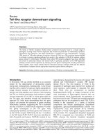
Figure 10
Summarizing picture: PPARα in lymphocytes
Summarizing picture: PPARα in lymphocytes. PPARα inhibits the actions of the pro-inflammatory transcription factors AP-1
and NFκB through protein-protein interactions and by up-regulating the expression of IκB. The nuclear hormone receptor is
inhibited by specific cytokines and by lymphocyte activation. In addition, we suggest that the imbalance of natural ligands in CF
leads to deficiencies in PPARα activation. Therefore, lymphocytes may be turned into cells that promote inflammation in CF.
We hypothesize that the addition of synthetic or natural ligands, such as fibrates and DHA respectively, may restore the activity and expression of PPARα, resulting in a more balanced lymphocytic immune response.
References
1.
2.
3.
4.
5.
6.
7.
Hubeau C, Lorenzato M, Couetil JP, Hubert D, Dusser D, Puchelle E,
Gaillard D: Quantitative analysis of inflammatory cells infiltrating the cystic fibrosis airway mucosa. Clin Exp Immunol
2001, 124:69-76.
Moss RB: Lymphocytes in cystic fibrosis lung disease: a tale of
two immunities. Clin Exp Immunol 2004, 135:358-360.
Reineck P, Artlich A, Hoeser C, Hüls G, Lindemann H: Leukotriene
im Atemkondensat von Kindern und Jugendlichen mit
Asthma bronchiale und zystischer Fibrose. Atemw -Lungenkrkh
2004, 3:103-108.
Ionescu AA, Nixon LS, Luzio S, Lewis-Jenkins V, Evans WD, Stone
MD, Owens DR, Routledge PA, Shale DJ: Pulmonary function,
body composition, and protein catabolism in adults with
cystic fibrosis. Am J Respir Crit Care Med 2002, 165:495-500.
Nixon LS, Yung B, Bell SC, Elborn JS, Shale DJ: Circulating immunoreactive interleukin-6 in cystic fibrosis. Am J Respir Crit Care
Med 1998, 157:1764-1769.
Poynter ME, Daynes RA: Peroxisome proliferator-activated
receptor alpha activation modulates cellular redox status,
represses nuclear factor-kappaB signaling, and reduces
inflammatory cytokine production in aging. J Biol Chem 1998,
273:32833-32841.
Delerive P, De Bosscher K, Besnard S, Vanden Berghe W, Peters JM,
Gonzalez FJ, Fruchart JC, Tedgui A, Haegeman G, Staels B: Peroxisome proliferator-activated receptor alpha negatively regu-
8.
9.
10.
11.
12.
lates the vascular inflammatory gene response by negative
cross-talk with transcription factors NF-kappaB and AP-1. J
Biol Chem 1999, 274:32048-32054.
Chung SW, Kang BY, Kim SH, Pak YK, Cho D, Trinchieri G, Kim TS:
Oxidized low density lipoprotein inhibits interleukin-12 production in lipopolysaccharide-activated mouse macrophages
via direct interactions between peroxisome proliferatoractivated receptor-gamma and nuclear factor-kappa B. J Biol
Chem 2000, 275:32681-32687.
Forman BM, Chen J, Evans RM: Hypolipidemic drugs, polyunsaturated fatty acids, and eicosanoids are ligands for peroxisome proliferator-activated receptors alpha and delta. Proc
Natl Acad Sci U S A 1997, 94:4312-4317.
Wahli W, Devchand PR, IJpenberg A, Desvergne B: Fatty acids,
eicosanoids, and hypolipidemic agents regulate gene expression through direct binding to peroxisome proliferator-activated receptors. Adv Exp Med Biol 1999, 447:199-209.
Tugwood JD, Issemann I, Anderson RG, Bundell KR, McPheat WL,
Green S: The mouse peroxisome proliferator activated
receptor recognizes a response element in the 5' flanking
sequence of the rat acyl CoA oxidase gene. EMBO J 1992,
11:433-439.
Shipley JM, Waxman DJ: Down-regulation of STAT5b transcriptional activity by ligand-activated peroxisome proliferatoractivated receptor (PPAR) alpha and PPARgamma. Mol Pharmacol 2003, 64:355-364.
Page 13 of 15
(page number not for citation purposes)
Respiratory Research 2006, 7:104
13.
14.
15.
16.
17.
18.
19.
20.
21.
22.
23.
24.
25.
26.
27.
28.
29.
30.
31.
32.
33.
Madej A, Okopien B, Kowalski J, Zielinski M, Wysocki J, Szygula B,
Kalina Z, Herman Z: [Levels of tumor necrosis factor alpha in
serum of patients with hyperlipoproteinemia IIB before and
after micronized fenofibrate therapy]. Pol Arch Med Wewn
1998, 99:308-313.
Despres JP, Lemieux I, Pascot A, Almeras N, Dumont M, Nadeau A,
Bergeron J, Prud'homme D: Gemfibrozil reduces plasma C-reactive protein levels in abdominally obese men with the atherogenic dyslipidemia of the metabolic syndrome. Arterioscler
Thromb Vasc Biol 2003, 23:702-703.
Marx N, Froehlich J, Siam L, Ittner J, Wierse G, Schmidt A, Scharnagl
H, Hombach V, Koenig W: Antidiabetic PPAR gamma-activator
rosiglitazone reduces MMP-9 serum levels in type 2 diabetic
patients with coronary artery disease. Arterioscler Thromb Vasc
Biol 2003, 23:283-288.
Staels B: [Glitazones and atherosclerosis]. Ann Endocrinol (Paris)
2005, 66:1S24-1S31.
Israelian-Konaraki Z, Reaven PD: Peroxisome proliferator-activated receptor-alpha and atherosclerosis: from basic mechanisms to clinical implications. Cardiol Rev 2005, 13:240-246.
Trifilieff A, Bench A, Hanley M, Bayley D, Campbell E, Whittaker P:
PPAR-alpha and -gamma but not -delta agonists inhibit airway inflammation in a murine model of asthma: in vitro evidence for an NF-kappaB-independent effect. Br J Pharmacol
2003, 139:163-171.
Birrell MA, Patel HJ, McCluskie K, Wong S, Leonard T, Yacoub MH,
Belvisi MG: PPAR-gamma agonists as therapy for diseases
involving airway neutrophilia. Eur Respir J 2004, 24:18-23.
Roulet M, Frascarolo P, Rappaz I, Pilet M: Essential fatty acid deficiency in well nourished young cystic fibrosis patients. Eur J
Pediatr 1997, 156:952-956.
Farrell PM, Mischler EH, Engle MJ, Brown DJ, Lau SM: Fatty acid
abnormalities in cystic fibrosis. Pediatr Res 1985, 19:104-109.
Sampson AP, Spencer DA, Green CP, Piper PJ, Price JF: Leukotrienes in the sputum and urine of cystic fibrosis children. Br
J Clin Pharmacol 1990, 30:861-869.
Zakrzewski JT, Barnes NC, Piper PJ, Costello JF: Detection of sputum eicosanoids in cystic fibrosis and in normal saliva by bioassay and radioimmunoassay. Br J Clin Pharmacol 1987, 23:19-27.
Carlstedt-Duke J, Bronnegard M, Strandvik B: Pathological regulation of arachidonic acid release in cystic fibrosis: the putative
basic defect. Proc Natl Acad Sci U S A 1986, 83:9202-9206.
Freedman SD, Blanco PG, Zaman MM, Shea JC, Ollero M, Hopper IK,
Weed DA, Gelrud A, Regan MM, Laposata M, Alvarez JG, O'Sullivan
BP: Association of cystic fibrosis with abnormalities in fatty
acid metabolism. N Engl J Med 2004, 350:560-569.
Loitsch SM, Kippenberger S, Dauletbaev N, Wagner TO, Bargon J:
Reverse transcription-competitive multiplex PCR improves
quantification of mRNA in clinical samples--application to
the low abundance CFTR mRNA. Clin Chem 1999, 45:619-624.
Kreuzer KA, Lass U, Landt O, Nitsche A, Laser J, Ellerbrok H, Pauli
G, Huhn D, Schmidt CA: Highly sensitive and specific fluorescence reverse transcription-PCR assay for the pseudogenefree detection of beta-actin transcripts as quantitative reference. Clin Chem 1999, 45:297-300.
Su JL, Simmons CJ, Wisely B, Ellis B, Winegar DA: Monitoring of
PPAR alpha protein expression in human tissue by the use of
PPAR alpha-specific MAbs. Hybridoma 1998, 17:47-53.
Dignam JD, Lebovitz RM, Roeder RG: Accurate transcription initiation by RNA polymerase II in a soluble extract from isolated mammalian nuclei. Nucleic Acids Res 1983, 11:1475-1489.
Dean TP, Dai Y, Shute JK, Church MK, Warner JO: Interleukin-8
concentrations are elevated in bronchoalveolar lavage, sputum, and sera of children with cystic fibrosis. Pediatr Res 1993,
34:159-161.
Balough K, McCubbin M, Weinberger M, Smits W, Ahrens R, Fick R:
The relationship between infection and inflammation in the
early stages of lung disease from cystic fibrosis. Pediatr Pulmonol 1995, 20:63-70.
Khan TZ, Wagener JS, Bost T, Martinez J, Accurso FJ, Riches DW:
Early pulmonary inflammation in infants with cystic fibrosis.
Am J Respir Crit Care Med 1995, 151:1075-1082.
Augarten A, Paret G, Avneri I, Akons H, Aviram M, Bentur L, Blau H,
Efrati O, Szeinberg A, Barak A, Kerem E, Yahav J: Systemic inflammatory mediators and cystic fibrosis genotype. Clin Exp Med
2004, 4:99-102.
/>
34.
35.
36.
37.
38.
39.
40.
41.
42.
43.
44.
45.
46.
47.
48.
49.
50.
51.
52.
Akbiyik F, Ray DM, Bozkaya H, Demirpence E: Ligand- and speciesdependent activation of PPARalpha. Cell Physiol Biochem 2004,
14:269-276.
Inoue I, Shino K, Noji S, Awata T, Katayama S: Expression of peroxisome proliferator-activated receptor alpha (PPAR alpha)
in primary cultures of human vascular endothelial cells. Biochem Biophys Res Commun 1998, 246:370-374.
Jones DC, Ding X, Daynes RA: Nuclear receptor peroxisome
proliferator-activated receptor alpha (PPARalpha) is
expressed in resting murine lymphocytes. The PPARalpha in
T and B lymphocytes is both transactivation and transrepression competent. J Biol Chem 2002, 277:6838-6845.
Greally P, Hussain MJ, Vergani D, Price JF: Serum interleukin-1
alpha and soluble interleukin-2 receptor concentrations in
cystic fibrosis. Arch Dis Child 1993, 68:785-787.
Dagli E, Warner JA, Besley CR, Warner JO: Raised serum soluble
interleukin-2 receptor concentrations in cystic fibrosis
patients with and without evidence of lung disease. Arch Dis
Child 1992, 67:479-481.
Waldmann TA: The structure, function, and expression of
interleukin-2 receptors on normal and malignant lymphocytes. Science 1986, 232:727-732.
Parmentier JH, Schohn H, Bronner M, Ferrari L, Batt AM, Dauca M,
Kremers P: Regulation of CYP4A1 and peroxisome proliferator-activated receptor alpha expression by interleukin1beta, interleukin-6, and dexamethasone in cultured fetal
rat hepatocytes. Biochem Pharmacol 1997, 54:889-898.
Beier K, Volkl A, Fahimi HD: TNF-alpha downregulates the peroxisome proliferator activated receptor-alpha and the
mRNAs encoding peroxisomal proteins in rat liver. FEBS Lett
1997, 412:385-387.
Andersson C, Ollero M, Junaidi O, Mergey M, Freedman SD: Selective block in DHA biosynthesis and PPARalpha expression in
airway epithelial cystic fibrosis cell lines. Pediatr Pulmonol 2003,
Supplement 25:236.
Ollero M, Junaidi O, Zaman MM, Tzameli I, Ferrando AA, Andersson
C, Blanco PG, Bialecki E, Freedman SD: Decreased expression of
peroxisome proliferator activated receptor gamma in cftr-/mice. J Cell Physiol 2004, 200:235-244.
Pall H, Zaman MM, Andersson C, Freedman SD: Decreased peroxisome proliferator activated receptor alpha is associated
with bile duct injury in cystic fibrosis transmembrane conductance regulator-/- mice. J Pediatr Gastroenterol Nutr 2006,
42:275-281.
Chinetti G, Griglio S, Antonucci M, Torra IP, Delerive P, Majd Z, Fruchart JC, Chapman J, Najib J, Staels B: Activation of proliferatoractivated receptors alpha and gamma induces apoptosis of
human monocyte-derived macrophages. J Biol Chem 1998,
273:25573-25580.
Devchand PR, Keller H, Peters JM, Vazquez M, Gonzalez FJ, Wahli W:
The PPARalpha-leukotriene B4 pathway to inflammation
control. Nature 1996, 384:39-43.
Marx N, Kehrle B, Kohlhammer K, Grub M, Koenig W, Hombach V,
Libby P, Plutzky J: PPAR activators as antiinflammatory mediators in human T lymphocytes: implications for atherosclerosis and transplantation-associated arteriosclerosis. Circ Res
2002, 90:703-710.
Lovett-Racke AE, Hussain RZ, Northrop S, Choy J, Rocchini A, Matthes L, Chavis JA, Diab A, Drew PD, Racke MK: Peroxisome proliferator-activated receptor alpha agonists as therapy for
autoimmune disease. J Immunol 2004, 172:5790-5798.
Linden A, Laan M, Anderson GP: Neutrophils, interleukin-17A
and lung disease. Eur Respir J 2005, 25:159-172.
McAllister F, Henry A, Kreindler JL, Dubin PJ, Ulrich L, Steele C,
Finder JD, Pilewski JM, Carreno BM, Goldman SJ, Pirhonen J, Kolls JK:
Role of IL-17A, IL-17F, and the IL-17 receptor in regulating
growth-related oncogene-alpha and granulocyte colonystimulating factor in bronchial epithelium: implications for
airway inflammation in cystic fibrosis. J Immunol 2005,
175:404-412.
Moss RB, Hsu YP, Olds L: Cytokine dysregulation in activated
cystic fibrosis (CF) peripheral lymphocytes. Clin Exp Immunol
2000, 120:518-525.
Moss RB, Bocian RC, Hsu YP, Dong YJ, Kemna M, Wei T, Gardner P:
Reduced IL-10 secretion by CD4+ T lymphocytes expressing
Page 14 of 15
(page number not for citation purposes)
Respiratory Research 2006, 7:104
53.
54.
/>
mutant cystic fibrosis transmembrane conductance regulator (CFTR). Clin Exp Immunol 1996, 106:374-388.
Hubeau C, Le Naour R, Abely M, Hinnrasky J, Guenounou M, Gaillard
D, Puchelle E: Dysregulation of IL-2 and IL-8 production in circulating T lymphocytes from young cystic fibrosis patients.
Clin Exp Immunol 2004, 135:528-534.
Sorensen RU, Stern RC, Chase PA, Polmar SH: Changes in lymphocyte reactivity to Pseudomonas aeruginosa in hospitalized patients with cystic fibrosis. Am Rev Respir Dis 1981,
123:37-41.
Publish with Bio Med Central and every
scientist can read your work free of charge
"BioMed Central will be the most significant development for
disseminating the results of biomedical researc h in our lifetime."
Sir Paul Nurse, Cancer Research UK
Your research papers will be:
available free of charge to the entire biomedical community
peer reviewed and published immediately upon acceptance
cited in PubMed and archived on PubMed Central
yours — you keep the copyright
BioMedcentral
Submit your manuscript here:
/>
Page 15 of 15
(page number not for citation purposes)