Saladin Anatomy and Physiology The Unity of Form and Function Episode 3 pps
Bạn đang xem bản rút gọn của tài liệu. Xem và tải ngay bản đầy đủ của tài liệu tại đây (4.49 MB, 70 trang )
Saladin: Anatomy &
Physiology: The Unity of
Form and Function, Third
Edition
3. Cellular Form and
Function
Text
© The McGraw−Hill
Companies, 2003
Chapter 3
Chapter 3 Cellular Form and Function 123
Dynein arms
Protofilaments
Tubulin
(c)
(b)
(a)
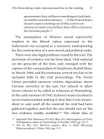
Figure 3.32 Microtubules. (a) A microtubule is composed of 13 protofilaments. Each protofilament is a spiral chain of globular proteins called
tubulin. (b) One of the nine microtubule pairs that form the axonemes of cilia and flagella. (c) One of the nine microtubule triplets that form a centriole.
Table 3.4 Summary of Organelles and Other Cellular Structures
Structure Appearance to TEM Function
Plasma membrane Two dark lines at cell surface, separated by narrow Prevents escape of cell contents; regulates exchange of
(figs. 3.3 and 3.6) light space materials between cytoplasm and extracellular fluid;
involved in intercellular communication
Microvilli Short, densely spaced, hairlike processes or scattered Increase absorptive surface area; some sensory roles
(figs. 3.10 and 3.11a–b) bumps on cell surface; interior featureless or with bundle (hearing, equilibrium, taste)
of microfilaments
Cilia Long hairlike projections of apical cell surface; axoneme Move substances along cell surface; some sensory roles
(figs. 3.11c–e and 3.12) with 9 ϩ 2 array of microtubules (hearing, equilibrium, smell, vision)
Flagellum Long, single, whiplike process with axoneme Sperm motility
Nucleus Largest organelle in most cells, surrounded by double unit Genetic control center of cell; directs protein synthesis
(figs. 3.3 and 3.25) membrane with nuclear pores
Rough ER Extensive sheets of parallel unit membranes with Protein synthesis and manufacture of cellular membranes
(fig. 3.26a) ribosomes on outer surface
Smooth ER Branching network of tubules with smooth surface Lipid synthesis, detoxification, calcium storage
(fig. 3.26b) (no ribosomes); usually broken into numerous small
segments in TEM photos
Ribosomes Small dark granules free in cytosol or on surface of Interpret the genetic code and synthesize polypeptides
(fig. 3.26a) rough ER
Golgi complex Several closely spaced, parallel cisternae with thick edges, Receives and modifies newly synthesized polypeptides,
(fig. 3.27) usually near nucleus, often with many Golgi vesicles nearby synthesizes carbohydrates, adds carbohydrates to
glycoproteins; packages cell products into Golgi vesicles
Golgi vesicles Round to irregular sacs near Golgi complex, usually with Become secretory vesicles and carry cell products to
(fig. 3.27) light, featureless contents apical surface for exocytosis, or become lysosomes
(continued)
Saladin: Anatomy &
Physiology: The Unity of
Form and Function, Third
Edition
3. Cellular Form and
Function
Text
© The McGraw−Hill
Companies, 2003
Chapter 3
124 Part One Organization of the Body
Table 3.4 Summary of Organelles and Other Cellular Structures,
(continued)
Structure Appearance to TEM Function
Lysosomes Round to oval sacs with single unit membrane, often a dark Contain enzymes for intracellular digestion, autophagy,
(fig. 3.28a) featureless interior but sometimes with protein layers programmed cell death, and glucose mobilization
or crystals
Peroxisomes Similar to lysosomes; often lighter in color Contain enzymes for detoxification of free radicals,
(fig. 3.28b) alcohol, and other drugs; oxidize fatty acids
Mitochondria Round, rod-shaped, bean-shaped, or threadlike structures ATP synthesis
(fig. 3.29) with double unit membrane and shelflike infoldings called
cristae
Centrioles Short cylindrical bodies, each composed of a circle of nine Form mitotic spindle during cell division; unpaired
(fig. 3.30) triplets of microtubules centrioles form basal bodies of cilia and flagella
Centrosome Clear area near nucleus containing a pair of centrioles Organizing center for formation of microtubules of
(fig. 3.5) cytoskeleton and mitotic spindle
Basal body Unpaired centriole at the base of a cilium or flagellum Point of origin, growth, and anchorage of a cilium or
(fig. 3.11e) flagellum; produces axoneme
Microfilaments Thin protein filaments (6 nm diameter), often in parallel Support microvilli; involved in muscle contraction and
(figs. 3.10 and 3.31) bundles or dense networks in cytoplasm other cell motility, endocytosis, and cell division
Intermediate filaments Thicker protein filaments (8–10 nm diameter) extending Give shape and physical support to cell; anchor cells to
(fig. 3.31) throughout cytoplasm or concentrated at cell-to-cell each other and to extracellular material; compartmentalize
junctions cell contents
Microtubules Hollow protein cylinders (25 nm diameter) Form axonemes of cilia and flagella, centrioles, basal
(figs. 3.31 and 3.32) bodies, and mitotic spindles; enable motility of cell parts;
direct organelles and macromolecules to their
destinations within a cell
Inclusions Highly variable—fat droplets, glycogen granules, protein Storage products or other products of cellular metabolism,
(fig. 3.26b) crystals, dust, bacteria, viruses; never enclosed in unit or foreign matter retained in cytoplasm
membranes
Insight 3.4 Evolutionary Medicine
Mitochondria—Evolution and
Clinical Significance
It is virtually certain that mitochondria evolved from bacteria that
invaded another primitive cell, survived in its cytoplasm, and became
permanent residents. Certain modern bacteria called ricketsii live in
the cytoplasm of other cells, showing that this mode of life is feasible.
The two unit membranes around the mitochondrion suggest that the
original bacterium provided the inner membrane and the host cell’s
phagosome provided the outer membrane when the bacterium was
phagocytized.
Several comparisons show the apparent relationship of mitochon-
dria to bacteria. Their ribosomes are more like bacterial ribosomes than
those of eukaryotic (nucleated) cells. Mitochondrial DNA (mtDNA) is a
small, circular molecule that resembles the circular DNA of other bac-
teria, not the linear DNA of the cell nucleus. It replicates independently
of nuclear DNA. mtDNA codes for some of the enzymes employed in
ATP synthesis. It consists of 16,569 base pairs (explained in chapter 4),
comprising 37 genes, compared to over a billion base pairs and about
35,000 genes in nuclear DNA.
When a sperm fertilizes an egg, any mitochondria introduced by the
sperm are quickly destroyed and only those provided by the egg are
passed on to the developing embryo. Therefore, all mitochondrial DNA
is inherited exclusively through the mother. While nuclear DNA is
reshuffled in every generation by sexual reproduction, mtDNA remains
unchanged except by random mutation. Biologists and anthropologists
have used mtDNA as a “molecular clock” to trace evolutionary lineages
in humans and other species. mtDNA has also been used as evidence in
criminal law and to identify the remains of soldiers killed in action.
mtDNA was used recently to identify the remains of the famed bandit
Jesse James, who was killed in 1882. Anthropologists have gained evi-
dence, although still controversial, that of all the women who lived in
Africa 200,000 years ago, only one has any descendents still living
today. This “mitochondrial Eve” is ancestor to us all.
mtDNA is very exposed to damage from free radicals normally gen-
erated in mitochondria by aerobic respiration. Yet unlike nuclear DNA,
mtDNA has no effective mechanism for repairing damage. Therefore, it
mutates about ten times as rapidly as nuclear DNA. Some of these
mutations are responsible for rare hereditary diseases. Tissues and
organs with the highest energy demands are the most vulnerable to
mitochondrial dysfunctions—nervous tissue, the heart, the kidneys,
and skeletal muscles, for example. Mitochondrial myopathy is a degen-
erative muscle disease in which the muscle displays “ragged red fibers,”
Saladin: Anatomy &
Physiology: The Unity of
Form and Function, Third
Edition
3. Cellular Form and
Function
Text
© The McGraw−Hill
Companies, 2003
Chapter 3 Cellular Form and Function 125
Concepts of Cellular Structure (p. 94)
1. Cytology is the study of cellular
structure and function.
2. All human structure and function is
the result of cellular activity.
3. Cell shapes are described as
squamous, polygonal, stellate,
cuboidal, columnar, spheroid, ovoid,
discoid, fusiform, and fibrous.
4. Most human cells are 10 to 15 m in
diameter. Cell size is limited in part
by the ratio of surface area to volume.
5. A cell is enclosed in a plasma
membrane and contains usually one
nucleus.
6. The cytoplasm is everything between
the plasma membrane and nucleus. It
consists of a clear fluid, the cytosol or
intracellular fluid (ICF), and
embedded organelles and other
structures. Fluid external to the cell
is extracellular fluid (ECF).
The Cell Surface (p. 98)
1. The plasma membrane is made of
lipid and protein.
2. The most abundant lipid molecules
in the membrane are phospholipids,
which form a bilayer with their
hydrophobic heads facing the ICF
and ECF. Other membrane lipids
include cholesterol and
glycolipids.
3. Membrane proteins are called integral
proteins if they are embedded in the
lipid bilayer and extend all the way
through it, and peripheral proteins if
they only cling to the intracellular
face of the lipid bilayer.
4. Membrane proteins serve as
receptors, second-messenger systems,
enzymes, channels, carriers,
molecular motors, cell-identity
markers, and cell-adhesion
molecules.
5. Channel proteins are called gates if
they can open and close. Gates are
called ligand-regulated, voltage-
regulated, or mechanically regulated
depending on whether they open and
close in response to chemicals,
voltage changes across the membrane,
or mechanical stress.
6. Second-messenger systems are
systems for generating an internal
cellular signal in response to an
external one. One of the best-known
examples results in the formation of
a second messenger, cyclic AMP
(cAMP), within the cell when
certain extracellular signaling
molecules bind to a membrane
receptor.
7. All cells are covered with a
glycocalyx, a layer of carbohydrate
molecules bound to membrane lipids
and proteins. The glycocalyx
functions in immunity and other
forms of protection, cell adhesion,
fertilization, and embryonic
development, among other roles.
8. Microvilli are tiny surface extensions
of the plasma membrane that increase
a cell’s surface area. They are
especially well developed on
absorptive cells, as in the kidney and
small intestine.
9. Cilia are longer, hairlike surface
extensions with a central axoneme,
composed of a 9 ϩ 2 arrangement of
microtubules. Some cilia are
stationary and sensory in function,
and some are motile and propel
substances across epithelial surfaces.
10. A flagellum is a long, solitary,
whiplike extension of the cell surface.
The only functional flagellum in
humans is the sperm tail.
Membrane Transport (p. 106)
1. The plasma membrane is selectively
permeable—it allows some
substances to pass through it but
prevents others from entering or
leaving a cell. There are several
methods of passage through a plasma
membrane.
2. Filtration is the movement of fluid
through a membrane under a physical
force such as blood pressure, while
the membrane holds back relatively
large particles.
3. Simple diffusion is the spontaneous
net movement of particles from a
place of high concentration to a place
of low concentration, such as
respiratory gases moving between the
pulmonary air sacs and the blood.
The speed of diffusion depends on
temperature, molecular weight,
concentration differences, and the
surface area and permeability of the
membrane.
4. Osmosis is the diffusion of water
through a selectively permeable
membrane from the more watery to
the less watery side. Channel
proteins called aquaporins allow
passage of water through plasma
membranes.
5. The speed of osmosis depends on the
relative concentrations, on the two
sides of a membrane, of solute
molecules that cannot penetrate the
membrane. Osmotic pressure, the
physical force that would be required
Chapter Review
Review of Key Concepts
cells with abnormal mitochondria that stain red with a particular his-
tological stain. Mitochondrial encephalomyopathy, lactic acidosis, and
strokelike episodes (MELAS) is a mitochondrial disease involving
seizures, paralysis, dementia, muscle deterioration, and a toxic accu-
mulation of lactic acid in the blood. Leber hereditary optic neuropathy
(LHON) is a form of blindness that usually appears in young adulthood
as a result of damage to the optic nerve. Kearns-Sayre syndrome (KSS)
involves paralysis of the eye muscles, degeneration of the retina, heart
disease, hearing loss, diabetes, and kidney failure. Damage to mtDNA
has also been implicated as a possible factor in Alzheimer disease,
Huntington disease, and other degenerative diseases of old age.
Chapter 3
Saladin: Anatomy &
Physiology: The Unity of
Form and Function, Third
Edition
3. Cellular Form and
Function
Text
© The McGraw−Hill
Companies, 2003
Chapter 3
126 Part One Organization of the Body
to stop osmosis, is proportional to the
concentration of nonpermeating
solutes on the side to which water is
moving.
6. An osmole is one mole of dissolved
particles in a solution. Osmolarity is
the number of osmoles of solute per
liter of solution. The osmolarity of
body fluids is usually expressed in
milliosmoles per liter (mOsm/L).
7. Tonicity is the ability of a solution to
affect the fluid volume and pressure
in a cell. A solution is hypotonic,
isotonic, or hypertonic to a cell if it
contains, respectively, a lower, equal,
or greater concentration of
nonpermeating solutes than the cell
cytoplasm does. Cells swell and burst
in hypotonic solutions and shrivel in
hypertonic solutions.
8. Carrier-mediated transport employs
membrane proteins to move solutes
through a membrane. A given carrier is
usually specific for a particular solute.
9. Membrane carriers can become
saturated with solute molecules and
then unable to work any faster. The
maximum rate of transport is the
transport maximum (T
m
).
10. A uniport is a carrier that transports
only one solute at a time; a symport
carries two or more solutes through
the membrane in the same direction
(a process called cotransport); and an
antiport carries two or more solutes
in opposite directions (a process
called countertransport).
11. Facilitated diffusion is a form of
carrier-mediated transport that moves
solutes through a membrane down a
concentration gradient, without an
expenditure of ATP.
12. Active transport is a form of carrier-
mediated transport that moves
solutes through a membrane up
(against) a concentration gradient,
with the expenditure of ATP.
13. The Na
ϩ
-K
ϩ
pump is an antiport that
moves Na
ϩ
out of a cell and K
ϩ
into
it. It serves for control of cell volume,
secondary active transport, heat
production, and maintenance of an
electrical membrane potential.
14. Vesicular transport is the movement
of substances in bulk through a
membrane in membrane-enclosed
vesicles.
15. Endocytosis is any form of vesicular
transport that brings material into a
cell, including phagocytosis,
pinocytosis, and receptor-mediated
endocytosis.
16. Exocytosis is a form of vesicular
transport that discharges material
from a cell. It functions in the release
of cell products and in replacement
of plasma membrane removed by
endocytosis.
The Cytoplasm (p. 115)
1. The cytoplasm is composed of a clear
gelatinous cytosol in which are
embedded organelles, the cytoskeleton,
and inclusions (table 3.4).
2. Organelles are internal structures in
the cytoplasm that carry out
specialized tasks for a cell.
3. Membranous organelles are enclosed
in one or two layers of unit
membrane similar to the plasma
membrane. These include the
nucleus, endoplasmic reticulum
(which has rough and smooth
portions), ribosomes, the Golgi
complex, lysosomes, peroxisomes,
and mitochondria. The centrioles and
ribosomes are nonmembranous
organelles.
4. The cytoskeleton is a supportive
framework of protein filaments and
tubules in a cell. It gives a cell its
shape, organizes the cytoplasmic
contents, and functions in
movements of cell contents and the
cell as a whole. It is composed of
microfilaments of the protein actin;
intermediate filaments of keratin or
other proteins; and cylindrical
microtubules of the protein
tubulin.
5. Inclusions are either stored cellular
products such as glycogen, pigments,
and fat, or foreign bodies such as
bacteria, viruses, and dust. Inclusions
are not vital to cell survival.
Selected Vocabulary
cytoplasm 96
plasma membrane 97
organelle 97
cytoskeleton 97
cytosol 97
intracellular fluid 97
extracellular fluid 97
receptor 100
channel protein 100
ligand-regulated gate 100
voltage-regulated gate 100
carrier 101
microvillus 103
cilium 103
filtration 106
simple diffusion 106
osmosis 107
osmolarity 108
hypotonic 108
hypertonic 108
isotonic 108
uniport 110
symport 110
antiport 110
facilitated diffusion 110
active transport 110
sodium-potassium pump 110
endocytosis 112
exocytosis 112
phagocytosis 112
endoplasmic reticulum 116
ribosome 118
Golgi complex 118
lysosome 119
peroxisome 119
mitochondrion 120
centriole 121
microfilament 120
intermediate filament 120
microtubule 121
Saladin: Anatomy &
Physiology: The Unity of
Form and Function, Third
Edition
3. Cellular Form and
Function
Text
© The McGraw−Hill
Companies, 2003
Chapter 3
Chapter 3 Cellular Form and Function 127
True or False
Determine which five of the following
statements are false, and briefly
explain why.
1. If a cell were poisoned so it could not
make ATP, osmosis through its
membrane would cease.
2. Material can move either into a cell
or out by means of active transport.
3. A cell’s second messengers serve
mainly to transport solutes through
the membrane.
4. The Golgi complex makes lysosomes
but not peroxisomes.
5. Some membrane channels are
peripheral proteins.
6. The plasma membrane consists
primarily of protein molecules.
7. The brush border of a cell is
composed of cilia.
8. Human cells swell or shrink in any
solution other than an isotonic
solution.
9. Osmosis is not limited by the
transport maximum (T
m
).
10. It is very unlikely for a cell to have
more centrosomes than ribosomes.
Answers in Appendix B
Answers in Appendix B
Testing Your Recall
1. The clear, structureless gel in a cell
is its
a. nucleoplasm.
b. protoplasm.
c. cytoplasm.
d. neoplasm.
e. cytosol.
2. The Na
ϩ
-K
ϩ
pump is
a. a peripheral protein.
b. an integral protein.
c. a G protein.
d. a glycolipid.
e. a phospholipid.
3. Which of the following processes
could occur only in the plasma
membrane of a living cell?
a. facilitated diffusion
b. simple diffusion
c. filtration
d. active transport
e. osmosis
4. Cells specialized for absorption of
matter from the ECF are likely to
show an abundance of
a. lysosomes.
b. microvilli.
c. mitochondria.
d. secretory vesicles.
e. ribosomes.
5. Osmosis is a special case of
a. pinocytosis.
b. carrier-mediated transport.
c. active transport.
d. facilitated diffusion.
e. simple diffusion.
6. Membrane carriers resemble enzymes
except for the fact that carriers
a. are not proteins.
b. do not have binding sites.
c. are not selective for particular
ligands.
d. change conformation when they
bind a ligand.
e. do not chemically change their
ligands.
7. The cotransport of glucose derives
energy from
a. a Na
ϩ
concentration gradient.
b. the glucose being transported.
c. a Ca
2ϩ
gradient.
d. the membrane voltage.
e. body heat.
8. The function of cAMP in a cell is
a. to activate a G protein.
b. to remove phosphate groups
from ATP.
c. to activate kinases.
d. to bind to the first messenger.
e. to add phosphate groups to
enzymes.
9. Most cellular membranes are made by
a. the nucleus.
b. the cytoskeleton.
c. enzymes in the peroxisomes.
d. the endoplasmic reticulum.
e. replication of existing membranes.
10. Matter can leave a cell by any of the
following means except
a. active transport.
b. pinocytosis.
c. an antiport.
d. simple diffusion.
e. exocytosis.
11. Most human cells are 10 to 15 ______
in diameter.
12. When a hormone cannot enter a cell,
it activates the formation of a/an
______ inside the cell.
13. ______ gates in the plasma membrane
open or close in response to changes
in the electrical charge difference
across the membrane.
14. The force exerted on a membrane by
water is called ______ .
15. A concentrated solution that causes a
cell to shrink is ______ to the cell.
16. Fusion of a secretory vesicle with the
plasma membrane, and release of the
vesicle’s contents, is called ______ .
17. Two organelles that are surrounded
by a double unit membrane are the
______ and the ______ .
18. Liver cells can detoxify alcohol with
two organelles, the ______ and ______.
19. An ion gate in the plasma membrane
that opens or closes when a chemical
binds to it is called a/an ______ .
20. The space enclosed by the unit
membrane of the Golgi complex
and endoplasmic reticulum is called
the ______ .
Saladin: Anatomy &
Physiology: The Unity of
Form and Function, Third
Edition
3. Cellular Form and
Function
Text
© The McGraw−Hill
Companies, 2003
Chapter 3
128 Part One Organization of the Body
Answers to Figure Legend Questions
3.9 Adenylate cyclase is integral. The G
protein is peripheral.
3.19 The Na
ϩ
-K
ϩ
pump requires ATP,
whereas osmosis does not. A dead
cell ceases to produce ATP.
3.23 Transcytosis is simply a
combination of endocytosis and
exocytosis.
3.25 Proteins and mRNA must be able to
move through the nuclear envelope.
These large molecules require large
pores for their passage.
3.30 A centriole has 27 microtubules—
9 groups of 3 each.
www.mhhe.com/saladin3
The Online Learning Center provides a wealth of information fully organized and integrated by chapter. You will find practice quizzes, interac-
tive activities, labeling exercises, flashcards, and much more that will complement your learning and understanding of anatomy and physiology.
Testing Your Comprehension
1. If someone bought a saltwater fish in
a pet shop and put it in a freshwater
aquarium at home, what would
happen to the fish’s cells? What
would happen if someone put a
freshwater fish in a saltwater
aquarium? Explain.
2. A farmer’s hand and forearm are
badly crushed in a hay bailer. Upon
hospital examination, his blood
potassium level is found to be
abnormal. Would you expect it to be
higher or lower than normal? Explain.
3. Many children worldwide suffer from
a severe deficiency of dietary protein.
As a result, they have very low levels
of blood albumin. How do you think
this affects the water content and
volume of their blood? Explain.
4. It is often said that mitochondria
make energy for a cell. Why is this
statement false?
5. Kartagener syndrome is a hereditary
disease in which dynein arms are
lacking from the axonemes of cilia
and flagella. Predict the effect of
Kartagener syndrome on a man’s
ability to father a child. Predict its
effect on his respiratory health.
Explain both answers.
Answers at the Online Learning Center
Saladin: Anatomy &
Physiology: The Unity of
Form and Function, Third
Edition
4. Genetics and Cellular
Function
Text
© The McGraw−Hill
Companies, 2003
The Nucleic Acids 130
• Organization of the Chromatin 130
• DNA Structure and Function 130
• RNA Structure and Function 133
Protein Synthesis and Secretion 134
• Preview 134
• The Genetic Code 134
• Transcription 136
• Translation 136
• Chaperones and Protein Structure 137
• Posttranslational Modification 138
• Packaging and Secretion 139
DNA Replication and the Cell Cycle 139
• DNA Replication 139
• Errors and Mutations 142
• The Cell Cycle 142
• Mitosis 143
• Timing of Cell Division 145
Chromosomes and Heredity 145
• The Karyotype 146
• Genes and Alleles 147
• Multiple Alleles, Codominance, and
Incomplete Dominance 148
• Polygenic Inheritance and Pleiotropy 148
• Sex Linkage 149
• Penetrance and Environmental Effects 149
• Dominant and Recessive Alleles at the
Population Level 149
Chapter Review 152
INSIGHTS
4.1 Medical History: Miescher and the
Discovery of DNA 130
4.2 Medical History: Discovery of the
Double Helix 132
4.3 Clinical Application: Can We
Replace Brain Cells? 143
4.4 Clinical Application: Cancer 151
4
CHAPTER
Genetics and
Cellular Function
A single DNA molecule spilling from a ruptured bacterial cell (TEM)
CHAPTER OUTLINE
Brushing Up
To understand this chapter, it is important that you understand or
brush up on the following concepts:
• Levels of protein structure (p. 80)
• Functions of proteins (p. 80)
• Exocytosis (p. 114)
• Ribosomes, rough endoplasmic reticulum, and Golgi
complex (pp. 116–119)
• Centrioles and microtubules (pp. 120, 121)
129
Saladin: Anatomy &
Physiology: The Unity of
Form and Function, Third
Edition
4. Genetics and Cellular
Function
Text
© The McGraw−Hill
Companies, 2003
Chapter 4
130 Part One Organization of the Body
S
ome of the basic ideas of heredity have been known since
antiquity, but a scientific understanding of how traits are
passed from parent to offspring began with the Austrian monk
Gregor Mendel (1822–84) and his famous experiments on garden
peas. In the early twentieth century, the importance of Mendel’s
work was realized and chromosomes were first seen with the
microscope. Cytogenetics now uses techniques of cytology and
microscopy to study chromosomes and their relationship to hered-
itary traits. Molecular genetics uses the techniques of biochem-
istry to study the structure and function of DNA. In this chapter,
we bring together some of the findings of molecular genetics,
cytogenetics, and mendelian heredity to explore what the genes
are, how they regulate cellular function, and how they are passed
on when cells divide and people reproduce. A few basic concepts
of heredity are introduced as a foundation for understanding con-
cepts ranging from color blindness to blood types in the chapters
that follow.
The Nucleic Acids
Objectives
When you have completed this section, you should be able to
• describe how DNA is organized in the nucleus; and
• compare the structures and functions of DNA and RNA.
With improvements in the microscope, nineteenth-century
cytologists saw that the nucleus divides in preparation for
cell division, and they came to regard the nucleus as the
most likely center of heredity. This led to a search for the
biochemical keys to heredity in the nucleus, and thus to
the discovery of deoxyribonucleic acid (DNA) (insight 4.1).
DNA directly or indirectly regulates all cellular form and
function.
Insight 4.1 Medical History
Miescher and the Discovery of DNA
Swiss biochemist Johann Friedrich Miescher (1844–95) was one of
the first scientists intent on identifying the hereditary material in
nuclei. In order to isolate nuclei with minimal contamination,
Miescher chose to work with cells that have large nuclei and very lit-
tle cytoplasm. At first he chose white blood cells extracted from the
pus in used bandages from a hospital; later, he used the sperm of
salmon—probably more agreeable to work with than used bandages!
Miescher isolated an acidic substance rich in phosphorus, which he
named nuclein. His student, Richard Altmann, later called it nucleic
acid—a term we now use for both DNA and RNA. Miescher correctly
guessed that “nuclein” (DNA) was the hereditary matter of the cell,
but he was unable to provide strong evidence for this conjecture,
and his work was harshly criticized. He died of tuberculosis at the
age of 51.
Organization of the Chromatin
A human cell usually has 46 molecules of DNA with an
average length of 44 mm (total slightly over 2 m). Each
molecule is 2 nm in diameter. To put this in perspective,
if a DNA molecule were the thickness of a telephone pole
(20 cm, or 8 in.), it would reach about 4,400 km (2,700 mi)
into space—far higher than the orbits of satellites and
space shuttles. Imagine trying to make a pole 20 cm thick
and 4,400 km long without breaking it! The problem for a
cell is even greater. It has 46 DNA molecules packed
together in a single nucleus, and it has to make an exact
copy of every one of them and distribute these equally to
its two daughter cells when the cell divides. Keeping the
DNA organized and intact is a tremendous feat.
Molecular biology and high-resolution electron
microscopy have provided some insight into how this task
is accomplished. Chromatin looks like a granular thread
(fig. 4.1a). The granules, called nucleosomes, consist of a
cluster of eight proteins called histones, with the DNA
molecule wound around the cluster. Histones serve as
spools that protect and organize the DNA. Other nuclear
proteins called nonhistones seem to provide structural
support for the chromatin and regulate gene activity.
Winding DNA around the nucleosomes makes the
chromatin shorter and more compact, but chromatin also
has higher orders of structure. The “granular thread,” about
10 nm wide, further twists into a coil about 30 nm wide.
When a cell prepares to undergo division, the chromatin
further supercoils into a fiber about 200 nm wide (fig. 4.1b).
Thus, the 2 m of DNA in each cell becomes shortened and
compacted in an orderly way that prevents tangling and
breakage without interfering with genetic function.
DNA Structure and Function
Nucleic acids are polymers of nucleotides (NEW-clee-oh-
tides). A nucleotide consists of a sugar, a phosphate group,
and a single- or double-ringed nitrogenous (ny-TRODJ-eh-
nus) base. Three bases—cytosine (C), thymine (T), and
uracil (U)—have a single carbon-nitrogen ring and are clas-
sified as pyrimidines (py-RIM-ih-deens). The other two
bases—adenine (A) and guanine (G)—have double rings
and are classified as purines (fig. 4.2). The bases of DNA are
C, T, A, and G, whereas the bases of RNA are C, U, A, and G.
The structure of DNA resembles a ladder (fig. 4.3a).
Each sidepiece is a backbone composed of phosphate
groups alternating with the sugar deoxyribose. The step-
like connections between the backbones are pairs of
nitrogenous bases. Imagine this as a soft rubber ladder that
you can twist, so that the two backbones become entwined
to resemble a spiral staircase. This is analogous to the
shape of the DNA molecule, described as a double helix.
The nitrogenous bases face the inside of the helix and
hold the two backbones together with hydrogen bonds.
Across from a purine on one backbone, there is a pyrimidine
Saladin: Anatomy &
Physiology: The Unity of
Form and Function, Third
Edition
4. Genetics and Cellular
Function
Text
© The McGraw−Hill
Companies, 2003
Chapter 4
131
Metaphase
chromosome
Chromatid
(700 nm in
diameter)
Supercoiled
structure
(200 nm in
diameter)
Chromatin
fiber
(10 nm in
diameter)
Nucleosome
Histones
DNA
(2 nm in
diameter)
(b)
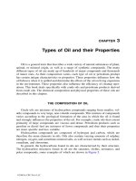
Figure 4.1 Chromatin Structure. (a) Nuclear contents of a germ cell
from an 8-week-old human embryo (colorized SEM). The center mass is the
nucleolus. It is surrounded by granular fibers of chromatin. Each granule is a
nucleosome. (b) The coiling of chromatin and its relationship to the
histones. Supercoiling beyond the 10-nm level occurs only during mitosis.
50 nm
(a)
HC
N
C
N
NH
2
N
H
C
C
CH
N
H
CH
2
O
C
NH
2
N
NH
C
CH
C
H
N
NC
HO
O
OH
P
H
HOH
HH
O
Adenine
Adenine (A)
Purines
C
O
N
NH
C
CH
CN
HN C
NH
2
Guanine (G)
H
C
NH
2
C
N
H
C
HC N
O
Cytosine (C)
Uracil (U)
C
C
O
C
O
CH
HN CH
N
H
N
H
C
C
HC
CH
3
NH
O
O
Thymine (T)
Phosphate Deoxyribose
Pyrimidines
(
b
)
(a)
Figure 4.2 Nucleotides and Nitrogenous Bases. (a) The
structure of a nucleotide, one of the monomers of DNA and RNA. In RNA,
the sugar is ribose. (b) The five nitrogenous bases found in DNA and RNA
nucleotides.
Saladin: Anatomy &
Physiology: The Unity of
Form and Function, Third
Edition
4. Genetics and Cellular
Function
Text
© The McGraw−Hill
Companies, 2003
Chapter 4
132 Part One Organization of the Body
on the other. A given purine cannot arbitrarily bind to just
any pyrimidine. Adenine and thymine form two hydrogen
bonds with each other, and guanine and cytosine form three,
as shown in figure 4.3b. Therefore, wherever there is an A
on one backbone, there is a T across from it, and every C is
paired with a G. A–T and C–G are called the base pairs. The
fact that one strand governs the base sequence of the other is
called the law of complementary base pairing. It enables us
to predict the base sequence of one strand if we know the
sequence of the complementary strand. The pairing of each
small, single-ringed pyrimidine with a large, double-ringed
purine gives the DNA molecule its uniform 2-nm width.
Think About It
What would be the base sequence of the DNA strand
across from ATTGACTCG? If a DNA molecule were
known to be 20% adenine, predict its percentage of
cytosine and explain your answer.
Insight 4.2 Medical History
Discovery of the Double Helix
The components of DNA were known by 1900—the sugar, phosphate,
and bases—but the technology did not exist then to determine how
they were put together. The credit for that discovery went mainly to
James Watson and Francis Crick in 1953 (fig. 4.4). The events sur-
rounding their discovery of the double helix represent one of the most
dramatic stories of modern science—the subject of many books and a
movie. When Watson and Crick came to share a laboratory at Cam-
bridge University in 1951, both had barely begun their careers. Watson,
age 23, had just completed his Ph.D. in the United States, and Crick, 11
years older, was a doctoral candidate. Yet the two were about to
become the most famous molecular biologists of the twentieth cen-
tury, and the discovery that won them such acclaim came without a
single laboratory experiment of their own.
Others were fervently at work on DNA, including Rosalind
Franklin and Maurice Wilkins at King’s College in London. Using a
technique called X-ray diffraction, Franklin had determined that
DNA had a repetitious helical structure with sugar and phosphate on
the outside of the helix. Without her permission, Wilkins showed one
of Franklin’s best X-ray photographs to Watson. Watson said, “The
instant I saw the picture my mouth fell open and my pulse began to
race.” It provided a flash of insight that allowed the Watson and
Crick team to beat Franklin to the goal. They were quickly able to
piece together a scale model from cardboard and sheet metal that
fully accounted for the known geometry of DNA. They rushed a
paper into print in 1953 describing the double helix, barely men-
tioning the importance of Franklin’s two years of painstaking X-ray
diffraction work in unlocking the mystery of life’s most important
molecule.
For this discovery, Watson, Crick, and Wilkins shared the Nobel Prize
in 1962. Nobel Prizes are awarded only to the living, and in the final
irony of her career, Rosalind Franklin had died in 1958, at the age of
37, of a cancer possibly induced by the X rays that were her window on
DNA architecture.
(a)
(b)
(c)
A
A
A
A
A
A
A
T
T
T
T
T
T
G
G
G
G
G
C
C
C
C
C
G
A
C
T
G
G
C
C
Sugar-phosphate
backbone
Sugar-phosphate
backbone
Complementary
base pairing
Hydrogen
bond
Figure 4.3 DNA Structure. (a) The “twisted ladder” structure. The two sugar-phosphate backbones twine around each other while complementary
bases (colored bars) face each other on the inside of the double helix. (b) A small segment of DNA showing the composition of the backbone and
complementary pairing of the nitrogenous bases. (c) A molecular space-filling model of DNA giving some impression of its actual geometry.
How would the uniform 2-nm diameter of DNA be affected if two purines or two pyrimidines could pair with each other?
Saladin: Anatomy &
Physiology: The Unity of
Form and Function, Third
Edition
4. Genetics and Cellular
Function
Text
© The McGraw−Hill
Companies, 2003
Chapter 4
Chapter 4 Genetics and Cellular Function 133
The essential function of DNA is to serve as a code
for the structure of polypeptides synthesized by a cell. A
gene is a DNA nucleotide sequence that codes for one
polypeptide. The next section of this chapter explains in
detail how the genes direct polypeptide synthesis. All the
genes of one person are called the genome (JEE-nome);
geneticists estimate that a human has about 35,000 genes.
These account for only 3% of our DNA; the other 97%
does not code for anything. Some of the noncoding DNA
serves important organizing roles in the chromatin, and
some of it is useless “junk DNA” that has accumulated
over the course of human evolution. The latest triumph of
molecular genetics is the human genome project, an enor-
mous multinational effort that led to the mapping of the
base sequence of the entire human genome. Its completion
(in all but some fine details) in June 2000 was hailed as a
scientific achievement comparable to putting the first man
on the moon.
RNA Structure and Function
DNA directs the synthesis of proteins by means of its
smaller cousins, the ribonucleic acids (RNAs). There are
three types of RNA: messenger RNA (mRNA), ribosomal
RNA (rRNA), and transfer RNA (tRNA). Their individual
roles are described shortly. For now we consider what
they have in common and how they differ from DNA
(table 4.1). The most significant difference is that RNA
is much smaller, ranging from about 70 to 90 bases in
tRNA to slightly over 10,000 bases in the largest mRNA.
DNA, by contrast, may be over a billion base pairs long.
Also, while DNA is a double helix, RNA consists of only
one nucleotide chain, not held together by complemen-
tary base pairs except in certain regions of tRNA where
the molecule folds back on itself. The sugar in RNA is
ribose instead of deoxyribose, and one of the pyrim-
idines of DNA, thymine, is replaced by uracil (U) in
RNA (see fig. 4.2).
The essential function of RNA is to interpret the
code in DNA and direct the synthesis of proteins. RNA
works mainly in the cytoplasm, while DNA remains
safely behind in the nucleus, “giving orders” from there.
This process is described in the next section of this
chapter.
Before You Go On
Answer the following questions to test your understanding of the
preceding section:
1. What is the difference between DNA and chromatin?
2. What are the three components of a nucleotide?
Which component varies from one nucleotide to another
in DNA?
3. What two factors govern the pattern of base pairing
in DNA?
4. Summarize the differences between DNA and RNA.
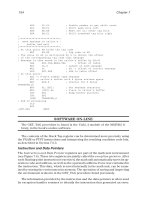
Figure 4.4 Discoverers of the Double Helix. (a) Rosalind
Franklin (1920–58), whose painstaking X-ray diffraction photographs
revealed important information about the basic geometry of DNA.
(b) One of Franklin’s X-ray photographs. (c) James Watson (1928–) (left)
and Francis Crick (1916–) (right), with their model of the double helix.
(a)
(b)
(c)
Saladin: Anatomy &
Physiology: The Unity of
Form and Function, Third
Edition
4. Genetics and Cellular
Function
Text
© The McGraw−Hill
Companies, 2003
Chapter 4
134 Part One Organization of the Body
Protein Synthesis and Secretion
Objectives
When you have completed this section, you should be able to
• define genetic code and describe how DNA codes for protein
structure;
• describe the process of assembling amino acids to form a
protein;
• explain what happens to a protein after its amino acid
sequence has been synthesized; and
• explain how DNA indirectly regulates the synthesis of
nonprotein molecules.
Everything a cell does ultimately results from the action of
its proteins; DNA directs the synthesis of those proteins.
Cells, of course, synthesize many other substances as
well—glycogen, fat, phospholipids, steroids, pigments,
and so on. There are no genes for these cell products, but
their synthesis depends on enzymes that are coded for by
the genes. For example, even though a cell of the testis has
no genes for testosterone, testosterone synthesis is indi-
rectly under genetic control (fig. 4.5). Since testosterone
strongly influences such behaviors as aggression and sex-
ual drive (in both sexes), we can see that genes also make
a significant contribution to behavior. In this section, we
examine how protein synthesis results from the instruc-
tions given in the genes.
Preview
Before studying the details of protein synthesis, it will
be helpful to consider the big picture. In brief, DNA con-
tains a genetic code that specifies which proteins a cell
can make. All the body’s cells except the sex cells con-
tain identical genes, but different genes are activated in
different cells; for example, the genes for digestive
enzymes are active in stomach cells but not in muscle
cells. When a gene is activated, a molecule of messenger
RNA (mRNA), a sort of mirror-image copy of the gene, is
made. Most mRNA migrates from the nucleus to the
cytoplasm, where its code is “read” by a ribosome. Ribo-
somes are composed of ribosomal RNA (rRNA) and
enzymes. Transfer RNA (tRNA) delivers amino acids to
the ribosome, and the ribosome chooses from among
these to assemble amino acids in the order directed by
the mRNA.
In summary, you can think of the process of protein
synthesis as DNA→mRNA→protein, with each arrow
reading as “codes for the production of.” The step from
DNA to mRNA is called transcription, and the step from
mRNA to protein is called translation. Transcription
occurs in the nucleus, where the DNA is, and most trans-
lation occurs in the cytoplasm. Recent research has
shown, however, that 10% to 15% of proteins are synthe-
sized in the nucleus, with both steps occurring there.
The Genetic Code
The body makes more than 2 million different proteins,
all from the same 20 amino acids and all encoded by
genes made of just 4 nucleotides (A, T, C, G)—a striking
illustration of how a great variety of complex structures
can be made from a small variety of simpler compo-
nents. The genetic code is a system that enables these 4
nucleotides to code for the amino acid sequences of all
proteins.
It is not unusual for simple codes to represent com-
plex information. Computers store and transmit complex
information, including pictures and sounds, in a binary
code with only the symbols 1 and 0. It is not surprising,
then, that a mere 20 amino acids can be represented by a
code of 4 nucleotides; all that is required is to combine
these symbols in varied ways. It requires more than 2
nucleotides to code for each amino acid, because A, U, C,
and G can combine in only 16 ways (AA, AU, AC, AG, UA,
UU, etc.). The minimum code to symbolize 20 amino acids
is 3 nucleotides per amino acid, and indeed this is the case
in DNA. A sequence of 3 DNA nucleotides that stands for
1 amino acid is called a base triplet. The “mirror image”
Table 4.1 Comparison of DNA and RNA
Feature DNA RNA
Sugar Deoxyribose Ribose
Nitrogenous bases A, T, C, G A, U, C, G
Number of nucleotide chains Two (double helix) One
Number of nitrogenous bases 10
8
to 10
9
base pairs 70 to 10,000 unpaired bases
Site of action Functions in nucleus; cannot leave Leaves nucleus; functions in cytoplasm
Function Codes for synthesis of RNA and protein Carries out the instructions in DNA; assembles proteins
Saladin: Anatomy &
Physiology: The Unity of
Form and Function, Third
Edition
4. Genetics and Cellular
Function
Text
© The McGraw−Hill
Companies, 2003
Chapter 4
Chapter 4 Genetics and Cellular Function 135
sequence in mRNA is called a codon. The genetic code is
expressed in terms of codons.
Table 4.2 shows a few representative triplets and
codons along with the amino acids they represent. You
can see from this listing that two or more codons can rep-
resent the same amino acid. The reason for this is easy to
explain mathematically. Four symbols (N) taken three at
a time (x) can be combined in N
x
different ways; that is,
there are 4
3
ϭ 64 possible codons available to represent
the 20 amino acids. Only 61 of these code for amino
acids. The other 3—UAG, UGA, and UAA—are called
stop codons; they signal “end of message,” like the
period at the end of a sentence. A stop codon enables the
cell’s protein-synthesizing machinery to sense that it has
reached the end of the gene for a particular protein. The
codon AUG plays two roles—it serves as a code for
methionine and as a start codon. This dual function is
explained shortly.
Translation
mRNA
DNA
Transcription
Enzyme
Activated
enzyme
Cholesterol
From pituitary
ICSH
Second
messenger
Interstitial cell of testis
HO
Testosterone
Secreted
CH
3
CH
3
CH
3
OH
O
1
2
4
3
5 6
Figure 4.5 Indirect Control of Testosterone Synthesis by DNA. There is no gene for testosterone, but DNA regulates its synthesis through the
enzymes for which it does code. (1) DNA codes for mRNA. (2) In the cytoplasm, mRNA directs the synthesis of an enzyme. (3) When testosterone is
needed, luteinizing hormone (LH) stimulates production of a second messenger within cells of the testis. (4) The second-messenger system activates the
enzyme encoded by the mRNA. (5) The enzyme converts cholesterol to testosterone. (6) Testosterone is secreted from the cell and exerts various
anatomical, physiological, and behavioral effects.
Table 4.2 Examples of the Genetic Code
Base Triplet Codon of Name of Abbreviation for
of DNA mRNA Amino Acid Amino Acid
CCT GGA Glycine Gly
CCA GGU Glycine Gly
CCC GGG Glycine Gly
CTC GAG Glutamic acid Glu
CGC GCG Alanine Ala
CGT GCA Alanine Ala
TGG ACC Threonine Thr
TGC ACG Threonine Thr
GTA CAU Valine Val
TAC AUG Methionine Met
Saladin: Anatomy &
Physiology: The Unity of
Form and Function, Third
Edition
4. Genetics and Cellular
Function
Text
© The McGraw−Hill
Companies, 2003
Chapter 4
136 Part One Organization of the Body
Transcription
Most protein synthesis occurs in the cytoplasm, but DNA
is too large to leave the nucleus. It is necessary, therefore,
to make a small RNA copy that can migrate through a
nuclear pore into the cytoplasm. Just as we might tran-
scribe (copy) a document, transcription in genetics means
the process of copying genetic instructions from DNA to
RNA. It is triggered by chemical messengers from the cyto-
plasm that enter the nucleus and bind to the chromatin at
the site of the relevant gene. An enzyme called RNA poly-
merase (po-LIM-ur-ase) then binds to the DNA at this
point and begins making RNA. Certain base sequences
(often TATATA or TATAAA) inform the polymerase where
to begin.
RNA polymerase opens up the DNA helix about 17
base pairs at a time. It transcribes the bases from one
strand of the DNA and makes a corresponding RNA.
Where it finds a C on the DNA, it adds a G to the RNA;
where it finds an A, it adds a U; and so forth. The enzyme
then rewinds the DNA helix behind it. Another RNA poly-
merase may follow closely behind the first one; thus, a
gene may be transcribed by several polymerase molecules
at once, and numerous copies of the same RNA are made.
At the end of the gene is a base sequence that serves as a
terminator, which signals the polymerase to release the
RNA and separate from the DNA.
The RNA produced by transcription is an “imma-
ture” form called pre-mRNA. This molecule contains
“sense” portions called exons that will be translated into
a peptide and “nonsense” portions called introns that
must be removed before translation. Enzymes remove the
introns and splice the exons together into a functional
mRNA molecule.
Translation
Just as we might translate a work from Spanish into Eng-
lish, genetic translation converts the language of
nucleotides into the language of amino acids (fig. 4.6).
This job is done by ribosomes, which are found mainly in
the cytosol and on the rough ER and nuclear envelope. A
ribosome consists of two granular subunits, large and
small, each made of several rRNA and enzyme molecules.
The mRNA molecule begins with a leader sequence
of bases that are not translated to protein but serve as a
binding site for the ribosome. The small ribosomal subunit
binds to it, the large subunit joins the complex, and the
ribosome begins pulling the mRNA through it like a rib-
bon, reading bases as it goes. When it reaches the start
codon, AUG, it begins making protein. Since AUG codes
for methionine, all proteins begin with methionine when
first synthesized, although this may be removed later.
Translation requires the participation of 61 types of
transfer RNA (tRNA), one for each codon (except stop
codons). Transfer RNA is a small RNA molecule that turns
back and coils on itself to form a cloverleaf shape, which
is then twisted into an angular L-shape (fig. 4.7). One end
of the L includes three nucleotides called an anticodon,
and the other end has a binding site specific for one amino
acid. Each tRNA picks up an amino acid from a pool of free
amino acids in the cytosol. One ATP molecule is used to
bind the amino acid to this site and provide the energy that
is used later to join that amino acid to the growing protein.
Thus, protein synthesis consumes one ATP for each pep-
tide bond formed.
When the small ribosomal subunit reads a codon
such as CGC, it must find an activated tRNA with the cor-
responding anticodon; in this case, GCG. This particular
tRNA would have the amino acid alanine at its other end.
The ribosome binds and holds this tRNA and then reads
the next codon—say GGU. Here, it would bind a tRNA
with anticodon CCA, which carries glycine.
The large ribosomal subunit contains an enzyme that
forms peptide bonds, and now that alanine and glycine are
side by side, it links them together. The first tRNA is no
longer needed, so it is released from the ribosome. The
second tRNA is used, temporarily, to anchor the growing
peptide to the ribosome. Now, the ribosome reads the third
codon—say GUA. It finds the tRNA with the anticodon
CAU, which carries the amino acid valine. The large sub-
unit adds valine to the growing chain, now three amino
acids long. By repetition of this process, the entire protein
is assembled. Eventually, the ribosome reaches a stop
codon and is finished translating this mRNA. The
polypeptide is turned loose, and the ribosome dissociates
into its two subunits.
One ribosome can assemble a protein of 400 amino
acids in about 20 seconds, but it does not work at the
task alone. After the mRNA leader sequence passes
through one ribosome, a neighboring ribosome takes it
up and begins translating the mRNA before the first
ribosome has finished. One mRNA often holds 10 or 20
ribosomes together in a cluster called a polyribosome
(fig. 4.8). Not only is each mRNA translated by all these
ribosomes at once, but a cell may have 300,000 identical
mRNA molecules undergoing simultaneous translation.
Thus, a cell may produce over 150,000 protein mole-
cules per second—a remarkably productive protein fac-
tory! As much as 25% of the dry weight of liver cells,
which are highly active in protein synthesis, is com-
posed of ribosomes.
Many proteins, when first synthesized, begin with a
chain of amino acids called the signal peptide. Like a
molecular address label, the signal peptide determines the
protein’s destination—for example, whether it will be sent
to the rough endoplasmic reticulum, a peroxisome, or a
mitochondrion. (Proteins used in the cytosol lack signal
peptides.) Some diseases result from errors in the signal
peptide, causing a protein to be sent to the wrong address,
Saladin: Anatomy &
Physiology: The Unity of
Form and Function, Third
Edition
4. Genetics and Cellular
Function
Text
© The McGraw−Hill
Companies, 2003
Chapter 4
Chapter 4 Genetics and Cellular Function 137
such as going to a mitochondrion when it should have
gone to a peroxisome, or causing it to be secreted from a
cell when it should have been stored in a lysosome.
Gunter Blöbel of Rockefeller University received the 1999
Nobel Prize for Physiology or Medicine for discovering
signal peptides in the 1970s.
Figure 4.9 summarizes transcription and translation
and shows how a nucleotide sequence translates to a
hypothetical peptide of 6 amino acids. A protein 500
amino acids long would have to be represented, at a min-
imum, by a sequence of 1,503 nucleotides (3 for each
amino acid, plus a stop codon). The average gene is prob-
ably around 1,200 nucleotides long; a few may be 10 times
this long.
Chaperones and Protein Structure
The amino acid sequence of a protein (primary structure)
is only the beginning; the end of translation is not the end
of protein synthesis. The protein now coils or folds into
its secondary and tertiary structures and, in some cases,
associates with other polypeptide chains (quaternary
structure) or conjugates with a nonprotein moiety, such as
a vitamin or carbohydrate. It is essential that these
processes not begin prematurely as the amino acid
sequence is being assembled, since the correct final shape
may depend on amino acids that have not been added yet.
Therefore, as new proteins are assembled by ribosomes,
they are sometimes picked up by older proteins called
Free amino acids
ATP ADP + P
i
Cytosol
Free tRNA
Nucleus
Translation
Ribosome
binds mRNA.
DNA
1
2
3
5
6
7
mRNA leaves
the nucleus.
tRNA binds an amino
acid; binding consumes
1 ATP.
4
tRNA anticodon binds
to complementary
mRNA codon.
The preceding tRNA hands off
the growing peptide to the new
tRNA, and the ribosome links
the new amino acid to the peptide.
tRNA is released
from the ribosome
and is available to
pick up a new amino
acid and repeat the
process.
After translating the entire
mRNA, ribosome dissociates
into its two subunits.
8
Ribosomal subunits rejoin to repeat the
process with the same or another mRNA.
Figure 4.6 Translation of mRNA.
Why would translation not work if ribosomes could bind only one tRNA at a time?
Saladin: Anatomy &
Physiology: The Unity of
Form and Function, Third
Edition
4. Genetics and Cellular
Function
Text
© The McGraw−Hill
Companies, 2003
Chapter 4
138 Part One Organization of the Body
chaperones. A chaperone prevents a new protein from
folding prematurely and assists in its proper folding once
the amino acid sequence has been completed. It may also
escort a newly synthesized protein to the correct destina-
tion in a cell, such as the plasma membrane, and help to
prevent improper associations between different pro-
teins. As in the colloquial sense of the word, a chaperone
is an older protein that escorts and regulates the behavior
of the “youngsters.” Some chaperones are also called
stress proteins or heat-shock proteins because they are
produced in response to heat or other stress on a cell and
help damaged proteins fold back into their correct func-
tional shapes.
Posttranslational Modification
If a protein is going to be used in the cytosol (for exam-
ple, the enzymes of glycolysis), it is likely to be made by
free ribosomes in the cytosol. If it is going to be packaged
into a lysosome or secreted from the cell, however, its sig-
nal peptide causes the entire polyribosome to migrate to
the rough ER and dock on its surface. Assembly of the
amino acid chain is then completed on the rough ER and
the protein is sent to the Golgi complex for final modifi-
cation. Thus, we turn to the functions of these organelles
in the modification, packaging, and secretion of a protein
(fig. 4.10).
Amino acid–
accepting end
Amino acid–
accepting end
Loop 1
Loop 1
Loop 4
Loop 4
Loop 2
Loop 2
Loop 3
UUA
UUA
Anticodon
Anticodon
A
C
C
A
C
C
(a) (b)
Figure 4.7 Transfer RNA (tRNA). (a) tRNA has an amino acid–accepting end that binds to one specific amino acid, and an anticodon that binds
to a complementary codon of mRNA. (b) The three-dimensional shape of a tRNA molecule.
60 nm
Figure 4.8 Several Ribosomes Attached to a Single mRNA Molecule, Forming a Polyribosome. The fine horizontal filament is mRNA;
the large granules attached to it are ribosomes; and the beadlike chains projecting from each ribosome are newly formed proteins.
Saladin: Anatomy &
Physiology: The Unity of
Form and Function, Third
Edition
4. Genetics and Cellular
Function
Text
© The McGraw−Hill
Companies, 2003
Chapter 4
Chapter 4 Genetics and Cellular Function 139
When a protein is produced on the rough ER, its signal
peptide threads itself through a pore in the ER membrane
and drags the rest of the protein into the cisterna. Enzymes
in the cisterna then remove the signal peptide and modify
the new protein in a variety of ways—removing some amino
acids segments, folding the protein and stabilizing it with
disulfide bridges, adding carbohydrate moieties, and so
forth. Such changes are called posttranslational modifica-
tion. Insulin, for example, is first synthesized as a polypep-
tide of 86 amino acids. In posttranslational modification, the
chain folds back on itself, three disulfide bridges are formed,
and 35 amino acids are removed. The final insulin molecule
is therefore made of two chains of 21 and 30 amino acids
held together by disulfide bridges (see fig. 17.15).
When the rough ER is finished with a protein, it
pinches off clathrin-coated transport vesicles. Like the
address on a letter, clathrin may direct the vesicle to its
destination, the Golgi complex. The Golgi complex
removes the clathrin, fuses with the vesicle, and takes the
protein into its cisterna. Here, it may further modify the
protein, for example by adding carbohydrate to it. Such
modifications begin in the cisterna closest to the rough ER.
Each cisterna forms transport vesicles that carry the pro-
tein to the next cisterna, where different enzymes may fur-
ther modify the new protein.
Packaging and Secretion
When the protein is processed by the last Golgi cisterna, far-
thest from the rough ER, that cisterna pinches off membrane-
bounded Golgi vesicles containing the finished product.
Some Golgi vesicles become secretory vesicles, which
migrate to the plasma membrane and release the product by
exocytosis. This is how a cell of the salivary gland, for exam-
ple, secretes mucus and digestive enzymes. The destina-
tions of these and some other newly synthesized proteins
are summarized in table 4.3.
Before You Go On
Answer the following questions to test your understanding of the
preceding section:
5. Define genetic code, codon, and genome.
6. Describe the genetic role of RNA polymerase.
7. Describe the genetic role of ribosomes and tRNA.
8. Why are chaperones important in ensuring correct tertiary
protein structure?
9. What roles do the rough ER and Golgi complex play in protein
production?
DNA Replication and the
Cell Cycle
Objectives
When you have completed this section, you should be able to
• describe how DNA is replicated;
• discuss the consequences of replication errors;
• describe the life history of a cell, including the events of
mitosis; and
• explain how the timing of cell division is regulated.
Before a cell divides, it must duplicate its DNA so it can
give a complete copy of the genome to each daughter cell.
Since DNA controls all cellular function, this replication
process must be very exact. We now examine how it is
accomplished and consider the consequences of mistakes.
DNA Replication
The law of complementary base pairing shows that we can
predict the base sequence of one DNA strand if we know
the sequence of the other. More importantly, it enables a
DNA
double helix
DNA
coding strand
Codons of
mRNA
Anticodons of
tRNA
Amino acids
1
2
3
4
5
Peptide
6
T A C C G C C C T T G C G T A C T C A C T
A U G G C G G G A A C G C A U G A G
UAC CGC CCU UGC GUA CUC
U G A
"Stop""Start"
Met Ala Gly Thr Val Glu
Met Ala Gly Thr Val Glu
Figure 4.9 Relationship of a DNA Base Sequence to Protein
Structure. (1) DNA. (2) A series of base triplets in the coding strand of
DNA. (3) The corresponding codons that would be in an mRNA molecule
transcribed from this DNA sequence. (4) Binding of mRNA to the
complementary anticodons of six tRNA molecules. (5) The amino acids
bound to these tRNAs. (6) Linkage of the amino acids into the peptide
that was encoded in the DNA.
Saladin: Anatomy &
Physiology: The Unity of
Form and Function, Third
Edition
4. Genetics and Cellular
Function
Text
© The McGraw−Hill
Companies, 2003
Chapter 4
140 Part One Organization of the Body
cell to reproduce one strand based on information in the
other. This immediately occurred to Watson and Crick
when they discovered the structure of DNA. Watson was
hesitant to make such a grandiose claim in their first pub-
lication, but Crick implored, “Well, we’ve got to say some-
thing! Otherwise people will think these two unknown
chaps are so dumb they don’t even realize the implications
of their own work!” Thus, the last sentence of their first
paper modestly stated, “It has not escaped our notice that
the specific pairing we have postulated . . . immediately
suggests a possible copying mechanism for the genetic
material.” Five weeks later they published a second paper
pressing this point more vigorously.
The basic idea of DNA replication is evident from its
base pairing, but the way in which DNA is organized in the
chromatin introduces some complications that were not
apparent when Watson and Crick first wrote. The funda-
mental steps of the replication process are as follows:
1. The double helix unwinds from the histones.
2. Like a zipper, an enzyme called DNA helicase
opens up a short segment of the helix, exposing
its nitrogenous bases. The point where one strand
of DNA is “unzipped” and separates from its
Cisterna
Nucleus
Rough endoplasmic
reticulum
Ribosomes
Secreted
protein
Golgi
vesicle
Lysosome
Transport
vesicle
Clathrin
coat
Golgi
vesicle
Secretory
vesicle
Exocytosis
Plasma
membrane
Protein synthesis
(translation)
Removal of
leader sequence
Protein folding
Golgi
complex
Figure 4.10 Protein Packaging and Secretion. Some proteins are synthesized by ribosomes on the rough ER and carried in transport vesicles to
the nearest cisterna of the Golgi complex. The Golgi complex modifies the structure of the protein, transferring it from one cisterna to the next, and
finally packages it in Golgi vesicles. Some Golgi vesicles may remain within the cell and become lysosomes, while others may migrate to the plasma
membrane and release the cell product by exocytosis.
Table 4.3 Some Destinations and
Functions of Newly
Synthesized Proteins
Destination or Function Proteins (examples)
Deposited as a structural protein Actin of cytoskeleton
within cells Keratin of epidermis
Used in the cytosol as a metabolic ATPase
enzyme Kinases
Returned to the nucleus for use in Histones of chromatin
nuclear metabolism RNA polymerase
Packaged in lysosomes for Numerous lysosomal enzymes
autophagy, intracellular digestion,
and other functions
Delivered to other organelles for Catalase of peroxisomes
intracellular use Mitochondrial enzymes
Delivered to plasma membrane to Hormone receptors
serve transport and other Sodium-potassium pumps
functions
Secreted by exocytosis for Digestive enzymes
extracellular functions Casein of breast milk
Saladin: Anatomy &
Physiology: The Unity of
Form and Function, Third
Edition
4. Genetics and Cellular
Function
Text
© The McGraw−Hill
Companies, 2003
Chapter 4
Chapter 4 Genetics and Cellular Function 141
complementary strand is called a replication fork
(fig. 4.11a).
3. An enzyme called DNA polymerase moves along
the opened strands, reads the exposed bases, and
like a matchmaker, arranges “marriages” with
complementary free nucleotides in the
nucleoplasm. If the polymerase finds the sequence
TCG, for example, it assembles AGC across from it.
One polymerase molecule moves away from the
replication fork replicating one strand of the opened
DNA, and another polymerase molecule moves in
the opposite direction, replicating the other strand.
Thus, from the old DNA molecule, two new ones
are made. Each new DNA consists of one new helix
synthesized from free nucleotides and one helix
conserved from the parent DNA (fig. 4.11b). The
process is therefore called semiconservative
replication.
4. While DNA is synthesized in the nucleus, new
histones are synthesized in the cytoplasm. Millions
of histones are transported into the nucleus within
a few minutes after DNA replication, and each new
DNA helix wraps around them to make new
nucleosomes.
Despite the complexity of this process, each DNA
polymerase works at an impressive rate of about 100 base
pairs per second. Even at this rate, however, it would take
weeks for one polymerase molecule to replicate even one
chromosome. But in reality, thousands of polymerase mol-
ecules work simultaneously on each DNA molecule and all
46 chromosomes are replicated in a mere 6 to 8 hours.
T
C
A
A
A
A
A
A
A
A
A
A
A
A
A
A
A
A
A
Old strand
A
T
T
T
T
T
T
T
T
T
T
T
T
T
T
T
G
G
G
G
G
G
G
G
G
G
G
G
G
G
G
G
G
G
G
G
C
C
C
C
C
C
C
C
C
C
C
C
C
C
C
C
C
C
C
New strand
a)
b)
Old DNA
Replication fork
New DNA
New DNA
DNA polymerase
Figure 4.11 Semiconservative DNA Replication. (a) At the replication fork, DNA helicase (not shown) unwinds the double helix and exposes
the bases. DNA polymerases begin assembling new bases across from the existing ones, moving away from the replication fork on one strand and toward
it on the other strand. (b) The result is two DNA double helices, each composed of one strand of the original DNA and one newly synthesized strand.
Saladin: Anatomy &
Physiology: The Unity of
Form and Function, Third
Edition
4. Genetics and Cellular
Function
Text
© The McGraw−Hill
Companies, 2003
Chapter 4
142 Part One Organization of the Body
Errors and Mutations
DNA polymerase is fast and accurate, but it makes mis-
takes. For example, it might read A and place a C across
from it where it should have placed a T. In Escherichia
coli, a bacterial species in which DNA replication has been
most thoroughly studied, about three errors occur for
every 100,000 bases copied. At this rate of error, every gen-
eration of cells would have about 1,000 faulty proteins,
coded for by DNA that had been miscopied. To help pre-
vent such catastrophic damage to the organism, the DNA
is continuously scanned for errors. After DNA polymerase
has replicated a strand, a smaller polymerase comes along,
“proofreads” it, and makes corrections where needed—for
example, removing C and replacing it with T. This
improves the accuracy of replication to one error per bil-
lion bases—only one faulty protein for every 10 cell divi-
sions (in E. coli).
Changes in DNA structure, called mutations,
1
can
result from replication errors or environmental factors.
Uncorrected mutations can be passed on to the descen-
dants of that cell, but some of them have no adverse effect.
One reason is that a new base sequence sometimes codes
for the same thing as the old one. For example, ACC and
ACG both code for threonine (see table 4.2), so a mutation
from C to G in the third place would not change protein
structure. Another reason is that a change in protein struc-
ture is not always critical to its function. For example,
humans and horses differ in 25 of the 146 amino acids that
make up their  hemoglobin, yet the hemoglobin is fully
functional in both species. Some mutations, however, may
kill a cell, turn it cancerous, or cause genetic defects in
future generations. When a mutation changes the sixth
amino acid of  hemoglobin from glutamic acid to valine,
for example, the result is a crippling disorder called
sickle-cell disease. Clearly some amino acid substitutions
are more critical than others, and this affects the severity
of a mutation.
The Cell Cycle
Most cells periodically divide into two daughter cells, so
a cell has a life cycle extending from one division to the
next. This cell cycle (fig. 4.12) is divided into four main
phases: G
1
, S, G
2
, and M.
G
1
is the first gap phase, an interval between cell
division and DNA replication. During this time, a cell syn-
thesizes proteins, grows, and carries out its preordained
tasks for the body. Almost all of the discussion in this book
relates to what cells do in the G
1
phase. Cells in G
1
also
begin to replicate their centrioles in preparation for the
next cell division and accumulate the materials needed to
replicate their DNA in the next phase. In cultured cells
called fibroblasts, which divide every 18 to 24 hours, G
1
lasts 8 to 10 hours.
S is the synthesis phase, in which a cell carries out
DNA replication. This produces two identical sets of DNA
molecules, which are then available, like the centrioles, to
be divided up between daughter cells at the next cell divi-
sion. This phase takes 6 to 8 hours in cultured fibroblasts.
G
2
, the second gap phase, is a relatively brief inter-
val (4 to 6 hours) between DNA replication and cell divi-
sion. In G
2
, a cell finishes replicating its centrioles and
synthesizes enzymes that control cell division.
M is the mitotic phase, in which a cell replicates its
nucleus and then pinches in two to form two new daugh-
ter cells. In cultured fibroblasts, the M phase takes 1 to 2
hours. The details of this phase are considered in the next
section. Phases G
1
, S, and G
2
are collectively called inter-
phase—the time between M phases.
The length of the cell cycle varies greatly from one
cell type to another. Stomach and skin cells divide rap-
idly, bone and cartilage cells slowly, and skeletal muscle
cells and nerve cells not at all (see insight 4.3). Some cells
leave the cell cycle for a “rest” and cease to divide for
days, years, or the rest of one’s life. Such cells are said to
be in the G
0
(G-zero) phase. The balance between cells
that are actively cycling and those standing by in G
0
is an
important factor in determining the number of cells in the
body. An inability to stop cycling and enter G
0
is charac-
teristic of cancer cells (see insight 4.4 at the end of the
chapter).
Prophase
Metaphase
Anaphase
Telophase
Growth
and
normal
metabolic
roles
DNA replication
Growth
and
preparation
for mitosis
I
n
t
e
r
p
h
a
s
e
M
i
t
o
t
i
c
p
h
a
s
e
(
M
)
F
i
r
s
t
g
a
p
p
h
a
s
e
(
G
1
)
S
e
c
o
n
d
g
a
p
p
h
a
s
e
(
G
2
)
S
y
n
t
h
e
s
i
s
p
h
a
s
e
(
S
)
Figure 4.12 The Cell Cycle.
1
muta ϭ change
Saladin: Anatomy &
Physiology: The Unity of
Form and Function, Third
Edition
4. Genetics and Cellular
Function
Text
© The McGraw−Hill
Companies, 2003
Chapter 4
Chapter 4 Genetics and Cellular Function 143
Think About It
What is the maximum number of DNA molecules ever
contained in a cell over the course of its life cycle?
(Assume the cell has only one nucleus.)
Insight 4.3 Clinical Application
Can We Replace Brain Cells?
Until recently, neurons (nerve cells) of the brain were thought to be
irreplaceable; when they died, we thought, they were gone forever. We
believed, indeed, that there was good reason for this. Motor skills and
memories are encoded in intricate neural circuits, and the growth of
new neurons might disrupt those circuits. Now we are not so sure.
A chemical called BrDU (bromodeoxyuridine) can be used to trace
the birth of new cells, because it becomes incorporated into their DNA.
BrDU is too toxic to use ordinarily in human research. However, in can-
cer patients, BrDU is sometimes used to monitor the growth of tumors.
Peter Eriksson, at Göteborg University in Sweden, obtained permission
from the families of cancer victims to examine the brain tissue of
BrDU-treated patients who had died. In the hippocampus, a region of
the brain concerned with memory, he and collaborator Fred Gage
found as many as 200 new neurons per cubic millimeter of tissue, and
estimated that up to 1,000 new neurons may be born per day even in
people in their 50s to 70s. These new neurons apparently arise not by
mitosis of mature neurons (which are believed to be incapable of mito-
sis), but from a reserve pool of embryonic stem cells. It remains
unknown whether new neurons are produced late in life in other
regions of the brain.
Mitosis
Mitosis (my-TOE-sis), in the sense used here, is the
process by which a cell divides into two daughter cells
with identical copies of its DNA. (Some define it as divi-
sion of the nucleus only and do not include the subse-
quent cell division.) Mitosis has four main functions:
1. formation of a multicellular embryo from a
fertilized egg;
2. tissue growth;
3. replacement of old and dead cells; and
4. repair of injured tissues.
Egg and sperm cells are produced by a combination
of mitosis and another form of cell division, meiosis,
described in chapter 27. Otherwise, all cells of the body
are produced entirely by mitosis. Four phases of mitosis
are recognizable—prophase, metaphase, anaphase, and
telophase (fig. 4.13).
In prophase,
2
at the outset of mitosis, the chromo-
somes supercoil into short, dense rods (fig. 4.14) which are
easier to distribute to daughter cells than the long, delicate
chromatin. A chromosome at this stage consists of two
genetically identical bodies called sister chromatids, joined
together at a pinched spot called the centromere. At
prophase, there are 46 chromosomes, two chromatids per
chromosome, and one molecule of DNA in each chromatid.
The nuclear envelope disintegrates during prophase and
releases the chromosomes into the cytosol. The centrioles
begin to sprout elongated microtubules, which push the
centrioles apart as they grow. Eventually, a pair of centrioles
lies at each pole of the cell.
In metaphase,
3
the chromosomes line up at random
along the midline of the cell. Microtubules grow toward
them from each centriole and some attach to the cen-
tromeres. This forms a football-shaped array called the
mitotic spindle. Shorter microtubules also radiate from
each centriole pair to form a star-shaped array called an
aster.
4
These microtubules anchor the centrioles to the
nearby plasma membrane.
In anaphase,
5
each centromere divides in two and
chromatids separate from each other. Each chromatid is
now a chromosome in its own right. These two daughter
chromosomes migrate to opposite poles of the cell, with
their centromeres leading the way and their arms trailing
behind. There is some evidence that the spindle fiber acts
a little like a railroad track, and a protein complex in the
centromere called the kinetochore
6
(kih-NEE-toe-core) acts
as a molecular motor that propels the chromosome along
the track. One of the kinetochore proteins is dynein, the
same motor molecule that causes movement of cilia and
flagella (see chapter 3). Since sister chromatids are geneti-
cally identical, and since each daughter cell receives one
chromatid from each metaphase chromosome, you can see
why the daughter cells of mitosis are genetically identical.
In telophase,
7
the chromosomes cluster on each side
of the cell. The rough ER produces a new nuclear envelope
around each cluster, and the chromosomes begin to uncoil
and return to the thinly dispersed chromatin form. The
mitotic spindle breaks up and vanishes. Each new nucleus
forms nucleoli, indicating it has already begun making
RNA and preparing for protein synthesis.
Telophase is the end of nuclear division but overlaps
with cytokinesis
8
(SY-toe-kih-NEE-sis), division of the cyto-
plasm. Cytokinesis is achieved by the motor protein myosin
pulling on microfilaments of actin in the membrane skele-
ton. This creates a crease called the cleavage furrow around
the equator of the cell, and the cell eventually pinches in
two. Interphase has now begun for these new cells.
2
pro ϭ first
3
meta ϭ next in a series
4
aster ϭ star
5
ana ϭ apart
6
kineto ϭ motion ϩ chore ϭ place
7
telo ϭ end, final
8
cyto ϭ cell ϩ kinesis ϭ action, motion
Saladin: Anatomy &
Physiology: The Unity of
Form and Function, Third
Edition
4. Genetics and Cellular
Function
Text
© The McGraw−Hill
Companies, 2003
Chapter 4
144 Part One Organization of the Body
Anaphase
Centromeres divide in two.
Spindle fibers pull sister chromatids to
opposite poles of cell.
Each pole (future daughter cell) now has an
identical set of genes.
Telophase
Chromosomes gather at each pole of cell.
Chromatin decondenses.
New nuclear envelope appears at each pole.
New nucleoli appear in each nucleus.
Mitotic spindle vanishes.
(Above photo also shows cytokinesis.)
Figure 4.13 Mitosis. The photographs show mitosis in whitefish eggs, where chromosomes are relatively easy to observe. The drawings show a
hypothetical cell with only two chromosome pairs; in humans, there are 23 pairs.
Aster
Mitotic spindle
Chromosomes
Centrioles
Prophase
Chromatin condenses into chromosomes.
Nucleoli and nuclear envelope break down.
Spindle fibers grow from centrioles.
Centrioles mi
g
rate to opposite poles of cell.
Metaphase
Chromosomes lie along midline of cell.
Some spindle fibers attach to kinetochores.
Fibers of aster attach to plasma membrane.
Saladin: Anatomy &
Physiology: The Unity of
Form and Function, Third
Edition
4. Genetics and Cellular
Function
Text
© The McGraw−Hill
Companies, 2003
Chapter 4
Chapter 4 Genetics and Cellular Function 145
Timing of Cell Division
One of the most important questions in biology is what
signals cells when to divide and when to stop. The acti-
vation and inhibition of cell division are subjects of
intense research for obvious reasons such as management
of cancer and tissue repair. Cells divide when (1) they
grow large enough to have enough cytoplasm to distribute
to their two daughter cells; (2) they have replicated their
DNA, so they can give each daughter cell a duplicate set
of genes; (3) they receive an adequate supply of nutrients;
(4) they are stimulated by growth factors, chemical sig-
nals secreted by blood platelets, kidney cells, and other
sources; or (5) neighboring cells die, opening up space in
a tissue to be occupied by new cells. Cells stop dividing
when nutrients or growth factors are withdrawn or when
they snugly contact neighboring cells. The cessation of
cell division in response to contact with other cells is
called contact inhibition.
Before You Go On
Answer the following questions to test your understanding of the
preceding section:
10. Describe the genetic roles of DNA helicase and DNA polymerase.
Contrast the function of DNA polymerase with that of RNA
polymerase.
11. Explain why DNA replication is called semiconservative.
12. Define mutation. Explain why some mutations are harmless and
others can be lethal.
13. List the stages of the cell cycle and summarize what occurs in
each one.
14. Describe the structure of a chromosome at metaphase.
Chromosomes and Heredity
Objectives
When you have completed this section, you should be able to
• describe the paired arrangement of chromosomes in the
human karyotype;
• define allele and discuss how alleles affect the traits of an
individual; and
• discuss the interaction of heredity and environment in
producing individual traits.
Heredity is the transmission of genetic characteristics
from parent to offspring. Several traits and diseases dis-
cussed in the forthcoming chapters are hereditary: bald-
ness, blood types, color blindness, and hemophilia, for
example. Thus it is appropriate at this point to lay the
groundwork for these discussions by introducing a few
basic principles of normal heredity. Hereditary defects
are described in chapter 29 along with nonhereditary
birth defects.
(c)
700 nm
Figure 4.14 Chromosome Structure. (a) A metaphase
chromosome. (b) Transmission electron micrograph. (c) Scanning electron
micrograph.
(b)
700 nm
photo
Centromere
Sister
chromatids
(a)
Saladin: Anatomy &
Physiology: The Unity of
Form and Function, Third
Edition
4. Genetics and Cellular
Function
Text
© The McGraw−Hill
Companies, 2003
Chapter 4
146 Part One Organization of the Body
Think About It
Why would a cell in metaphase be more useful than a
cell in interphase for producing a karyotype?
The paired state of the homologous chromosomes
results from the fact that a sperm cell bearing 23 chromo-
somes fertilizes an egg, which also has 23. Sperm and egg
cells, and the cells on their way to becoming sperm and
eggs, are called germ cells. All other cells of the body are
called somatic cells. Somatic cells are described as
diploid
10
because their chromosomes are in homologous
pairs, whereas germ cells beyond a certain stage of devel-
opment are haploid,
11
meaning they contain half as many
chromosomes as the somatic cells. In meiosis (see chapter
27), homologous chromosomes become segregated from
each other into separate daughter cells leading to the hap-
Figure 4.15 Karyotype of a Normal Human Male. This is a false-color micrograph of chromosomes stained to accentuate their banding
patterns. The two chromosomes of each homologous pair exhibit similar size, shape, and banding.
How would this karyotype differ if it were from a female?
The Karyotype
A karyotype (fig. 4.15) is a chart of the chromosomes iso-
lated from a cell at metaphase, arranged in order by size
and structure. It reveals that most human cells, with the
exception of germ cells (described shortly), contain 23
pairs of similar-looking chromosomes (except for X and Y
chromosomes). The two chromosomes in each pair are
called homologous
9
(ho-MOLL-uh-gus) chromosomes.
One is inherited from the mother and one from the father.
Two chromosomes, designated X and Y, are called sex
chromosomes and the other 22 pairs are called autosomes
(AW-toe-somes). A female normally has a homologous pair
of X chromosomes, whereas a male has one X chromosome
and a much smaller Y chromosome.
10
diplo ϭ double
11
haplo ϭ half
9
homo ϭ same ϩ log ϭ relation