Saladin Anatomy and Physiology The Unity of Form and Function Episode 8 ppt
Bạn đang xem bản rút gọn của tài liệu. Xem và tải ngay bản đầy đủ của tài liệu tại đây (3.29 MB, 70 trang )
Saladin: Anatomy &
Physiology: The Unity of
Form and Function, Third
Edition
12. Nervous Tissue Text
© The McGraw−Hill
Companies, 2003
Chapter 12
476 Part Three Integration and Control
Overview of the Nervous System (p. 444)
1. The nervous and endocrine systems
are the body’s two main systems of
internal communication and
physiological coordination. Study of
the nervous system, or neuroscience,
includes neurophysiology,
neuroanatomy, and clinical neurology.
2. The nervous system receives
information from receptors, integrates
information, and issues commands to
effectors.
3. The nervous system is divided into
the central nervous system (CNS) and
peripheral nervous system (PNS). The
PNS has sensory and motor divisions,
and each of these has somatic and
visceral subdivisions.
4. The visceral motor division is also
called the autonomic nervous system,
which has sympathetic and
parasympathetic divisions.
Nerve Cells (Neurons) (p. 445)
1. Neurons have the properties of
excitability, conductivity, and
secretion.
2. A neuron has a soma where its
nucleus and most other organelles are
located; usually multiple dendrites
that receive signals and conduct them
to the soma; and one axon (nerve
fiber) that carries nerve signals away
from the soma.
3. The axon branches at the distal end
into a terminal arborization, and each
branch ends in a synaptic knob. The
synaptic knob contains synaptic
vesicles, which contain
neurotransmitters.
4. Neurons are described as multipolar,
bipolar, or unipolar depending on the
number of dendrites present, or
anaxonic if they have no axon.
5. Neurons move material along the axon
by axonal transport, which can be fast
or slow, anterograde (away from the
soma) or retrograde (toward the soma).
Supportive Cells (Neuroglia) (p. 449)
1. Supportive cells called neuroglia
greatly outnumber neurons. There are
six kinds of neuroglia:
oligodendrocytes, astrocytes,
ependymal cells, and microglia in the
CNS, and Schwann cells and satellite
cells in the PNS.
2. Oligodendrocytes produce the myelin
sheath around CNS nerve fibers.
3. Astrocytes play a wide variety of
protective, nutritional, homeostatic,
and communicative roles for the
neurons, and form scar tissue when
CNS tissue is damaged.
4. Ependymal cells line the inner
cavities of the CNS and secrete and
circulate cerebrospinal fluid.
5. Microglia are macrophages that
destroy microorganisms, foreign
matter, and dead tissue in the CNS.
6. Schwann cells cover nerve fibers in
the PNS and produce myelin around
many of them.
7. Satellite cells surround somas of the
PNS neurons and have an uncertain
function.
8. Myelin is a multilayered coating of
oligodendrocyte or Schwann cell
membrane around a nerve fiber, with
periodic gaps called nodes of Ranvier
between the glial cells.
9. Signal transmission is relatively slow
in small nerve fibers, unmyelinated
fibers, and at nodes of Ranvier. It is
much faster in large nerve fibers and
myelinated segments (internodes) of a
fiber.
10. Damaged nerve fibers in the PNS can
regenerate if the soma is unharmed.
Repair requires a regeneration tube
composed of neurilemma and
endoneurium, which are present only
in the PNS.
Electrophysiology of Neurons (p. 455)
1. An electrical potential is a difference
in electrical charge between two
points. When a cell has a charge
difference between the two sides of
the plasma membrane, it is
polarized. The charge difference is
called the resting membrane
potential (RMP). For a resting
neuron, it is typically Ϫ70 mV
(negative on the intracellular side).
2. A current is a flow of charge particles—
especially, in living cells, Na
ϩ
and K
ϩ
.
Resting cells have more K
ϩ
inside than
outside the cell, and more Na
ϩ
outside
than inside. A current occurs when
gates in the plasma membrane open
and allow these ions to diffuse across
the membrane, down their
concentration gradients.
3. When a neuron is stimulated on the
dendrites or soma, Na
ϩ
gates open
and allow Na
ϩ
to enter the cell. This
slightly depolarizes the membrane,
creating a local potential. Short-
distance diffusion of Na
ϩ
inside the
cell allows local potentials to spread
to nearby areas of membrane.
4. Local potentials are graded,
decremental, reversible, and can be
excitatory or inhibitory.
5. The trigger zone and unmyelinated
regions of a nerve fiber have voltage-
regulated Na
ϩ
and K
ϩ
gates that open
in response to changes in membrane
potential and allow these ions through.
6. If a local potential reaches threshold,
voltage-regulated gates open. The
inward movement of Na
ϩ
followed by
the outward movement of K
ϩ
creates
a quick voltage change called an
action potential. The cell depolarizes
as the membrane potential becomes
less negative, and repolarizes as it
returns toward the RMP.
7. Unlike local potentials, action
potentials follow an all-or-none law
and are nondecremental and
irreversible. Following an action
potential, a patch of cell membrane
has a refractory period in which it
cannot respond to another stimulus.
8. One action potential triggers another
in the plasma membrane just distal to
it. By repetition of this process, a
chain of action potentials, or nerve
signal, travels the entire length of an
unmyelinated axon. The refractory
period of the recently active
membrane prevents this signal from
traveling backward toward the soma.
9. In myelinated fibers, only the nodes
of Ranvier have voltage-regulated
Chapter Review
Review of Key Concepts
Saladin: Anatomy &
Physiology: The Unity of
Form and Function, Third
Edition
12. Nervous Tissue Text
© The McGraw−Hill
Companies, 2003
Chapter 12
Chapter 12 Nervous Tissue 477
gates. In the internodes, the signal
travels rapidly by Na
ϩ
diffusing along
the intracellular side of the
membrane. At each node, new action
potentials occur, slowing the signal
somewhat, but restoring signal
strength. Myelinated nerve fibers are
said to show saltatory conduction
because the signal seems to jump
from node to node.
Synapses (p. 463)
1. At the distal end of a nerve fiber is a
synapse where it meets the next cell
(usually another neuron or a muscle
or gland cell).
2. The presynaptic neuron must release
chemical signals called
neurotransmitters to cross the
synaptic cleft and stimulate the next
(postsynaptic) cell.
3. Neurotransmitters include
acetylcholine (ACh), monoamines
such as norepinephrine (NE) and
serotonin, amino acids such as
glutamate and GABA, and
neuropeptides such as -endorphin
and substance P. A single
neurotransmitter can affect different
cells differently, because of the
variety of receptors for it that various
cells possess.
4. Some synapses are excitatory, as when
ACh triggers the opening of Na
ϩ
-K
ϩ
gates and depolarizes the postsynaptic
cell, or when NE triggers the synthesis
of the second messenger cAMP.
5. Some synapses are inhibitory, as
when GABA opens a Cl
Ϫ
gate and the
inflow of Cl
Ϫ
hyperpolarizes the
postsynaptic cell.
6. Synaptic transmission ceases when
the neurotransmitter diffuses away
from the synaptic cleft, is reabsorbed
by the presynaptic cell, or is
degraded by an enzyme in the cleft
such as acetylcholinesterase (AChE).
7. Hormones, neuropeptides, nitric
oxide (NO), and other chemicals can
act as neuromodulators, which alter
synaptic function by altering
neurotransmitter synthesis, release,
reuptake, or breakdown.
Neural Integration (p. 468)
1. Synapses slow down communication
in the nervous system, but their role
in neural integration (information
processing and decision making)
overrides this drawback.
2. Neural integration is based on the
relative effects of small depolarizations
called excitatory postsynaptic
potentials (EPSPs) and small
hyperpolarizations called inhibitory
postsynaptic potentials (IPSPs) in the
postsynaptic membrane. EPSPs make
it easier for the postsynaptic neuron to
fire, and IPSPs make it harder.
3. Some combinations of
neurotransmitter and receptor produce
EPSPs and some produce IPSPs. The
postsynaptic neuron can fire only if
EPSPs override IPSPs enough for the
membrane voltage to reach threshold.
4. One neuron receives input from
thousands of others, some producing
EPSPs and some producing IPSPs.
Summation, the adding up of these
potentials, occurs in the trigger zone.
Two types of summation are temporal
(based on how frequently a
presynaptic neuron is stimulating the
postsynaptic one) or spatial (based on
how many presynaptic neurons are
simultaneously stimulating the
postsynaptic one).
5. One presynaptic neuron can facilitate
another, making it easier for the
second to stimulate a postsynaptic
cell, or it can produce presynaptic
inhibition, making it harder for the
second one to stimulate the
postsynaptic cell.
6. Neurons encode qualitative and
quantitative information by means of
neural coding. Stimulus type
(qualitative information) is
represented by which nerve cells are
firing. Stimulus intensity (quantitative
information) is represented both by
which nerve cells are firing and by
their firing frequency.
7. The refractory period sets an upper
limit on how frequently a neuron
can fire.
8. Neurons work in groups called
neuronal pools.
9. A presynaptic neuron can, by itself,
cause postsynaptic neurons in its
discharge zone to fire. In its
facilitated zone, it can only get a
postsynaptic cell to fire by
collaborating with other presynaptic
neurons (facilitating each other).
10. Signals can travel diverging,
converging, reverberating, or parallel
after-discharge circuits of neurons.
11. Memories are formed by neural
pathways of modified synapses. The
ability of synapses to change with
experience is called synaptic
plasticity, and changes that make
synaptic transmission easier are
called synaptic potentiation.
12. Immediate memory may be based on
reverberating circuits. Short-term
memory (STM) may employ these
circuits as well as synaptic
facilitation, which is thought to
involve an accumulation of Ca
2ϩ
in
the synaptic knob.
13. Long-term memory (LTM) involves
the remodeling of synapses, or
modification of existing synapses so
that they release more
neurotransmitter or have more
receptors for a neurotransmitter. The
two forms of LTM are declarative and
procedural memory.
Selected Vocabulary
central nervous system 444
peripheral nervous system 444
afferent neuron 446
interneuron 446
efferent neuron 446
soma 446
dendrite 446
axon 448
synapse 448
synaptic vesicle 448
oligodendrocyte 450
astrocyte 450
ependymal cell 450
microglia 451
Schwann cell 451
myelin sheath 451
node of Ranvier 453
resting membrane
potential 455
depolarization 456
local potential 456
hyperpolarize 458
action potential 458
repolarize 458
excitatory postsynaptic
potential 468
inhibitory postsynaptic
potential 469
synaptic potentiation 473
Saladin: Anatomy &
Physiology: The Unity of
Form and Function, Third
Edition
12. Nervous Tissue Text
© The McGraw−Hill
Companies, 2003
Chapter 12
478 Part Three Integration and Control
Testing Your Recall
1. The integrative functions of the
nervous system are performed
mainly by
a. afferent neurons.
b. efferent neurons.
c. neuroglia.
d. sensory neurons.
e. interneurons.
2. The highest density of voltage-
regulated ion gates is found on the
______ of a neuron.
a. dendrites
b. soma
c. nodes of Ranvier
d. internodes
e. synaptic knobs
3. The soma of a mature neuron
lacks
a. a nucleus.
b. endoplasmic reticulum.
c. lipofuscin.
d. centrioles.
e. ribosomes.
4. The glial cells that destroy
microorganisms in the CNS are
a. microglia.
b. satellite cells.
c. ependymal cells.
d. oligodendrocytes.
e. astrocytes.
5. Posttetanic potentiation of a synapse
increases the amount of ______ in the
synaptic knob.
a. neurotransmitter
b. neurotransmitter receptors
c. calcium
d. sodium
e. NMDA
6. An IPSP is ______ of the postsynaptic
neuron.
a. a refractory period
b. an action potential
c. a depolarization
d. a repolarization
e. a hyperpolarization
7. Saltatory conduction occurs only
a. at chemical synapses.
b. in the initial segment of an axon.
c. in both the initial segment and
axon hillock.
d. in myelinated nerve fibers.
e. in unmyelinated nerve fibers.
8. Some neurotransmitters can have
either excitatory or inhibitory effects
depending on the type of
a. receptors on the postsynaptic
neuron.
b. synaptic vesicles in the axon.
c. synaptic potentiation that occurs.
d. postsynaptic potentials on the
synaptic knob.
e. neuromodulator involved.
9. Differences in the volume of a sound
are likely to be encoded by
differences in ______ in nerve fibers
from the inner ear.
a. neurotransmitters
b. signal conduction velocity
c. types of postsynaptic potentials
d. firing frequency
e. voltage of the action potentials
10. Motor effects that depend on
repetitive output from a neuronal
pool are most likely to use
a. parallel after-discharge circuits.
b. reverberating circuits.
c. facilitated circuits.
d. diverging circuits.
e. converging circuits.
11. Neurons that convey information to
the CNS are called sensory, or ______ ,
neurons.
12. To perform their role, neurons must
have the properties of excitability,
secretion, and ______ .
13. The ______ is a period of time in
which a neuron is producing an
action potential and cannot respond
to another stimulus of any strength.
14. Neurons receive incoming signals by
way of specialized processes called
______ .
15. In the central nervous system, cells
called ______ perform one of the same
functions that Schwann cells do in
the peripheral nervous system.
16. A myelinated nerve fiber can produce
action potentials only in specialized
regions called ______ .
17. The trigger zone of a neuron consists
of its ______ and ______.
18. The neurotransmitter secreted at an
adrenergic synapse is ______ .
19. A presynaptic nerve fiber cannot
cause other neurons in its ______ to
fire, but it can make them more
sensitive to stimulation from other
presynaptic fibers.
20. ______ are substances released along
with a neurotransmitter that modify
the neurotransmitter’s effect.
True or False
Determine which five of the following
statements are false, and briefly
explain why.
1. A neuron never has more than one
axon.
2. Oligodendrocytes perform the same
function in the brain as Schwann
cells do in the peripheral nerves.
3. A resting neuron has a higher
concentration of K
ϩ
in its cytoplasm
than in the extracellular fluid
surrounding it.
4. During an action potential, a neuron
is repolarized by the outflow of
sodium ions.
5. Excitatory postsynaptic potentials
lower the threshold of a neuron
and thus make it easier to
stimulate.
6. The absolute refractory period sets an
upper limit on how often a neuron
can fire.
7. A given neurotransmitter has the
same effect no matter where in the
body it is secreted.
8. Nerve signals travel more rapidly
through the nodes of Ranvier than
through the internodes.
Answers in Appendix B
Saladin: Anatomy &
Physiology: The Unity of
Form and Function, Third
Edition
12. Nervous Tissue Text
© The McGraw−Hill
Companies, 2003
Chapter 12
Chapter 12 Nervous Tissue 479
Testing Your Comprehension
1. Schizophrenia is sometimes treated
with drugs such as chlorpromazine
that inhibit dopamine receptors. A
side effect is that patients begin to
develop muscle tremors, speech
impairment, and other disorders
similar to Parkinson disease.
Explain.
2. Hyperkalemia is an excess of
potassium in the extracellular fluid.
What effect would this have on the
resting membrane potentials of the
nervous system and on neuronal
excitability?
3. Suppose the Na
ϩ
-K
ϩ
pumps of nerve
cells were to slow down because of
some metabolic disorder. How would
this affect the resting membrane
potentials of neurons? Would it make
neurons more excitable than normal,
or make them more difficult to
stimulate? Explain.
4. The unity of form and function is an
important concept in understanding
synapses. Give two structural reasons
why nerve signals cannot travel
backward across a chemical synapse.
What might be the consequences if
signals did travel freely in both
directions?
5. The local anesthetics tetracaine and
procaine (Novocain) prevent voltage-
regulated Na
ϩ
gates from opening.
Explain why this would block the
conduction of pain signals in a
sensory nerve.
Answers to Figure Legend Questions
12.9 It would become lower (more
negative).
12.16 They are axosomatic.
12.21 One EPSP is a voltage change of
only 0.5 mV or so. It requires a
change of about 15 mV to bring a
neuron to threshold.
12.25 The CNS interprets a stimulus as
more intense if it receives signals
from high-threshold sensory
neurons than if it receives signals
only from low-threshold neurons.
12.27 A reverberating circuit, because a
neuron early in the circuit is
continually restimulated
www.mhhe.com/saladin3
The Online Learning Center provides a wealth of information fully organized and integrated by chapter. You will find practice quizzes,
interactive activities, labeling exercises, flashcards, and much more that will complement your learning and understanding of anatomy
and physiology.
9. The synaptic contacts in the nervous
system are fixed by the time of birth
and cannot be changed thereafter.
10. Mature neurons are incapable of
mitosis.
Answers in Appendix B
Answers at the Online Learning Center
Saladin: Anatomy &
Physiology: The Unity of
Form and Function, Third
Edition
13. The Spinal Cord, Spinal
Nerves, and Somatic
Reflexes
Text
© The McGraw−Hill
Companies, 2003
The Spinal Cord 482
• Functions 482
• Gross Anatomy 482
• Meninges of the Spinal Cord 482
• Cross-Sectional Anatomy 485
• Spinal Tracts 486
The Spinal Nerves 490
• General Anatomy of Nerves and Ganglia 490
• Spinal Nerves 492
• Nerve Plexuses 494
• Cutaneous Innervation and Dermatomes 503
Somatic Reflexes 503
• The Nature of Reflexes 503
• The Muscle Spindle 504
• The Stretch Reflex 504
• The Flexor (Withdrawal) Reflex 506
• The Crossed Extensor Reflex 507
• The Golgi Tendon Reflex 508
Chapter Review 510
INSIGHTS
13.1 Clinical Application: Spina
Bifida 484
13.2 Clinical Application: Poliomyelitis
and Amyotrophic Lateral
Sclerosis 490
13.3 Clinical Application: Shingles 493
13.4 Clinical Application: Spinal Nerve
Injuries 494
13.5 Clinical Application: Spinal Cord
Trauma 508
13
CHAPTER
The Spinal Cord,
Spinal Nerves, and
Somatic Reflexes
Cross section through two fascicles (bundles) of nerve fibers in a nerve
CHAPTER OUTLINE
Brushing Up
To understand this chapter, it is important that you understand or
brush up on the following concepts:
• Function of antagonistic muscles (p. 329)
• Parallel after-discharge circuits (p. 472)
481
Saladin: Anatomy &
Physiology: The Unity of
Form and Function, Third
Edition
13. The Spinal Cord, Spinal
Nerves, and Somatic
Reflexes
Text
© The McGraw−Hill
Companies, 2003
Chapter 13
W
e studied the nervous system at a cellular level in chapter 12.
In these next two chapters, we move up the structural hier-
archy to study the nervous system at the organ and system levels
of organization. The spinal cord is an “information highway”
between your brain and your trunk and limbs. It is about as thick as
a finger, and extends through the vertebral canal as far as your first
lumbar vertebra. At regular intervals, it gives off a pair of spinal
nerves that receive sensory input from the skin, muscles, bones,
joints, and viscera, and that issue motor commands back to muscle
and gland cells. The spinal cord is a component of the central nerv-
ous system and the spinal nerves a component of the peripheral
nervous system, but these central and peripheral components are
so closely linked structurally and functionally that it is appropriate
that we consider them together in this chapter. The brain and cra-
nial nerves will be discussed in chapter 14.
The Spinal Cord
Objectives
When you have completed this section, you should be able to
• name the two types of tissue in the central nervous system
and state their locations;
• describe the gross and microscopic anatomy of the spinal
cord; and
• name the major conduction pathways of the spinal cord and
state their functions.
Functions
The spinal cord serves three principal functions:
1. Conduction. The spinal cord contains bundles of
nerve fibers that conduct information up and down
the cord, connecting different levels of the trunk
with each other and with the brain. This enables
sensory information to reach the brain, motor
commands to reach the effectors, and input
received at one level of the cord to affect output
from another level.
2. Locomotion. Walking involves repetitive,
coordinated contractions of several muscle groups
in the limbs. Motor neurons in the brain initiate
walking and determine its speed, distance, and
direction, but the simple repetitive muscle
contractions that put one foot in front of another,
over and over, are coordinated by groups of
neurons called central pattern generators in the
cord. These neuronal circuits produce the
sequence of outputs to the extensor and flexor
muscles that cause alternating movements of
the legs.
3. Reflexes. Reflexes are involuntary stereotyped
responses to stimuli. They involve the brain, spinal
cord, and peripheral nerves.
Gross Anatomy
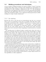
The spinal cord (fig. 13.1) is a cylinder of nervous tissue
that begins at the foramen magnum and passes through the
vertebral canal as far as the inferior margin of the first lum-
bar vertebra (L1). In adults, it averages about 1.8 cm thick
and 45 cm long. Early in fetal development, the spinal
cord extends for the full length of the vertebral column.
However, the vertebral column grows faster than the
spinal cord, so the cord extends only to L3 by the time of
birth and to L1 in an adult. Thus, it occupies only the
upper two-thirds of the vertebral canal; the lower one-
third is described shortly. The cord gives rise to 31 pairs of
spinal nerves that pass through the intervertebral foram-
ina. Although the spinal cord is not visibly segmented, the
part supplied by each pair of spinal nerves is called a seg-
ment. The cord exhibits longitudinal grooves on its ventral
and dorsal sides—the ventral median fissure and dorsal
median sulcus, respectively.
The spinal cord is divided into cervical, thoracic,
lumbar, and sacral regions. It may seem odd that it has a
sacral region when the cord itself ends well above the
sacrum. These regions, however, are named for the level of
the vertebral column from which the spinal nerves
emerge, not for the vertebrae that contain the cord itself.
In the inferior cervical region, a cervical enlarge-
ment of the cord gives rise to nerves of the upper limbs. In
the lumbosacral region, there is a similar lumbar enlarge-
ment where nerves to the pelvic region and lower limbs
arise. Inferior to the lumbar enlargement, the cord tapers
to a point called the medullary cone. The lumbar enlarge-
ment and medullary cone give rise to a bundle of nerve
roots that occupy the canal of vertebrae L2 to S5. This bun-
dle, named the cauda equina
1
(CAW-duh ee-KWY-nah) for
its resemblance to a horse’s tail, innervates the pelvic
organs and lower limbs.
Think About It
Spinal cord injuries commonly result from fractures of
vertebrae C5 to C6, but never from fractures of L3 to
L5. Explain both observations.
Meninges of the Spinal Cord
The spinal cord and brain are enclosed in three fibrous
membranes called meninges (meh-NIN-jeez)—singular,
meninx
2
(MEN-inks). These membranes separate the soft
tissue of the central nervous system from the bones of the
vertebrae and skull. From superficial to deep, they are the
dura mater, arachnoid mater, and pia mater.
482
Part Three Integration and Control
1
cauda ϭ tail ϩ equin ϭ horse
2
menin ϭ membrane
Saladin: Anatomy &
Physiology: The Unity of
Form and Function, Third
Edition
13. The Spinal Cord, Spinal
Nerves, and Somatic
Reflexes
Text
© The McGraw−Hill
Companies, 2003
Chapter 13
Chapter 13 The Spinal Cord, Spinal Nerves, and Somatic Reflexes 483
The dura mater
3
(DOO-ruh MAH-tur) forms a loose-
fitting sleeve called the dural sheath around the spinal
cord. It is a tough collagenous membrane with a thickness
and texture similar to a rubber kitchen glove. The space
between the sheath and vertebral bone, called the epidural
space, is occupied by blood vessels, adipose tissue, and
loose connective tissue (fig. 13.2a). Anesthetics are some-
times introduced to this space to block pain signals during
childbirth or surgery; this procedure is called epidural
anesthesia.
The arachnoid
4
(ah-RACK-noyd) mater adheres to the
dural sheath. It consists of a simple squamous epithelium,
the arachnoid membrane, adhering to the inside of the dura,
and a loose mesh of collagenous and elastic fibers spanning
the gap between the arachnoid membrane and the pia mater.
This gap, called the subarachnoid space, is filled with cere-
brospinal fluid (CSF), a clear liquid discussed in chapter 14.
The pia
5
(PEE-uh) mater is a delicate, translucent
membrane that closely follows the contours of the spinal
cord. It continues beyond the medullary cone as a fibrous
Cervical
spinal
nerves
Thoracic
spinal
nerves
Lumbar
spinal
nerves
Sacral
spinal
nerves
Cervical
enlargement
Dura mater
and arachnoid
mater
Lumbar
enlargement
Cauda equina
Coccygeal
ligament
Medullary
cone
Figure 13.1 The Spinal Cord, Dorsal Aspect.
3
dura ϭ tough ϩ mater ϭ mother, womb
4
arachn ϭ spider, spider web ϩ oid ϭ resembling
5
pia ϭ tender, soft
Saladin: Anatomy &
Physiology: The Unity of
Form and Function, Third
Edition
13. The Spinal Cord, Spinal
Nerves, and Somatic
Reflexes
Text
© The McGraw−Hill
Companies, 2003
Chapter 13
strand, the terminal filum, forming part of the coccygeal
ligament that anchors the cord to vertebra L2. At regular
intervals along the cord, extensions of the pia called den-
ticulate ligaments extend through the arachnoid to the
dura, anchoring the cord and preventing side-to-side
movements.
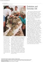
Insight 13.1 Clinical Application
Spina Bifida
About one baby in 1,000 is born with spina bifida (SPY-nuh BIF-ih-
duh), a congenital defect resulting from the failure of one or more ver-
tebrae to form a complete vertebral arch for enclosure of the spinal
cord. This is especially common in the lumbosacral region. One form,
spina bifida occulta,
6
involves only one to a few vertebrae and causes
no functional problems. Its only external sign is a dimple or hairy pig-
mented spot. Spina bifida cystica
7
is more serious. A sac protrudes
from the spine and may contain meninges, cerebrospinal fluid, and
parts of the spinal cord and nerve roots (fig. 13.3). In extreme cases,
inferior spinal cord function is absent, causing lack of bowel control
and paralysis of the lower limbs and urinary bladder. The last of these
conditions can lead to chronic urinary infections and renal failure.
Pregnant women can significantly reduce the risk of spina bifida by
taking supplemental folic acid (a B vitamin) during early pregnancy.
Good sources of folic acid include green leafy vegetables, black beans,
lentils, and enriched bread and pasta.
6
bifid ϭ divided, forked ϩ occult ϭ hidden
7
cyst ϭ sac, bladder
484 Part Three Integration and Control
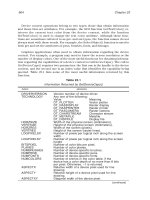
Posterior median sulcus
Anterior
median fissure
(b)
Dorsal horn
Lateral
column
Gray
commissure
Ventral column
Central
canal
Dorsal
column
Ventral root of
spinal nerve
Dorsal root
ganglion
Spinal
nerve
Lateral horn
Ventral horn
Dorsal root of
spinal nerve
Figure 13.2 Cross Section of the Thoracic Spinal Cord. (a) Relationship to the vertebra, meninges, and spinal nerve. (b) Anatomy of the spinal
cord itself.
Fat in epidural space
Dural sheath
Arachnoid mater
Pia mater
Spinal nerve
Bone of vertebra
Spinal cord
Denticulate ligament
Subarachnoid
space
(a)
Saladin: Anatomy &
Physiology: The Unity of
Form and Function, Third
Edition
13. The Spinal Cord, Spinal
Nerves, and Somatic
Reflexes
Text
© The McGraw−Hill
Companies, 2003
Chapter 13
Chapter 13 The Spinal Cord, Spinal Nerves, and Somatic Reflexes 485
Cross-Sectional Anatomy
Figure 13.2a shows the relationship of the spinal cord to a
vertebra and spinal nerve, and figure 13.2b shows the cord
itself in more detail. The spinal cord, like the brain, con-
sists of two kinds of nervous tissue called gray and white
matter. Gray matter has a relatively dull color because it
contains little myelin. It contains the somas, dendrites,
and proximal parts of the axons of neurons. It is the site of
synaptic contact between neurons, and therefore the site
of all synaptic integration (information processing) in the
central nervous system. White matter contains an abun-
dance of myelinated axons, which give it a bright, pearly
white appearance. It is composed of bundles of axons,
called tracts, that carry signals from one part of the CNS to
another. In fixed and silver-stained nervous tissue, gray
matter tends to have a darker brown or golden color and
white matter a lighter tan to yellow color.
Gray Matter
The spinal cord has a central core of gray matter that looks
somewhat butterfly- or H-shaped in cross sections. The
core consists mainly of two dorsal (posterior) horns,
which extend toward the dorsolateral surfaces of the cord,
and two thicker ventral (anterior) horns, which extend
toward the ventrolateral surfaces. The right and left sides
are connected by a gray commissure. In the middle of the
commissure is the central canal, which is collapsed in
most areas of the adult spinal cord, but in some places
(and in young children) remains open, lined with ependy-
mal cells, and filled with CSF.
As a spinal nerve approaches the cord, it branches
into a dorsal root and ventral root. The dorsal root carries
sensory nerve fibers, which enter the dorsal horn of the
cord and sometimes synapse with an interneuron there.
Such interneurons are especially numerous in the cervical
and lumbar enlargements and are quite evident in histo-
logical sections at these levels. The ventral horns contain
the large somas of the somatic motor neurons. Axons from
these neurons exit by way of the ventral root of the spinal
nerve and lead to the skeletal muscles. The spinal nerve
roots are described more fully later in this chapter.
In the thoracic and lumbar regions, an additional lat-
eral horn is visible on each side of the gray matter. It con-
tains neurons of the sympathetic nervous system, which
send their axons out of the cord by way of the ventral root
along with the somatic efferent fibers.
White Matter
The white matter of the spinal cord surrounds the gray
matter and consists of bundles of axons that course up
and down the cord and provides avenues of communi-
cation between different levels of the CNS. These bun-
dles are arranged in three pairs called columns or funi-
culi
8
(few-NIC-you-lie)—a dorsal (posterior), lateral,
and ventral (anterior) column on each side. Each col-
umn consists of subdivisions called tracts or fasciculi
9
(fah-SIC-you-lye).
Figure 13.3 Spina Bifida Cystica.
8
funicul ϭ little rope, cord
9
fascicul ϭ little bundle
Saladin: Anatomy &
Physiology: The Unity of
Form and Function, Third
Edition
13. The Spinal Cord, Spinal
Nerves, and Somatic
Reflexes
Text
© The McGraw−Hill
Companies, 2003
Chapter 13
Spinal Tracts
Knowledge of the locations and functions of the spinal
tracts is essential in diagnosing and managing spinal cord
injuries. Ascending tracts carry sensory information up
the cord and descending tracts conduct motor impulses
down. All nerve fibers in a given tract have a similar ori-
gin, destination, and function.
Several of these tracts undergo decussation
10
(DEE-
cuh-SAY-shun) as they pass up or down the brainstem and
spinal cord—meaning that they cross over from the left
side of the body to the right, or vice versa. As a result, the
left side of the brain receives sensory information from the
right side of the body and sends its motor commands to
that side, while the right side of the brain senses and con-
trols the left side of the body. A stroke that damages motor
centers of the right side of the brain can thus cause paral-
ysis of the left limbs and vice versa. When the origin and
destination of a tract are on opposite sides of the body, we
say they are contralateral
11
to each other. When a tract
does not decussate, so the origin and destination of its
fibers are on the same side of the body, we say they are
ipsilateral.
12
The major spinal cord tracts are summarized in
table 13.1 and figure 13.4. Bear in mind that each tract is
repeated on the right and left sides of the spinal cord.
Ascending Tracts
Ascending tracts carry sensory signals up the spinal cord.
Sensory signals typically travel across three neurons from
their origin in the receptors to their destination in the sen-
sory areas of the brain: a first-order neuron that detects a
stimulus and transmits a signal to the spinal cord or brain-
stem; a second-order neuron that continues as far as a
“gateway” called the thalamus at the upper end of the
brainstem; and a third-order neuron that carries the signal
the rest of the way to the sensory region of the cerebral cor-
tex. The axons of these neurons are called the first-
through third-order nerve fibers. Deviations from the path-
way described here will be noted for some of the sensory
systems to follow.
The major ascending tracts are as follows. The names
of most ascending tracts consist of the prefix spino- followed
by a root denoting the destination of its fibers in the brain.
• The gracile
13
fasciculus (GRAS-el fah-SIC-you-lus)
carries signals from the midthoracic and lower parts of
the body. Below vertebra T6, it composes the entire
dorsal column. At T6, it is joined by the cuneate
fasciculus, discussed next. It consists of first-order
nerve fibers that travel up the ipsilateral side of the
spinal cord and terminate at the gracile nucleus in the
medulla oblongata of the brainstem. These fibers carry
486
Part Three Integration and Control
Table 13.1 Major Spinal Tracts
Tract Column Decussation Functions
Ascending (sensory) Tracts
Gracile fasciculus Dorsal In medulla Limb and trunk position and movement, deep touch, visceral pain, vibration,
below level T6
Cuneate fasciculus Dorsal In medulla Same as gracile fasciculus, from level T6 up
Spinothalamic Lateral and ventral In spinal cord Light touch, tickle, itch, temperature, pain, and pressure
Dorsal spinocerebellar Lateral None Feedback from muscles (proprioception)
Ventral spinocerebellar Lateral In spinal cord Same as dorsal spinocerebellar
Descending (motor) Tracts
Lateral corticospinal Lateral In medulla Fine control of limbs
Ventral corticospinal Ventral None Fine control of limbs
Tectospinal Lateral and ventral In midbrain Reflexive head-turning in response to visual and auditory stimuli
Lateral reticulospinal Lateral None Balance and posture; regulation of awareness of pain
Medial reticulospinal Ventral None Same as lateral reticulospinal
Vestibulospinal Ventral None Balance and posture
10
decuss ϭ to cross, form an X
11
contra ϭ opposite
12
ipsi ϭ the same ϩ later ϭ side
13
gracil ϭ thin, slender
Saladin: Anatomy &
Physiology: The Unity of
Form and Function, Third
Edition
13. The Spinal Cord, Spinal
Nerves, and Somatic
Reflexes
Text
© The McGraw−Hill
Companies, 2003
Chapter 13
Chapter 13 The Spinal Cord, Spinal Nerves, and Somatic Reflexes 487
signals for vibration, visceral pain, deep and
discriminative touch (touch whose location one can
precisely identify), and especially proprioception
14
from the lower limbs and lower trunk. (Proprioception
is a nonvisual sense of the position and movements of
the body.)
• The cuneate
15
(CUE-nee-ate) fasciculus (fig. 13.5a)
joins the gracile fasciculus at the T6 level. It occupies
the lateral portion of the dorsal column and forces the
gracile fasciculus medially. It carries the same type of
sensory signals, originating from level T6 and up
(from the upper limb and chest). Its fibers end in the
cuneate nucleus on the ipsilateral side of the medulla
oblongata. In the medulla, second-order fibers of the
gracile and cuneate systems decussate and form the
medial lemniscus
16
(lem-NIS-cus), a tract of nerve
fibers that leads the rest of the way up the brainstem
to the thalamus. Third-order fibers go from the
thalamus to the cerebral cortex. Because of
decussation, the signals carried by the gracile and
cuneate fasciculi ultimately go to the contralateral
cerebral hemisphere.
• The spinothalamic (SPY-no-tha-LAM-ic) tract
(fig. 13.5b) and some smaller tracts form the
anterolateral system, which passes up the anterior
and lateral columns of the spinal cord. The
spinothalamic tract carries signals for pain,
temperature, pressure, tickle, itch, and light or crude
touch. Light touch is the sensation produced by
stroking hairless skin with a feather or cotton wisp,
without indenting the skin; crude touch is touch
whose location one can only vaguely identify. In this
pathway, first-order neurons end in the dorsal horn of
the spinal cord near the point of entry. Second-order
neurons decussate to the opposite side of the spinal
cord and there form the ascending spinothalamic
tract. These fibers lead all the way to the thalamus.
Third-order neurons continue from there to the
cerebral cortex.
• The dorsal and ventral spinocerebellar (SPY-no-
SERR-eh-BEL-ur) tracts travel through the lateral
column and carry proprioceptive signals from the
limbs and trunk to the cerebellum, a large motor
control area at the rear of the brain. The first-order
neurons of this system originate in the muscles and
tendons and end in the dorsal horn of the spinal cord.
Second-order neurons send their fibers up the
spinocerebellar tracts and end in the cerebellum.
Fibers of the dorsal tract travel up the ipsilateral side
of the spinal cord. Those of the ventral tract cross over
and travel up the contralateral side but then cross back
in the brainstem to enter the ipsilateral cerebellum.
Both tracts provide the cerebellum with feedback
needed to coordinate muscle action, as discussed in
chapter 14.
Descending
tracts
Ascending
tracts
Lateral
corticospinal tract
Lateral reticulospinal tract
Vestibulospinal tract
Medial reticulospinal tract
Lateral tectospinal tract
Medial tectospinal tract
Ventral corticospinal tract
Ventral spinocerebellar tract
Dorsal spinocerebellar tract
Dorsal column
Gracile fasciculus
Cuneate fasciculus
Anterolateral system
(containing
spinothalamic tract)
Figure 13.4 Tracts of the Spinal Cord. All of the illustrated tracts occur on both sides of the cord, but only the ascending sensory tracts are
shown on the left (red ), and only the descending motor tracts on the right (green).
If you were told that this cross section is either at level T4 or T10, how could you determine which is correct?
14
proprio ϭ one’s own ϩ cept ϭ receive, sense
15
cune ϭ wedge
16
lemniscus ϭ ribbon
Saladin: Anatomy &
Physiology: The Unity of
Form and Function, Third
Edition
13. The Spinal Cord, Spinal
Nerves, and Somatic
Reflexes
Text
© The McGraw−Hill
Companies, 2003
Chapter 13
488 Part Three Integration and Control
Somesthetic cortex
(postcentral gyrus)
Third-order
neuron
Medial
lemniscus
Medial
lemniscus
Second-order
neuron
Cuneate
nucleus
Gracile
nucleus
First-order
neuron
(a) (b)
Gracile fasciculus
Cuneate fasciculus
Midbrain
Medulla
Spinal cord
Thalamus
Receptors for body movement, limb positions,
fine touch discrimination, and pressure
Somesthetic cortex
(postcentral gyrus)
Third-order
neuron
Second-order
neuron
Spinothalamic
tract
First-order
neuron
Anterolateral system
Midbrain
Medulla
Spinal cord
Thalamus
Receptors for pain, heat, and cold
Figure 13.5 Some Ascending Pathways of the CNS. The spinal cord, medulla, and midbrain are shown in cross section and the cerebrum and
thalamus (top) in frontal section. Nerve signals enter the spinal cord at the bottom of the figure and carry somatosensory information up to the cerebral
cortex. (a) The cuneate fasciculus and medial lemniscus; (b) the spinothalamic tract.
Saladin: Anatomy &
Physiology: The Unity of
Form and Function, Third
Edition
13. The Spinal Cord, Spinal
Nerves, and Somatic
Reflexes
Text
© The McGraw−Hill
Companies, 2003
Chapter 13
Chapter 13 The Spinal Cord, Spinal Nerves, and Somatic Reflexes 489
Descending Tracts
Descending tracts carry motor signals down the brainstem
and spinal cord. A descending motor pathway typically
involves two neurons called the upper and lower motor
neuron. The upper motor neuron begins with a soma in
the cerebral cortex or brainstem and has an axon that ter-
minates on a lower motor neuron in the brainstem or
spinal cord. The axon of the lower motor neuron then
leads the rest of the way to the muscle or other target
organ. The names of most descending tracts consist of a
word root denoting the point of origin in the brain, fol-
lowed by the suffix -spinal. The major descending tracts
are described here.
• The corticospinal (COR-tih-co-SPY-nul) tracts carry
motor signals from the cerebral cortex for precise,
finely coordinated limb movements. The fibers of this
system form ridges called pyramids on the ventral
surface of the medulla oblongata, so these tracts were
once called pyramidal tracts. Most corticospinal fibers
decussate in the lower medulla and form the lateral
corticospinal tract on the contralateral side of the
spinal cord. A few fibers remain uncrossed and form
the ventral corticospinal tract on the ipsilateral side
(fig. 13.6). Fibers of the ventral tract decussate lower
in the spinal cord, however, so even they control
contralateral muscles.
• The tectospinal (TEC-toe-SPY-nul) tract begins in
a midbrain region called the tectum and crosses to
the contralateral side of the brainstem. In the
lower medulla, it branches into lateral and medial
tectospinal tracts of the upper spinal cord. These
are involved in reflex movements of the head,
especially in response to visual and auditory
stimuli.
• The lateral and medial reticulospinal (reh-TIC-you-lo-
SPY-nul) tracts originate in the reticular formation of
the brainstem. They control muscles of the upper and
lower limbs, especially to maintain posture and
balance. They also contain descending analgesic
pathways that reduce the transmission of pain signals
to the brain (see chapter 16).
• The vestibulospinal (vess-TIB-you-lo-SPY-nul) tract
begins in a brainstem vestibular nucleus that receives
impulses for balance from the inner ear. The tract
passes down the ventral column of the spinal cord and
controls limb muscles that maintain balance and
posture.
Rubrospinal tracts are prominent in other mammals,
where they aid in muscle coordination. Although often
pictured in illustrations of human anatomy, they are
almost nonexistent in humans and have little functional
importance.
Internal
capsule
Motor cortex
(precentral gyrus)
Midbrain
Medulla
Spinal cord
Spinal cord
Cerebral peduncle
Upper motor
neurons
Decussation in
medulla
Lateral corticospinal
tract
Ventral corticospinal
tract
Lower motor
neurons
To skeletal musclesTo skeletal muscles
Medullary pyramid
Figure 13.6 Two Descending Pathways of the CNS. The lateral
and ventral corticospinal tracts, which carry signals for voluntary muscle
contraction. Nerve signals originate in the cerebral cortex at the top of
the figure and carry motor commands down the spinal cord.
Saladin: Anatomy &
Physiology: The Unity of
Form and Function, Third
Edition
13. The Spinal Cord, Spinal
Nerves, and Somatic
Reflexes
Text
© The McGraw−Hill
Companies, 2003
Chapter 13
Think About It
You are blindfolded and either a tennis ball or an iron
ball is placed in your right hand. What spinal tract(s)
would carry the signals that enable you to
discriminate between these two objects?
Insight 13.2 Clinical Application
Poliomyelitis and Amyotrophic Lateral
Sclerosis
Poliomyelitis
17
and amyotrophic lateral sclerosis
18
(ALS) are two dis-
eases that involve destruction of motor neurons. In both diseases, the
skeletal muscles atrophy from lack of innervation.
Poliomyelitis is caused by the poliovirus, which destroys motor neu-
rons in the brainstem and ventral horn of the spinal cord. Signs of polio
include muscle pain, weakness, and loss of some reflexes, followed by
paralysis, muscular atrophy, and sometimes respiratory arrest. The virus
spreads by fecal contamination of water. Historically, polio afflicted
mainly children, who sometimes contracted the virus in the summer by
swimming in contaminated pools. The polio vaccine has nearly elimi-
nated new cases.
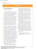
ALS is also known as Lou Gehrig disease after the baseball player who
contracted it. It is marked not only by the degeneration of motor neu-
rons and atrophy of the muscles, but also sclerosis of the lateral regions
of the spinal cord—hence its name. In most cases of ALS, neurons are
destroyed by an inability of astrocytes to reabsorb glutamate from the
tissue fluid, allowing this neurotransmitter to accumulate to a toxic
level. The early signs of ALS include muscular weakness and difficulty in
speaking, swallowing, and using the hands. Sensory and intellectual
functions remain unaffected, as evidenced by the accomplishments of
astrophysicist and best-selling author Stephen Hawking, who was
stricken with ALS while he was in college. Despite near-total paralysis, he
remains highly productive and communicates with the aid of a speech
synthesizer and computer. Tragically, many people are quick to assume
that those who have lost most of their ability to communicate their ideas
and feelings have no ideas and feelings to communicate. To a victim, this
may be more unbearable than the loss of motor function itself.
17
polio ϭ gray matter ϩ myel ϭ spinal cord ϩ itis ϭ inflammation
18
a ϭ without ϩ myo ϭ muscle ϩ troph ϭ nourishment
Before You Go On
Answer the following questions to test your understanding of the
preceding section:
1. Name the four major regions and two enlargements of the
spinal cord.
2. Describe the distal (inferior) end of the spinal cord and the
contents of the vertebral canal from level L2 to S5.
3. Sketch a cross section of the spinal cord showing the dorsal and
ventral horns. Where are the gray and white matter? Where are
the columns and tracts?
4. Give an anatomical explanation as to why a stroke in the right
cerebral hemisphere can paralyze the limbs on the left side of
the body.
The Spinal Nerves
Objectives
When you have completed this section, you should be able to
• describe the attachment of a spinal nerve to the spinal cord;
• trace the branches of a spinal nerve distal to its attachment;
• name the five plexuses of spinal nerves and describe their
general anatomy;
• name some major nerves that arise from each plexus; and
• explain the relationship of dermatomes to the spinal nerves.
General Anatomy of Nerves
and Ganglia
The spinal cord communicates with the rest of the body by
way of the spinal nerves. Before we discuss those specific
nerves, however, it is necessary to be familiar with the
structure of nerves and ganglia in general.
A nerve is a cordlike organ composed of numerous
nerve fibers (axons) bound together by connective tissue
(fig. 13.8). If we compare a nerve fiber to a wire carrying an
electrical current in one direction, a nerve would be com-
parable to an electrical cable composed of thousands of
wires carrying currents in opposite directions. A nerve
contains anywhere from a few nerve fibers to more than a
million. Nerves usually have a pearly white color and
resemble frayed string as they divide into smaller and
smaller branches.
490
Part Three Integration and Control
Figure 13.7 Stephen Hawking (1942– ), Lucasian Professor
of Mathematics at Cambridge University.
Saladin: Anatomy &
Physiology: The Unity of
Form and Function, Third
Edition
13. The Spinal Cord, Spinal
Nerves, and Somatic
Reflexes
Text
© The McGraw−Hill
Companies, 2003
Chapter 13
Chapter 13 The Spinal Cord, Spinal Nerves, and Somatic Reflexes 491
Spinal cord
(a)
Dorsal root
Dorsal root ganglion
Ventral root
Spinal
nerve
Epineurium
Fascicle
Blood
vessels
Perineurium
Axon
Endoneurium
around
individual
axon
Schwann cell
of myelinated
axon
Unmyelinated
axon
Figure 13.8 Anatomy of a Nerve. (a) A spinal nerve and its association with the spinal cord. (b) Cross section of a nerve (SEM). Myelinated nerve
fibers appear as white rings and unmyelinated fibers as solid gray. Credit for b: Richard E. Kessel and Randy H. Kardon, Tissues and Organs: A Text-Atlas
of Scanning Electron Microscopy, 1979, W. H. Freeman and Company.
(b)
Epineurium
Perineurium
Blood vessels
Endoneurium
Fascicle
Nerve fiber
Nerve fibers of the peripheral nervous system are
ensheathed in Schwann cells, which form a neurilemma
and often a myelin sheath around the axon (see chapter 12).
External to the neurilemma, each fiber is surrounded by a
basal lamina and then a thin sleeve of loose connective tis-
sue called the endoneurium. In most nerves, the nerve
fibers are gathered in bundles called fascicles, each
wrapped in a sheath called the perineurium. The per-
ineurium is composed of one to six layers of overlapping,
squamous, epithelium-like cells. Several fascicles are then
Saladin: Anatomy &
Physiology: The Unity of
Form and Function, Third
Edition
13. The Spinal Cord, Spinal
Nerves, and Somatic
Reflexes
Text
© The McGraw−Hill
Companies, 2003
Chapter 13
bundled together and wrapped in an outer epineurium to
compose the nerve as a whole. The epineurium is com-
posed of dense irregular fibrous connective tissue and pro-
tects the nerve from stretching and injury. Nerves have a
high metabolic rate and need a plentiful blood supply.
Blood vessels penetrate as far as the perineurium, and oxy-
gen and nutrients diffuse through the extracellular fluid
from there to the nerve fibers.
Think About It
How does the structure of a nerve compare to that of
a skeletal muscle? Which of the descriptive terms for
nerves have similar counterparts in muscle histology?
Peripheral nerve fibers are of two kinds: sensory
(afferent) fibers carry signals from sensory receptors to the
CNS, and motor (efferent) fibers carry signals from the CNS
to muscles and glands. Both sensory and motor fibers can
also be described as somatic or visceral and as general or
special depending on the organs they innervate (table 13.2).
A mixed nerve consists of both sensory and motor
fibers and thus transmits signals in two directions,
although any one nerve fiber within the nerve transmits
signals one way only. Most nerves are mixed. Purely
sensory nerves, composed entirely of sensory axons, are
less common; they include the olfactory and optic
nerves discussed in chapter 14. Nerves that carry only
motor fibers are called motor nerves. Many nerves often
described as motor are actually mixed because they
carry sensory signals of proprioception from the muscle
back to the CNS.
If a nerve resembles a thread, a ganglion
19
resem-
bles a knot in the thread. A ganglion is a cluster of cell
bodies (somas) outside the CNS. It is enveloped in an
epineurium continuous with that of the nerve. Among
the somas are bundles of nerve fibers leading into and out
of the ganglion. Figure 13.9 shows a type of ganglion
called the dorsal root ganglion associated with the spinal
nerves.
Spinal Nerves
There are 31 pairs of spinal nerves: 8 cervical (C1–C8), 12
thoracic (T1–T12), 5 lumbar (L1–L5), 5 sacral (S1–S5), and
1 coccygeal (Co) (fig. 13.10). The first cervical nerve
emerges between the skull and atlas, and the others
emerge through intervertebral foramina, including the
anterior and posterior foramina of the sacrum.
Proximal Branches
Each spinal nerve has two points of attachment to the
spinal cord (fig. 13.11). Dorsally, a branch of the spinal
nerve called the dorsal root divides into six to eight nerve
rootlets that enter the spinal cord (fig. 13.12). A little dis-
tal to the rootlets is a swelling called the dorsal root gan-
glion, which contains the somas of afferent neurons. Ven-
trally, another row of six to eight rootlets leave the spinal
cord and converge to form the ventral root.
The dorsal and ventral roots merge, penetrate the
dural sac, enter the intervertebral foramen, and there form
the spinal nerve proper.
Spinal nerves are mixed nerves, with a two-way traf-
fic of afferent (sensory) and efferent (motor) signals. Affer-
ent signals approach the cord by way of the dorsal root and
enter the dorsal horn of the gray matter. Efferent signals
begin at the somas of motor neurons in the ventral horn
and leave the spinal cord via the ventral root. Some
viruses invade the central nervous system by way of these
roots (see insight 13.3).
The dorsal and ventral roots are shortest in the cervi-
cal region and become longer inferiorly. The roots that
arise from segments L2 to Co of the cord form the cauda
equina.
Distal Branches
Distal to the vertebrae, the branches of a spinal nerve are
more complex (fig. 13.13). Immediately after emerging
from the intervertebral foramen, the nerve divides into a
dorsal ramus,
20
a ventral ramus, and a small meningeal
branch. The meningeal branch (see fig. 13.11) reenters the
vertebral canal and innervates the meninges, vertebrae,
and spinal ligaments. The dorsal ramus innervates the
muscles and joints in that region of the spine and the skin
492
Part Three Integration and Control
Table 13.2 The Classification of
Nerve Fibers
Class Description
Afferent fibers Carry sensory signals from receptors to the CNS
Efferent fibers Carry motor signals from the CNS to effectors
Somatic fibers Innervate skin, skeletal muscles, bones, and joints
Visceral fibers Innervate blood vessels, glands, and viscera
General fibers Innervate widespread organs such as muscles,
skin, glands, viscera, and blood vessels
Special fibers Innervate more localized organs in the head,
including the eyes, ears, olfactory and taste
receptors, and muscles of chewing, swallowing,
and facial expression
19
gangli ϭ knot
20
ramus ϭ branch
Saladin: Anatomy &
Physiology: The Unity of
Form and Function, Third
Edition
13. The Spinal Cord, Spinal
Nerves, and Somatic
Reflexes
Text
© The McGraw−Hill
Companies, 2003
Chapter 13
Chapter 13 The Spinal Cord, Spinal Nerves, and Somatic Reflexes 493
of the back. The ventral ramus innervates the ventral and
lateral skin and muscles of the trunk and gives rise to
nerves of the limbs.
Think About It
Do you think the meningeal branch is sensory, motor,
or mixed? Explain your reasoning.
The ventral ramus differs from one region of the
trunk to another. In the thoracic region, it forms an inter-
costal nerve that travels along the inferior margin of a rib
and innervates the skin and intercostal muscles (thus con-
tributing to breathing), as well as the internal oblique,
external oblique, and transversus abdominis muscles. All
other ventral rami form the nerve plexuses described next.
Insight 13.3 Clinical Application
Shingles
Chickenpox (varicella), a common disease of early childhood, is caused
by the varicella-zoster virus. It produces an itchy rash that usually
clears up without complications. The virus, however, remains for life in
the dorsal root ganglia. The immune system normally keeps it in check.
If the immune system is compromised, however, the virus can travel
down the sensory nerves by fast axonal transport and cause shingles
(herpes zoster). This is characterized by a painful trail of skin discol-
oration and fluid-filled vesicles along the path of the nerve. These signs
usually appear in the chest and waist, often on just one side of the
body. Shingles usually occurs after the age of 50. While it can be very
painful and may last 6 months or longer, it eventually heals sponta-
neously and requires no special treatment other than aspirin and
steroidal ointment to relieve pain and inflammation.
Dorsal root ganglion
Direction of signal
transmission
Ventral root
Spinal cord
Epineurium of ganglion
Epineurium of dorsal root
Dorsal root
Fibers of somatosensory
(afferent) neurons
Connective
tissue
Ventral root
Somas of somatosensory
(afferent) neurons
Spinal nerve
Fibers of motor
(efferent) neurons
Blood vessels
Direction of signal
transmission
Dorsal root ganglion
Figure 13.9 Anatomy of a Ganglion. The dorsal root ganglion contains the somas of unipolar sensory neurons conducting signals to the spinal
cord. To the left of it is the ventral root of the spinal nerve, which conducts motor signals away from the spinal cord. (The ventral root is not part of the
ganglion.)
Where are the somas of the motor neurons located?
Saladin: Anatomy &
Physiology: The Unity of
Form and Function, Third
Edition
13. The Spinal Cord, Spinal
Nerves, and Somatic
Reflexes
Text
© The McGraw−Hill
Companies, 2003
Chapter 13
Nerve Plexuses
Except in the thoracic region, the ventral rami branch and
anastomose (merge) repeatedly to form five weblike nerve
plexuses: the small cervical plexus deep in the neck, the
brachial plexus near the shoulder, the lumbar plexus of
the lower back, the sacral plexus immediately inferior to
this, and finally the tiny coccygeal plexus adjacent to the
lower sacrum and coccyx. A general view of these
plexuses is shown in figure 13.10; they are illustrated and
described in tables 13.3 through 13.6. The muscle actions
controlled by these nerves are described in the muscle
tables in chapter 10.
Insight 13.4 Clinical Application
Spinal Nerve Injuries
The radial and sciatic nerves are especially vulnerable to injury. The
radial nerve, which passes through the axilla, may be compressed
against the humerus by improperly adjusted crutches, causing crutch
paralysis. A similar injury often resulted from the now-discredited prac-
tice of trying to correct a dislocated shoulder by putting a foot in a per-
son’s armpit and pulling on the arm. One consequence of radial nerve
injury is wrist drop—the fingers, hand, and wrist are chronically flexed
because the extensor muscles supplied by the radial nerve are paralyzed.
Because of its position and length, the sciatic nerve of the hip and
thigh is the most vulnerable nerve in the body. Trauma to this nerve pro-
duces sciatica, a sharp pain that travels from the gluteal region along the
posterior side of the thigh and leg as far as the ankle. Ninety percent of
cases result from a herniated intervertebral disc or osteoarthritis of the
lower spine, but sciatica can also be caused by pressure from a pregnant
uterus, dislocation of the hip, injections in the wrong area of the buttock,
or sitting for a long time on the edge of a hard chair. Men sometimes suf-
fer sciatica from the habit of sitting on a wallet carried in the hip pocket.
494 Part Three Integration and Control
Atlas (first cervical vertebra)
Cervical nerves (8 pairs)
Cervical enlargement
1st thoracic vertebra
Thoracic nerves (12 pairs)
Lumbar enlargement
1st lumbar vertebra
Medullary cone
Lumbar nerves (5 pairs)
Cauda equina
Ilium
Sacral nerves (5 pairs)
Coccygeal nerves (1 pair)
C1
C2
C3
C4
C5
C6
C7
C8
T1
T2
T3
T4
T5
T6
T7
T8
T9
T10
T11
T12
L1
L2
L3
L4
L5
S1
S2
S3
S4
S5
Cervical plexus (C1–C5)
Brachial plexus (C5–T1)
Intercostal (thoracic) nerves
Lumbar plexus (L1–L4)
Sacral plexus (L4–S4)
Sciatic
nerve
Figure 13.10 The Spinal Nerve Roots and Plexuses, Dorsal View.
Saladin: Anatomy &
Physiology: The Unity of
Form and Function, Third
Edition
13. The Spinal Cord, Spinal
Nerves, and Somatic
Reflexes
Text
© The McGraw−Hill
Companies, 2003
Chapter 13
Chapter 13 The Spinal Cord, Spinal Nerves, and Somatic Reflexes 495
Ventral root
Dorsal root
Spinal nerve
Dorsal root
ganglion
Sympathetic
ganglion
Communicating rami
Dorsal ramus
Ventral ramus
Meningeal branch
Body of vertebra
Anterior
Posterior
Spine of vertebra
Deep muscles of back
Spinal cord
Figure 13.11 Branches of a Spinal Nerve in Relation to the Spinal Cord and Vertebra (cross section).
Posterior median
sulcus
Gracile fasciculus
Cuneate
fasciculus
Lateral column
Segment C5
Cross section
Arachnoid
mater
Dura mater
Neural arch of
vertebra C3 (cut)
Vertebral artery
Spinal nerve C5
Rootlets
Dorsal root
Dorsal root
ganglion
Ventral root
Figure 13.12 The Point of Entry of Two Spinal Nerves into the Spinal Cord. Dorsal view with vertebrae cut away. Note that each dorsal
root divides into several rootlets that enter the spinal cord. A segment of the spinal cord is the portion receiving all the rootlets of one spinal nerve.
In the labeled rootlets of spinal nerve C5, are the nerve fibers afferent or efferent? How do you know?
Saladin: Anatomy &
Physiology: The Unity of
Form and Function, Third
Edition
13. The Spinal Cord, Spinal
Nerves, and Somatic
Reflexes
Text
© The McGraw−Hill
Companies, 2003
Chapter 13
496 Part Three Integration and Control
Thoracic cavity
Spinal nerve
Communicating rami
Dorsal ramus
Ventral ramus
Intercostal nerve
Sympathetic
chain ganglion
Anterior
cutaneous nerve
Lateral
cutaneous nerve
(b)
Figure 13.13 Rami of the Spinal Nerves. (a) Anterolateral view of the spinal nerves and their subdivisions in relation to the spinal cord and
vertebrae. (b) Cross section of the thorax showing innervation of muscles of the chest and back.
Ventral root
Dorsal root
Dorsal and ventral rootlets
of spinal nerve
Dorsal root ganglion
Dorsal ramus
of spinal nerve
Spinal nerve
Communicating rami
Sympathetic chain
ganglion
Ventral ramus
of spinal nerve
(a)
Saladin: Anatomy &
Physiology: The Unity of
Form and Function, Third
Edition
13. The Spinal Cord, Spinal
Nerves, and Somatic
Reflexes
Text
© The McGraw−Hill
Companies, 2003
Chapter 13
497
Table 13.3 The Cervical Plexus
The cervical plexus (fig. 13.14) receives fibers from the ventral rami of nerves C1 to C5 and gives rise to the nerves listed, in order from superior to inferior.
The most important of these are the phrenic
21
nerves, which travel down each side of the mediastinum, innervate the diaphragm, and play an essential role
in breathing. In addition to the major nerves listed here, there are several motor branches that innervate the geniohyoid, thyrohyoid, scalene, levator
scapulae, trapezius, and sternocleidomastoid muscles.
Lesser Occipital Nerve
Composition: Somatosensory
Innervation: Skin of lateral scalp and dorsal part of external ear
Great Auricular Nerve
Composition: Somatosensory
Innervation: Skin of and around external ear
Transverse Cervical Nerve
Composition: Somatosensory
Innervation: Skin of ventral and lateral aspect of neck
Segmental branch
Hypoglossal nerve (XII)
Lesser occipital nerve
Anterior root
Ansa cervicalis
Roots
Great auricular nerve
Posterior root
Supraclavicular nerve
Branch to brachial plexus
C1
C2
C3
C4
C5
Transverse cervical nerve
Phrenic nerve
Figure 13.14 The Cervical Plexus.
21
phren ϭ diaphragm
Ansa Cervicalis
Composition: Motor
Innervation: Omohyoid, sternohyoid, and sternothyroid muscles
Supraclavicular Nerve
Composition: Somatosensory
Innervation: Skin of lower ventral and lateral neck, shoulder, and ventral chest
Phrenic (FREN-ic) Nerve
Composition: Motor
Innervation: Diaphragm
Saladin: Anatomy &
Physiology: The Unity of
Form and Function, Third
Edition
13. The Spinal Cord, Spinal
Nerves, and Somatic
Reflexes
Text
© The McGraw−Hill
Companies, 2003
Chapter 13
498 Part Three Integration and Control
Table 13.4 The Brachial Plexus
The brachial plexus (figs. 13.15 and 13.16) is formed by the ventral rami of nerves C4 to T2. It passes over the first rib into the axilla and innervates the
upper limb and some muscles of the neck and shoulder. It gives rise to nerves for cutaneous sensation, muscle contraction, and proprioception from the
joints and muscles.
The subdivisions of this plexus are called roots, trunks, divisions, and cords (color-coded in figure 13.15). The five roots are the ventral rami of nerves C5 to
T1, which provide most of the fibers to this plexus (C4 and T2 contribute partially). The five roots unite to form the upper, middle, and lower trunks. Each
trunk divides into an anterior and posterior division, and finally the six divisions merge to form three large fiber bundles—the posterior, medial, and
lateral cords.
Axillary Nerve
Composition: Motor and somatosensory
Origin: Posterior cord of brachial plexus
Sensory innervation: Skin of lateral shoulder and arm; shoulder joint
Motor innervation: Deltoid and teres minor
Scapula
Clavicle
Lateral cord
Posterior cord
Medial cord
Axillary nerve
Musculocutaneous
nerve
Radial nerve
Radial nerve
Median nerve
Median nerve
Ulnar nerve
Digital
branch
of median
nerve
Superficial branch
of ulnar nerve
Digital branch
of ulnar nerve
Ulna
Radius
Humerus
Dorsal scapular nerve
Long thoracic nerve
Suprascapular nerve
Subclavian nerve
Posterior
divisions
Roots
Anterior
divisions
Posterior cord
Musculocutaneous nerve
C5
C6
C7
C8
Trunks
T1
Axillary nerve
Subscapular nerve
Thoracodorsal nerve
Lateral cord
Radial nerve
Medial and lateral pectoral nerves
Median nerve
Ulnar nerve
Medial cutaneous antebrachial nerve
Medial brachial cutaneous nerve
Medial
cord
Figure 13.15 The Brachial Plexus.
Radial Nerve
Composition: Motor and somatosensory
Origin: Posterior cord of brachial plexus
Sensory innervation: Skin of posterior aspect of arm, forearm, and wrist;
joints of elbow, wrist, and hand
Motor innervation: Muscles of posterior arm and forearm: triceps brachii,
supinator, anconeus, brachioradialis, extensor carpi radialis brevis,
extensor carpi radialis longus, and extensor carpi ulnaris
(continued)
Saladin: Anatomy &
Physiology: The Unity of
Form and Function, Third
Edition
13. The Spinal Cord, Spinal
Nerves, and Somatic
Reflexes
Text
© The McGraw−Hill
Companies, 2003
Chapter 13
Chapter 13 The Spinal Cord, Spinal Nerves, and Somatic Reflexes 499
Table 13.4 The Brachial Plexus (continued)
Musculocutaneous Nerve
Composition: Motor and somatosensory
Origin: Lateral cord of brachial plexus
Sensory innervation: Skin of lateral aspect of forearm
Motor innervation: Muscles of anterior arm: coracobrachialis, biceps brachii, and brachialis
Median Nerve
Composition: Motor and somatosensory
Origin: Medial cord of brachial plexus
Sensory innervation: Skin of lateral two-thirds of hand, joints of hand
Motor innervation: Flexors of anterior forearm; thenar muscles; first and second lumbricals
Ulnar Nerve
Composition: Motor and somatosensory
Origin: Medial cord of brachial plexus
Sensory innervation: Skin of medial part of hand; joints of hand
Motor innervation: Flexor carpi ulnaris, flexor digitorum profundus, adductor pollicis, hypothenar muscles, interosseous muscles, and third and fourth
lumbricals
Accessory n.
Hypoglossal n.
Trapezius m.
Vagus n.
Superior
thyroid a.
Larynx
Sympathetic
paravertebral
ganglion
Brachial
plexus
Vagus n.
Phrenic n.
Subclavian a.
Thyroid gland
First rib
Figure 13.16 Photograph of the Brachial Plexus. Anterior view of the right shoulder, also showing three of the cranial nerves, the
sympathetic trunk, and the phrenic nerve (a branch of the cervical plexus). Most of the other structures resembling nerves in this photograph are
blood vessels. (a. ϭ artery; m. ϭ muscle; n. ϭ nerve.)
Saladin: Anatomy &
Physiology: The Unity of
Form and Function, Third
Edition
13. The Spinal Cord, Spinal
Nerves, and Somatic
Reflexes
Text
© The McGraw−Hill
Companies, 2003
Chapter 13
Table 13.5 The Lumbar Plexus
The lumbar plexus (fig. 13.17) is formed from the ventral rami of nerves L1 to L4 and some fibers from T12. With only five roots and two divisions, it is less
complex than the brachial plexus.
Iliohypogastric Nerve
Composition: Motor and somatosensory
Sensory innervation: Skin of anterior abdominal wall
Motor innervation: Internal and external obliques and transversus
abdominis
Ilioinguinal Nerve
Composition: Motor and somatosensory
Sensory innervation: Skin of upper medial thigh; male scrotum and root of
penis; female labia majora
Motor innervation: Joins iliohypogastric nerve and innervates the same
muscles
Genitofemoral Nerve
Composition: Somatosensory
Sensory innervation: Skin of middle anterior thigh; male scrotum and
cremaster muscle; female labia majora
Lateral Femoral Cutaneous Nerve
Composition: Somatosensory
Sensory innervation: Skin of lateral aspect of thigh
Iliohypogastric nerve
Anterior view
Ilioinguinal nerve
Genitofemoral nerve
Lateral femoral cutaneous nerve
Posterior divisions
Roots
Anterior divisions
Femoral nerve
Saphenous nerve
Obturator nerve
Lumbosacral trunk
L1
L2
L3
L4
L5
From sacral plexus
From lumbar plexus
Os coxae
Sacrum
Pudendal nerve
Femoral nerve
Sciatic nerve
Femur
Tibial nerve
Common fibular
nerve
Tibia
Fibula
Superficial fibular
nerve
Deep fibular nerve
Medial plantar nerve
Lateral plantar nerve
Tibial nerve
Posterior view
Figure 13.17 The Lumbar Plexus.
Femoral Nerve
Composition: Motor and somatosensory
Sensory innervation: Skin of anterior and lateral thigh; medial leg and foot
Motor innervation: Anterior muscles of thigh and extensors of leg; iliacus,
psoas major, pectineus, quadriceps femoris, and sartorius
Saphenous (sah-FEE-nus) Nerve
Composition: Somatosensory
Sensory innervation: Skin of medial aspect of leg and foot; knee joint
Obturator Nerve
Composition: Motor and somatosensory
Sensory innervation: Skin of superior medial thigh; hip and knee joints
Motor innervation: Adductor muscles of leg: external obturator, pectineus,
adductor longus, adductor brevis, adductor magnus, and gracilis