Báo cáo y học: "CD23+/CD21hi B-cell translocation and ipsilateral lymph node collapse is associated with asymmetric arthritic flare in TNF-Tg mice" potx
Bạn đang xem bản rút gọn của tài liệu. Xem và tải ngay bản đầy đủ của tài liệu tại đây (1.97 MB, 12 trang )
RESEARC H ARTIC L E Open Access
CD23
+
/CD21
hi
B-cell translocation and ipsilateral
lymph node collapse is associated with
asymmetric arthritic flare in TNF-Tg mice
Jie Li
1,2
, Quan Zhou
3
, Ronald W Wood
4,5
, Igor Kuzin
6
, Andrea Bottaro
2,6
, Christopher T Ritchlin
1,6
, Lianping Xing
1
and Edward M Schwarz
1,2,5*
Abstract
Introduction: Rheumatoid arthritis (RA) is a chronic autoimmune disease with episodic flares in affected joints.
However, how arthritic flare occurs only in select joints during a systemic autoimmune disease remains an enigma.
To better understand these observations, we developed longitudinal imaging outcomes of synovitis and lymphatic
flow in mouse models of RA, and identified that asymmetric knee flare is associated with ipsila teral popliteal lymph
node (PLN) collapse and the translocation of CD23
+
/CD21
hi
B-cells (B-in) into the paracortical sinus space of the
node. In order to understand the relationship between this B-in translocation and lymph drainage from flaring
joints, we tested the hypothesis that asymmetric tumor necrosis factor (TNF)-induced knee arthritis is associated
with ipsilateral PLN and iliac lymph node (ILN) collapse, B-in translocation, and decreased afferent lymphatic flow.
Methods: TNF transgenic (Tg) mice with asymmetric knee arthritis were identified by contrast-enhanced (CE)
magnetic resonance imagi ng (MRI), and PLN were phenotyped as “expanding ” or “ collapsed” using LNcap
threshold = 30 (Arbitrary Unit (AU)). Inflammatory-erosive arthritis was confirmed by histology. Afferent lymphatic
flow to PLN and ILN was quantified by near infrared imaging of injected indocyanine green (NIR-ICG). The B-in
population in PLN and ILN was assessed by immunohistochemistry (IHC) and flow cytometry. Linear regression
analyses of ipsilateral knee synovial volume and afferent lymphatic flow to PLN and ILN were performed.
Results: Afferent lymph flow to collapsed nodes was significantly lower (P < 0.05) than flow to expanding nodes by
NIR-ICG imaging, and this occurred ipsilaterally. While both collapsed and expanding PLN and ILN had a significant
increase (P < 0.05) of B-in compared to wild type (WT) and pre-arthritic TNF-Tg nodes, B-in of expanding lymph nodes
(LN) resided in follicular areas while B-in of collapsed LN were present within LYVE-1+ lymphatic vessels. A significant
correlation (P < 0.002) was noted in afferent lymphatic flow between ipsilateral PLN and ILN during knee synovitis.
Conclusions: Asymmetric knee arthritis in TNF-Tg mice occurs simultaneously with ipsilateral PLN and ILN collapse.
This is likely due to translocation of the expanded B-in population to the lumen of the lymphatic vessels, resulting in a
dramatic decrease in afferent lymphatic flow. PLN collapse phenotype can serve as a new biomarker of knee flare.
Introduction
One of the most intriguing features of rheumatoid
arthritis (RA) is t he fluctuating disease activity charac-
terized by disease flares and quiescence observed in
most patients over time [1,2]. Indeed, despite the
advances in treatment over the last decade, control of
disease flare remains a major challenge in rheumatology
practice [3,4]. The factors responsible for the cyclical
exacerbation of joint inflammation are poorly under-
stood, but environmental factors such as pregnancy,
changes in weather, stress, smoking and infection have
received attention as potential triggers [5-8]. In contrast,
relatively little attention has been directed towards the
possible role of local factors in RA flare. The fact that
an RA flare often occurs asymmetrically in the setting of
systemic immune mediated inflammation suggests that
* Correspondence:
1
Center for Musculoskeletal Research, University of Rochester School of
Medicine and Dentistry, 601 Elmwood Avenue, Rochester, NY 14642, USA
Full list of author information is available at the end of the article
Li et al. Arthritis Research & Therapy 2011, 13:R138
/>© 2011 Li et al.; licensee BioMed Central Ltd. This is an open access article distributed under the terms of the Creative Commons
Attribution License ( which permits unrestricted use, distribution, and reproduction in
any medium, provided the original work is properly cited.
events in and around the joint may be of central impor-
tance akin to t he interplay of osteitis and r egional bio-
mechanical forces that lead to enthesopathy in
spondyloarthritis [9].
One potential key variable in the development of
arthritic flare is regional efferent lymphatic flow from
RA joints. It has been known for almost 75 years that
lymphatic vessels proliferate at sites of inflammation
[10], but the contribution of lymph clearance has been
largely overlooked until recently [11]. Studies show that
lymphatic clearance serves as a compensatory mechan-
ism to mobilize and transport cells, interstitial fluid and
catabolic factors p roduced during a chronic inflamma-
tory response [12,13]. It is important not to overlook
previous observations on rheumatoid lymphedema [14],
and classic clinical studies that demonstrated the effi-
cacy of thoracic duct drainage on lymphocyte popula-
tions and reduction of clinical symptoms in RA [15].
However, a major obstruction to progress in this field
has been the lack of quantitative measures of lymphatic
flow. Although case reports with lymphoscintigraphy
have posited that patients with tenosynovial inflamma-
tion and normal lymphatic drainage demonstrate
improved pharmacologic responses and improved clini-
cal outcomes compared to patients with chronic lym-
phatic vascular damage and persistent oedema [16,17],
this theory has yet to be tested in animal models or
clinical trials.
Lymphatic research performed on animal models pro-
vides a novel opportunity to systematically examine the
natural history of inflammatory-erosive arthritis. For
example, the critical role of vascular endothelial growth
factor C (VEGF-C) and its receptor VEGFR-3 in the for-
mation of new lymphatic vessels (lymphangiogenesis)
opened new avenues of research [18,19]; and the dra-
matic changes in the pulse of efferent lymphatic vessels
during the acute (five pulses per minut e) and chronic
(one pulse per minute) phases of the inflammatory
arthritis emphasized the contribution of local variables
that had previousl y been largely unknown [20]. Of criti-
cal importance from a translational perspective is the
developm ent of novel therapies (for example, flavonoids,
VEGF-C) that specifically target lymphangiogenesis and
increase lymphatic flow [21]. Equally important is the
availability of new methods to assess lymph node (LN)
draining function and lymphatic flow in vivo,which
have the potential to serve as biomarkers of arthritic
flare and response to therapy.
CE-MRI is of particular interest because it takes
advantage of the redistribution of intravenously deliv-
ered gadolinium (Gd-DTPA) to the open sinus spaces of
LN [22]. For a rea dily accessible LN like the popliteal
(PLN), CE-MRI can be used to quantify volume (LNvol),
the difference between pre and post contrast
enhancement (LNCE), and their product (LNcap), which
is an estimate of the node’s draining capacity [22]. Real
time indocyanine green near-infrared (ICG-NIR) lym-
phatic imaging, a clinically validated approach to map
sentinel lymph nodes during tumor resection [23], has
been used to quantify various parameters of lymphatic
flow over a 1 hr study period and the residual ICG at
the injection site 24 hr later [19,20,24].
We applied these longitudinal outcome measures to
study the natural history of inflam matory-erosive arthri-
tis in the TNF-Tg [25] and K/BxN [26] murine models
of RA, and noted severa l observations about PLN beha-
vior in relation to the development of ankle and knee
synovitis in the animals [19,20,22,27-29]. The PLN dis-
plays a significant increase in volume (LNvol; from < 2
to > 10 mm
3
), contrast enhancement ( LNCE; from
approximately 2 to approximately 4 AU) and capaci-
tance (LNcap; from < 3 to > 40 AU), prior to disease
onset, which continues during ankle arthritis in TNF-Tg
mice from two to nine months of age [22]. A similar
PLN behavior is seen in K/BxN mice as they develop
inflammat ory arthritis [20], despite the dist inct patholo-
gies and triggering events in these two models (tenosy-
novitis in TNF-Tg [30], versus Fc-receptor and
complement activation in K/BxN [31]). Flow cytometry
and histology analyses confirmed that the increased
volume results from accumulation of lymphatic fluid,
associated with the expression of LYVE 1+, a lymph spe-
cific hylauronic acid receptor, on the surface of lympha-
tic vessels, and the influx of a unique subset of CD23
+
/CD21
hi
Bcellsininflamed nodes (B-in)
[19,20,22,27-29]. Based on these dynamic volume fluxes,
we refer to these nodes as “expanding” PLN.
Synovitis with focal erosions in the knees of these ani-
mals typically occurs several months after onset of ankle
arthritis, and is concomitant with a significant decrease
in PLN LN vol, LNCE and LNcap [22,29], which we
refer to as “collapsed” PLN. Moreover, arthritic flare in
the knee and variations in PLN volumes often occur
asymmetrically in the same animal, and some TNF-Tg
mice never (> 1 yr) develop knee arthritis in lower limbs
that sustain expan ding PLN, although they all have
severe ankle arthritis [22,29]. These findings indicate
that systemic effects such as autoimmunity or aging
alone are insufficient to trigger knee flare, and raise the
possibility that local factors maybe important. Of note
was that flow cy tometry of PLN confirmed that the
decreased volume is due to the loss of fluid, because no
significant difference in cell numbers were detected in
the “collapsing ” vs. “expanding” PLN [29]. We also failed
to detect any significant difference in the B-in popula-
tion in terms of cell numbers, gene expression and mar-
kers of activation and proliferation [29]. Thus, B-in are
not an indicator of collapsed PLN and subsequent knee
Li et al. Arthritis Research & Therapy 2011, 13:R138
/>Page 2 of 12
flare, whose mechanism remains unknown. However,
detailed immunohistochemistry (IHC) studies revealed
that the B-in population translocates from the follicular
areas in th e expanding PLN, to the paracortical lympha-
tic sinuses of the collapsed PLN [29]. Additionally, B
cell depletion therapy with anti-CD20 antibodies sus-
tained high LNCE of PLN, and anti-CD20 antibody trea-
ted TNF-Tg mice did not display asymmetric arthritis
similar to the arthritic flare observed in the placebo
group [29]. Collectively, these results suggest that: i)
expanding PLN pr otect the adjacent knee from arthritis;
and ii) the loss of lymphatic drainage due to PLN col-
lapse precipitates the accumulation of inflammation in
the afferent joint that manifests as an arthritic flare.
Interestingly, there have been no published studies that
specifically focus on lymphatic drainage of the knee.
Although some work has been done in this field [32,33],
it remains unclear if lymph from the synovium drains
directly to PLN or ILN in mice. Here we examined the
association of asymmetric TNF-induced knee arthritis
with: i) ipsilateral PLN and ILN collapse, ii) transloca-
tion of CD23
+
/CD21
hi
B cells (B-in), and iii) decreased
afferent lymphatic flow from the lower limb.
Materials and methods
Animals
The3,647lineofTNF-transgenicmiceinaC57BL/6
background were originally obtained from Dr. George
Kollias (Institute of Immunology, Alexander Fleming
Biomedical Sciences Research Center, Vari, Greece). The
TNF-Tg mice are maintained as heterozygotes, such that
non-transgenic littermates are used as aged-matched
wild type (WT) controls. All animal studies were per-
formed under protocols approved by the University of
Rochester Committee for Animal Resources.
CE-MRI and MR data analysis
Two cohorts of gender mixed TNF-Tg mice at different
ages were studied. The first (young) cohort was identi-
fied by studying three-month-old TNF-Tg mice (n = 10)
with frank ankle to ensure disease initiation. At this
stage, TNF-Tg mice do not have knee arthriti s yet, thus
they were longitudinally assessed to capture the initial
knee flare These animals received bilateral CE-MRI of
their lower limbs every two weeks until asymmetric
PLN collapse was detected as previously described [29].
To examine the relationship between knee synovial
volume and lymphatic flow to the PLN and ILN, a sec-
ond cohort of TNF-Tg mice (n = 12) with a broad
range of arthritis severity (knee synovial volume range
of < 1 to > 6 mm
3
) was obtained by studying animals
fromthreetomorethansevenmonthsofagebyCE-
MRI. Briefly, anesthetized mice were positioned with
their knee inserted into a customized knee coil, and MR
images were obtained on a 3 Tesla Siemens Trio MRI
(Sieme ns Medical Solutions, Erlangen, Germany). Amira
(TGS Unit, Mercury Computer Systems, San Diego, CA,
USA) was used for segmentation and quantification of
ankle synovial volume, knee synovial volume, LNvol,
LNCE and LNcap as previously described [22,28].
Histology and Immunohistochemistry
Knee joints were fixed in 4.5% phosphate-buffered for-
malin and deca lcified in 14% EDTA for seven days.
Histology sections were stained with Orange G and
Alcian Blue (OG/AB) or for tartrate-resistant acid
phosphatase as previously described [34]. LNs were
processed using two different protocols. For multicolor
immunofluorescence microscopy, fresh frozen LNs
were cut into 6-μm-thick sections. PLN sections were
fixed with 4% paraforma ldehyde, rehydrated in PBS,
and blocked with rat serum, prior to incubation with
PE-conjugated anti-IgM (clone II/41; eBioscience, San
Diego, CA, USA) and anti-LYVE-1 (ab14917, Abcam,
Cambridge, MA, USA) together with secondary anti-
body FITC-anti-rabbit IgG (Invitrogen, Carlsbad, CA,
USA) or PE-anti-rabbit IgG (Invitrogen). To assess co-
localization of B cells within lymphatic vessels, IHC
photographs were taken and then analyzed by dividing
the number of yellow pixels by the total number of
pixels in the manually segmented LN to determine the
% of overlapping red and green pixels (Image-Pro Plus
Version 5.4.0.2.9 (Media Cybernetics, Inc. Bethesda,
MD, USA)). For standard histology, fresh frozen PLN
sections were directly used for hematoxylin and eosin
(H&E) staining.
Flow cytometry
A separate cohort of m ice was used to quantify the B-in
population in PLN and IL from WT, one- to two-
month-old TNF-Tg mice (pre-expanding PLN stage)
and older TNF-Tg mice with established disease
(expanding and collapsed PLN) via multicolor flow cyto-
metry as previously described [29]. Briefly, single-cell
suspensions were incubated with a combination of the
following fluorochrome-labeled Abs: APC-Alexa 750
anti-B220 (clone RA3-6B2; eBioscience); PE anti-IgM
(clone II/41; eBioscience); Pacific Blue anti-CD21/35
(clone 7E9; BioLegend, San Diego, California); and PE-
Cy7 anti-CD23 (clone B3B4; BioLegend). Samples were
run on an LSRII cytometer and analyzed by FlowJo soft-
ware (BD Pharmingen, San Diego, California). To con-
trol for nonspecific Ab b inding, isotyp e control
experiments were condu cted and resulted in nonsignifi-
cant background stains. To quantify the B-in population,
an initial gating on the B220+/IgM+ population was
performed. The cells within this gate were analyzed for
CD21 and CD23 expression
Li et al. Arthritis Research & Therapy 2011, 13:R138
/>Page 3 of 12
Indocyanine green near-IR (ICG-NIR) lymphatic imaging
Lymphatic drainage was quantified by ICG-NIR using a
Spy1000 system (Novadaq Technologies, Bonita Springs,
Florida) as previously described [20]. The video outputs
of the camera were a ttached to the network (Axis
241SA video server, Lund, Sweden); the image streams
were captured (Security Spy by Ben Bird) as Qu ickTime
movies (Apple Computers, Cupertino, California). Indi-
vidual JPEG image sequences were then exported for
further analysis with ImageJ. Indocy anine green (Acorn)
was dissolved in distilled water at 0.1 μg/μl, and 6 μlof
the green solution was inject ed intradermally into the
mouse footpad or knee joint using a 30-gauge needle.
ICG-NIR imaging was performed for 1 hour immedi-
ately after ICG injection, and again for 5 minutes 24
hours later. Sequential imagesfromthemoviefilewere
exported, and the ICG fluorescence intensity of the
injection site and PLN was determined using Image J
software (Developed by National Institutes of Health,
Bethesda, Maryland) to quantify: i) T-initial (T-in),
which is the time it takes for the injected ICG to be
detected in lymphatic vessels in the leg; ii) S-max, which
is the maximum ICG signal intensity observed in PLN
during the first hour imaging session; iii) T-max, which
is the time it takes for the PLN to achieve S-max; and
iv) percent clearance, which is an assessment of ICG
washout through the lymphatics and is quantified as the
percent difference in ICG signal intensity at the injec-
tion site immediately after administration and 24 hours
later. To quantify lymphatic draining in ILN, 6 μlof
ICG solution was injecte d intraarticularly into the knee
cavity. Ten minutes after injection the mouse was eutha-
nized, dissected to expose ILNs, and the signal intensity
(SI) of the node was determined using Image J.
Statistical analysis
Two-tailed t-tests were used to make comparisons
between groups. Correlations between measures were
estimated using Pearson’ s correlation coefficient and
tested for significance using a two-sided t-test test. P-
values less than 0.05 were considered significant and P-
values less than 0.01 were considered highly significant.
Results
Afferent lymphatic flow to collapsed PLN is significantly
decreased compared to expanding PLN
TNF-Tg mice (n = 10) with asymmetric knee arthritis
were identified by CE-MRI as previousl y des cribed [29],
and the data are presented in Table 1. Figure 1 is also
presented to illustrate the dramatically different pheno-
types of expanding vs. collapsed PLN and synovitis in
the adjacent knee, as assessed by the primary CE-MRI
and 3D reconstructed images. Although the phenotype
ofthePLNcouldbesubjectivelydeterminedbygross
assessment of the images, analysis of the CE-MRI data
presented in Table 1 revealed non-overlapping threshold
values (LN CE = 5 AU and LNcap = 30 AU) that were
subsequently used as objective criteria to define the
PLN as expanding or collapsed. Based on these criteria,
we found that the synovial volume of the knees adjacent
to collapsed PLN is significantly greater than expanding
PLN (Table 1).
To further confirm the association of asymmetric lym-
phatic defects and arthritic flare in TNF-Tg knees with
collapsed PLN vs. expanding PLN, we performed ICG-
NIR imaging and subsequent histological analyses as
illustrated in Figure 2. The ICG-NIR results demon-
strated that lymph flow to collapsed PLN is significantly
decreased in all of the parameters tested (Figure 3). His-
tology of the knees of these mice confirmed that
advanced inflammatory-erosive arthritis was only pre-
sent in joints adjacent to collapsed PLN, and that there
was little or no evidence of arthritis in the knees adja-
cent to expanding PLN (Figure 2). Collectively, the find-
ings suggest that the volume of the draining lymph
node may be an important variable in the onset of an
arthritic flare.
Lymph from the knee joint primarily drains to ILN
To directly address the issue of primary efferent lym-
phatic drainage from the knee joint, we injected ICG
intra-articularly into the knee cavity of WT mice, and
monitored particle migration by whole body NIR ima-
ging (Figure 4). The results demonstrated that most of
the migr ating ICG resided in ILN 30 minutes after
injection, while there was no detectable signal in PLN at
this time. Therefore, since both ILN and PLN drain the
lower limb, the mo st likely explanation for the coinci-
dence between PLN collapse and knee flare is that ipsi-
lateral PLN and ILN collapse occurs simultaneously
through some unknown limb-specific mechanism.
B-in expansion in ILN is similar to that in ipsilateral PLN
In order to test our hypothesis that ipsilateral ILN and
PLN collapse simultaneously, we first analyzed the B-in
population of ipsilateral PLN and ILN fro m WT, two-
Table 1 Expanding vs. collapsed PLN
Parameter Expanding Collapsed
LN volume range (mm
3
) 5.0 to 12.7 3.6 to 7.7
LN CE range (arbitrary units) 5.7 to 8.2 2.1 to 4.3
LN capacity range (arbitrary units) 30.5 to 83.5 8.4 to 27.5
Knee synovitis volume (mm
3
) 3.5 ± 0.7 5.5 ± 1.2*
LN CE (arbitrary units) 6.8 ± 0.8 3.4 ± 0.7*
LN capacity (arbitrary units) 52.9 ± 19.1 18.6 ± 6.8*
CE, contrast enhancement; LN, lymph node; PLN, popliteal lymph node. Values
are mean ± SD, n = 10 for each group. *P < 0.05 vs. expanding.
Li et al. Arthritis Research & Therapy 2011, 13:R138
/>Page 4 of 12
B
D
A
C
E
H
F
G
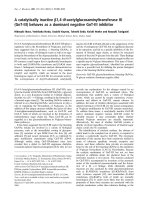
Figure 1 CE-MRI phenotypi ng of collapsed vs. expanding PLN and asymmetric knee arthr itis in TNF-Tg mice. TNF-Tg mice (n =10;20
legs) with ankle arthritis were monitored by CE-MRI to phenotype their PLN as expanding or collapsed. The quantitative data are presented in
Table 1. 2D CE-MRI (A-D), and 3D reconstructed volumes (E-H), of the left (A, B, E, F) and right (C, D, G, H) legs of a representative TNF-Tg mouse
with asymmetric arthritis are presented to illustrate the phenotypic differences between collapsed PLN (A, E), which are smaller and have limited
contrast enhancement vs. expanding PLN (C, G), which are larger and have saturated contrast enhancement throughout most of the node. The
asymmetric arthritic phenotype in this animal is also apparent from the contrast enhancing pannus tissue that surrounds the femoral chondyles
in only one knee (red arrows in B), and the larger synovial volume 5.6 mm
3
(F) vs. 3.8 mm
3
(H).
A
B
E
F
C
*
*
*
D
G
H
Figure 2 Asymmetric TNF-induced knee arthritis is associated with ipsilateral PLN collapse and decreased afferent lymphatic flow. The
mice described in Figure 1 were subjected to NIR-ICG imaging to quantify lymphatic drainage from their lower limbs, prior to sacrifice for
histology, and data from a representative animal are shown. The NIR-ICG images of the left (A) and right (B) lower limb of the mouse obtained
30 minutes after the ICG injection into the footpad (red arrows) illustrates the dramatic difference in afferent lymphatic flow to the PLN (green
arrows) as evidenced by the lack of signal in the collapsed (A) versus the bright signal in the expanding (B) PLN. Micrographs (5x) of the H&E
stained histology of the PLN reveal the shrunken phenotype of the collapsed PLN (C), compared to the expanding PLN with enlarged
paracortical sinuses (* in D). Micrographs of the H&E (E, F) and TRAP (G, H) stained histology of the knees taken at 5x and 10x respectively,
confirmed the presence of extensive synovitis (arrows in E) and focal erosions (G) in the left knee ipsilateral to the collapsed PLN, in contrast to
the very early stage arthritis observed in the right knee ipsilateral to the expanding PLN (F, H).
Li et al. Arthritis Research & Therapy 2011, 13:R138
/>Page 5 of 12
month-old TNF-Tg mice prior to the onset of ankle
arthritis, and TNF-Tg mice with bilateral ankle and
asymmetric knee arthritis (Figure 5). The flow cytometry
results showed that both expanding and collapsed PLN
and ILN have a similar three-fold increase in total B-in
numbers vs. aged matched WT controls. Moreover, this
increase was disease specific, as no differences in B-in
numbers were detected between WT and pre-arthritic
100
T-max
*
S-max
80
100
%-clearance
100
100
T-initial
20
40
60
80
0
20
40
60
80
*
20
40
60
80
*
20
40
60
*
80
0
Exp
Col
Exp Col
0
Exp
Col
0
0
Ex
p
Col
Figure 3 Afferent lymphatic drainage in the lower limb is significantly decreased after PLN collapse. NIR-ICG imaging was performed on
the TNF-Tg mice described in Figure 1 to quantify lymphatic flow in the lower limb. The real time video of the NIR-ICG imaging session was
used to quantify the four outcome measures of lymphatic flow from the foot to expanding (Exp) and collapsed (Col) PLN, and the data are
presented as the mean +/- SD for the group (n =3,*P < 0.05 vs. Exp).
ILN
AB
PLN
ILNs
C
Figure 4 Ipsilateral ILN drains ICG from WT/pre-arthritic knees not from arthritic knees with collapsed PLN. Non-recovery ICG-NIR
imaging of PLN and ILN was performed on WT mice (n = 2) following an intraarticular ICG injection into the knee, in which their abdominal
cavity was opened to expose the ILN. NIR-ICG images obtained 10 minutes after injection from one of the animals are shown highlighting the
ICG drainage from the injection site (black arrows) to the PLN (A dorsal view) versus the ILN (B ventral view). Note the absence of ICG signal in
the PLN (circled region), and its presence in the ILN (white arrow). Similar non-recovery ICG-NIR imaging was performed on TNF-Tg mice (n =8)
with asymmetric knee arthritis, and a ventral view image of a representative animal with collapsed (left) and expanding (right) PLN is shown (C).
Black arrows indicate the ICG injection site in the knee, and white arrows point to the ILN. Note that ICG has migrated to the right ILN but not
the left ILN resulting in a dramatic difference in SI (179 vs. 253).
Li et al. Arthritis Research & Therapy 2011, 13:R138
/>Page 6 of 12
Expand
i
ng
Collapsed
WT
A
71.4
0.81
12.6
35.7
4.44
45.4
45.2
3.87
37.8
CD23
B-in
Fo
MZ
PL
N
6.16
77.3
2.44
18.3
57.4
8.93
19.2
56.1
4.25
B
CD23
ILN
CD21
40
50
***
*
***
D
50
60
70
***
***
s
%
C
***
PLN ILN
0
10
20
30
40
***
**
WT Pre Exp Exp Col
10
20
30
40
50
0
***
B-in cell
s
WT Pre Exp Exp Col
c
ells # (x10
-6
)
2
3
4
5
***
***
**
**
0.2
0.3
0.4
0.5
E
F
**
*
B-in
c
0
1
0.0
0.1
WT Pre Exp Exp Col
WT Pre Ex
p
Ex
p
Col
Figure 5 B-in expansion in Ipsilateral PLN and ILN of TNF-Tg mice with inflammatory arthritis. PLN and ILN (n ≥ 4) were harvested from
wild-type (WT) mice, one- to two-month old TNF-Tg mice before the onset of ankle arthritis and PLN expansion (Pre Exp), and older TNF-Tg
mice with established disease after PLN expansion (Exp), or after PLN collapse (Col), and used for multicolor flow cytometry as described in
Materials and methods. The B-in population was quantified from the B220+/IgM+ fraction based on CD21 and CD23 staining, as illustrated by
representative histograms of ipsilateral PLN (A), and ILN (B), from each group. This gating approach segregates the phenotypic follicular B cells
(Fo), the marginal zone B cells (MZ), and the B-in population. The percentage of each population is shown. The percentage of B-in cells within
this B220+/IgM+ fraction from PLN (C), and ILN ( D); and the absolute number of B-in cells from PLN (E) and ILN (F) are presented as the mean
+/- SD for each group (*P < 0.05, **P < 0.01, ***P < 0.001).
Li et al. Arthritis Research & Therapy 2011, 13:R138
/>Page 7 of 12
TNF-Tg PLN and ILN. Finally, we observed similar per-
centages of hematopoietic cell populations between ipsi-
lateral PLN and ILN (multicolor flow for CD1d, CD3,
CD4, CD5, CD8, CD11b, CD11c, CD19, CD24, CD25,
CD80, CD86, CD69, CD93, IgD and GL7, data not
shown), which is consistent with our previous findings
[29]. Thus, B-in expansion in ipsilateral PLN and ILN
occurs simultaneously.
B cell translocation into LYVE-1+ sinuses in collapsed
ipsilateral PLN and ILN
Previously, we showed that expanding and collapsed
PLN display distinct lymphoid architecture [29].
Expanding PLN have normal B cell follicles and T cell
zone, with dilated paracortical sinuses filled with lymph
that are mostly free of cells, suggesting active draining
function. In collapsed PLN, the architecture of the B
cell follicles and T cell zone are totally disrupted by B-
in translocation into the paracorti cal sinuses in the cen-
ter of the node, consistent with decreased draining func-
tion. As this B-in translocation and de creased interstitial
space within the lymphatic vessels are the prominent
histological differences between expanding and collapsed
PLN, we investigated these features in ipsilateral PLN
and ILN. Tissue sections were immunostained for both
B cells and lymphatic endothelial cells with labeled anti-
bodies against IgM and LYVE-1 respectively. Selective
imaging of the IgM
hi
cells, which i ncludes the B-in
population, was performed by signal intensity threshold-
ing, and subsequent co-localization within lymphatic
endothelium was assessed by superimposition two-color
fluorescence microscopy images (Figure 6). The results
demonstrated consistent association: all of expanding
PLN were ipsilateral to ILN with wide lymphatic vessels
that were void of IgM
hi
cells (< 0.1% overlap with
LYVE-1). Conversely, all of the collapsed PLN were ipsi-
lateral to ILN whose lymphatic vessels were filled with
IgM
hi
cells. These IHC results support the hypothesis
that asymmetric knee flare is mediated by simultaneous
ipsilateral ILN and PLN collapse due to the transloca-
tion of B-in to the lumen of the lymphatic vessels of the
nodes. We predict that these translocated B-in cells
obstruct the lymphatics of the lower limb resulting in
decreased afferent flow from the knee to the ILN.
Afferent lymphatic flow to ipsilateral lymph nodes is
associated with knee synovitis
To assess the direct association between knee synovitis
and afferent lymphatic flow to ipsilateral lymph nodes, a
cohort of TNF-Tg mice with a broad range of knee
arthritis was identified by performing CE-MRI on ani-
mals three to less than nine months of age to quantify
knee synovial volume. Subsequently, ICG-NIR imaging
was performed to quantify the signal intensity of PLN
or ILN independently. Linear regression analysis of
these data revealed highly significant correlations (Figure
7). These results support a model in which approxi-
mately 70% of knee flare in TNF-Tg mice can be
explained by decreased lymphatic flow.
Discussion
Our understanding of the events that lead to joint flare
is incomplete, and critical questions remain answered.
Specifically, the mechanisms that promote t he develop-
ment of asymmetric arthritis in the setting of a systemic
immune mediated inflammatory disease have yet to be
identi fied. We have previously demonstrated that altera-
tions in PLN correlate with knee flare in TNF-Tg mice
[22,28,29]. These intriguing observati ons provoked us to
interrogate this potential biomarker, which predictably
increases in size and contrast enhancement during a
prolonged expansion phase, followed by a sudden col-
lapse (Figure 1). Since these descriptive phenotypes are
determined by a quantitat ive outcome measure, we set
out to find an empirical CE-MRI threshold value that
can objectively segregate expanding vs. collapsed PLN.
Here we demonstrate this value to be LNcap = 30
(Table 1). Interestingly, LNCE = 5 also proved to be a
reliable threshold value, while LN vo lume did not,
demonstrating the importance of perfusion over size in
this biomarker of arthritic flare.
The observation that asymmetric PLN colla pse occurs
concomitantly with arthritic flare in the adjacent knee
(Figure 2), leads to the prediction that there also must be
asymmetry in lymphatic draining function in the lower
limb. Our ICG-NIR imaging results demonstrate this to
be true (Figure 3), and support our conclusion from the
CE-MRI data that the functional s ignificance of the PLN
in the arthritic flare process in the adjacent knee is
dependent on lymphatic flow and not on node size.
The importance of lymphatic flow is also underscore d
by the fact that there are no significant cellular differ-
ences between expanding and collapsed PLN as deter-
mined by assessment of surface markers, proliferation
and B cell heterogeneity, although they bot h have a sig-
nificantly increased B-in population [29]. However, we
did observe a histological difference between these phe-
notypes in that expanding PLN c ontain large paracorti-
cal sinuses devoid of IgM+ cells, while in collapsed PLN
the paracortical sinuses were filled with IgM+ cells. This
tissue morphology is consistent with “clogging” of lym-
phatic vessels by B cell aggregates and resultant, dimin-
ished lymphatic flow as observed in collapsed vs.
expanding PLN (Figure 2).
The biggest surprise of this study was the finding that
the ILN drains the knee (Figure 4), which initially
appeared to be inconsistent with a model where collapse
of PLN-induces an arthritic flare i n the ipsilateral knee.
Li et al. Arthritis Research & Therapy 2011, 13:R138
/>Page 8 of 12
One potential explanation is that ipsilateral PLN and
ILN collapse is triggered by the same stimuli and occurs
simultaneously. In support of this theory, we found that
B-in expansion (Figure 5) and translocation (Figure 6)
also occur simultaneously in ipsilateral PLN and ILN.
Moreover,wefoundthatlymphaticdrainagetoboth
PLN and ILN significantly correlate with knee synovial
volume in TNF-Tg mice (Figure 7), suggesting that a
single mechanism may be responsible for LN collapse in
the same limb. It is important to note that these experi-
ments were limited by the facts that PLN drain to ILN
sequentially, thus making quantification of lymphatic
flow to ipsilateral PLN and ILN impossible; and that
ICG-NIR imaging of ILN requires euthanasia to expos e
the abdominal cavity, which limits quantitative assess-
ment to a single time point. Nevertheless, the linkage of
PLN and ILN collapse with the onset of ipsilateral k nee
synovitis strongly supports the existence of regional
lymphatic factors that mediate joint flare during chronic
inflammatory arthritis. Although purely speculative at
this time, we find that this experimental evidence points
to a central neuromuscular cascade that innervates the
lymphatics along the axial plane o f the limb, and domi-
nates the local intrinsic lymphatic pumps that are
known to be under adrenergic, cholinergic and peptiner-
gic control [35]. Experiments to elucidate this central
neuromuscular signal are ongoing.
The emerging paradig m to explain the pathogen esis of
inflammatory arthritis posits th at the disease initiat es in
the small distal joints of the flanges as a tenosynovitis,
which rapidly spreads to the adjacent joint due to the
immediate proximity of the inflamed synovial sheath and
the synovium [ 30]. The chronic inflammation in these
small joint s stimula tes lymphangiogenesis to limit the
A
B
0.07 %
0.57 %
IgM LYVE
IgM LYVE
0.93 %
0.02 %
C
D
Figure 6 Ipsilateral PLN and ILN collapse is associated with B cell translocation into LYVE+ sinuses. Ipsilateral pairs of PLN and ILN (n =
4) from the mice described in Figure 1 were processed for IHC with fluorescently labeled antibodies against IgM and LYVE-1 to image the B
cells (red) and lymphatic endothelium (green) respectively. Multicolor fluorescent micrographs (5x) of representative ILN (A, B) and PLN (C, D)
are presented to illustrate the distinct staining in expanding nodes (A, C), versus the apparent co-localization (yellow) in the collapsed nodes (B,
D), due to the B cells that have translocated into the paracortical lymphatic sinuses. The images were analyzed in Image-Pro Plus and the
percentage of yellow pixels representing the overlapping signal is indicated.
Li et al. Arthritis Research & Therapy 2011, 13:R138
/>Page 9 of 12
progression of synovitis and pannus formation by remov-
ing the immune cells and catabolic factors. Thus, disease
spreads to the large-proximal joints only when the lym-
phatic drainage capacity of the limb is severely impaired ,
or a yet to be identified incident triggers LN collapse.
Here we provide the first evidence that LN collapse occurs
in series along an ipsilateral axis. The potential clinical sig-
nificance of t his is that the underappreciated enlarged
efferent LN of RA joints that are often palpable on exam,
or evident on imaging studies, may reflect disease activity
and potentially a response to therapy. To explore this pos-
sibility we are currently evaluating the potential of MRI
and ultrasound i maging to phenotype LN in RA patients
as expanding or collapsed (Cli nicalTrials.gov ID#
NCT01098201, NCT01083563). More over, thi s model
predicts that at least a component of the efficacy of BCDT
is derived from its ability to clear B-in from lymphatic
endothelium and thus “ unclog” the sinuses and restore
lymphatic flow. Certainly this hypothesis is testable in ani-
mal models and clinical trials, and future studies will
determine the overall importance of this process in the
etiology of arthritic flare.
Conclusions
Asymmetric knee arthritis in TNF-Tg mice is triggered
by simultaneous collapse of ipsilateral PLN and ILN,
I
ILN IC
G
S
R
2
=0.7411
P 0 006
I
Knee synovial volume mm
3
P
=
0
.
006
PLN ICG S
I
R
2
=0.6467
P=0.0017
Knee s
y
novial volume mm
3
Figure 7 Decreased lymphatic flow to ipsilateral PLN and ILN correlates with increased knee synovitis. The knee synovial volume of TNF-
Tg mice (ages three to less than nine months) at different stages of disease was quantified by CE-MRI. The lymphatic draining function of PLN
and ILN in these mice was quantified independently by ICG-NIR imaging 30 minutes and 10 minutes after injection respectively by determining
the signal intensity (SI) of the node. These data were used to assess the direct relationship between knee synovitis and lymph draining function
in the ipsilateral ILN and PLN via linear regression analyses, in which the correlation coefficient (R
2
) with its statistical significance (P) is shown.
Li et al. Arthritis Research & Therapy 2011, 13:R138
/>Page 10 of 12
which is likely due to B-in translocat ion to the lumen of
lymphatic vessels of both lymph nodes and result in a
dramatic decrease in afferent lymphatic flow in the
lower limb. As PLN and ILN function in series, PLN
function serves as a new biomarker of arthritic flare in
the adjacent knee.
Abbreviations
AU: arbitrary unit; B-in: B cells in inflamed node; CE-MRI: contrast-enhanced
magnetic resonance imaging; ICG-NIR: indocyanine green near-infrared; ILN:
iliac lymph node; LN cap: lymph node capacity; LN CE: lymph node contrast
enhancement; LN vol: lymph node volume; PLN: popliteal lymph node; RA:
rheumatoid arthritis; TNF-Tg: tumor necrosis factor transgenic.
Acknowledgements
The authors would like to thank Ryan Tierney and Patricia Weber for
technical assistance with the histology and CE-MRI respectively. This work
was supported by research grants from the National Institutes of Health PHS
awards (AR48697 and AR53586 to LX, AI78907, DE17096 and AR54041 to
EMS).
Author details
1
Center for Musculoskeletal Research, University of Rochester School of
Medicine and Dentistry, 601 Elmwood Avenue, Rochester, NY 14642, USA.
2
Department of Microbiology and Immunology, University of Rochester
School of Medicine and Dentistry, 601 Elmwood Avenue, Rochester, NY
14642, USA.
3
Department of Pathology and Laboratory Medicine, University
of Rochester School of Medicine and Dentistry, 601 Elmwood Avenue,
Rochester, NY 14642, USA.
4
Department of Obstetrics and Gynecology,
University of Rochester School of Medicine and Dentistry, 601 Elmwood
Avenue, Rochester, NY 14642, USA.
5
Department of Urology, University of
Rochester School of Medicine and Dentistry, 601 Elmwood Avenue,
Rochester, NY 14642, USA.
6
Division of Allergy, Immunology, Rheumatology,
Department of Medicine, University of Rochester School of Medicine and
Dentistry, 601 Elmwood Avenue, Rochester, NY 14642, USA.
Authors’ contributions
JL performed most of the experiments, analyzed the data and participated
in the manuscript draft. QZ participated in part of the ICG-NIR lymphatics
imaging and data analysis. IK helped with flow cytometry. RWW, AB, LX and
CTR provided scientific input and helped with manuscript editing. EMS
designed the study, and drafted and finalized the manuscript. All authors
read and approved the final manuscript.
Competing interests
The authors declare that they have no competing interests.
Received: 4 June 2011 Revised: 26 July 2011 Accepted: 31 August 2011
Published: 31 August 2011
References
1. Fransen J, Hauselmann H, Michel BA, Caravatti M, Stucki G: Responsiveness
of the self-assessed rheumatoid arthritis disease activity index to a flare
of disease activity. Arthritis Rheum 2001, 44:53-60.
2. Brunner HI, Lovell DJ, Finck BK, Giannini EH: Preliminary definition of
disease flare in juvenile rheumatoid arthritis. J Rheumatol 2002,
29:1058-1064.
3. Bingham CO, Pohl C, Woodworth TG, Hewlett SE, May JE, Rahman MU,
Witter JP, Furst DE, Strand CV, Boers M, Alten RE: Developing a
standardized definition for disease “flare” in rheumatoid arthritis
(OMERACT 9 Special Interest Group). J Rheumatol 2009, 36:2335-2341.
4. Ringold S, Bittner R, Neogi T, Wallace CA, Singer NG: Performance of
rheumatoid arthritis disease activity measures and juvenile arthritis
disease activity scores in polyarticular-course juvenile idiopathic arthritis:
Analysis of their ability to classify the American College of
Rheumatology pediatric measures of response and the preliminary
criteria for flare and inactive disease. Arthritis Care Res (Hoboken) 2010,
62:1095-1102.
5. Lin HC, Chen SF, Chen YH: Increased risk of adverse pregnancy outcomes
in women with rheumatoid arthritis: a nationwide population-based
study. Ann Rheum Dis 2009, 69:715-717.
6. Gio-Fitman J: The role of psychological stress in rheumatoid arthritis.
Medsurg Nurs 1996, 5:422-426.
7. Kallberg H, Ding B, Padyukov L, Bengtsson C, Ronnelid J, Klareskog L,
Alfredsson L: Smoking is a major preventable risk factor for rheumatoid
arthritis: estimations of risks after various exposures to cigarette smoke.
Ann Rheum Dis 2010, 70:508-511.
8. Firestein GS: Evolving concepts of rheumatoid arthritis. Nature 2003,
423:356-361.
9. McGonagle D, Marzo-Ortega H, O’Connor P, Gibbon W, Pease C, Reece R,
Emery P: The role of biomechanical factors and HLA-B27 in magnetic
resonance imaging-determined bone changes in plantar fascia
enthesopathy. Arthritis Rheum 2002, 46:489-493.
10. Pullinger BD, Florey HW: Proliferation of lymphatics in inflammation. J
Pathol Bacteriol 1937, 45:157-170.
11. Schwarz EM, Proulx ST, Ritchlin CT, Boyce BF, Xing L: The role of bone
marrow edema and lymphangiogenesis in inflammatory-erosive arthritis.
Adv Exp Med Biol 2010, 658:1-10.
12. Olszewski WL, Pazdur J, Kubasiewicz E, Zaleska M, Cooke CJ, Miller NE:
Lymph draining from foot joints in rheumatoid arthritis provides insight
into local cytokine and chemokine production and transport to lymph
nodes. Arthritis Rheum 2001, 44:541-549.
13. Halin C, Detmar M: Chapter 1. Inflammation, angiogenesis, and
lymphangiogenesis. Methods Enzymol 2008, 445:1-25.
14. Grillet B, Dequeker J: Rheumatoid lymphedema.
J Rheumatol 1987,
14:1095-1097.
15.
Ueo T, Tanaka S, Tominaga Y, Ogawa H, Sakurami T: The effect of thoracic
duct drainage on lymphocyte dynamics and clinical symptoms in
patients with rheumatoid arthritis. Arthritis Rheum 1979, 22:1405-1412.
16. Quarta L, Corrado A, d’Onofrio F, Maruotti N, Cantatore FP: Two cases of
distal extremity swelling with pitting oedema in psoriatic arthritis: the
different pathological mechanisms. Rheumatol Int 2010, 30:1367-1370.
17. Bohm M, Riemann B, Luger TA, Bonsmann G: Bilateral upper limb
lymphoedema associated with psoriatic arthritis: a case report and
review of the literature. Br J Dermatol 2000, 143:1297-1301.
18. Zhang Q, Guo R, Lu Y, Zhao L, Zhou Q, Schwarz EM, Huang J, Chen D,
Jin ZG, Boyce BF, Xing L: VEGF-C, a lymphatic growth factor, is a RANKL
target gene in osteoclasts that enhances osteoclastic bone resorption
through an autocrine mechanism. J Biol Chem 2008, 283:13491-13499.
19. Guo R, Zhou Q, Proulx ST, Wood R, Ji RC, Ritchlin CT, Pytowski B, Zhu Z,
Wang YJ, Schwarz EM, Xing L: Inhibition of lymphangiogenesis and
lymphatic drainage via vascular endothelial growth factor receptor 3
blockade increases the severity of inflammation in a mouse model of
chronic inflammatory arthritis. Arthritis Rheum 2009, 60:2666-2676.
20. Zhou Q, Wood R, Schwarz EM, Wang YJ, Xing L: Near-infrared lymphatic
imaging demonstrates the dynamics of lymph flow and
lymphangiogenesis during the acute versus chronic phases of arthritis
in mice. Arthritis Rheum 2010, 62:1881-1889.
21. Rovensky J, Stancikova M, Rovenska E, Stvrtina S, Stvrtinova V, Svik K:
Treatment of rat adjuvant arthritis with flavonoid (Detralex),
methotrexate, and their combination. Ann N Y Acad Sci 2009,
1173:798-804.
22. Proulx ST, Kwok E, You Z, Beck CA, Shealy DJ, Ritchlin CT, Boyce BF, Xing L,
Schwarz EM: MRI and quantification of draining lymph node function in
inflammatory arthritis. Ann N Y Acad Sci 2007, 1117:106-123.
23. Troyan SL, Kianzad V, Gibbs-Strauss SL, Gioux S, Matsui A, Oketokoun R,
Ngo L, Khamene A, Azar F, Frangioni JV: The FLARE intraoperative near-
infrared fluorescence imaging system: a first-in-human clinical trial in
breast cancer sentinel lymph node mapping. Ann Surg Oncol 2009,
16:2943-2952.
24. Xing L, Ji R: Lymphangiogenesis, myeloid cells and inflammation. Expert
Rev Clin Immunol 2008, 4:599-613.
25. Keffer J, Probert L, Cazlaris H, Georgopoulos S, Kaslaris E, Kioussis D,
Kollias G: Transgenic mice expressing human tumour necrosis factor: a
predictive genetic model of arthritis. EMBO J 1991, 10:4025-4031.
26. Kouskoff V, Korganow AS, Duchatelle V, Degott C, Benoist C, Mathis D:
Organ-specific disease provoked by systemic autoimmunity. Cell 1996,
87:811-822.
Li et al. Arthritis Research & Therapy 2011, 13:R138
/>Page 11 of 12
27. Zhang Q, Lu Y, Proulx S, Guo R, Yao Z, Schwarz EM, Boyce BF, Xing L:
Increased lymphangiogenesis in joints of mice with inflammatory
arthritis. Arthritis Res Ther 2007, 9:R118.
28. Proulx ST, Kwok E, You Z, Papuga MO, Beck CA, Shealy DJ, Ritchlin CT,
Awad HA, Boyce BF, Xing L, Schwarz EM: Longitudinal assessment of
synovial, lymph node, and bone volumes in inflammatory arthritis in
mice by in vivo magnetic resonance imaging and microfocal computed
tomography. Arthritis Rheum 2007, 56:4024-4037.
29. Li J, Kuzin I, Moshkani S, Proulx ST, Xing L, Skrombolas D, Dunn R, Sanz I,
Schwarz EM, Bottaro A: Expanded CD23(+)/CD21(hi) B cells in inflamed
lymph nodes are associated with the onset of inflammatory-erosive
arthritis in TNF-transgenic mice and are targets of anti-CD20 therapy. J
Immunol 2010, 184:6142-6150.
30. Hayer S, Redlich K, Korb A, Hermann S, Smolen J, Schett G: Tenosynovitis
and osteoclast formation as the initial preclinical changes in a murine
model of inflammatory arthritis. Arthritis Rheum 2007, 56:79-88.
31. Ji H, Ohmura K, Mahmood U, Lee DM, Hofhuis FM, Boackle SA, Takahashi K,
Holers VM, Walport M, Gerard C, Ezekowitz A, Carroll MC, Brenner M,
Weissleder R, Verbeek JS, Duchatelle V, Degott C, Benoist C, Mathis D:
Arthritis critically dependent on innate immune system players.
Immunity 2002, 16:157-168.
32. Dardzinski BJ, Schmithorst VJ, Holland SK, Boivin GP, Imagawa T,
Watanabe S, Lewis JM, Hirsch R: MR imaging of murine arthritis using
ultrasmall superparamagnetic iron oxide particles. Magn Reson Imaging
2001, 19:1209-1216.
33. Inoue Y, Kiryu S, Watanabe M, Oyaizu N, Ohtomo K: Fluorescence lymph
node mapping in living mice using quantum dots and a compression
technique. J Fluoresc 2010, 20:599-606.
34. Tsutsumi R, Hock C, Bechtold CD, Proulx ST, Bukata SV, Ito H, Awad HA,
Nakamura T, O’Keefe RJ, Schwarz EM: Differential effects of biologic versus
bisphosphonate inhibition of wear debris-induced osteolysis assessed by
longitudinal micro-CT. J Orthop Res 2008, 26:1340-1346.
35. Zawieja D: Lymphatic biology and the microcirculation: past, present and
future. Microcirculation 2005, 12:141-150.
doi:10.1186/ar3452
Cite this article as: Li et al.: CD23
+
/CD21
hi
B-cell translocation and
ipsilateral lymph node collapse is associated with asymmetric arthritic
flare in TNF-Tg mice. Arthritis Research & Therapy 2011 13:R138.
Submit your next manuscript to BioMed Central
and take full advantage of:
• Convenient online submission
• Thorough peer review
• No space constraints or color figure charges
• Immediate publication on acceptance
• Inclusion in PubMed, CAS, Scopus and Google Scholar
• Research which is freely available for redistribution
Submit your manuscript at
www.biomedcentral.com/submit
Li et al. Arthritis Research & Therapy 2011, 13:R138
/>Page 12 of 12