Báo cáo y học: "Equipment review: Gastric intramucosal pH measurement" pptx
Bạn đang xem bản rút gọn của tài liệu. Xem và tải ngay bản đầy đủ của tài liệu tại đây (263.95 KB, 5 trang )
REVIEW
Equipment review: Gastric intramucosal pH
measurement
Francisco Baigorri, Xavier Calvet, Domenec Joseph
64cc-1-2-061
Introduction
Gastric tonometry has emerged as an attractive, rela-
tively noninvasive technology for assessing gastrointest-
inal perfusion and oxygenation by detecting acidosis in
the gut wall. Several clinical studies have shown that
gastric intramucosal acidosis detected by this procedure
predicts increased mortality of critcally ill adults in med-
ical and surgical intensive care unit (ICU) settings [1-3],
and that it is a better predictor of mortality from critical
illness than other mesures of global oxygen delivery and
systemic hemo dynamics [4]. It has also been suggested
that correcting intramucosal acidosis may increase survi-
val in selected critically ill patients [5].
The purpose of this review is to discuss factors influ-
encing in vivo reliability and variability of gastric tono-
metry, and to analyze the causes of the occasional
misinterpretation of its results.
The gastric tonometry technique — causes of
misinterpretation of the results
The measurement of gastric mucosal acidosis by gastric
tonometry is based on the principle that the fluid in a
hollow viscus can be used to estimate gas tensions in
the surrounding tissues. The main assumption is that,
after a given equilibration time, luminal and mucosal
CO
2
partial pressures (PCO
2
) will be similar. Conse-
quently, the increased tissue production of CO
2
during
hypoxia (from the reaction between hydrogen anions
and bicarbonate) can be detected by analyzing the liquid
inside the gastric lumen.
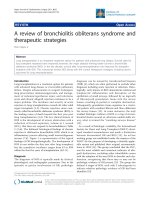
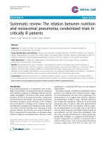
Conventional gastric tonometry involves the place-
ment of a modified nasogastric (NG) tube, equipped
with a gas-permeable, saline-filled silicone balloon at its
tip, into the stomach [6,7] (Fig 1). Allowing enough
time for the equilibration of CO
2
between the fluid in
the balloon and the gastric lumen (30– 90 min), the
saline is then aspirated and its PCO
2
determined using a
blood gas analyzer. Thus, gastric tonometry can deter-
mine intraluminal PCO
2
(PiCO
2
) which is assumed to
be in equilbrium with gastric mucosal PCO
2
. Intramuco-
sal pH (pHim) can be calculated by the Henderson-Has-
selbach equation, using the PiCO
2
value determined by
gastric tonometry and the arterial bicarbonate concen-
tration, assuming that the tissue bicarbonate concentra-
tion is in the equilibrium with that in the capil laries,
which is further assumed to be the same as determined
for arterial blood.
Consequently, c auses of misleading interpretations of
gastric tonometry can be divided as follows:
1. those wh ich ‘originat e from the patient’, and actu-
ally confound the logical interpretation based on clinical
determinations (mainly, d isturbances in systemic acid
base balance);
2. local factors in the gastric lumen which can alter
the relationship between PiCO
2
and mucosal PCO
2
, and
3. factors inherent in the technique which can cause
erroneous determinations of PiCO
2
.
Patient factors - from pHim to DPCO
2
According to one report [8], gastric tonometry failed
to accurately estimate the magnitude of the decrease
in tissue pH in conditions of low perfusion (total and
partial occlusi on of the superior mesenteric ar tery);
direct measurement of pH with microelec trodes was
found to be more accurate. One probable explanation
for the inaccuracy of gastric tonometry in these low
perfusion conditions is the fact that tissue bicarbonate
levels are overestimated when determined via arterial
concentration. This is because tissue b icarbonate is
consumed by buffering protons which are generated by
ischemic tissue, therefore reducing the input of fresh
bicarbonate. As a clinical example of this phenomenon,
Benjamin et al [9]reportedthatcalculatedpHim
became normal with sodium bicarbonate administra-
tion in the treatment of a patient with severe systemic
acidosis despite laparotomy-proven massive mesenteric
Intensive Care Service, Hospital de Sabadell, Parc Tauli s/n, 08208 Sabadell,
Spain
Baigorri et al. Critical Care 1997, 1:61
©1997CurrentScienceLtd
ischemia, resulting in a correction of the calculated
pHim.
However, an examination of the contribution of the
stomach mucosa to its own acid-base status must
include an assessment of the arterial blood perfusing
that area. If arterial bicarbonate is low due to acidosis
arising somewhere other than the gastrointestinal tract,
then calculated pHim may be low despite normal
PiCO
2
.
Indeed, studies in crit ically ill patients have shown a
striking correlation between pHim and measurements of
metabolic acidosis [4,10]. Therefore, measuring PiCO
2
alone is simpler and eliminates arterial bicarbonate as
one source of error.
Tissue PCO
2
— the parameter we aim to determin e
by gastric tonometry — equilbrates almost exactly with
capillary PCO
2
and, according to the Fick principle, is
related to tissue CO
2
production and arterial CO
2
Figure 1 Gastric tonometry determines intraluminal PCO
2
which is assumed to be in equilibrium with PCO
2
in the gastric mucosa. Intramucosal
pH (pHim) can be calculated by the Henderson-Hasselbach equation using the PCO
2
value determined by gastric tonometry and the
bicarbonate concentration in arterial blood.
Baigorri et al. Critical Care 1997, 1:61
Page 2 of 5
content, and is inversely related to regional blood flow.
Therefore, changes in arterial PCO
2
(PaCO
2
) should
affect tissue PCO
2
. It has been shown that respiratory
acidosis leads to tissue hypercarbia in animals [11]. This
observation is in agreement with clinical studies show-
ing that patients with hypercapnia have a significantly
higher PiCO
2
than those without, and that their pHim
is also signigficantly lower [12]. The r elationship
between PaCO
2
and PiCO
2
has also been observed in
individual patients whose PaCO
2
was modified by
changes in dead space [13]. Thus, pHim assesses not
only splanchnic oxygenation, but also arterial a cid-base
status.
The gradient between PiCO
2
and PaCO
2
(DPCO
2
),
which is solely determined by the ratio between blood
flow and CO
2
production in the tissue, shoul d be a bet-
ter measure of mucosal perfusion. A new question arises
from this assertion - how much does DPCO
2
have to
increase in order to indicate not just low g astric blood
flow, but also anaerobic generation of CO
2
[14]?
Schlichtig and Bowles [15] recently reported that the
onset of intestinal anaerobiosis in normal dogs occurred
when PiCO
2
had risen to 65 mmHg and DPCO
2
had
increased to 25–35 mmHg.
The use of the gradient between intramucosal and
arterial pH [16] seems to be more cumbersome and less
precise than DPCO
2
[17].
Finally, we should ask whether gastric mucosal acido-
sis always indicates tissue hypoperfusion. Like most stu-
dies validating gastric tonometry, the aforementioned
article by Schlichtig and Bowles [15] used a model of
progressive flow reduction. However, there is eviden ce
from animal sepsis models of intestinal mucosal acidosis
that is unrelated to tissue hypoxia [18,19]. Therefore,
tissue acidosis in sepsis may result from causes other
than cellular dysoxia. It has been suggested that a pre-
ferential increase in anaerobic glucose utilization at the
expense of oxidative glucose metabolism, even in the
presence of adequate or even supranormal oxygen levels,
could lead to t issue acidosis [20,21]. This concept has
important clinical implications - we should take particu-
lar care when attending to septic patients with gastric
mucosal acidosis because it may not in fact be incontro-
vertible evidence of tissue hypoperfusion [21].
Local factors that can influence ga stric tonometry
The relationship between high PiCO
2
and mucosal
ischemia in the stomach is invalidated in cases where
CO
2
is produced in the lumen. Buffering of gastric acid
by bicarbonate, either from an exogeneous source, or
from gastric or duodenal secretions, is a major cause of
increased intraluminal CO
2
.
Studies in human volunteers have shown that admin-
istration of ranitidine, an H
2
receptor blocking agent,
reduces the error in PiCO
2
measurement [22-24]. Con-
sequently, inhibition of acid secretion is now considered
to be mandatory for proper assessment of intraluminal
PCO
2
. However, this recommendation has not been vali-
dated in critically ill patients; studies suggest that the
use of H
2
-blockers in the critically ill has no effect on
the assessment of intraluminal PCO
2
[25,26]. Discrepan-
cies between results in healthy volunteers and critically
ill patients may be related to a reduced gastric acid
secretion in the latter as a result of compromised visc-
eral perfusion [27-29]. However, these studies of criti-
cally ill patients were performed with small patient
samples and over a short period, without changes in
their hemodynamic status. The results may, therefore,
not be applicable to hemodynamically unstable patients.
The effect of other treatments commonly administered
via an NG tube on the measurement of pHi by gastric
tonometry remains unclear. Elsewhere [30] we studied
the effect of sucralfate, which is widely used for stress
ulcer bleeding prophylaxis because it does not signifi-
cantly reduce gastric pH and tends to decrease the addi-
tional risk of gastric bacterial overgrowth. Our results
suggested that enteral administration of sucralfate does
not alter the determination of pHim by ga stric tonome-
try in critically ill patients.
Enteral feeding may also affect accurate assessment of
PiCO
2
. Once food enters the stomach it stim ulates
secretion of gastric juice and bicarbonate ions. This
combination, along with the digestion of nutrients, may
generate CO
2
inside the gastric lumen. In animals, it has
been shown that gastric instraluminal PCO
2
increases
after feeding [31]. This effect has also b een observed in
asymptomatic subjects [32] and in critically ill patients
[33]. Consequently, it is currently recommended that
ent eral feedings be discontinued fo r about 1–2hbefore
measuring pHim. This period may need to be longer in
patients with delayed gastric emptying.
In normal conditioins, blood flow to each portion of
the gastrointestinal tract is proportional to the level of
local activity. Blood flow increases after feeding by 100–
150% for 3–6 h. Consequently, if the flow cannot
increase appropriately, enteral feeding may result in gas-
trointestinal hypoxia with mucosal acidosis. In fact, the
presence of mucosal acidosis after feeding has been used
to detect chronic gastric ischemia [32].
Factors related to the technique — from saline to
air
Gastric tonometr y presents the main sources of problem
— the time required from equilibration, the measure-
ment of saline PCO
2
, and the potenti al loss of CO
2
dur-
ing transport of the sample.
The first of these, the time required for equilibration,
is an important factor. Equilibration follows Fick’slaw
Baigorri et al. Critical Care 1997, 1:61
Page 3 of 5
of diffusion. Complete equilibration of the tonometer
solution with mucosal PCO
2
requires at least 60–90
min, with shorter times resulting in the measurement
becoming significantly more variable.
Measurement of saline PCO
2
is also an important
source of error and, as shown by Takala et al, depends
on both the analyzer used and the actual PCO
2
level
[34]. Most analyzers underestimated saline PCO
2
by 5–
19%. Notably, the performance of all analyzers markedly
improved when a buffer solution was used. Why, then,
not use a buffer solution instead of saline? The problem
is that due to the higher CO
2
-binding capacity of the
buffer, more time is required for the equilibration of tis-
sue and sample CO
2
, reducing the ability of t he intra-
gastric tonometer to respond to changing tissue PCO
2
.
Another alternative to the use of saline is air. The use
of ‘balloonless’ air tonometry has been reported in ani-
mals, and Salzman et al have demonstrated a good cor-
relation between tonometric PCO
2
measurements
obtained simultaneously from samples of air and saline
solution [11]. Although, in the above study the air was
analyzed by a blood gas analyzer, its use has opened by
the possibility of determining intramucosal PCO
2
by
capnography.
Capnography is the basis of some new systems for
nearly continuous monitoring of intramucosal PCO
2
.
One r ecently validated system allows continuous recir-
culation of gas through the balloon of the tonometer
[35]. The new system was compared with a conventional
tonometer in a n in vivo experiment on dogs wit h
indu ced hypoxia. The air system showed a high er sensi-
tivity in detecting tissue hypoxia. The probable explana-
tion for the greater sensitivity of the continuous
monitoring system was that the recirculating gas was
already in equilibrium with PiCO
2
immediately before
the induction of hypoxia.
An automated tonometric system which also uses cap-
nography with a conventional tonometer is now com-
mercially available [36 ]. This system allows concomitant
determination of end-tid al CO
2
with PiCO
2
to es timate
DPCO
2
. This system works by introducing a certain
amount of air into the balloon, which is periodically
aspirated in order to determine PCO
2
.Thesameairis
sent back to the balloon after d etermining tonometric
PCO
2
. Therefore, as with the system described above
that uses recirculating gas, it increases sensitivity to
changes in intramucosal PCO
2
,allowingsamplingtimes
shorter than 30 min.
In addition to higher sensitivity, the expected advan-
tages of these systems are:
1. shorter sampling times;
2. the s elected time for equilibration is always con-
stant, and
3. the fact that there is no need for saline aspiration
and transport to a blood gas analyzer, avoiding the risk
of CO
2
loss during transport and, thus, reducing the
actual number of error sources in the technique.
Conclusion
In summary, gastric tonometry is relatively simple tech-
nique, but obtaining reliable results and interpreting
them accurately requires a comprehensive knowledge of
the technique and careful attention to the smallest
detail. Frequency of measurement is limited by the time
required and staff intervention involved. The use of air
instead of saline, and PCO
2
determination by capnogra-
phy seem to be promising ways of avoiding some of the
problems that the technique presents.
Published: 26 November 1997
References
1. Gys T, Hubens A, Neels H, Lauwers LF, Peeters R: Prognostic value of
gastric intramural pH in surgical intensive care patients. Crit Care Med
1988, 16:1222-1224.
2. Doglio GR, Pusajo JF, Egurrola MA, et al: Gastric mucosal pH as a
prognostic Index of mortality in critically ill patients. Crit Care Med 1991,
19:1037-1040.
3. Gutierrez G, Bismar H, Dantzker Dr, Silva N: Comparison of gastric
intramucosal pH with measures of oxygen transport and consumption
in critically ill patients. Crit Care Med 1992, 20:451-457.
4. Maynard N, Bihari D, Beale R, et al: Assesment of splanchnic oxygenation
by gastric tonometry in patients with acute circulatory failure. JAMA
1993, 270:1203-1210.
5. Gutierrez G, Palizas F, Doglio G, et al: Gastric Intramucosal pH as a
therapeutic index of tissue oxygenation in critically ill patients. Lancet
1992, 339:195-199.
6. Fiddian-Green RG, McGough E, Pittenger G, et al: Predictive value of
intramural pH and other risk factors for massive bleeding for stress
ulceration. Gastroenterology 1983, 85:613-620.
7. Fiddian-Green RG, Baker S: Predictive value of the stomach wall pH for
complications after cardiac operations: comparison with other
monitoring. Crit Care Med 1987, 15:153-156.
8. Antonsson JB, Boyle CC III, Kruithoff KL, et al: Validation of tonometric
measurement of gut intramural pH during endotoxemia and mesenteric
occlusion in pigs. Am J Physiol 1990, 259:G519-G523.
9. Benjamin E, Polokoff E, Oropello JM, Leibowitz AB, Lberti TJ: Sodium
bicarbonate administration affects the diagnostic accuracy of
gastrointestinal tonometry in acute mesenteric ischemia. Crit Care Med
1992, 20:1181-1183.
10. Boyd O, Mackay CJ, Lamb G, Bland JM, Grounds RM, Bennett ED:
Comparison of clinical information gained from routine blood-gas
analysis and from gastric tonometry for intramural pH. Lancet 1993,
341:142-146.
11. Salzman AL, strong KE, Wang H, Wollert S, Vandermeer TJ, Fink MP:
Intraluminal ‘balloonless’ air tonometry: a new method for
determination gastrointestinal mucosal carbon dioxide tension. Crit Care
Med 1994, 22:126-134.
12. Baigorri F, Mas A, Calvet X, et al: Efecto de la hipercapnia sobre la
determinación del pH intramucoso gástrico. Med Intensiva 1995,
19:239-244.
13. Mas A, Baigorri F, Joseph D, et al: Effect of acute changes of PaCO
2
in the
assessment of gastric perfusion by tonometry. Intensive Care Med 1996,
22 (suppl 3):S437
14. Russell JA: Gastric tonometry: does it work? Intensive Care Med 1997,
23:3-6.
Baigorri et al. Critical Care 1997, 1:61
Page 4 of 5
15. Schlichtig R, Bowles SA: Distinguishing between aerobic and anaerobic
appearance of dissolved CO
2
in intestine during low flow. J Appl Physiol
1994, 76:2443-2451.
16. Fiddian-Green RG: Management of gastric intramucosal acidosis. In:
Yearbook of Intensive Care and Emergency Medicine. Edited by Vincent JL.
Berlin: Springer-Verlag, 1994, 217-224.
17. Schlichtig R, Metha N, Gayowski TJP: Tissue-arterial PCO
2
difference is a
better marker of ischemia than intramural pH (pHi) or arterial pH-pHi
difference. J Crit Care 1996, 11:51-56.
18. Heard SO, Baum TD, Wang HL, Rothschild HR, Fink MP: Systemic and
mesenteric O
2
metabolism in endotoxic pigs:effect of graded
hemorrhage. Circ Shock 1991, 35:44-52.
19. Van der Meer TJ, Wang H, Fink MP: Endotoxemia causes ileal mucosal
acidosis in the absence of mucosal hypoxia in a normodynamic porcine
model of septic shock. Crit Care Med 1995, 23:1217-1226.
20. Russell JA, Baigorri F: Is serum lactate a clinically useful parameter? Crit
Care Alert 1995, 3:14-16.
21. Fink MP: Does tissue acidosis in sepsis indicate tissue hypoperfusion?
Intensive Care Med 1996, 22 :1144-1146.
22. Heard SO, Helsmoortel CM, Kent JC, Shahnarian A, Fink MP: Gastric
tonometry in healthy volunteers:effect of ranitidine on calculated
intramural pH. Crit Care Med 1991, 19:271-274.
23. Kolkman JJ, Groeneveld ABJ, Meuwissen SGM: Effect of ranitidine on basal
and bicarbonate-enhanced intragastric pCO
2
: a tonometric study. GUT
1994, 35:737-741.
24. Parviainen I, Vaisänen O, Ruokonen E, Takala J: Effect of nasogastric
suction and ranitidine on the calculated gastric intramucosal pH.
Intensive Care Med 1996, 22 :319-323.
25. Maynard N, Atkinson S, Mason R, Smithies M, Bihari D: Influence of
intravenous ranitidine on gastric intramucosal pH in critically ill patients.
Crit Care Med 1994, 22 :A79.
26. Baigorri F, Calvet X, Duarte M, et al: Effect of ranitidine treatment in
gastric mucosal pH determinations in critically ill patients. Intensive Care
Med 1994, 20 (suppl 1):S3.
27. Stannard A, Hutchinson A, Morris DL, Byrne A: Gastric exocrine ‘failure’ in
critically ill patients: incidence and associated features. Br Med J 1988,
296:155-156.
28. Hunt RH: Acid, bacteria and the mucosal barrier: aspiration or
translocation as the cause of pneumonia? Eur J Gastroenterol Hepatol
1992, 4:881-884.
29. Higgins D, Mythen MG, Webb AR: Low intramucosal pH is associated with
failure to acidify the gastric lumen in response to pentagastrin. Intensive
Care Med 1994, 20:105-108.
30. Calvet X, Baigorri F, Duarte M, et al: Effect of sucralfate on gastric
intramucosal pH in critically ill patients. Intensive Care Med 1997,
23:738-742.
31. Rune SJ, Henriksen FW: Carbon dioxide tensions in the proximal part of
the canine gastrointenstinal tract. Gastroentrology 1969, 56:758-762.
32. Fiddian-Green RG, Stanley JC, Nostrant T, Phillips D: Chronic gastric
ischemia. A cause of abdominal pain or bleeding identified from the
presence of gastric mucosal acidosis. J Cardiovasc Surg 1989, 30:852-859.
33. Marik PE, Lorenzana A: Effect of tube feedings on the measurement of
gastric intramucosal pH. Crit Care Med 1996, 24:1498-1500.
34. Takala J, Parviainen I, Siloaho M, Ruokonen E, Hamalainen E: Saline PCO
2
is
an important source of error in the assessment of gastric intramucosal
pH. Crit Care Med 1994, 22:1877-1879.
35. Guzman JA, Kruse JA: Development and validation of a technique for
continuous monitoring of gastric intramucosal pH. Am J Respir Crit Care
Med 1996, 153:694-700.
36. Lahtinen P, Valta P, Takala J: Gas tonometry - evaluation of a new method
and comparison with saline tonometry. Intensive Care Med 1996, 22
(suppl 3):S362.
doi:10.1186/cc104
Cite this article as: Baigorri et al.: Equipment review: Gastric
intramucosal pH measurement. Critical Care 1997 1:61.
Submit your next manuscript to BioMed Central
and take full advantage of:
• Convenient online submission
• Thorough peer review
• No space constraints or color figure charges
• Immediate publication on acceptance
• Inclusion in PubMed, CAS, Scopus and Google Scholar
• Research which is freely available for redistribution
Submit your manuscript at
www.biomedcentral.com/submit
Baigorri et al. Critical Care 1997, 1:61
Page 5 of 5