Báo cáo y học: "The effects of dopamine and epinephrine on hemodynamics and oxygen metabolism in hypoxic anesthetized piglets" potx
Bạn đang xem bản rút gọn của tài liệu. Xem và tải ngay bản đầy đủ của tài liệu tại đây (199.98 KB, 9 trang )
Primary research
The effects of dopamine and epinephrine on hemodynamics and
oxygen metabolism in hypoxic anesthetized piglets
Po-Yin Cheung
*
and Keith J Barrington
†
*
University of Alberta, Edmonton, Alberta, Canada
†
McGill University, Montreal, Quebec, Canada
Correspondence: KJ Barrington, MBChB, FRCP(C), MRCP(UK), Room C7.68, Royal Victoria Hospital, 687 Pine Ave W, Montreal, Quebec, Canada
H3A 1A1. Tel: 514 842 1231 (ext 4876); fax: 514 843 1741; e-mail:
CI = cardiac index; EO
2
= oxygen extraction; HAFI = hepatic arterial flow index; hepatic DO
2
= hepatic oxygen delivery; hepatic DO
2
ratio = ratio of
hepatic arterial oxygen delivery to total hepatic oxygen delivery; MVRI = mesenteric vascular resistance index; PAP = mean pulmonary arterial pres-
sure; PVFI = portal venous flow index; PVRI = pulmonary vascular resistance index; S
a
O
2
= arterial saturation; SAP = mean systemic arterial pres-
sure; S
p
O
2
= portal venous saturation; S
v
O
2
= mixed venous saturation; SVRI = systemic vascular resistance index; VO
2
= oxygen consumption;
THFI = total hepatic flow index.
Available online />Abstract
Background: The most appropriate inotropic agent for use in the newborn is uncertain. Dopamine and
epinephrine are commonly used, but have unknown effects during hypoxia and pulmonary
hypertension; the effects on the splanchnic circulation, in particular, are unclear.
Methods: The effects on the systemic, pulmonary, hepatic, and mesenteric circulations of infusions of
dopamine and epinephrine (adrenaline) were compared in 17 newborn piglets. Three groups [control
(n = 5), dopamine (n = 6) and epinephrine (n = 6)] of fentanyl anesthetized newborn piglets were
instrumented to measure cardiac index (CI), hepatic arterial and portal venous blood flow, mean
systemic arterial pressure (SAP), mean pulmonary arterial pressure (PAP), and arterial, portal and
mixed venous oxygen saturations. Systemic, pulmonary, and mesenteric vascular resistance indices
[systemic vascular resistance index (SVRI), pulmonary vascular resistance index (PVRI), mesenteric
vascular resistance index (MVRI)], and systemic and splanchnic oxygen extraction and consumption
were calculated. Alveolar hypoxia was induced, with arterial oxygen saturation being maintained at
55–65%. After 1 h of stabilization during hypoxia, each animal received either dopamine or
epinephrine; randomly administered doses of 2, 10, and 32 µgkg
–1
min
–1
and 0.2, 1.0, and
3.2 µgkg
–1
min
–1
respectively were infused for 1 h at each dose. Results were compared with the 1 h
hypoxia values by two-way analysis of variance.
Results: Epinephrine increased CI at all doses, with no significant effects on SAP and SVRI. Although
epinephrine increased PAP at 3.2 µg kg
–1
min
–1
, it had no effect on PVRI. Dopamine had no effect on
CI, SAP, and SVRI, but increased PAP at all doses and PVRI at 32 µgkg
–1
min
–1
. The SAP/PAP ratio
was decreased with 32 µgkg
–1
min
–1
dopamine, whereas epinephrine did not affect the ratio. In the
mesenteric circulation, dopamine at 32 µgkg
–1
min
–1
increased portal venous flow and total hepatic
blood flow and oxygen delivery, and decreased MVRI; epinephrine had no effect on these variables.
Epinephrine increased hepatic arterial flow at 0.2 µgkg
–1
min
–1
; dopamine had no effect on hepatic
arterial flow at any dose. Despite these hemodynamic changes, there were no differences in systemic
or splanchnic oxygen extraction or consumption at any dose of dopamine or epinephrine.
Conclusions: Epinephrine is more effective than dopamine at increasing cardiac output during hypoxia
in this model. Although epinephrine preserves the SAP/PAP ratio, dopamine shows preferential
pulmonary vasoconstriction, which might be detrimental if it also occurs during the management of
infants with persistent fetal circulation. Dopamine, but not epinephrine, increases portal flow and total
hepatic flow during hypoxia.
Keywords: inotropes, regional flow, oxygen extraction, piglets
Received: 10 November 2000
Revisions requested: 14 December 2000
Revisions received: 28 February 2001
Accepted: 12 April 2001
Published: 26 April 2001
Critical Care 2001, 5:158–166
This article may contain supplementary data which can only be found
online at />© 2001 Cheung and Barrington et al, licensee BioMed Central Ltd
(Print ISSN 1364-8535; Online ISSN 1466-609X)
Available online />commentary review reports
primary research
Introduction
Among the inotropes available for cardiovascular support
in critically ill newborns, dopamine and epinephrine
(adrenaline) are commonly used in neonatal intensive care
units [1]. With increasing clinical and animal data showing
that hemodynamic responses to inotropes in newborns
differ from those in adults and older children [2–4], it is
uncertain whether these agents are appropriate in the
treatment of shock or hypotension in sick newborns who
are at risk for the development of persistent fetal circula-
tion and necrotizing enterocolitis. Indeed, the appropriate
catecholamine in various clinical situations also remains
undetermined for the critically ill adult.
The adrenoceptors in the pulmonary and mesenteric vascu-
lature mature differently. For example, the neonatal pul-
monary vasculature appears to be deficient in
dopaminergic receptors [2,5], whereas α, β and dopamin-
ergic receptors are present in the mature mesenteric vas-
culature [6]. The functional maturity and expression of the
various adrenoceptors in the newborn vary greatly [7]. We
have previously reported the responses of the pulmonary
and mesenteric circulation to dopamine and epinephrine
infusions in anesthetized normoxic [8] and hypoxic [9]
piglets. In this acutely instrumented hypoxic model, epi-
nephrine, at a low dose (0.2 µgkg
–1
min
–1
), produced a
pulmonary vasodilatation; in comparison, dopamine had no
such effect. However, there are no data on the effects on
mesenteric hemodynamics and oxygen metabolism of infu-
sions of either dopamine or epinephrine during hypoxia.
The objectives of this study were to evaluate the effects of
dopamine and epinephrine infusions in hypoxic piglets on
systemic, pulmonary, and mesenteric circulations, and on
systemic and splanchnic oxygen metabolism.
Materials and methods
Seventeen newborn piglets (1–3 days of age), weighing
1.4–2.4 kg (mean 1.89 kg), were obtained. Anesthesia
was induced with inhaled halothane (5%, decreasing to
2%). A double lumen external jugular catheter and a
common carotid arterial line were inserted. A right atrial
catheter was established through the right external jugular
vein. After tracheotomy and the commencement of
assisted ventilation, anesthesia was maintained by a
10 µgkg
–1
dose of fentanyl and the piglets were paral-
ysed with 0.1 mg kg
–1
doses of pancuronium; halothane
was discontinued after a maximum of 20 min. Dextrose-
saline solution was infused at a rate of 15–20 ml kg
–1
h
–1
while the skin incisions were open. Piglets were ventilated
at pressures of 16/4 cmH
2
O at a rate of 12–18 breaths
per minute.
A left thoracotomy was then performed in the 4th inter-
costal space. The pericardium was opened and a
20-gauge catheter was inserted into the root of the
pulmonary artery for the measurement of pulmonary artery
pressure. A 6 mm transit time ultrasound flow probe (Tran-
sonic Corporation, Ithaca, NY, USA) was placed around
the main pulmonary artery to measure cardiac output. A
midline laparotomy was performed. A 5-Fr Argyle catheter
was inserted through the umbilical vein into the portal
venous system. Two Transonic transit time ultrasound flow
probes (2 mm and 1 mm) were placed around the portal
vein and the common hepatic artery respectively. The neck
incision, thoracotomy, and laparotomy were closed with
sutures after these procedures had finished. Blood gases
were drawn and 15 min of recording was done to ensure
that the animal was stable. Stability, which usually
occurred 20–30 min after completion of the surgical pro-
cedure, was defined as (1) heart rate and blood pressure
within 10% of the post-anesthetic presurgical values, (2)
right atrial pressure of 3–8 mmHg, (3) arterial P
a
O
2
75–120 mmHg, P
a
CO
2
37–43 mmHg and pH 7.35–7.45.
The surgical procedure usually finished within 75 min.
Fentanyl infusion at 5 µgkg
–1
h
–1
was used for analgesia
and sedation for the rest of the experiment. Rectal temper-
ature was maintained between 38.0 and 38.5°C by means
of a heating blanket and an infrared heating lamp.
Five piglets were used as controls. After a baseline moni-
toring period of at least 15 min, simultaneous blood
samples were drawn for determination of arterial, mixed
venous and portal venous oxygen saturation by co-oxime-
ter (Hemoximeter, Copenhagen, Denmark). The inspired
oxygen concentration was decreased to 12% and then
adjusted to achieve an arterial saturation of between 55%
and 65% (P
a
O
2
usually 40–50 mmHg); blood gas estima-
tion was repeated at 30 min intervals. The following hemo-
dynamic variables were monitored continuously for 4 h of
hypoxia: mean systemic arterial pressure (SAP), mean pul-
monary arterial pressure (PAP), right atrial pressure (RAP),
heart rate, pulse oximetry oxygen saturation (Nellcor,
Hayward, CA, USA), pulmonary blood flow, portal venous
flow and hepatic arterial flow. Analog outputs of the pres-
sure amplifiers and flow monitors were digitized by a DT
2801-A analog to digital converter board (Data Transla-
tion, Mississauga, Ontario, Canada) in a Dell 425E per-
sonal computer. Software was custom written using the
Asyst programming environment. All signals were acquired
continuously at 24 Hz and saved on hard disk. Three-
minute averages of the hemodynamic variables and
oxygen saturation variables [arterial (S
a
O
2
), mixed venous
(S
v
O
2
), and portal venous (S
p
O
2
) saturations] were mea-
sured at 60 min intervals during the 4 h of hypoxia.
Cardiac index (CI), portal venous flow index (PVFI), and
hepatic arterial flow index (HAFI) were calculated by divid-
ing the non-indexed variables by body weight.
Six piglets were prepared for each of the dopamine and
epinephrine infusion groups. Hypoxia, with an arterial
oxygen saturation between 55% and 65%, was induced
Critical Care Vol 5 No 3 Cheung and Barrington
as above. After 1 h of systemic hypoxia, baseline record-
ings of the above hemodynamic and oxygenation variables
were made. Each piglet received either dopamine or epi-
nephrine and was administered all three doses, which
were selected in random order as determined by a Latin-
Square method. Dopamine and epinephrine were infused
at doses of 2, 10, and 32 µgkg
–1
min
–1
and 0.2, 1.0, and
3.2 µgkg
–1
min
–1
respectively. The total intravenous fluid
rate was kept constant throughout the infusions. The drug
infusion was continued for 60 min. The hemodynamic
(3 min averaged values) and oxygen saturation variables at
30 and 60 min of each infusion dose were collected for
analysis. Blood lactate was measured after 60 min of
hypoxia and after 60 min at each dose of the drug.
We calculated the following variables at individual doses:
1. Systemic vascular resistance index (SVRI) =
(SAP – RAP)/CI.
2. Pulmonary vascular resistance index (PVRI) = PAP/CI.
3. Mesenteric vascular resistance index (MVRI) =
SAP/PVFI.
4. Total hepatic flow index (THFI) = PVFI + HAFI.
5. Systemic oxygen extraction (systemic EO
2
) =
[(S
a
O
2
– S
v
O
2
)/S
a
O
2
] × 100%.
6. Splanchnic oxygen extraction (splanchnic EO
2
) =
[(S
a
O
2
– S
p
O
2
)/S
a
O
2
] × 100%.
7. Systemic oxygen consumption (systemic VO
2
) =
CI × (S
a
O
2
– S
v
O
2
) × 1.34 × [Hb].
8. Splanchnic oxygen consumption (splanchnic VO
2
) =
PVFI × (S
a
O
2
– S
p
O
2
) × 1.34 × [Hb].
9. Hepatic oxygen delivery (hepatic DO
2
) =
(HAFI × S
a
O
2
+ PVFI × S
p
O
2
) × 1.34 × [Hb].
10.Ratio of hepatic arterial oxygen delivery to total hepatic
DO
2
(hepatic DO
2
ratio) = [HAFI × S
a
O
2
/(HAFI ×
S
a
O
2
+ PVFI × S
p
O
2
)] × 100%.
The protocol was approved by the Laboratory Animal Care
Committee of University of Alberta, and complied with the
guidelines of the Canadian Council on Animal Care.
Statistical analysis
One-way repeated-measures analysis of variance
(ANOVA) was used to analyze the variables at different
doses within groups. Two-way ANOVA was used to iden-
tify the difference between groups at different doses. The
data were analyzed with a software program (Sigma Stat
version 1.01; Jandel Scientific, San Rafael, CA, USA).
Dunnett’s post-hoc test was used, if the overall ANOVA
was significant, to compare differences with the values
obtained after 1 h of hypoxia (the ‘hypoxia baseline’).
P < 0.05 was considered significant. All results are
expressed as means ± SD.
Results
Controls (
n
=5)
After 1 h of systemic hypoxia, significant increases in
PAP, PVRI, and CI were found (Table 1). No significant
changes in SAP, SVRI, PVFI, HAFI, THFI, or MVRI were
demonstrated. Hypoxia increased systemic EO
2
and
splanchnic EO
2
significantly. Hepatic DO
2
ratio was not
affected. The control animals had no significant change
in any of the recorded hemodynamic and metabolic vari-
ables over the subsequent 3 h of the study in compari-
son with the 1 h values. At 1 h of hypoxia the control
group values for the above variables were not signifi-
cantly different from the hypoxia baseline values in the
other two groups.
Table 1
Effects (means ± SD) of prolonged hypoxia in five anesthetized control piglets
Normoxia 1 h hypoxia 2 h hypoxia 3 h hypoxia 4 h hypoxia
Cardiac index (ml kg
–1
min
–1
) 136 ± 25 151 ± 49* 154 ± 50* 151 ± 35* 142 ± 43
Arterial saturation (%) 99 ± 0.5 61 ± 6* 63 ± 3* 59 ± 4* 60 ± 4*
Systemic DO
2
(ml kg
–1
min
–1
) 20 ±3.4 14 ± 2.2* 13 ± 1.6* 14 ± 3.7* 13 ± 3.7*
SAP (mmHg) 83 ± 20 79 ± 15 79 ± 15 75 ± 15 73 ± 16*
PAP (mmHg) 25 ± 2 37 ± 7* 37 ± 7* 38 ± 7* 40 ± 6*
PVRI (mmHg ml
–1
kg
–1
min
–1
) 0.19 ± 0.04 0.27 ± 0.11* 0.25 ± 0.09* 0.26 ± 0.07* 0.30 ± 0.06*
SAP/PAP ratio 3.3 ± 0.6 2.2 ± 0.5* 2.2 ± 0.5* 2.0 ± 0.4* 1.8 ± 0.4*
Systemic EO
2
(%) 27 ± 5.5 46 ± 14* 44 ± 13* 43 ± 13* 44 ± 11*
EO
2
, oxygen extraction; DO
2
, oxygen delivery. *P < 0.05 compared with normoxic baseline.
Dopamine (
n
= 6) (Table 2)
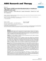
There was no significant effect on SAP, CI (Fig. 1) or cal-
culated SVRI (Fig. 2) with any dose of dopamine, PAP
was elevated at all doses, and a significant increase in
calculated PVRI was demonstrated only at
10 µgkg
–1
min
–1
dopamine. The SAP/PAP ratio was
lowered significantly with 32 µgkg
–1
min
–1
dopamine
(Table 2). The effects on PAP and the SAP/PAP ratio
were sustained throughout the infusions. There were sig-
nificant increases in PVFI and THFI (Fig. 3), with
decreases in calculated MVRI, at a dose of
32 µgkg
–1
min
–1
dopamine. The SAP/PAP ratio during
32 µgkg
–1
min
–1
dopamine, at both the initial and final
30 min, was significantly lower than at the 1 h baseline,
and was lower than all doses of epinephrine. The
changes in PVFI and calculated MVRI with
32 µgkg
–1
min
–1
dopamine at the final 30 min were sig-
nificantly different from these variables at the 1 h baseline
and at all doses of the epinephrine group.
The decrease in mesenteric vascular resistance and the
increase in hepatic venous flow during the highest dose
of dopamine, with a stable CI, led to an increase in the
total hepatic blood flow as a proportion of cardiac
output.
No significant changes in systemic EO
2
, systemic VO
2
,
splanchnic EO
2
, splanchnic VO
2
, and hepatic DO
2
ratio
were found with any dose of dopamine infusion. At
32 µgkg
–1
min
–1
dopamine, hepatic DO
2
increased signif-
icantly from the 1 h baseline. Serum lactate concentration
was elevated by hypoxia but was not significantly affected
by dopamine.
Available online />commentary review reports
primary research
Table 2
Effects (means ± SD) of dopamine infusions in six anesthetized hypoxic piglets
Normoxia 1 h hypoxia 2i 2f 10i 10f 32i 32f
SAP/PAP ratio 3.4 ± 0.65
§
2.0 ± 0.27 1.7 ± 0.32 1.6 ± 0.21 1.7 ± 0.33 1.8 ± 0.60 1.4 ± 0.12*† 1.4±0.20*†
MVRI (mmHg ml
–1
kg
–1
min
–1
) 1.86 ± 0.49 2.00 ± 0.55 2.07±0.82 1.98 ± 0.84 1.99 ± 0.86 2.07 ± 0.86 1.45 ±.74*† 1.21 ±.46*†
Systemic DO
2
(ml kg
–1
min
–1
) 33 ± 5
§
19 ± 5 23 ± 8 24 ± 8 21 ± 6 21 ± 6 20 ± 4 19 ± 4
Systemic EO
2
(%) 28 ± 11.7
§
43 ± 12.6 42 ± 12.7 42 ± 13.8 39 ± 7.5 39 ± 10.8 42 ± 11.4 40 ± 13.1
Splanchnic EO
2
(%) 20 ± 6.7
§
39 ± 9.3 37 ± 13.2 35 ± 14.6 33 ± 11.3 30 ± 9.1 24 ± 11.4 26 ± 13.9
Systemic VO
2
(ml kg
–1
min
–1
) 7.02 ± 3.36 6.16 ± 2.14 6.78 ± 2.80 7.13 ± 2.57 6.54 ± 3.40 6.04 ± 2.00 6.27 ± 2.14 5.80 ± 2.33
Splanchnic VO
2
(ml kg
–1
min
–1
) 1.19 ± 0.42 1.07 ± 0.15 1.10 ± 0.32 1.11 ± 0.22 1.07 ± 0.36 0.97 ± 0.36 0.84 ± 0.49 1.02 ± 0.44
Hepatic DO
2
(ml kg
–1
min
–1
) 5.86 ± 0.92
§
2.29 ± 0.87 2.56 ± 1.07 2.88 ± 1.26 2.73 ± 0.85 2.82 ± 0.90 3.32 ± 1.26 3.61 ± 1.25*
Hepatic DO
2
ratio (%) 17 ± 14.2 20 ± 13.1 18 ± 13.2 19 ± 14.2 18 ± 12.1 16 ± 11.1 13 ± 11.3 10 ± 8.6
(HAFI + PVFI)/CI (%) 28 ± 4
§
23 ± 3 23 ± 5 23 ± 4 26 ± 12 27 ± 11 27 ± 5 32 ± 6!
Arterial lactate (mM) 9.2 ± 6.6 9.4 ± 4.7 12.0 ± 7.1 10.2 ± 4.9
i, initial (3 min average at 30 min of infusion); f, final (3 min average at 60 min of infusion).
*
P < 0.05 compared with variables at 1 h of hypoxia (one-way repeated measures ANOVA);
†
P < 0.05
compared with variables during all doses of epinephrine infusion (two-way ANOVA);
§
P < 0.05 for difference between normoxia baseline and 1 h of hypoxia. EO
2
, oxygen extraction; DO
2
,
oxygen delivery; MVRI, mesenteric vascular resistance index; hepatic DO
2
ratio, the proportion of hepatic DO
2
accounted for by hepatic arterial oxygen delivery; (HAFI + PVFI)/CI, total hepatic
blood flow as a proportion of the cardiac index.
Figure 1
Effects of hypoxia and dopamine infusion on cardiac index, and
systemic and pulmonary artery pressures. i, initial (3 min average at 30
min of infusion at that dose); f, final (3 min average at 60 min of
infusion). *P < 0.05 compared with effects of hypoxia.
Experimental Period
NormoxiaHypoxia 2i 2f 10i 10f 32i 32f
0
20
40
60
80
100
150
200
250
300
350
Mean arterial blood pressure (mmHg)
Mean pulmonary artery pressure (mmHg)
Cardiac Index (mL/kg.min)
Dopamine infusion rate
Hypoxia
*
*
*
**
*
*
Critical Care Vol 5 No 3 Cheung and Barrington
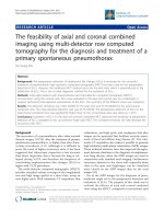
Epinephrine (
n
= 6) (Table 3)
PAP was significantly increased at the final 30 min of
3.2 µgkg
–1
min
–1
epinephrine infusion (Fig. 4). There was
no significant increase in SAP at this epinephrine dose.
The SAP/PAP ratio was not changed with epinephrine
infusions (Table 3). Sustained and significant increases in
CI were found at all doses of epinephrine. Calculated
SVRI was decreased significantly with lower doses of epi-
nephrine (0.2 and 1.0 µgkg
–1
min
–1
), but calculated PVRI
was not different from the 1 h hypoxia value at any dose
Figure 2
Effects of hypoxia and dopamine infusion on systemic and pulmonary
vascular resistance indices. i, initial (3 min average at 30 min of
infusion at that dose); f, final (3 min average at 60 min of infusion).
*P < 0.05 compared with effects of hypoxia.
Experimental period
NormoxiaHypoxia 2i 2f 10i 10f 32i 32f
Vascular resistance index (mmHg/mL/kg.min)
0,0
0,1
0,2
0,3
0,4
0,5
0,6
0,7
0,8
Systemic vascular resistance
Pulmonary vascular resistance
Dopamine infusion rate
Hypoxia
*
*
Figure 3
Effects of hypoxia and dopamine infusion on portal venous, hepatic
arterial, and total hepatic blood flows. i, initial (3 min average at 30 min
of infusion at that dose); f, final (3 min average at 60 min of infusion).
*P < 0.05 compared with effects of hypoxia.
Experimental Period
NormoxiaHypoxia 2i 2f 10i 10f 32i 32f
Blood flow (mL/kg.min)
0
2
4
6
8
10
12
14
40
60
80
Portal Venous Flow Index
Hepatic Arterial Flow Index
Total Hepatic Flow Index
Dopamine infusion rate
Hypoxia
*
Table 3
Effects (means ± SD) of epinephrine infusions in six anesthetized hypoxic piglets
Normoxia 1 h hypoxia 0.2i 0.2f 1.0i 1.0f 3.2i 3.2f
SAP/PAP ratio 3.4 ± 0.52
§
2.2 ± 0.46 2.0 ± 0.48 2.0 ± 0.40 2.0 ± 0.23 2.0 ± 0.34 2.1 ± 0.43 2.0 ± 0.44
MVRI (mmHg ml
–1
kg
–1
min
–1
) 1.67 ± 0.39 2.07 ± 0.80 2.15 ± 1.10 2.22 ± 1.31 2.43 ± 1.53 2.71 ± 2.20 2.37 ± 1.11 2.37 ± 1.28
Systemic DO
2
(ml kg
–1
min
–1
) 35 ± 12
§
20 ± 7 23 ± 6 20 ± 9 21 ± 6 18 ± 5 22 ± 4 20 ± 7
Systemic EO
2
(%) 29 ± 5.4
§
43 ± 9.8 42 ± 5.7 40 ± 3.8 42 ± 10.7 38 ± 3.8 39 ± 6.2 42 ± 3.6
Splanchnic EO
2
(%) 20 ± 6.2
§
37 ± 6.8 36 ± 7.0 41 ± 9.7 40 ± 6.7 41 ± 8.4 35 ± 11.4 42 ± 10.8
Systemic VO
2
(ml kg
–1
min
–1
) 7.65 ± 1.53 7.36 ± 2.13 8.16 ± 2.65 7.30 ± 1.35 8.21 ± 2.24 7.23 ± 1.28 7.63 ± 2.47 7.87 ± 1.63
Splanchnic VO
2
(ml kg
–1
min
–1
) 1.25 ± 0.41 1.37 ± 0.36 1.27 ± 0.57 1.23 ± 0.36 1.29 ± 0.48 1.33 ± 0.27 1.19 ± 0.59 1.32 ± 0.40
Hepatic DO
2
(ml kg
–1
min
–1
) 5.97 ± 1.24
§
2.93 ± 0.76 3.07 ± 0.61 2.79 ± 0.89 2.61 ± 0.52 2.59 ± 0.62 2.83 ± 0.69 2.62 ± 0.71
Hepatic DO
2
ratio (%) 16 ± 7.6 19 ± 9.8 28 ± 15.7 32 ± 17.8*‡ 29 ± 14.6 23 ± 12.8 24 ± 12.1 29 ± 18.8
(HAFI + PVFI)/CI (%) 33 ± 10
§
26 ± 7 23 ± 3 22 ± 4 20 ± 3 20 ± 5 21 ± 5 21 ± 3
Arterial lactate (mM) 8.9 ± 3.4 12.6 ± 5.2 14.0 ± 7.0* 13.5 ± 5.4*
i, initial (3 min average at 30 min of infusion); f, final (3 min average at 60 min of infusion).
*
P < 0.05 compared with variables at 1 h of hypoxia (one-way repeated measures ANOVA);
‡
P < 0.05
compared with variables during all doses of dopamine infusion (two-way ANOVA);
§
P < 0.05 for difference between normoxia baseline and 1 h of hypoxia. EO
2
, oxygen extraction; DO
2
, oxygen
delivery; MVRI, mesenteric vascular resistance index; hepatic DO
2
ratio, the proportion of hepatic DO
2
that is accounted for by hepatic arterial oxygen delivery; (HAFI + PVFI)/CI, total hepatic
blood flow as a proportion of the cardiac index.
(Fig. 5). No significant change was found in PVFI, THFI
(Fig. 6), and calculated MVRI with any dose of epinephrine.
HAFI was increased significantly with 0.2 µgkg
–1
min
–1
epinephrine. The CI with 3.2 µgkg
–1
min
–1
epinephrine
was significantly higher than during dopamine infusion at
any dose. The increase in HAFI, at 0.2 µgkg
–1
min
–1
, was
significantly higher than that produced by any dose of
dopamine.
There was no change in mesenteric vascular resistance
and increase in CI with epinephrine; there was therefore a
trend to a decrease in the total hepatic blood flow when
expressed as a proportion of cardiac output, which was
not statistically significant.
There were no significant changes in systemic EO
2
, sys-
temic VO
2
, splanchnic EO
2
, splanchnic VO
2
, and hepatic
DO
2
during epinephrine infusion in comparison with the 1 h
baseline. A significant elevation in hepatic DO
2
was found
at the final recording obtained during 0.2 µgkg
–1
min
–1
epi-
nephrine (at 1 h), and this was significantly elevated com-
pared with the baseline hypoxia and all doses of dopamine.
The serum lactate was elevated by 1 h of hypoxia to a level
equivalent to that in the dopamine group, and was further
elevated by either 1.0 or 3.2 µgkg
–1
min
–1
epinephrine (but
not by 0.2 µgkg
–1
min
–1
).
Discussion
Both dopamine and epinephrine are commonly used med-
ications in the treatment of shock and hypotension in sick
newborns. Our study is the first that compares the effects
of dopamine and epinephrine infusions on regional hemo-
dynamics and oxygen metabolism in a newborn mammal. It
is also important to realize that all previous studies of the
effects of inotropes in the newborn have used infusions for
a maximum of 15–20 min. The prolonged inotrope infu-
sions in our experiment are unique and are somewhat
more relevant to the problem of cardiovascular support for
the critically ill newborn, who might receive these drugs for
hours or days.
Similarly, many newborns receiving these drugs are
hypoxic, receive large doses of opiates to reduce instabil-
Available online />commentary review reports
primary research
Figure 4
Effects of hypoxia and epinephrine infusion on cardiac index, and
systemic and pulmonary artery pressures. i, initial (3 min average at 30
min of infusion at that dose); f, final (3 min average at 60 min of
infusion). *P<0.05 compared with effects of hypoxia.
Experimental Period
NormoxiaHypoxia 0.2i 0.2f 1.0i 1.0f 3.2i 3.2f
0
20
40
60
80
100
150
200
250
300
Mean Arterial Blood Pressure (mmHg)
Mean pulmonary artery pressure (mmHg)
Cardiac Index (mL/kg.min)
Hypoxia
Epinephrine infusion rate
*
*
*
*
*
*
*
*
*
Figure 5
Effects of hypoxia and epinephrine infusion on systemic and pulmonary
vascular resistance indices. i, initial (3 min average at 30 min of
infusion at that dose); f, final (3 min average at 60 min of infusion).
*P<0.05 compared with effects of hypoxia.
Experimental period
NormoxiaHypoxia 0.2i 0.2f 1.0i 1.0f 3.2i 3.2f
Vascular resistance index (mmHg/mL/kg.min)
0,0
0,1
0,2
0,3
0,4
0,5
0,6
0,7
0,8
Systemic vascular resistance
Pulmonary vascular resistance
Hypoxia
Epinephrine infusion rate
*
*
*
*
*
Figure 6
Effects of hypoxia and epinephrine infusion on portal venous, hepatic
arterial, and total hepatic blood flows. i, initial (3 min average at 30 min
of infusion at that dose); f, final (3 min average at 60 min of infusion).
*P<0.05 compared with effects of hypoxia.
Experimental Period
NormoxiaHypoxia 0.2i 0.2f 1.0i 1.0f 3.2i 3.2f
Blood flow (mL/kg.min)
0
2
4
6
8
10
12
14
20
30
40
50
60
70
Portal Venous Flow Index
Hepatic Arterial Flow Index
Total Hepatic Flow Index
Hypoxia
Epinephrine infusion rate
*
*
ity, are critically ill and stressed, and might have recently
had major surgery. Thus, although acutely instrumented
models are often criticized for being ‘unphysiologic,’ the
stress of surgery might, in some ways, represent the clini-
cal situation in which these drugs are actually used more
accurately than healthy, chronically instrumented, models.
Nevertheless, the animal model employed in the present
study does not completely mirror the conditions in criti-
cally ill newborn humans. Although sick hypoxic newborns
are usually hypotensive as well, it is also important to
realize that the animals had no underlying disease condi-
tion; some such conditions, for example sepsis, might
modify responses to infused catecholamines. Because of
potential differences in drug metabolism, the number,
affinity, and maturation of adrenergic receptors, and car-
diovascular reflexes, the responses described to any
inotropic agent in a non-human mammal should be taken
as only a guide to potential effects, which must be con-
firmed in human newborns.
We chose the empirical doses in this comparison study on
the basis of our previous paper showing that a tenfold
higher dose of dopamine achieves a similar increase in CI
to that of epinephrine [9]. The random order of administra-
tion of the doses was designed to eliminate the possible
effects of bias related to progressive cumulative doses and
the duration of systemic hypoxia. There is no commercially
available co-oximetry system specifically designed for piglet
blood. The only commonly used oximeter with animal coeffi-
cients, the IL282, does not include piglet blood settings.
However, we have previously shown, when using these
devices, that the apparent carboxyhemoglobin is erro-
neously elevated when using blood with very different
optical characteristics [10]; the apparent carboxyhemoglo-
bin levels in our piglets were almost always less than 2%,
suggesting that the oxygen saturation values should be reli-
able. Furthermore, the trends shown are likely to be accu-
rate, even if the actual values are somewhat imprecise.
This study confirms the differential responses in systemic,
pulmonary, and mesenteric circulations with dopamine and
epinephrine infusions that we have previously reported in
anesthetized normoxic piglets [8]. Such responses differ
from responses seen in adult subjects; these differences
might be related to differential maturation of adrenoceptors
and functional immaturity of the receptor mechanisms in
newborns [11,12], as well as to differences in the ultra-
structure and metabolism of the myocardium [13,14]. The
ontogeny of the adrenoceptors seems to vary in the
regional circulations and therefore the responses to
inotropes are different in different vascular beds [15–17].
Our findings suggest that epinephrine, being both an α
and a β adrenoceptor agonist, would be a more appropri-
ate agent for use in inotropic support for hypoxic new-
borns if the same effects are present in the human infant.
We demonstrated an increase in oxygen delivery conse-
quent on the use of epinephrine during hypoxia; SAP was
maintained and CI increased throughout the dose range
(0.2–3.2 µgkg
–1
min
–1
). An increase in cardiac output
and oxygen delivery would be important in shocked
hypoxic newborns. Dopamine did not affect the SAP and
CI at any dose, although it might increase SAP and CI at
a dose of 32 µgkg
–1
min
–1
in normoxic conditions, as
previously described in other studies [11,18–20]. This is
consistent with clinical reports showing that dopamine
might increase blood pressure in hypotensive newborns
but with no increase in cardiac output; indeed cardiac
output seems to decrease [21]. In our previous experi-
ment we did not demonstrate any further increase in CI
with either dopamine or epinephrine infusions during
hypoxia with arterial oxygen saturation between 45% and
50% [9]. The difference in the effects of hypoxia on the
responses to inotropes of cardiac output in this and the
previous study might well be related to the difference in
the severity of the hypoxia [22].
O’Laughlin et al demonstrated an increase in cardiac
output during dopamine infusion in hypoxic unanesthetized
newborn lambs at a mean postnatal age of 6.5 days [23].
The differences in the results of the two studies might rep-
resent a species difference, a postnatal age effect, an
anesthesia effect, or some other detail of the experimental
maneuvers. The drug infusions in O’Laughlin’s study were
begun after 30 min of hypoxia; we have shown in a piglet
model that 30 min is an insufficient period for the stabiliza-
tion of cardiac output after initiation of this degree of
hypoxia [24]. It could therefore be that the dopamine infu-
sion in O’Laughlin’s study was begun at a time when the
cardiac output was still increasing. The doses also seem to
have been given in sequential rather than random order,
which can lead to apparent effects that are due to the
order of administration rather than a true dosage effect.
O’Laughlin also reported drug effects after 15 min; we did
not measure hemodynamics at this time, so we might have
missed transient effects of the drugs.
The relative effects of epinephrine on systemic and pul-
monary pressures are potentially favourable if they can be
reproduced in newborns with persistent pulmonary hyper-
tension. The SAP/PAP ratio is crucially important for the
direction of shunting across the ductus arteriosus, which
determines the oxygen content of the blood distributed to
various organs. In the presence of a lowered SAP/PAP
ratio, owing to hypoxic pulmonary vasoconstriction, epi-
nephrine did not alter the ratio but did increase cardiac
output and therefore oxygen delivery. However, dopamine
infusion at a high dose (32 µgkg
–1
min
–1
) had a detrimen-
tal effect on the SAP/PAP ratio [25]; with no significant
effect on CI this could lead to a decrease in tissue oxygen
delivery if ductal shunt were reversed and systemic oxygen
saturations fell as a consequence. The differences
Critical Care Vol 5 No 3 Cheung and Barrington
between the two drugs might well be because
epinephrine is a potent β
2
agonist, whereas dopamine has
little effect at this receptor, and there seems to be
enhanced β
2
-adrenoceptor responsiveness in the pul-
monary vasculature during hypoxia [26].
Dopamine increased mesenteric flow at the highest dose
(32 µgkg
–1
min
–1
). In our previous normoxic experiments
there were no significant changes in PVFI and calculated
MVRI with dopamine infusion [7,8]. We have shown, with
a selective agonist, active vasodilatation mediated by spe-
cific dopamine receptors in the mesenteric circulation of
the newborn piglet [7]. Thus hypoxia seems to have
enhanced the vasodilatory efficacy of dopamine in the cir-
culation of the bowel, which might be via downregulation
of α receptors [27] in the mesenteric circulation and/or
increased effects of stimulating dopaminergic receptors.
However, despite this apparent beneficial effect in the
mesenteric blood flow, we did not investigate the mucosal
blood flow in the gut, which is particularly vulnerable to
hypoxic–ischemic insult. Indeed, a harmful effect of
dopamine infusion on the mucosal blood flow has been
reported [28]. Whereas epinephrine infusion showed a
vasoconstrictive effect on the mesenteric vasculature in
the previous normoxic experiment [8], this decrease in
mesenteric flow was not apparent in this hypoxic model;
the possible mechanisms for this difference include an
effect of hypoxia on the activity of α receptors, and an
enhanced responsiveness to β
2
stimulation. Thus the dif-
ferences in both the epinephrine and dopamine responses
during hypoxia would be explained by a reduction in α-
mediated vasoconstriction.
Epinephrine infusions should be used cautiously despite
the lack of effects on the bowel circulation seen in this
study, in view of the results of the previous study, which
did show a reduction in bowel perfusion during epineph-
rine infusion at high dose [8]. Vasoconstriction with high
doses of epinephrine could subject the hypoxic bowel in
sick newborns to ischemic injury and increase the risk for
the development of necrotizing enterocolitis [29,30].
Dopamine demonstrates a potentially hepatoprotective
effect at its highest dose. At 32 µgkg
–1
min
–1
, dopamine
improved hepatic DO
2
as a result of mesenteric vasodi-
latation without a concomitant increase in splanchnic EO
2
or splanchnic VO
2
. The increase in HAFI and hepatic DO
2
ratio with 0.2 µgkg
–1
min
–1
epinephrine infusion is inter-
esting. It demonstrates a probable β
2
-vasodilatation effect
with epinephrine at low dose during hypoxia (as also
reflected in the decrease in calculated SVRI); a low dose
of epinephrine could also be protective and improve
hepatic perfusion and oxygen delivery in hypoxic new-
borns. Further studies on hepatic perfusion and oxygen
metabolism in systemic hypoxia are required for an evalua-
tion of the hepatoprotective role of inotropes.
No effect on systemic or splanchnic VO
2
or EO
2
was
demonstrated with either inotrope despite the increase in
systemic oxygen delivery with epinephrine infusions.
Anaerobic metabolism is the main source of ATP produc-
tion during hypoxia. It is advantageous for the tissue to
minimize oxygen consumption during systemic hypoxia
[31]. Although we require cautious interpretation of the
negative findings because of the small sample size and
thus the limited statistical power, we did not show an
effect on oxygen metabolism with either catecholamine. A
dopamine-related increase in oxygen consumption has
been shown in a study of endotoxic dogs during normoxia
[32]. In the same experiment, during a 30 min hypoxic
challenge, a decrease in systemic VO
2
with no improve-
ment in systemic EO
2
was demonstrated. We did not
confirm this in our study, which might be related to the dif-
ference in oxygen metabolism in isolated hypoxia as
opposed to hypoxia and sepsis, and to the duration of
hypoxia between studies.
Conclusion
During severe alveolar hypoxia in the newborn piglet, epi-
nephrine increases cardiac output whereas dopamine has
no effect. Epinephrine preserves the SAP/PAP ratio,
whereas dopamine causes pulmonary vasoconstriction.
Epinephrine has no effect on splanchnic blood flow,
whereas dopamine increases both portal and total hepatic
flow. A reconsideration of the approach to the sick
newborn infant is warranted.
Acknowledgement
This study was supported by the Heart and Stroke Foundation of
Canada and Perinatal Research Centre, University of Alberta, Edmon-
ton, Canada.
References
1. Zaritsky A, Chernow B: Use of catecholamines in pediatrics. J
Pediatr 1984, 15:341–350.
2. Driscoll DJ, Pinsky WW, Entman ML: How to use inotropic
drugs in children. Drug Ther 1979, 9:124–134.
3. Driscoll DJ, Gillette PC, Lewis RM, Hartley CJ, Schwartz A: Com-
parative hemodynamic effects of isoproterenol, dopamine
and dobutamine in the newborn dog. Pediatr Res 1979, 13:
1006–1009.
4. Roze J, Tohier C, Maingneneau C, Lefevre M, Mouzard A:
Response to dobutamine and dopamine in the hypotensive
very preterm infant. Arch Dis Child 1993, 69:59–63.
5. Polak M, Drummond WH: Systemic and pulmonary vascular
effects of selective dopamine receptor blockade and stimula-
tion in lambs. Pediatr Res 1993, 33:181–184.
6. Pawlik W, Mailman D, Shanbour LL, Jacobson ED: Dopamine
effects on the intestinal circulation. Am Heart J 1976, 91:325–
331.
7. Pearson RJ, Barrington KJ, Jirsch DW, Cheung PY: Dopaminer-
gic receptor-mediated effects in the mesenteric vasculature
and renal vasculature of the chronically instrumented
newborn piglet. Crit Care Med 1996, 24:1706–1712.
8. Cheung PY, Barrington KJ, Pearson RJ, Bigam DL, Finer NN, Van
Aerde JE: Systemic, pulmonary and mesenteric perfusion and
oxygenation effects of dopamine and epinephrine. Am J
Respir Crit Care Med 1997, 155:32–37.
9. Barrington KJ, Finer NN, Chan WKY: A blind, randomized com-
parison of the circulatory effects of dopamine and epineph-
rine infusions in the newborn piglet during normoxia and
hypoxia. Crit Care Med 1995, 23:740–48.
Available online />commentary review reports
primary research
Critical Care Vol 5 No 3 Cheung and Barrington
10. Ryan CA, Barrington KJ, Vaughan D, Finer NN: Directly mea-
sured arterial oxygen saturation in the newborn infant. J
Pediatr 1986, 109:526–529.
11. Gootman PM, Buckley NM, Gootman N: Postnatal maturation of
the central neural cardiovascular regulatory system. In: Fetal
and Newborn Cardiovascular Physiology. Edited by Longo LD,
Reneau DD. New York: Garland Press; 1978: vol 1, 93–152.
12. Vapaavouri EK, Shinebourne EA, Williams RL, Heymann MA,
Rudolph AM: Development of cardiovascular responses to
autonomic blockade in intact fetal and neonatal lambs. Biol
Neonate 1973, 22:177–188.
13. Smith RE, Page E: Ultrastructural changes in rabbit heart mito-
chondria during the perinatal period. Dev Biol 1977, 57:109–
117.
14. Lopaschuk GD, Collins-Nakai RL, Toshiyuki I: Develomental
changes in energy substrate use by the heart. Cardiovasc Res
1992, 26:1172–1180.
15. Gootman N, Budley BJ, Gootman PM, Nagelberg JS: Age related
effects of single injections of dopamine on cardiovascular
function in developing swine. Dev Pharmacol Ther 1982, 4:
139–150.
16. Gootman N, Budley BJ, Gootman PM, Griswold PG, Mell JD,
Nudel DB: Maturation in related changes in regional circulat-
ing effects of dopamine infusion in swine. Dev Pharmacol Ther
1983, 6:9–22.
17. Feltes T, Hansen TN, Martin CG, Leblanc AL, Smith S, Giesler
ME: The effects of dopamine infusion on regional blood flow
in newborn lambs. Pediatr Res 1987, 21:131–136.
18. Vane D, Weber TR, Caresky J, Grosfeld JL: Systemic and renal
effects of dopamine in the infant pig. J Surg Res 1982, 32:
477–483.
19. Fiser DH, Fewell JE, Hill DE, Brown AL: Cardiovascular and
renal effects of dopamine and dubutamine in healthy con-
scious piglets. Crit Care Med 1988, 16:340–3445.
20. Girardin E, Berner M, Rouge JC, Rivest RW, Friedli B, Paunier L:
Effect of low dose dopamine on hemodynamic and renal
function in children. Pediatr Res 1989, 26:200–203.
21. Roze JC, Tohier C, Maingueneau C, Lefevre M, Mouzard A:
Response to dobutamine and dopamine in the hypotensive
very preterm infant. Arch Dis Child 1993, 69:59–63.
22. Ng ML, Levy MN, DeGeest H, Zieske H: Effects of myocardial
hypoxia on left ventricular performance. Am J Physiol 1966,
211:43–50.
23. O’Laughlin MP, Fisher DJ, Dreyer WJ, O’Brian ES: Augmentation
of cardiac output with intravenous catecholamines in unanes-
thetized hypoxemic newborn lambs. Pediatr Res 1987, 22:
667–674.
24. Cheung PY, Barrington KJ, Bigam DL: Temporal effects of pro-
longed hypoxaemia and reoxygenation on systemic, pul-
monary and mesenteric perfusions in newborn piglets.
Cardiovasc Res 1998, 39:451–458.
25. Mentzer R, Alegre CA, Nolan SP: The effect of dopamine and
isoproterenol on the pulmonary circulation. J Thorac Cardiovas
Surg 1976, 71:807.
26. Lock JE, Olley PM, Coceani F: Enhanced
ββ
adrenergic receptor
responsiveness in hypoxic neonatal pulmonary circulation.
Am J Physiol 1981, 240:H697–H703.
27. Tateishi J, Faber JE: ATP-sensitive K
+
channels mediate
αα
2D-
adrenergic receptor contraction of arteriolar smooth muscle
and reversal of contraction by hypoxia. Circ Res 1995, 76:53–
63.
28. Neviere R, Mathieu D, Chagnon JL, Lebleu N, Wattel F: The con-
trasting effects of dobutamine and dopamine on gastric
mucosal perfusion in septic patients. Am J Respir Crit Care
Med 1996, 154:1684–1688.
29. Ballance WA, Dahms BB, Shenker N, Kliegman RM: Pathology
of neonatal necrotizing enterocolitis: a ten year experience. J
Pediatr 1990, 117:S6–S13.
30. Konto WP Jr, Wilson R: Epidemiology of necrotizing enterocoli-
tis with etiologic implications. Perinatol Neonatol 1983, 7:63–68.
31. Suguihara C, Bancalari E, Hehre D, Duara S, Gerhardt T:
Changes in ventilation and oxygen consumption during acute
hypoxia in sedated newborn piglets. Pediatr Res 1994, 35:
536–540.
32. Cain SM, Curtis SE: Systemic and regional oxygen uptake and
delivery and lactate flux in endotoxic dogs infused with
dopexamine. Crit Care Med 1991, 19:1552–1560.