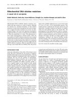
Báo cáo y học: " Bench-to-bedside review: Toll-like receptors and their role in septic shock" pps
Bạn đang xem bản rút gọn của tài liệu. Xem và tải ngay bản đầy đủ của tài liệu tại đây (459.86 KB, 12 trang )
18W = 18-Wheeler; DIF = Dorsal-like immunity factor; DN = dominant-negative; ECSIT = evolutionarily conserved signaling intermediate in Toll
pathways; HSP = heat shock protein; IL = interleukin; IL-1R = IL-1 receptor; IL-1R1 = type 1 IL-1 receptor; IRAK = IL-1 receptor-associated kinase;
LBP = lipopolysaccharide-binding protein; LPS = lipopolysaccharide; MHC = major histocompatibility complex; MyD88 = myeloid differentiation
factor; NF = nuclear factor; PRR = pattern recognition receptor; TIR = Toll/IL-1 receptor; TLR = Toll-like receptor; TNF = tumor necrosis factor.
Available online />The recent recognition of the TLRs as the principal inducers
of the innate immune system is among the most important
and fundamental discoveries in microbial pathogenesis in the
past decade. This discovery fills an essential gap in our
understanding of the molecular events that follow microbial
infection and the initial host defense to invasive pathogens.
The TLRs are the critical pattern recognition molecules that
alert the host to presence of a microbial pathogen [1,2]. The
initial engagement of TLRs to specific and highly conserved
microbial constituents largely determines the ultimate fate of
each host–pathogen interaction. Once an organism breaches
the integument or mucous membrane barriers, the innate
immune system must recognize this potential threat and
orchestrate an appropriate response to surround and eradi-
cate the pathogen.
The remarkable ability of the human immune system to rapidly
respond to a myriad of potential microbial invaders remains
essential for survival. This fact is amply demonstrated in
patients with a variety of inherited or acquired immune defi-
ciencies. The mechanisms that underlie this antimicrobial
defense mechanism have fascinated researchers for over a
century. This vigorous early warning and microbial clearance
mechanism must have been of fundamental evolutionary
importance to our early hominid ancestors. The adoption of a
predatory lifestyle and the loss of the thick coating of fur that
separates Homo sapiens from our primate relatives must
have produced heavy selection pressure, favoring a highly
primed innate immune system because frequent injuries to
our thin integument would inevitably occur.
Humans retain one of the most active host immune responses
to microbial antigens known in the entire animal kingdom. The
price paid for this defense system is a heightened sensitivity
to endotoxin that makes humans more susceptible to
lipopolysaccharide (LPS)-induced shock than almost all other
mammalian species [3]. The essential role that the TLRs play
in this early defense system, and the impact of the signaling
system of the innate immune response in human septic shock
are the focus of this review.
Review
Bench-to-bedside review: Toll-like receptors and their role in
septic shock
Steven M Opal* and Christian E Huber
†
*Professor of Medicine, Infectious Disease Division, Brown University Medical School, Providence, Rhode Island, USA
†
Research Fellow, Infectious Disease Division, Brown University Medical School, Providence, Rhode Island, USA
Correspondence: Steven M Opal,
Published online: 15 February 2002 Critical Care 2002, 6:125-136
© 2002 BioMed Central Ltd (Print ISSN 1364-8535; Online ISSN 1466-609X)
Abstract
The Toll-like receptors (TLRs) are essential transmembrane signaling receptors of the innate immune
system that alert the host to the presence of a microbial invader. The recent discovery of the TLRs has
rapidly expanded our knowledge of molecular events that initiate host–pathogen interactions. These
functional attributes of the cellular receptors provide insights into the nature of pattern recognition
receptors that activate the human antimicrobial defense systems. The fundamental significance of the
TLRs in the generation of systemic inflammation and the pathogenesis of septic shock is reviewed. The
potential clinical implications of therapeutic modulation of these recently characterized receptors of
innate immunity are also discussed.
Keywords CpG motifs, sepsis, septic shock, Toll-like receptors, Toll receptors
Critical Care April 2002 Vol 6 No 2 Opal and Huber
The microbial mediators implicated in the
pathogenesis of bacterial sepsis
While bacterial endotoxin expressed in Gram-negative bacte-
ria is generally viewed as the principal mediator of Gram-neg-
ative septic shock, recent evidence indicates that LPS works
in concert with a variety of other microbial mediators that con-
tribute to systemic inflammation [4,5]. The current listing of
those microbial mediators implicated in the pathogenesis of
septic shock and the principal receptors that recognize them
is presented in Table 1.
These mediators have several structural or functional attrib-
utes that are essential for microbial survival and pathogenesis.
Importantly, these microbial elements are unique to prokary-
otes or lower eukaryotes and thus are logical targets for
recognition of these microbial invaders by the innate immune
system. These mediators work in combination with each other
and probably synergize to induce systemic inflammation and
contribute to the pathogenesis of septic shock [3,4].
While LPS is the best studied and probably the most impor-
tant microbial mediator of sepsis, it is sufficient but not nec-
essary to induce shock. The most direct evidence that LPS is
not essential for the induction of an inflammatory signal to
host phagocytic cells is found with the LPS-deficient Neisseria
meningitidis strain [6]. Deletion of lipid A synthetic genes in
this strain of N. meningitidis results in a viable bacterium that
lacks LPS in its outer membrane. Lipid A deletion in enteric
Gram-negative bacteria such as Escherichia coli is a lethal
mutation. The LPS-deficient strain of N. meningitidis is still
capable of inducing inflammatory reactions (although at lower
levels) via the TLR2 receptors found on macrophages. This
indicates that Gram-negative bacteria have other cell wall
components that induce inflammation even in the absence of
bacterial LPS [6].
Host response to microbial mediators
through the innate immune system
The innate immune system (macrophages, neutrophils,
natural killer cells and the alternative complement pathway)
has evolved as an early, rapid response system to microbial
invasion. The actions against the invading pathogens are
either direct (e.g. phagocytosis and killing) or indirect
through the release of cytokines or other stimulatory mole-
cules, which trigger the adaptive immune system by activat-
ing B cells and T cells.
Janeway [4] and Poltorak et al. [7] proposed a central
concept in the understanding of the innate immune system:
the identification of infectious agents by means of conserved
structural features through pattern recognition receptors
(PRRs). The components expressed by microbial agents that
trigger the immune response are termed pathogen-associ-
ated molecular patterns. The discovery of the TLRs of the
innate immune system provides the PRRs that detect these
pathogen-associated molecular patterns.
Comparative molecular biology has succeeded in unlocking
the mystery of the cellular receptors to endotoxin and a large
number of other microbial elements (Table 1). This evolution-
arily conserved receptor system has allowed multicellular
organisms (including plants as well as invertebrates and ver-
tebrates) to rapidly recognize the presence of microbial
invaders. Microbial pathogens represent an immediate threat
to survival, and this requires a vigorous and coordinated
immune response in defense of the viability of the host [5].
The following section summarizes the major findings that
defined the TLR family as the central transmembrane receptor
of the innate immune system.
IL-1 receptor/TLR superfamily
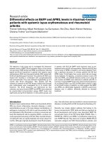
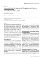
The IL-1 receptor (IL-1R)/TLR superfamily members (Fig. 1)
are found in many plants and in vertebrate and invertebrate
animal species. Those members with known function share
the feature of being involved in host responses to injury and
infection [8]. Most notably, in humans these include not only
the receptor and accessory protein for IL-1, but also the IL-18
receptor and its accessory protein, and the long-sought sig-
naling receptor for LPS, TLR4 [1,8].
Many proteins from diverse systems show homology to the
cytoplasmic domain of the type 1 interleukin-1 receptor
Table 1
Microbial products implicated in the pathogenesis of sepsis
Mediator products Host receptor
Lipopolysaccharide LBP-CD14-TLR4-MD2
Peptidoglycan CD14-TLR2, CD14-TLR2/6, CD14-TLR2/1, CD14-TLR2/?
Lipopeptides, lipoteichoic acid CD14-TLR2, CD14-TLR2/6, CD14-TLR2/1, CD14-TLR2/?
Microbial DNA (unmethylated CpG motifs) TLR9
Bacterial flagella TLR5
Double-stranded viral RNA TLR3
LBP, lipopolysaccharide-binding protein; TLR, Toll-like receptor.
(IL-1R1). This expanding IL-1R1-like family includes murine
and human proteins, Drosophila (fruit fly) proteins, and a plant
(tobacco) protein [7,8]. In Drosophila, Toll is involved in the
rapid and transient transcriptional induction of several genes
encoding potent antimicrobial peptides on septic injury [9]. A
noteworthy plant member of the IL-1R1 family is the tobacco
N gene [10]. The N gene encodes a protein with an amino-
terminal domain that has significant homology to Toll and the
cytoplasmic domain of the IL-1R1. Introduction of the N gene
into tobacco mosaic virus-sensitive strains of tobacco confers
resistance to tobacco mosaic virus via the ability to mount a
hypersensitive response to the virus at the site of infection
within the plant. This system is fundamentally similar to the
acute inflammatory response pathway stimulated by injury
and mediated by IL-1 in mammals [1].
A growing number of mammalian homologs of IL-1R1 have
recently been identified that contain highly conserved regions
in their cytosolic domains. Homologous regions were also
found in a receptor-like protein of the Drosophila fly called
Toll. This has resulted in the defining of the IL-1R/TLR super-
family, the conserved region being termed the Toll/IL-1R (TIR)
domain [9].
Drosophila
Toll members
Toll was originally described in Drosophila as a type I trans-
membrane receptor that controls dorsal–ventral polarity
during embryogenesis [11]. Nüesslein-Volhard and
Wieschaus discovered the first Toll mutants in fruit fly
embryos. Wieschaus noted that the Toll mutant embryos
failed to hatch and developed no ventral or lateral cell types.
When Nüesslein-Volhard saw the particular embryos lacking
the entire mesoderm and nervous system, she exclaimed
‘Toll!’ (German for jazzy or cool). The new gene was thus
given its name.
Four members of the Toll family have been identified in
Drosophila thus far: Toll, 18-Wheeler (18W), Mst and
STSDm2245. Toll and 18W share the greatest similarity to
each other and also to the cytoplasmic tail of the mammalian
IL-1R1. The Drosophila 18W is required for morphogenesis
and has many similarities to Toll. The extracellular regions of
Toll and 18W contain multiple leucine-rich repeats and
carboxyl-terminal cysteine-rich domains [12].
Drosophila
Toll signaling pathway
Current understanding of the Toll pathway in Drosophila
immune response came from studies of promoters of genes
induced in response to infection. Insects respond to infection
with antimicrobial peptides produced by the fat body and
hemocytes. All of the antimicrobial peptide genes include
NF-κB or Rel proteins in their upstream regions. The first
insect protein discovered to regulate transcription through
NF-κB sites was Dorsal. Subsequent genetic studies further
identified Spätzle, Toll, Pelle, Tube and Cactus as necessary
partners to induce Drosomycin, an antifungal Drosophila
peptide, in response to fungal infection (Fig. 1) [9].
It has been demonstrated by genetic complementation tests
that Spätzle is the Toll ligand. Spätzle is endogenously
secreted as an inactive precursor molecule. Protease Easter
creates an active form through proteolysis into the biologically
active carboxyl-terminal polypeptide fragment [13]. Binding of
Spätzle to Toll activates the receptor by ligand-dependent
receptor dimerization. This is of interest because there might
be an endogenous ligand for the mammalian homologs of Toll.
Activated Toll recruits the adapter protein known as Tube and
a protein kinase referred to as Pelle to the intracellular part of
the Toll protein [9]. No true homolog to Tube has yet been
defined in mammals, but the myeloid differentiation factor
MyD88 appears to have a similar function.
The mammalian IL-1R-associated kinase (IRAK) appears to be a
homolog to Drosophila Pelle, which interacts with another
protein, the Drosophila homolog of mammalian TRAF6
(dTRAF6). The close relationship within Toll-related signaling
also becomes evident from the fact that MyD88, IRAK and
TRAF6 participate not only in Toll or TLR signaling, but also in
IL-1 and IL-18 signaling, leading to the activation of NF-κB [16].
The next downstream step in the signaling cascade is a direct
association of the Drosophila homolog of TRAF6 with the
Drosophila homolog to evolutionarily conserved signaling
intermediate in Toll pathways (ECSIT). Transfection of
dECSIT in insect cells leads to production of diptericin,
attacin and defensin (antimicrobial peptides) [15].
Available online />Figure 1
The homologies and interconnections between the Toll-like receptors
(TLRs) and the type 1 interleukin-1 receptor (IL-1R1) complex. AcP,
accessory protein; Ig, immunoglobulin; MyD88, myeloid differentiation
factor; R, receptor; TIR, Toll/IL-1 receptor.
The final members of the signal transduction family in
Drosophila are Dorsal and Dorsal-like immunity factor (DIF).
These are released from Cactus, which is a cytoplasmic
anchoring protein and the Drosophila homolog to I-κB;
Dorsal and DIF are the Drosophila NF-κB homologs [9].
After release from Cactus, Dorsal and DIF translocate into the
nucleus and induce gene transcription. There is also evi-
dence for a Drosophila homolog to the mammalian I-κB
kinase Drosophila LPS-activated kinase, which phosphory-
lates Cactus. In transfection models, Drosophila LPS-acti-
vated kinase-deficient cell lines fail to respond to LPS
stimulation [12].
The role of the Toll receptor in the adult fly is the induction of
immune response in fungal infection. Toll-deficient flies fail to
express Drosomycin. In infection models, Toll-mutant flies died
from overwhelming growth of Aspergillus fumigatus [16].
The immune response of Toll-deficient flies is not altered
against bacteria, suggesting a different receptor and a differ-
ent pathway. Williams et al. demonstrated that 18W-deficient
flies have an increased mortality from E. coli infections [17].
This might be due to a reduced expression of the antibacter-
ial Drosophila protein attacin.
Another clue to the pathogen specificity is that antifungal and
antibacterial signaling is also divergent further downstream:
18W-deficient flies in bacterial infection have a reduced
translocation of DIF into the nucleus, whereas Dorsal translo-
cation is unaffected [18]. Toll, however, does not require DIF
for intracellular signaling. This suggests that there might be a
selective immune response to particular microbial pathogens.
Homologies between
Drosophila
Toll and the TLR family
All members of the Toll family are membrane proteins that
cross the membrane once and share similar extracellular
domains [14]. Typical examples are 18–31 leucine-rich
repeats. The extracellular domain of human TLR4 contains 22
copies of the leucine-rich repeats. The extracellular domains
are very divergent among the different members: TLR2 and
TLR4 share only 24% of identical sequences. This makes it
probable that they bind different ligands.
The TLRs of different species are very different: mouse TLR4
and human TLR4 are only 53% identical. Genetic studies of
leucine-rich repeat structures among different individuals also
revealed that polymorphisms are responsible for a different
reaction on microbial challenge. The intracellular part con-
tains a cytoplasmic domain of approximately 200 amino acids
that is evolutionarily conserved. This highly conserved region
is known as the TIR domain [14].
Mammalian Toll homologs
The mammalian homologs of Drosophila Toll are termed TLR
proteins and have been most intensively studied [14,17]. To
date, 10 human TLRs (TLR1–TLR9) have been described
and their structures have been published [19–22].
TLR1–TLR6 have been characterized by their distinctive
expression patterns with mRNA detection assays.
TLR1 is expressed ubiquitously and at rather high levels.
TLR2 has a particular strong expression pattern in peripheral
blood mononuclear cells, but is also expressed in lymphoid
tissue [23]. TLR3 mRNA is expressed in the lung, muscle, the
heart, the brain and intestinal cells. Alternative splicing vari-
ants have been reported from the pancreas and the placenta.
Among peripheral blood cells, TLR3 is selectively expressed
in specific subsets of dendritic cells [24]. TLR4 is expressed
in lymphocytes, the spleen and the heart. TLR5 mRNA is
detectable in peripheral blood monocytes, leukocytes, the
ovary and the prostate. TLR6 expression was located in the
spleen, the thymus, the ovary and the lung [21].
The fact that TLR mRNA is also expressed in cells of epithe-
lial surfaces like intestinal epithelial cells [25] suggests that
they act as sentinels for invading microbes. Among these
human TLRs, the microbial ligands for TLR2, TLR4, TLR9
and, most recently, for TLR5, and perhaps TLR3, have been
identified (see following sections).
Understanding the regulation of TLR expression will provide
further insight into their tissue distribution and their role in
combating infection. There has been an initial report of the
TLR4 promoter region. The upstream region shares similari-
ties with many myeloid-specific genes: it contains purine-rich
sequences, recognized through transcription factors of the
Ets family. Through supershift analysis, the Ets member PU.1
was shown to have affinity to the promoter region [26]. In
another report, IL-4 was shown to downregulate TLR expres-
sion. The T helper 2-type adaptive immune response may
thus inhibit TLR activation [27].
LPS tolerance is a phenomenon where pre-exposure to LPS
induces a reduced sensitivity to a subsequent challenge of
LPS [28]. In vivo, this phenomenon is related to a decreased
febrile response and to a diminished response to subsequent
severe infection. LPS tolerance, also termed LPS refractori-
ness, might be related to differential regulation of the TLR
family [28,29]. There are contradictory reports about the reg-
ulation of TLR mRNA and surface TLR protein on stimulation
with its ligands. Nomura et al. [28] reported that LPS stimula-
tion leads to a decrease in TLR4 mRNA levels and surface
expression of the TLR4–MD2 complex in mouse
macrophages. The LPS pretreated monocytes also secreted
less proinflammatory cytokines. Other reports show an
increased amount of intracellular TLR mRNA on stimulation
with LPS [29,30].
These contradictory findings may be related to differences in
experimental conditions used in each laboratory. The potential
impact of endotoxin tolerance (or reprogramming) in human
Critical Care April 2002 Vol 6 No 2 Opal and Huber
sepsis is of critical importance. The differential expression of
TLRs in patients over the course of different stages of early
and late severe sepsis is a priority in current sepsis research.
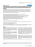
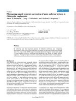
TLR signaling and homology with the IL-1R
As already mentioned, the intracellular part of members of
the TLR family has a high degree of homology to the intra-
cellular domain of the IL-1R. This remarkable level of homol-
ogy suggests similar intracellular signaling pathways (see
Fig. 2) [19,31].
Postreceptor signaling of the IL-1R is well understood.
Binding of the ligand causes receptor dimerization and, with
involvement of IL-1RAP (an accessory protein), the intracellu-
lar part of the receptor forms a complex of MyD88 (an adapter
protein) and IRAK (a kinase). IRAK then phosphorylates
TRAF6, which leads to NF-κB-inducing kinase and I-κB
kinase activation. The I-κB kinase phosphorylates I-κB, which
then dissociates from NF-κB [14]. NF-κB translocates to the
nucleus and initiates gene transcription. When human
homologs to the Drosophila Toll were discovered, there was
great interest regarding their role in innate immunity. Medzhi-
tov et al. reported NF-κB activation as well as cytokine release
through TLR4 in transfected monocytic cell lines, which were
able to express TLR4 constitutively at a high level [32].
TLR2 and TLR4 have so far been the most intensively
studied members of mammalian homologs to Drosophila
Toll. Currently recognized ligands for TLR4 are presented in
Table 2, and the known ligands for TLR2 are presented in
Table 3. Both TLR2 and TLR4 require the adapter protein
MyD88 for signaling, and immunoprecipitation studies
showed direct interaction of MyD88 and IRAK [32]. MyD88
was originally isolated and characterized as a myeloid differ-
entiation primary response gene. MyD88 itself consists of a
carboxyl-terminal TIR domain, making it a member of the TLR
family. IRAK has been shown both to interact with both
MyD88 and TRAF6 [20].
Further evidence for the role of TRAF6 and NF-κB-inducing
kinase in TLR4 signaling came from the study of TRAF6 and
NF-κB-inducing kinase dominant-negative (DN) variants,
which could not activate NF-κB [33]. Another very interesting
observation came from this study; activation of the c-Jun N-
terminal kinase activation through TLR4 is prevented by the
MyD88-DN variant, but not by the TRAF6-DN variant. This
indicates an alternative signaling pathway that diverges at the
MyD88 level.
Two novel members of the IL-1R/TLR intracellular complex
have recently been reported: Tollip, and the TIR domain-con-
taining adapter protein TIRAP. Tollip is present in a complex
with IRAK and, during activation of the IL-1R/TLR complex,
Tollip associates with IL-1RAcP. IRAK autophosphorylation is
then triggered by MyD88, which leads to rapid dissociation of
IRAK from Tollip and the receptor complex [34].
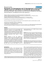
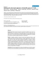
Horng et al. defined TIRAP, another adapter that mediates
MyD88-independent signaling in response to TLR4 stimula-
tion in MyD88-deficient mice [35]. It appears that the TIRAP-
dependent, MyD88-independent signaling pathway is
particularly important in the expression of MHC class II mole-
cules and accessory molecules (B.7 antigens) on antigen-
presenting cells and T-cell activation (see Fig. 3).
The protein ECSIT is specific for TLR/IL-1 signaling, and is
involved in the proteolytic cleavage and activation of the
mitogen-activated protein kinase MEKK-1 after TRAF6 activa-
tion [15]. ECSIT-DN mice fail to activate NF-κB through
MEK-1 and MEKK-1.
Recent observations by Miyake and colleagues demonstrated
the necessity of another cell surface molecule for TLR4 signal
transduction [36]. The protein MD-2 has no intracellular domain,
but on co-expression with TLR4 enhances LPS sensitivity in
transfection models. MD-2 cotransfection with TLR2 had no
effect on LPS response. Recent data support a direct binding of
MD-2 to LPS. This effect was independent of CD14 or LPS-
Available online />Figure 2
Comparisons between the signaling pathways in the insect and
mammalian IL-1/Toll-like receptor (TLR) pathways. (d), Drosophilia
homolog; ECSIT, evolutionarily conserved signaling intermediate of
Toll; HSP, heat shock protein; IKB, inhibitory subunit κB; IKK, I-κB
kinase; IRAK, IL-1 receptor-associated kinase; LBP, lipopolysaccharide
binding protein; LPS, lipopolysaccharide; LTA, lipoteichoic acid;
MEKK, mitogen-activated protein extracellular activated receptor
kinase kinase; MyD88, myeloid differentiation factor; NF-κB, nuclear
factor-κB; TNF, tumor necrosis factor; TRAF6, tumor necrosis factor
receptor-associated factor 6.
binding protein (LBP) and suggests a specific and unique role
for MD-2 in LPS recognition that contributes to modulation of
the proinflammatory response of effector cells [37].
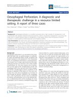
TLR2 signaling shares many similarities with IL-1R/TLR4 sig-
naling. TLR2-dependent NF-κB activation requires the TIR
domain, which is the epitope for the TIR–MyD88–IRAK
complex that induces TRAF6 [23]. Nonetheless, there are
severe unique features involved in TLR2 signaling. TLR2
forms heterodimeric structures with other TLR members such
as TLR1 and TLR6. A recent report demonstrated that the
p85 regulatory subunit of phosphatidylinositol-3′-kinase can
directly associate with the intracellular domain of TLR2 [38].
The Rho-type GTPase Rac1 also appears to be associated
with TLR2-mediated signaling. This alternative signaling
pathway activates a number of phosphorylated lipids and
results in the generation of the intracellular protein kinase Akt.
This pathway directly activates NF-κB, independent of the
phosphorylation and degradation of I-κB [38] (see Fig. 4).
TLR4 as the LPS receptor
Another exciting development in the history of TLR research
has been the report that TLR4 is utilized by LPS and there-
fore is the long-sought LPS receptor [7,39].
Probably the most powerful microbial stimulant of innate
immune responses is LPS. LPS has been known to induce
signals very similar to IL-1, and also to bind to CD14 on
macrophages. The best-characterized interaction is between
LPS and LBP. LPS is first bound by LBP, a plasma lipid trans-
fer protein that moves LPS monomers from aggregates or
bacterial membranes to a binding site on surface receptor
CD14. Alternatively, the LBP–LPS complex can be recog-
nized by a soluble version of CD14 that subsequently acti-
vates non-myeloid cells [40].
CD14 is a known PRR on the surface of monocytes/
macrophages. It has been clear for many years that CD14 has
a major role for the effects of LPS on macrophages, mono-
cytes, and neutrophils, and that CD14 increases the sensitivity
of macrophages to LPS [46]. But the precise role of CD14 in
LPS signaling remains unclear. There has to be a molecule
with the ability to identify the binding partner and to discrimi-
nate LPS from host lipids and to transduce signals across the
membrane. Neither LBP nor CD14 have been shown to have
the binding specificity to discriminate LPS from host lipids.
The interest in TLR biology increased greatly as evidence
accumulated that these proteins participate in intracellular sig-
naling initiated by LPS and Gram-negative bacteria.
Medzhitov et al. showed in 1997 that a constitutively active
mutant of human TLR4 induces the expression of the NF-κB-
controlled cytokines IL-1, IL-6 and IL-8. This implicated the
role of TLR4 in innate immunity [19]. The precise nature of
the transmembrane receptor was complicated by the fact that
alternative evidence developed in parallel by other investiga-
tors implicated TLR2 as the LPS receptor. Researchers
demonstrated in 1998 that overexpression of TLR2 in mam-
malian cells renders cells responsive to LPS in a CD14-
dependent manner. TLR2 was stably transfected in human
kidney cell lines and did activate NF-κB in the presence of
CD14 and LBP, and after LPS stimulation [23].
Critical Care April 2002 Vol 6 No 2 Opal and Huber
Table 2
Currently recognized ligands for Toll-like receptor 4
Microorganism Microbial product
Gram-negative bacteria Lipopolysaccharide, lipid A
Gram-positive bacteria Lipoteichoic acid
Mycobacteria Live Mycobacteria tuberculosis
Spirochetes Treponema brennaborense glycolipids
Virus Respiratory syncytial virus protein F
Other ligands Heat shock protein 60, Taxol
Table 3
Currently recognized ligands for Toll-like receptor 2
Organism Product
Gram-negative bacteria Lipopolysaccharide (LPS) from various sources; possibly contaminated with peptides (Salmonella, Shigella spp.,
Escherichia coli). Purified LPS from Leptospira interrogans and Porphyromonas gingivalis LPS
Gram-positive bacteria Listeria monocytogenes, Bacillus, Streptococcus spp., Staphylococcus aureus-lipoteichoic acid, peptidoglycan,
lipopeptides
Mycobacteria Heat-killed Mycobacteria tuberculosis, Mycobacteria avium lipopeptides, lipoarabinomannan, mannosylated
phosphatidylinositol
Spirochetes Borrelia burgdorferi, Treponema pallidum, Treponema maltophilum (lipopeptides, glycolipids, outer surface
protein A)
Mycoplasma Mycoplasma fermentans (R-MALP, lipopeptides)
Yeast Zymosan
A second approach by Beutler and colleagues proved on a
genetic level that TLR4 is involved in LPS signaling [7]. These
proofs were carried out in genetic analysis in two strains of
mice, C3H/HeJ and C57BL/10Scr, both known for their
hyporesponsiveness to LPS and their increased mortality
from Gram-negative sepsis. Over the past 20 years it has
been shown that hyporesponsiveness to LPS maps to a
single autosomal locus (lps
d
), and impaired responses could
be documented both in whole animals and in cells taken from
these animals.
Through extensive genetic mapping work performed by
Poltorak et al. and Qureshi et al., the lps
d
allele was shown to
map to the gene encoding TLR4. The tlr4 locus located to the
target region in chromosome 4 [7,39].
A missense mutation in the Tlr4 gene locus was demonstrated
to be responsible for the altered LPS responsiveness in the
C3H/HeJ strain. A point mutation converts a proline residue at
position 712 to histidine (P712H), thus rendering the receptor
inactive [7]. A further proof is that C57BL/10ScCr mice carry
a large deletion mutation and do not express TLR4 mRNA.
Additional evidence of the ability of TLR4 to bind to LPS is
found in a recent report about a newly identified soluble form
of TLR4 [41]. Soluble TLR4 can be spliced and shed follow-
ing LPS stimulation and can prevent LPS signaling at the cell
membrane of LPS-sensitive target cells.
After the initial observations with TLR2 as a physiologic LPS
receptor [23], the relative role of TLR2 in LPS signaling has
been re-examined. Hirschfeld et al. [42] reported that many
LPS preparations in early experiments had bioactive contami-
nants that activate TLR2. Thorough purification of LPS elimi-
nated signaling through TLR2. This paradigm has been
challenged again by the report that purified LPS from Prophy-
romonas gingivalis and Leptopira interrogans only signals
through TLR2, and not through TLR4 [43]. Therefore, the role
of TLR2 in LPS signaling still remains ambiguous. TLR2
exerts a cellular response to LPS, but with a lower affinity
than TLR4. TLR2 does not therefore appear to be essential
for LPS signaling in native cells that also express TLR4. TLR2
could also be an alternative LPS receptor in TLR4-deficient
cells [44]. Finally, TLR2 might be the genuine LPS receptor
for LPS from non-enteric Gram-negative bacteria [43]. TLR2
has subsequently been demonstrated to be a broad spec-
trum PRR that interacts with a wide variety of other bacterial
components besides LPS. TLR2 has thus far been involved in
the recognition of multiple microbial products from Gram-
Available online />Figure 3
The intracellular signaling pathways of the Toll-like receptor 4 (TLR4) complex. IKB, inhibitory subunit κB; IKK, I-κB kinase; IRAK, IL-1 receptor-
associated kinase; LBP, lipopolysaccharide binding protein; LPS, lipopolysaccharide; Mal, myeloid differentiation factor MyD88-like; MyD88,
myeloid differentiation factor; NF-κB, nuclear factor-κB; NIK, nuclear factor-κB inducing kinase; PKR, RNA binding protein kinase; TIR, Toll IL-1
receptor; TIRAP, Toll IL-1 receptor adapter protein; Tollip, Toll interactive protein; TRAF6, tumor necrosis factor receptor-associated factor 6.
positive bacteria, mycobacteria, spirochetes, mycoplasma
and yeast antigens [1,45] (Table 3).
Other ligands in TLR signaling
The concept of innate immunity demands the presence of
non-clonal receptors that recognize a variety of highly con-
served bacterial structures. More evidence has been gener-
ated that the TLR family represents this key group of innate
immune response receptors. TLR2 has also been recog-
nized as a signal transducer for numerous bacterial prod-
ucts. Spirochetes lack LPS, but possess membrane
lipoproteins in the cell wall. TLR2 antibody administered to
human peripheral blood mononuclear cells prevented
cytokine release after stimulation with lipoproteins and
lipopeptides from Mycoplasma fermentans, Borrelia
burgdorferi and Treponema pallidum [45]. Chinese hamster
ovary cells (CHO-K1 cell lines) fail to express a functional
TLR2 due to a frame-shift mutation in their TLR2 mRNA
sequence. These cells were transfected with TLR2 and
tested for many bacterial products. B. burgdorferi lipopro-
tein has also been shown to stimulate NF-κB through TLR2
in transfected Chinese hamster ovary cells and also in TLR-
2-transfected 293 cells [1,45].
TLR4 signaling is primarily activated after lipid A/LPS chal-
lenge, but is also sensitive to lipoteichoic acid from Gram-posi-
tive bacteria and live Mycobacteria tuberculosis bacteria
(Table 2). Purified glycolipids from Treponema brennaborense,
a spirochete that causes a bovine infectious disease, have
been associated with TLR4-dependent signaling [46].
Ohashi et al. reported the potential first endogenous ligand
for the TLR4 [47]. Recent studies have shown that heat
shock protein (HSP) is a danger signal to the innate immune
system. Macrophages respond with proinflammatory cytokine
response to stimulation with both autologous HSP60 and
microbial HSP60/65. Macrophages from C3H/HeJ
responded with nitric oxide formation and tumor necrosis
factor (TNF) secretion, whereas they remained unresponsive
to LPS [48]. A recent publication has linked TLR4 signaling
and expression in injured myocardium. Since there was no
infection present in this model, these findings open up the
perspective that TLR4 may also be responding to non-infec-
tious and endogenous ligands in inflammation [49].
Viral particles also act as a ligand for TLR4. Kurt-Jones et al.
showed that the innate immune response to respiratory syn-
cytial virus coat protein F is mediated by signaling through
Critical Care April 2002 Vol 6 No 2 Opal and Huber
Figure 4
The intracellular signaling pathways of the Toll-like receptor 2 (TLR2) complex. Akt, intracellular protein kinase; IKB, inhibitory subunit κB; IKK, I-κB
kinase; IRAK, IL-1 receptor-associated kinase; MyD88, myeloid differentiation factor; NF-κB, nuclear factor-κB; NIK, nuclear factor-κB inducing
kinase; PG, peptidoglycan; PI3K, phosphatidyl inositol-3′-kinase; Rac, Rho-type GTPase; TIR, Toll IL-1 receptor; Tollip, Toll interactive protein;
TRAF6, tumor necrosis factor receptor-associated factor 6.
TLR4 and CD14. Respiratory syncytial virus infection per-
sisted longer in the lungs of TLR4-deficient mice compared
with normal mice [49].
Toll-like receptor 9
The third member of the mammalian TLR family for which
ligand specificity has been defined is TLR9. Hemmi et al. [50]
first defined, in Japan, the TLR9 molecule as the receptor for
bacterial DNA. The newly recognized TLR9 was identified by
a blast search of the murine gene library with sequence
homology to the TIR regions of other known TLRs. Hemmi et
al. determined that abundant mRNA transcripts of the TLR9
gene were found in many tissues, indicating that this TLR was
likely to be expressed and to have a physiological role. This
was confirmed by the generation of a TLR9
–/–
knockout
mouse strain. These animals were shown to be incapable of
responding to unmethylated CpG motifs of synthetic oligo-
nucleotides.
Bacterial DNA has been known as a potent immunostimulant
for mammalian cells for several years from the work of Krieg
[51] and Sparwasser et al. [52]. Bacterial DNA stimulates
proinflammatory cytokines, nitric oxide and MHC class II
expression by macrophage/monocyte cell lines, promotes B
cell activation, and induces a T helper 1-type cytokine response
by T cells [51–53]. The mechanisms that underlie the recogni-
tion of microbial DNA are now increasingly understood.
Unmethylated CpG motifs are found in microbial DNA, while
these sequences are relatively rare in human DNA. When
these sequences do occur in mammalian DNA they are
usually modified by methylation. Human cells are capable of
recognizing sequences of DNA that are common to bacterial
genomes but are rare in human DNA. When these unmethy-
lated CpG sequences are flanked by two purines on the 5′
side and two pyrimidines on the 3′ end of the immunostimula-
tory nucleic acid, sequences in bacterial DNA induce a
strong proinflammatory signal for human immune effector
cells [51]. The specific sequences that are optimally recog-
nized by human cells are GT-C-p-G-TT, while murine cells
recognize GA-C-p-G-TT [53].
The molecular mechanism responsible for the ability to dis-
criminate bacterial DNA from human DNA remained obscure
until recent discoveries with TLR9. TLR9-DN mutants had no
TNF, IL-12, IL-6 and interferon-γ response on exposure to
oligonucleotides containing CpG motifs found in microbial
DNA [50]. Moreover, TLR9 knockout mice are refractory to
lethal shock from synthetic oligonucleotides bearing unmethy-
lated CpG motifs that rapidly induce refractory hypotension
and lethality in wild-type mice.
The precise mechanism by which TLR9 can recognize the
subtle differences between mammalian and prokaryotic DNA
with such exquisite precision is currently unknown. It may be
necessary to solve the three-dimensional crystal structure of
the ectodomain of TLR9 to determine the recognition
sequences of this DNA receptor molecule.
The intracellular signaling pathways responsible for CpG DNA
immunostimulation share some similarities with other pattern
recognition molecules such as TLR4. All TLRs identified thus
far signal in concert with MyD88 [33], although accessory
pathways are known to exist at least for TLR2 [38] and TLR4
[35]. Engagement of TLRs to their appropriate ligands is fol-
lowed by activation of a specific sequence of protein tyrosine
kinases, mitogen-activated protein kinases and extracellular-
activated receptor kinase molecules. The end result is nuclear
translocation of the signal transducer NF-κB. This transcrip-
tional activator promotes the increase in transcription fre-
quency of numerous proinflammatory genes. Specific
members of the HSP family (HSP90) appear to be critical in
orchestrating these intracellular signaling pathways [54].
Despite these similarities, it now appears that important dif-
ferences exist in the cellular processes that follow ligand
recognition by TLR4 and TLR9. The TLR4 signal is greatly
enhanced by the simultaneous surface expression of the
pattern recognition molecule CD14. The signaling induced by
prokaryotic DNA is largely independent of CD14 expression.
There is also a significant delay in cytokine generation follow-
ing CpG DNA stimulation compared with LPS stimulation
[58]. This delay may be accounted for by the necessity for
endoctyosis of the CpG DNA–TLR9 complex before signal-
ing begins. TLR4–MD2–LPS intracellular signaling occurs
directly at the cell membrane surface.
Current evidence indicates that TLR9 is surface expressed
on the membrane of immune effector cells, yet intracellular
signaling may occur only after the TLR9–CpG sequence has
been internalized. Oligonucleotides immobilized on solid sur-
faces fail to stimulate mammalian cells. Inhibitors of cellular
uptake disrupt signaling by CpG DNA, indicating the need to
internalize the prokaryotic DNA within the endosomal com-
partment before initiation of the specific signaling cascade.
The essential adaptor molecule MyD88 colocalizes with
tagged TLR9 at the endosomal compartment by confocal
imaging [50].
LPS and CpG DNA synergistically activate immune effector
cells [55]. Both microbial ligands promote cytokine gene
expression at the transcriptional level. It has recently been
shown that the combinations of these TLR ligands synergize
at the post-transcriptional level by prolonging mRNA stability
within the cytoplasm of macrophages. Undoubtedly, many
other potential interactions may be found in the near future
between multiple TLRs that are co-expressed on the cell sur-
faces of human cells.
Toll-like receptor 5
The major ligand for TLR5 has recently been discovered by
Hayashi et al. [56]. Bacterial flagellin from either Gram-posi-
Available online />tive or Gram-negative bacteria has been found to induce
mobilization of NF-κB and to promote expression of pro-
inflammatory cytokines from mammalian mononuclear cells.
Motility is an important virulence property of numerous bacte-
rial pathogens including Salmonella spp., Pseudomonas
aeruginosa, and Listera monocytogenes. Flagellin proteins
are highly conserved among bacterial pathogens and consid-
erable structural homology is essential to maintain the
integrity of the locomotion system of bacterial organisms. Iso-
lated and purified flagella protein itself, or flagellin proteins
expressed on the cell surface of either Gram-positive or
Gram-negative bacteria, stimulate monocyte/macrophage
cells in a TLR5-specific, CD14-independent manner. The
TLR5 receptor thus appears to be the principal means by
which the innate immune system recognizes flagellated bac-
terial pathogens [56].
TLR protein heterodimers
While the precise biochemical nature of the 10 recognized
human TLR proteins are being defined, there is great interest in
discovering their organization and interactions in response to
microbial infection. One hypothesis is that each TLR recognizes
a distinct lipoprotein or glycolipid to elicit a specific response. In
this manner, different pathogen-associated molecular patterns
could activate different Toll family members, leading to activation
of particular target genes. Results from Drosophila studies
support this hypothesis. The 18W of the Drosophila system
responds to bacterial stimuli and produces the antibacterial
peptide gene attacin, while Toll itself induces the antifungal
peptide drosomycin but no antibacterial peptides [8,9].
A similar mechanism could be true in humans or, more likely,
the signaling mechanisms are more complex and interactive.
The possibility of heterodimers with differing contributions of
TLRs on their cell surface responding to different microbial
mediators appears to be more probable. There has been con-
certed effort to answer the question of whether TLR het-
erodimers exist in vivo and induce different intracellular signals.
Underhill et al. constructed a mutation for TLR2 that is equiv-
alent to the P712H mutation of TLR4 in C3H/HeJ mice. In
TLR2-P681H, the proline at position 681 is replaced by a his-
tidine, thus creating a DN mutation. This group also geneti-
cally engineered the TLR4-P712H mutation [57]. These
mutant TLRs were transfected into RAW-TT10 murine
macrophages. The cell line with the mutant TLR2 showed
impaired TNF-α production when stimulated with Staphylo-
coccus aureus, whereas there was normal reaction to LPS
and Salmonella minnesota. Expression of the TLR4 mutant
allowed a normal cytokine response to S. aureus, but strong
inhibition of the TNF-α response to LPS and moderate inhibi-
tion of the response to S. minnesota.
There is no evidence to date that supports the importance of
TLR2/TLR4 heterodimers in LPS signaling. It has recently
been suggested that TLR6 may form heterodimers with TLR2
in response to bacterial peptidoglycan but not with lipopep-
tides where TLR2 homodimers are found [38]. Combinations
of TLR signals could mediate the variable host response
signals induced by a variety of different microbial pathogens
and mediators.
Blocking TLR function as a potential
therapeutic option?
The development of antagonists for TLR proteins may serve
as a useful tool in counteracting the harmful proinflammatory
response that complicates systemic microbial infections.
There are at least three basic strategies for reducing signal
transduction of TLRs with the specific goal of reducing the
consequences of their biological effects.
First, the development of specific soluble TLR family members
to bind and neutralize their respective microbial or mammalian
ligands. Examples would be soluble TLR4 in Gram-negative
sepsis [41], or soluble TLR2 for the treatment of toxic shock
syndromes caused by staphylococcal exotoxins.
Second, the development of small molecules or antibody mol-
ecules that interfere with the extracellular domains of the
TLRs. This approach could prevent interaction with distal
intracellular signaling molecules before a natural ligand binds
to TLR at the cell surface of effector cells. An example for this
strategy would be an LPS antagonist molecule that binds to
TLR4 but fails to activate an intracellular signal [1].
This strategy will be greatly facilitated once the ligand binding
pockets of the TLRs are precisely characterized. The X-ray
crystallographic structure analysis of the TLR protein with a
specific ligand has so far not been successful. This structural
information would allow for the design of novel therapeutic
agonists and antagonists that could influence the outcome of
a wide range of fatal infectious states.
Third, the development of small molecules that interfere with
the intracellular domains of the TLRs. This approach could
prevent interaction with distal intracellular signaling molecules
after ligand binding to TLRs. An example for this strategy would
be small molecules that might prevent recruitment of MyD88, a
central intracellular member of the IL-1R/TLR family [32].
Concluding remarks
The human immune system is well endowed with potent
detection and alarm systems to respond to the ever present
threat of microbial pathogens. The recent discovery of the
TLR family now permits a detailed evaluation of the molecular
pathogenesis of sepsis. The availability of human and micro-
bial functional genomics should allow us to more fully under-
stand the complex interactions that exist between host and
pathogen in septic patients in the future.
Competing interests
None declared.
Critical Care April 2002 Vol 6 No 2 Opal and Huber
References
1. Means TK, Golenbock DT, Fenton MJ: The biology of Toll-like
receptors. Cytokine Growth Factor Rev 2000, 11:219-232.
2. Casadevall A, Pirofski L-A: Host–pathogen interactions:
redefining the basic concepts of virulence and pathogenicity.
Infect Immun 1999, 67:3703-3713.
3. Heumann D, Glauser MP, Calandra T: Molecular basis of
host–pathogen interaction in septic shock. Curr Opin Microbiol
1998, 1:49-55.
4. Janeway CA Jr: The immune system evolved to discriminate
infectious nonself from noninfectious self. Immunol Today
1992, 13:11-16.
5. Finlay BB, Falcow S: Common themes in microbial patho-
genicity revisited. Microbiol Mol Biol Rev 1997, 61:136-169.
6. Steeghs L, den Hartog R, den Boer A, Zomer B, Roholl P, van der
Ley P: Meningitis bacterium is viable without endotoxin.
Nature 1998, 392:449-450.
7. Poltorak A, He X, Smirnova I, Liu M-Y, Van Huffel C, Du X, Bird-
well D, Alejos E, Silva M, Galanos C, Freudenberg M, Ricciardi-
Castagnoli P, Layton B, Beutler B: Defective LPS signaling in
C3H/HeJ and C57BL/10ScCr mice: mutations in TLr4 gene.
Science 1998, 282:2085-2088.
8. Hoffmann JA, Kafatos FC, Janeway CA, Ezekowitz RAB: Phyloge-
netic perspectives in innate immunity. Science 1999, 284:
1313-1317.
9. Hoffmann JA, Reichhart J-M: Drosophila immunity. Trends Cell
Biol 1997, 7:309-316.
10. Whitham S, Dinesh-Kumar SP, Choi D, Hehl R, Corr C, Baker B:
The product of the tobacco mosaic virus resistance gene N:
similarity to toll and the interleukin-1 receptor. Cell 1994, 78:
1101-1115.
11. Stein D, Roth S, Vogelsang E, Nusslein-Volhard C: The polarity
of the dorsoventral axis in the Drosophila embryo is defined
by an extracellular signal. Cell 1991, 65:725-735.
12. Gay NJ, Keith FJ: Drosophila Toll and IL-1 receptor. Nature
1991, 351:355-356.
13. DeLotto Y, DeLotto R: Proteolytic processing of the Drosophila
Spatzle protein by Easter generates a dimeric NGF-like mole-
cule with ventralising activity. Mech Dev 1998, 72:141-148.
14. O’Neill LA, Dinarello CA: The IL-1 receptor/toll-like receptor
superfamily: crucial receptors for inflammation and host
defense. Immunol Today 2000, 21:206-209.
15. Kopp E, Medzhitov R, Carothers J, Xiao C, Douglas I, Janeway
CA, Ghosh S: ECSIT is an evolutionarily conserved intermedi-
ate in the Toll/IL-1 signal transduction pathway. Genes Dev
1999, 13:2059-2071.
16. Lemaitre B, Nicolas E, Michaut L, Reichhart JM, Hoffmann JA: The
dorsoventral regulatory gene cassette Spatzle/Toll/Cactus
controls the potent antifungal response in Drosophila adults.
Cell 1996, 86:973-983.
17. Williams MJ, Rodriguez A, Kimbrell DA, Eldon ED: The 18-
wheeler mutation reveals complex antibacterial gene regula-
tion in Drosophila host defense. EMBO J 1997, 16:6120-6130.
18. Wu LP, Anderson KV: Regulated nuclear import of Rel proteins
in the Drosophila immune response. Nature 1998, 392:93-97.
19. Medzhitov R, Preston-Hurlburt P, Janeway CA Jr: A human
homologue of the Drosophila Toll protein signals activation of
adaptive immunity. Nature 1997, 388:394-397.
20. Chaudhary PM, Ferguson C, Nguyen V, Nguyen O, Massa HF,
Eby M, Jasmin A, Trask BJ, Hood L, Nelson PS: Cloning and
characterization of two Toll/Interleukin-1 receptor-like genes
TIL3 and TIL4: evidence for a multi-gene receptor family in
humans. Blood 1998, 91:4020-4027.
21. Takeuchi O, Kawai T, Sanjo H, Copeland NG, Gilbert DJ, Jenkins
NA, Takeda K, Akira S: TLR6: A novel member of an expanding
toll-like receptor family. Gene 1999, 231:59-65.
22. Du X, Poltorak A, Wei Y, Beutler B: Three novel mammalian
toll-like receptors: gene structure, expression, and evolution.
Eur Cytokine Network 2000, 11:362-371.
23. Yang RB, Mark MR, Gurney AL, Godowski PJ: Signaling events
induced by lipopolysaccharide-activated toll-like receptor 2. J
Immunol 1999, 163:639-643.
24. Kadowaki N, Ho S, Antonenko S, Malefyt RW, Kastelein RA, Liu
YJ: Subsets of human dendritic cell precursors express differ-
ent toll-like receptors and respond to different microbial anti-
gens. J Exp Med 2001, 194:863-869.
25. Cario E, Rosenberg IM, Brandwein SL, Beck PL, Reinecker HC,
Podolsky DK: Lipopolysaccharide activates distinct signaling
pathways in intestinal epithelial cell lines expressing Toll-like
receptors. J Immunol 2000, 164: 966-972.
26. Rehli M, Poltorak A, Schwarzfischer L, Krause SW, Andreesen R,
Beutler B: PU.1 and interferon consensus sequence-binding
protein regulate the myeloid expression of the human Toll-
like receptor 4 gene. J Biol Chem 2000, 275:9773-9781.
27. Staege H, Schaffner A, Schneemann M: Human toll-like recep-
tors 2 and 4 are targets for deactivation of mononuclear
phagocytes by interleukin-4. Immunol Lett 2000, 71:1-3.
28. Nomura F, Akashi S, Sakao Y, Sato S, Kawai T, Matsumoto M,
Nakanishi K, Kimoto M, Miyake K, Takeda K, Akira S: Cutting
edge: endotoxin tolerance in mouse peritoneal macrophages
correlates with down-regulation of surface toll-like receptor 4
expression. J Immunol 2000, 164:3476-3479.
29. Liu S, Salyapongse AN, Geller DA, Vodovotz Y, Billiar TR: Hepa-
tocyte toll-like receptor 2 expression in vivo and in vitro: role
of cytokines in induction of rat TLR2 gene expression by
lipopolysaccharide. Shock 2000, 14:361-365.
30. Huber CE, Rehli M, Kießling S, Schölmerich J, Rogler G, Glück T:
Regulation of human toll-like receptor mRNA in septic
patients [abstract 345.24]. Shock 2000, 13(suppl):24.
31. Bowie A, O’Neill LA: The interleukin-1 receptor/Toll-like recep-
tor superfamily: signal generators for pro-inflammatory inter-
leukins and microbial products. J Leukoc Biol 2000, 67:
508-514.
32. Medzhitov R, Preston-Hurlburt P, Kopp E, Stadlen A, Chen C,
Ghosh S, Janeway CA Jr: MyD88 is an adapter protein in the
hToll/IL-1 receptor family signaling pathways. Mol Cell 1998,
2:253-258.
33. Muzio M, Natoli G, Saccani S, Levrero M, Mantovani A: The
human Toll signaling pathway: divergence of nuclear factor
κκ
B and JNK/SAPK activation upstream of tumor necrosis
factor receptor-associated factor 6 (TRAF6). J Exp Med 1998,
187:2097-2101.
34. Burns K, Clatworthy J, Martin L, Martinon F, Plumpton C,
Maschera B, Lewis A, Ray K, Tschopp J, Volpe F: Tollip, a new
component of the IL-1RI pathway, links IRAK to the IL-1
receptor. Nat Cell Biol 2000, 2:346-351.
35. Horng T, Barton GM, Medzhitov R: TIRAP: an adapter molecule
in the Toll signaling pathway. Nat Immunol 2001, 2:835-841.
36. Shimazu R, Akashi S, Ogata H, Nagai Y, Fukudome K, Miyake K,
Kimoto M: MD-2, a molecule that confers lipopolysaccharide
responsiveness on Toll-like receptor 4. J Exp Med 1999, 189:
1777-1782.
37. Viriyakosol S, Tobias PS, Kitchens RL, Kirkland TN: MD-2 binds
to bacterial lipopolysaccharide. J Biol Chem 2001, 276:38044-
38051.
38. Arbibe L, Mira JP, Teusch N, Kline L, Guha M, Mackman N,
Godowski PJ, Ulevitch RJ, Knaus UG: Toll-like receptor 2-medi-
ated NF-kappa B activation requires a Rac1-dependent
pathway. Nat Immunol 2000, 1:533-540.
39. Qureshi ST, Lariviere L, Leveque G, Clermont S, Moore KJ, Gros
P, Malo D: Endotoxin-tolerant mice have mutations in Toll-like
receptor 4. J Exp Med 1999, 189:615-625.
40. Fenton MJ, Golenbock DT: LPS-binding proteins and receptors.
J Leukoc Biol 1998, 64:25-32.
41. Iwami K, Matsuguchi T, Masada A, Kikuchi T, Musikacharoen T,
Yoshikai Y: Cutting edge: Naturally occurring soluble form of
mouse toll like receptor 4 inhibits lipopolysaccharide signal-
ing. J Immunol 2001, 165: 6682-6686.
42. Hirschfeld M, Ma Y, Weis JH, Vogel SN, Weis JJ: Cutting edge:
repurification of lipopolysaccharide eliminates signaling
through both human and murine toll-like receptor 2.
J Immunol 2000, 165:618-622.
43. Werts C, Tapping RI, Mathison JC, Chuang TH, Kravchenko V,
Saint Girons I, Haake DA, Godowski PJ, Hayashi F, Ozinsky A,
Underhill DM, Kirschning CJ, Wagner H, Aderem A, Tobias PS,
Ulevitch RJ: Leptospiral lipopolysaccharide activates cells
through a TLR2-dependent mechanism. Nat Immunol 2001,
2:346-352.
44. Takeuchi O, Hoshino K, Kawai T, Sanjo H, Takada H, Ogawa T,
Takeda K, Akira S: Differential roles of TLR2 and TLR4 in recog-
nition of Gram-negative and Gram-positive bacterial cell wall
components. Immunity 1999, 11:443-451.
Available online />45. Lien E, Sellati TJ, Yoshimura A, Flo TH, Rawadi G, Finberg RW,
Carroll JD, Espevik T, Ingalls RR, Radolf JD, Golenbock DT: Toll-
like receptor 2 functions as a pattern recognition receptor for
diverse bacterial products. J Biol Chem 1999, 274:33419-
33425.
46. Schröder NW, Opitz B, Lamping N, Michelsen KS, Zahringer U,
Gobel UB, Schumann RR: Involvement of lipopolysaccharide
binding protein, CD14, and Toll-like receptors in the initiation
of innate immune responses by Treponema glycolipids. J
Immunol 2000, 165:2683-2693.
47. Ohashi K, Burkart V, Flohe S, Kolb H: Heat shock protein 60 is a
putative endogenous ligand of the Toll-like receptor-4
complex. J Immunol 2000, 164:558-561.
48. Frantz S, Kobzik L, Kim YD, Fukazawa R, Medzhitov R, Lee RT,
Kelly RA: Toll4 (TLR4) expression in cardiac myocytes in
normal and failing myocardium. J Clin Invest 1999, 104:271-
280.
49. Kurt-Jones EA, Popova L, Kwinn L, Haynes LM, Jones LP, Tripp
RA, Walsh EE, Freeman MW, Golenbock DT, Anderson LJ,
Finberg RW: Pattern recognition receptors TLR4 and CD14
mediate response to respiratory syncytial virus. Nat Immunol
2000, 1:398-401.
50. Hemmi H, Takeuchi O, Kawai T, Kaisho T, Sato S, Sanjo H, Mat-
sumoto M, Hoshino K, Wagner H, Takeda K, Akira S: A Toll-like
receptor recognizes bacterial DNA. Nature 2000, 408:740-744.
51. Kreig AM: Mechanisms and applications of immune stimula-
tory CpG oligodeoxynucleotides. Biochim Biophys Acta 1999,
1489:107-116.
52. Sparwasser T, Miethke T, Lipford G, Erdmann A, Hacker H, Heeg
K, Wagner H: Macrophages sense pathogens via DNA motifs:
induction of tumor necrosis factor-
αα
-mediated shock. Eur J
Immunol 1997, 27:1671-1674.
53. Bauer S, Kirschning CJ, Hacker H, Redecke V, Hausmann S,
Akira S, Wagner H, Lipford GB: Human TLR9 confers respon-
siveness to bacterial DNA via specific CpG motif recognition.
Proc Natl Acad Sci USA 2001, 98:9237-9242.
54. Zhu FG, Pisetsky DS: Role of heat shock protein 90 in immune
response stimulation by bacterial DNA and synthetic oligonu-
cleotides. Infect Immun 2001, 69:5536-5552.
55. Gao JJ, Xue Q, Papasian CJ, Morrison DC: Bacterial DNA and
lipopolysaccharide induce synergistic production of TNF-
αα
through a post-transcriptional mechanism. J Immunol 2001,
166:6855-6860.
56. Hayashi F, Smith KD, Ozinsky A, Hawn TR, Yi EC, Goodlett DR,
Eng JK, Akira S, Underhill DM, Aderem A: The innate immune
response to bacterial flagellin is mediated by Toll-like recep-
tor 5. Nature 2001, 420:1099-1103.
57. Underhill DM, Ozinsky A, Hajjar AM, Stevens A, Wilson CB, Bas-
setti M, Aderem A: The Toll-like receptor 2 is recruited to
macrophage phagosomes and discriminates between
pathogens. Nature 1999, 401:811-815.
Critical Care April 2002 Vol 6 No 2 Opal and Huber