Báo cáo y học: "Clinical review: Splanchnic ischaemia" ppsx
Bạn đang xem bản rút gọn của tài liệu. Xem và tải ngay bản đầy đủ của tài liệu tại đây (112.38 KB, 7 trang )
CPB = cardiopulmonary bypass; HABR = hepatic arterial buffer response.
Critical Care August 2002 Vol 6 No 4 Jakob
Under conditions of low systemic blood flow or haemor-
rhage, perfusion of vital organs is maintained at the
expense of perfusion of visceral organs [1–3]. If blood flow
to the splanchnic tissues is sufficiently low, ischaemia and
(if it is prolonged) tissue damage and necrosis may occur.
As a result of splanchnic ischaemia the gut may become
permeable, and endotoxin and other bacterial products can
pass through the gut wall into lymph nodes and blood
vessels [4,5], thereby causing injury to local and distant
organs [6,7]. The splanchnic organs may also be at risk in
septic shock, even when splanchnic blood flow is normal
or elevated, because of a major increase in metabolic
demand [8,9].
There are only a few methods with which to measure
splanchnic perfusion in the clinical setting, and interpretation
of the obtained results can be difficult. Once detected, the
treatment of splanchnic ischaemia is not straightforward
[10]. There is no drug available that selectively improves
splanchnic perfusion in a clinically significant way. On the
other hand, a number of drugs may actually worsen splanch-
nic perfusion and/or metabolism [11,12].
The present review discusses important pathophysiological
aspects of splanchnic vasoregulation and presents recently
published experimental and clinical trials in the field of
impaired splanchnic blood flow and metabolism.
Splanchnic perfusion in low-flow states and
mechanisms of impairment
A number of studies have demonstrated disproportionately
impaired perfusion of the gut and colon in low-flow states
[1–3,13–15]. However, redistribution of blood flow away from
the splanchnic organs has been demonstrated mainly in exper-
imental haemorrhagic shock. We [16] and others [17] have
provided evidence that blood flow to the splanchnic region is
reduced in proportion to systemic blood flow under different
conditions of low cardiac output. Varying study conditions and
consequent pathophysiological reactions may explain these
inconsistent findings. Nevertheless, there is evidence that
even a reduction in splanchnic blood flow in proportion to
other regional flows may have severe consequences.
The vasoconstrictive response to circulatory shock is medi-
ated by the sympathetic nervous system, the renin–
Review
Clinical review: Splanchnic ischaemia
Stephan M Jakob
Consultant, Department of Intensive Care Medicine, University Hospital, Bern, Switzerland
Correspondence: Stephan Jakob,
Published online: 8 April 2002 Critical Care 2002, 6:306-312
This article is online at />© 2002 BioMed Central Ltd (Print ISSN 1364-8535; Online ISSN 1466-609X)
Abstract
Inadequate splanchnic perfusion is associated with increased morbidity and mortality, particularly if
liver dysfunction coexists. Heart failure, increased intra-abdominal pressure, haemodialysis and the
presence of obstructive sleep apnoea are among the multiple clinical conditions that are associated
with impaired splanchnic perfusion in critically ill patients. Total liver blood flow is believed to be
relatively protected when gut blood flow decreases, because hepatic arterial flow increases when
portal venous flow decreases (the hepatic arterial buffer response [HABR]). However, there is
evidence that the HABR is diminished or even abolished during endotoxaemia and when gut blood
flow becomes very low. Unfortunately, no drugs are yet available that increase total hepato-splanchnic
blood flow selectively and to a clinically relevant extent. The present review discusses old and new
concepts of splanchnic vasoregulation from both experimental and clinical viewpoints. Recently
published trials in this field are discussed.
Keywords gastric mucosal pH, hepatic arterial buffer response, lactate, splanchnic blood flow
Available online />angiotensin system and vasopressin [14]. When α-adrener-
gic receptors on postcapillary mesenteric venules and veins
are stimulated, the resulting autotransfusion will improve the
performance of the heart by increasing cardiac filling. Selec-
tive vasoconstriction of the afferent mesenteric arterioles
serves to sustain systemic vascular resistance and therefore
to maintain arterial blood pressure. This response is depen-
dent to a limited degree on the sympathetic nervous system,
but is mainly mediated by the renin–angiotensin axis and
vasopressin [14]. This has recently been demonstrated in
pigs, in which sustained periods of cardiac tamponade after
mild haemorrhage followed by resuscitation were associ-
ated with selective splanchnic vasospasm and ischaemic
hepatic injury [13]. These manifestations of splanchnic
vasoconstriction and the resulting biochemical and histolog-
ical signs of postischaemic liver injury were not ameliorated
by α-adrenergic blockade, but were attenuated either by
prior nephrectomy or by angiotensin-converting enzyme
inhibition. Similar results were reported during graded
haemorrhagic shock [15].
Mechanisms of inadequate splanchnic blood
flow in septic states
Splanchnic tissue oxygenation may also be at risk in septic
shock, even though total hepato-splanchnic blood flow may be
normal or elevated. This is due to a major increase in meta-
bolic demand, reflected by increased tissue oxygen consump-
tion and impaired oxygen extraction [8,9]. The increase in
oxygen consumption has been related to cytokine production
[18] and to diversion of oxygen to the generation of reactive
oxygen species [19]. It has been proposed that the relative
hypoxia of mesenteric organs during sepsis may actually
account for a large proportion of the vasodilatation that is seen
in this condition.
Dangers associated with inadequate
splanchnic blood flow
Low blood flow to the gut with and without reperfusion is
associated with increased permeability of the gut wall [20],
endotoxaemia, presence of bacteria in abdominal lymph
nodes and the thoracic duct [4,5], and possibly bacteraemia
[21]. Furthermore, leucocyte-activating factors are released
during ischaemia and reperfusion of splanchnic organs [22].
Inadequate splanchnic perfusion is associated with multiple
organ failure and death [23,24]. However, studies have
failed to demonstrate this complete sequence of events in
humans. Nevertheless, low gastric mucosal pH is clearly
associated with increased morbidity and mortality in critically
ill patients [24–27].
Another line of evidence for the deleterious effects of inade-
quate splanchnic perfusion comes from sepsis trials. Liver
dysfunction was associated with a markedly higher mortality
rate [28], and the ability to increase splanchnic oxygen
delivery in septic conditions correlated with a lower mortal-
ity rate [29].
When is the splanchnic region at risk for
inadequate perfusion?
Compromised cardiac function is a main reason why blood
flow becomes inadequate in critically ill patients. This may be
particularly true when the metabolic demands are increased,
for example after cardiac surgery. Such patients may already
exhibit signs of tissue hypoxia when they arrive in the inten-
sive care unit after cardiopulmonary bypass (CPB). A recent
retrospective study attempted to identify risk factors for peri-
operative hyperlactataemia in 124 patients after elective
cardiac surgery with extracorporeal bypass [30]. Patients
with increased postoperative lactate concentrations had
longer CPB times and lower blood pressures in the initial
phase of extracorporeal circulation. Hyperlactataemia in such
patients may have been the result of increased lactate pro-
duction, decreased lactate uptake by the liver and other
organs, or a combination of both. In animals subjected to low
cardiac output [16] mesenteric, but not prehepatic, lactate
exchange increased; this suggests that splanchnic tissue
supplied by the coeliac trunk (e.g. spleen, pancreas, duode-
num) is able to utilize or store lactate. Hyperlactataemia
during low systemic perfusion was found to be a result of
both an increased lactate production and a lack of ability to
increase hepatic lactate uptake [16].
Tissue hypoxia in patients during CPB may be induced by low
blood pressure or by insufficient tissue perfusion due to
limited pump flow, or both. The relative importance of flow
versus pressure in splanchnic perfusion during CPB was
recently studied in rabbits [31]. Simultaneous measurements
of tissue blood flow in four different splanchnic areas (gastric,
jejunum, ileum, liver) were taken using laser Doppler flow-
metry before and during CPB. Blood pressure and flow were
maintained high or low in random order. The flow was better
preserved in all organs but the liver when CPB flow was high,
and this was independent of the pressure. Hence, it seems
reasonable to focus more on preserving CPB flow rather than
pressure to avoid splanchnic ischaemia during CPB. In
humans undergoing elective coronary artery bypass grafting,
jejunal mucosal perfusion appeared to be well maintained
during mild hypothermic CPB, when pump flow was main-
tained at around 2.5 l/min per m
2
[32].
Blood flow may become insufficient after cardiac surgery
because of increasing metabolic demands in combination
with compromised myocardial function. We measured sys-
temic, splanchnic and femoral blood flows, metabolism, and
markers of the adequacy of tissue perfusion in 17 patients
after elective cardiac surgery, from arrival in the intensive care
unit until extubation [33]. Cardiac output and femoral blood
flows increased by 12% and 28%, respectively, whereas the
fraction of cardiac output distributed to the splanchnic region
decreased by 20%. At the same time, splanchnic oxygen
extraction increased by 16%. In certain patients splanchnic
oxygen extraction was in the range at which signs of organ
dysfunction or damage are likely to occur [34,35]. Hepatic
Critical Care August 2002 Vol 6 No 4 Jakob
venous lactate : pyruvate ratio was high after admission to the
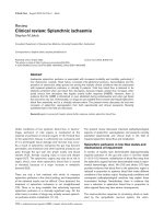
intensive care unit and decreased subsequently (Fig. 1). The
high splanchnic lactate : pyruvate ratios at admission to the
intensive care unit resulted from low pyruvate rather than high
lactate levels, and are therefore unlikely to indicate anaerobic
metabolism. On the other hand, only excessive anaerobic
mesenteric metabolism or frank liver hypoxia will result in sys-
temic hyperlactataemia because of the high lactate extraction
capability of the liver. Initially high concentrations of glu-
tathione transferase-α decreased during the postoperative
period, and indocyanine green extraction was well preserved.
It is likely that the observed increase in splanchnic oxygen
extraction was sufficient to compensate for the lack of
increase in blood flow and to maintain aerobic metabolism
and cellular integrity in the splanchnic region in these patients
with a normal cardiac reserve.
Surgical, medical and nursing procedures in critically ill
patients may also interfere with the ability to maintain ade-
quate systemic and regional oxygen delivery. We recently
showed that haemodialysis with ultrafiltration is associated
with a significant reduction in systemic, splanchnic and
femoral blood flows [36]. Nine patients with acute renal
failure and stable systemic haemodynamics were studied.
The need for extracorporeal renal replacement therapy was
defined as excess of extracellular water and insufficient
urinary output despite the use of diuretic drugs; serum urea
concentration greater than 20 mmol/l; and creatinine concen-
tration greater than 400 µmol/l. Haemodialysis was per-
formed according to standard clinical practice, and
hypotension (systolic blood pressure < 90 mmHg) was
treated by reducing the filtration rate and infusion of
100–200 ml Ringers acetate if necessary. Systemic, hepato-
splanchnic and femoral haemodynamics and oxygen trans-
port, and gastric mucosal partial carbon dioxide tension were
measured before haemodialysis, 2 hours after initiating a 4-
hour period of haemodialysis, and again 2 hours after comple-
tion of haemodialysis.
The median amount of fluid removed during 4 hours of
haemodialysis was 2000 ml (range 300–2500 ml) [36].
Haemodialysis was associated with a parallel decrease in
cardiac output, stroke volume and splanchnic blood flow
despite stable arterial blood pressure. All flows returned to
baseline values after dialysis without therapeutic interven-
tions, suggesting that this mode of renal replacement therapy
induces acute but only temporary reduction in splanchnic
perfusion in intensive care patients with stable haemodynam-
ics. In such patients haemodialysis can therefore still be rec-
ommended. However, the systemic and regional response to
acute intermittent haemodialysis may be different in patients
who are hypotensive or who need vasoactive drugs to main-
tain a sufficient blood pressure. These patients may not toler-
ate intermittent haemodialysis.
The abdominal compartment syndrome is another clinical
condition during which splanchnic blood flow is at risk
(recently reviewed by Morken and West [37]). It may occur
under various surgical and medical conditions, such as blunt
and penetrating abdominal trauma, retroperitoneal haemor-
rhage, ruptured abdominal aortic aneurysm, peritonitis and
pancreatitis, among others. Inadequate gastric mucosal per-
fusion has recently also been described in patients with
obstructive sleep apnoea [38] and during periods of weaning
from mechanical ventilation [39,40].
Mechanisms to preserve hepato-splanchnic
blood flow
In contrast to the gut, the liver is believed to be relatively well
protected against hypoperfusion because of the HABR [41].
The HABR describes the hydrodynamic interaction between
portal venous and hepatic arterial blood flow. Table 1 summa-
rizes important findings of studies in HABR [41–47]. When
mesenteric and, consequently, portal venous blood flow
decreases, hepatic arterial blood flow increases. The com-
pensation of hepatic arterial blood flow for the decreased
portal venous blood flow is in the range 20–30% [41,46,48].
Compensation in terms of oxygen delivery is substantially
higher because of the much greater oxygen content in the
hepatic artery as compared with the portal vein. The current
concept is the adenosine washout hypothesis; namely,
adenosine in the Mall’s space is washed out when portal
venous blood flow is normal but not when it is low. Under
these circumstances, adenosine causes hepatic arterial
vasodilatation [41]. It has been shown that liver oxygen
supply is maintained during haemorrhage until the blood loss
exceeds 30% [49]. The HABR is abolished early during
endotoxaemia and recovers partially after several hours
[47,50–52].
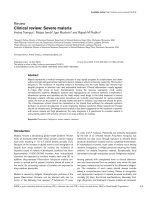
Figure 1
Lactate :pyruvate ratio in relation to lactate concentrations in
17 patients after cardiac surgery. Data points are pooled values from
three different time points from arrival to the intensive care unit until
extubation. (Adapted from [33].)
We tested the HABR under conditions of low mesenteric
blood flow in pigs [53]. A total of 14 animals were random-
ized either to partial superior mesenteric artery occlusion or
to serve as controls. Superior mesenteric artery flow was
reduced by a clamp to a median flow of 2 ml/kg per min
(range 1–3 ml/kg per min) for 120 min in ischaemic animals.
After 2 hours the clamp was released and the measurements
were continued for another 60 min. The HABR was assessed
four times at hourly intervals during ischaemia and reperfusion
by acute and intermittent reduction in portal vein blood flow.
The absolute increase in hepatic arterial blood flow in response
to portal vein occlusion decreased significantly during
ischaemia. A decrease in the efficiency but not an exhaustion of
the HABR was also seen in control animals. During reperfu-
sion, hepatic arterial blood flow changes induced by portal vein
occlusion increased again. We hypothesize that the repeated
ischaemia/reperfusion events caused by testing the buffer
response may have interfered with hepatic adenosine produc-
tion or transport. Alternatively, an effect of the surgical proce-
dure and anaesthesia may have contributed to the observed
changes in both groups of animals.
The exhaustion of the HABR has important clinical implications.
If a reduction in splanchnic blood flow has already resulted in a
compensatory increase in hepatic arterial blood flow, then an
acute further decrease in splanchnic blood flow may no longer
be compensated for by the HABR, especially when systemic
blood flow and/or pressure decrease concomitantly.
Our experimental design, which is similar to that employed by
most investigators to test the HABR, may modify both quanti-
tative and qualitative aspects of the HABR elicited under con-
ditions of low systemic blood flow. This is because clamping
the superior mesenteric artery results in increased systemic
vascular resistance and increased perfusion pressure across
the coeliac trunk–hepatic artery axis. If the systemic blood
flow is low, then a low coeliac trunk blood flow may not
increase sufficiently to compensate for an acute decrease in
portal venous blood flow.
In order to address this question, we designed a study in which
systemic blood flow was decreased in two steps by cardiac
tamponade [16]. At baseline and every 30 min thereafter, the
portal vein was intermittently occluded. Cardiac output
decreased by 21% and 55% and systemic arterial blood pres-
sure by 40% and 64% during the first stage (moderate) and
second stage (severe) of tamponade, respectively, and it
remained constant in a control group. During moderate tam-
ponade, hepatic arterial blood flow increased in both groups by
approximately 50%. During severe tamponade hepatic arterial
blood flow decreased in tamponade animals. Nevertheless,
fractional splanchnic blood flow was preserved. The acute
compensation of hepatic arterial blood flow for a decrease in
portal venous flow decreased during moderate tamponade and
disappeared during severe tamponade. These data demon-
strate that the liver is only initially protected during low systemic
perfusion. Later, the HABR is exhausted. In the same experi-
ment, we demonstrated that the splanchnic organs are not
among the first to produce lactate because fractional splanch-
nic blood flow is preserved. In contrast, the capacity of the liver
to increase lactate uptake was exhausted early.
Is splanchnic blood flow impaired in low
blood pressure?
It has been proposed that decreasing arterial blood pressure
is associated with impaired perfusion of splanchnic organs
and disturbed cellular integrity [54]. Andel and coworkers
[55] tested the effect of deliberate hypotension on splanchnic
perfusion balance with the use of either isoflurane or a combi-
nation of esmolol and nitroglycerin. Sixteen anaesthetized
Available online />Table 1
Important studies on hepatic arterial buffer response, with main findings
Reference Species Main finding
Lautt (1985) [41] Cat Antagonism of HABR by the adenosine antagonist 8-phenyltheophylline
Lautt et al. (1988) [42] Cat Hepatic arterial vascular response to intravenous drugs dependent on direct action of
the drug on hepatic artery and on indirect effects of drug-induced changes in portal
venous blood flow
Lautt and McQuaker (1989) [43] Cat Protective dilatation of hepatic artery during haemorrhage is mediated by adenosine
Lautt et al. (1990) [44] Cat During high portal venous blood flow, hepatic artery is nearly fully constricted; during low
portal venous blood flow, hepatic artery is nearly fully dilated
Henderson et al. (1992) [45] Human Intact HABR in liver transplant patients
Ayuse et al. (1994) [46] Pig Change in portal venous blood flow alters hepatic arterial resistance upstream from the
site of a constant arterial back pressure
Ayuse et al. (1995) [47] Pig HABR is abolished during endotoxaemia independently of nitric oxide or α-adrenergic
receptor antagonists
HABR = hepatic arterial buffer response. Adapted from Jakob [10].
patients undergoing elective maxillofacial surgery were ran-
domly allocated to one of the two drug regimens. Systolic
blood pressure was decreased to approximately 30% below
preoperative values while maintaining mean arterial blood
pressure at levels greater than 50 mmHg. Gastric tonometers
were used to assess the adequacy of mucosal perfusion, and
arterial lactate was measured in order to assess the overall
adequacy of oxygen delivery. Those investigators found that
neither method that had been used to decrease blood pres-
sure compromised splanchnic tissue oxygen balance in these
patients. Overall organ perfusion was sufficient in both
groups because none of the patients demonstrated an
increase in blood lactate concentration. Maintaining arterial
blood pressure above 50 mmHg in defined patient groups
therefore appears to be safe in terms of splanchnic tissue
oxygenation if hypovolaemia is prevented.
Gastric mucosal pH in early goal-directed
therapy for critically ill patients
In 1992 Gutierrez and coworkers [56] proposed the use of
treatment titrated against gastric intramucosal pH in the man-
agement of critically ill patients. However, three consecutive
studies did not confirm these findings [57–59]. The trial by
Gutierrez and coworkers has been criticized because of the
unexplained high mortality in the control group, in which no
attempts were made to standardize the treatment. It is impor-
tant to realize in this context that changes in gastric intramu-
cosal pH do not necessarily reflect similar changes in
hepato-splanchnic blood flow.
How can the splanchnic blood flow be
increased?
Recently, the haemodynamic effects of fenoldopam (a
dopamine-1 receptor agonist) were studied before and after
induction of splanchnic ischaemia by haemorrhage [60]. After
haemorrhage, this drug restored portal vein blood flow to
near baseline, maintained the splanchnic fraction of cardiac
output, and attenuated the rise in gut mucosal partial carbon
dioxide tension. Fenoldopam also redistributed the blood flow
away from the serosal to the mucosal layer both at baseline
and during haemorrhage. Whether this drug also exerts its
beneficial effects under clinical conditions of low splanchnic
blood flow has yet to be demonstrated.
In patients with septic shock, dobutamine was compared with
the phosphodiesterase inhibitor enoximone [61]. In 48
patients either one of the drugs was infused randomly after
fluid resuscitation. Liver blood flow was estimated using the
continuous indocyanine green infusion technique and hepatic
venous catheterization. Liver function was assessed using
monoethylglycine xylidide formation after lidocaine injection,
and inflammation was quantified by release of hepatic tumour
necrosis factor-α. Cardiac output and total hepato-splanchnic
blood flow increased in both groups after 12 and 48 hours of
the respective drug infusions. The fractional hepato-splanch-
nic blood flow decreased slightly in dobutamine-treated
patients and remained unchanged in the enoximone group.
Hepato-splanchnic oxygen consumption and release of
tumour necrosis factor-α were increased in both groups after
12 hours of vasoactive drug infusion, but arterial monoethyl-
glycine xylidide concentrations increased only in the enoxi-
mone group. Because of methodological problems in that
study (lack of control patients, incomplete assessment of
monoethylglycine xylidide kinetics and multiple comparisons
without correction), it is hard to conclude whether one drug is
superior to the other in terms of preserved liver function or
attenuation of inflammation.
Even relatively small increases in intra-abdominal pressure
during carbon dioxide laparoscopy are associated with
impaired systemic, portal venous and hepatic arterial blood
flow [62]. Although small doses of dobutamine appear to
restore gut mucosal perfusion and improve hepatic arterial
blood flow in this setting, total hepato-splanchnic blood
flow cannot be maintained with either dobutamine or
dopamine [62].
Dopamine is used to support cardiac output and blood pres-
sure in patients with cardiac failure and septic shock [63,64].
Dopamine at a low rate is still frequently used in patients with
renal failure, with the aim of increasing both renal perfusion
and urinary output, although there is a lack of data demon-
strating subsequent improvement in renal function [65,66].
We measured the effects of dopamine on systemic and
splanchnic blood flow and metabolism in septic and cardiac
surgery patients [12]. Dopamine infusion was started at a
dose of 1 µg/kg per min and then gradually increased until
the thermodilution cardiac output was 25% higher than at
baseline. Dopamine infusion (1–9 µg/kg per min) caused a
parallel increase in systemic and splanchnic blood flow.
Although systemic oxygen consumption did not change in
either group, splanchnic oxygen consumption decreased sig-
nificantly during dopamine infusion in septic patients and
increased in cardiac surgery patients.
A decrease in splanchnic oxygen consumption during
dopamine infusion has recently been reported in patients with
acute hepatic failure [67]. A reduction in hepato-splanchnic
oxygen uptake despite an increase in regional perfusion may
indicate blood flow redistribution. Alternatively, some meta-
bolic functions of the hepato-splanchnic region may have been
impaired. Dopamine may directly inhibit isoenzymes of the
cytochrome P450 complex [68]. The different effects of
dopamine infusion on splanchnic oxygen consumption in the
two patient groups could possibly be explained by different
baseline activities of P450 isoenzymes. The indications and
safety of dopamine in sepsis should therefore be re-evaluated.
Conclusion
Inadequate splanchnic perfusion in critically ill patients is
associated with increased morbidity and mortality. The under-
lying pathophysiological mechanisms are still not well under-
Critical Care August 2002 Vol 6 No 4 Jakob
stood. Splanchnic blood flow may become insufficient as a
result of a multitude of different diseases and treatment
modalities. Splanchnic vasoregulation is complex and is also
altered by disease and treatment. Unfortunately, many of the
available monitoring tools for hepato-splanchnic blood flow
and metabolism are difficult to apply in the clinical setting,
and interpretation of the results obtained is not straightfor-
ward. Thus, concepts of splanchnic resuscitation are not
established. Future research projects should focus on the
interplay between the physiological regulatory mechanisms in
splanchnic organs, disease and treatment.
Competing interests
None declared.
References
1. Bulkley GB, Kvietys PR, Perry MA, Granger DN: Effects of
cardiac tamponade on colonic hemodynamics and oxygen
uptake. Am J Physiol 1983, 244:G604-G612.
2. Schlichtig RA, Kramer DJ, Pinsky MR: Flow redistribution during
progressive hemorrhage is a determinant of critical O
2
deliv-
ery. J Appl Physiol 1992, 70:169-178.
3. Vatner SF: Effects of hemorrhage on regional blood flow dis-
tribution in dogs and primates. J Clin Invest 1974, 54:225-235.
4. Grotz MR, Ding J, Guo W, Huang Q, Deitch EA: Comparison of
plasma cytokine levels in rats subjected to superior mesen-
teric artery occlusion or hemorrhagic shock. Shock 1995, 3:
362-368.
5. Ljungdahl M, Lundholm M, Katouli M, Rasmussen I, Engstrand L,
Haglund U: Bacterial translocation in experimental shock is
dependent on the strains in the intestinal flora. Scand J Gas-
troenterol 2000, 35:389-397.
6. Aranow JS, Fink MP: Determinants of intestinal barrier failure
in critical illness. Br J Anaesth 1996, 77:71-81.
7. Baker JW, Deitch EA, Li M, Berg RD, Specian RD: Hemorrhagic
shock induces bacterial translocation from the gut. J Trauma
1988, 28:896-906.
8. Arvidsson D, Rasmussen I, Almqvist P, Niklasson F, Haglund U:
Splanchnic oxygen consumption in septic and hemorrhagic
shock. Surgery 1991, 109:190-197.
9. Dahn MS, Lange P, Lobdell K, Hans B, Jacobs LA, Mitchell RA:
Splanchnic and total body oxygen consumption differences in
septic and injured patients. Surgery 1987, 101:69-80.
10. Jakob SM: Splanchnic vasoregulation and metabolism: new
insights into physiology [PhD thesis]. Kuopio, Finland: Kuopio
University Publications D, Medical Sciences 240; 2001.
11. Reinelt H, Radermacher P, Kiefer P, Fischer G, Wachter U, Vogt J,
Georgieff M: Impact of exogenous beta-adrenergic receptor
stimulation on hepatosplanchnic oxygen kinetics and meta-
bolic activity in septic shock. Crit Care Med 1999, 27:325-331.
12. Jakob SM, Ruokonen E, Takala J: Effects of dopamine on sys-
temic and regional blood flow and metabolism in septic and
cardiac surgery patients. Shock 2002 (in press).
13. Bailey RW, Bregman ML, Fuh KC, Hamilton SR, Herlong HF,
Bulkley GB: Hemodynamic pathogenesis of ischemic hepatic
injury following cardiogenic shock/resuscitation. Shock 2000,
14:451-459.
14. Reilly PM, Wilkins KB, Fuh KC, Haglund U, Bulkley GB: The
mesenteric hemodynamic response to circulatory shock: an
overview. Shock 2001, 15:329-343.
15. Toung T, Reilly PM, Fuh KC, Ferris R, Bulkley GB: Mesenteric
vasoconstriction in response to hemorrhagic shock. Shock
2000, 13:267-273.
16. Jakob SM, Tenhunen JJ, Laitinen S, Heino A, Alhava E, Takala J:
Effects of systemic arterial hypoperfusion on splanchnic
hemodynamics and hepatic arterial buffer response in pigs.
Am J Physiol Gastrointest Liver Physiol 2001, 280:G819-G827.
17. Riddez L, Hahn RG, Brismar B, Strandberg A, Svensen C, Heden-
stierna G: Central and regional hemodynamics during acute
hypovolemia and volume substitution in volunteers. Crit Care
Med 1997, 25:635-640.
18. Fong Y, Marano MA, Moldawer LL, Wei H, Calvano SE, Kenney
JS, Allison AC, Cerami A, Shires GT, Lowry SF: The acute
splanchnic and peripheral tissue metabolic response to endo-
toxin in humans. J Clin Invest 1990, 85:1896-1904.
19. Taylor DE, Kantrow SP, Piantadosi CA: Mitochondrial respira-
tion after sepsis and prolonged hypoxia. Am J Physiol 1998,
275:L139-L144.
20. Khanna A, Rossman JE, Fung H-L, Caty MG: Intestinal and
hemodynamic impairment following mesenteric ischemia/
reperfusion. J Surg Res 2001, 99:114-119.
21. Bone RC: Toward an epidemiology and natural history of
SIRS. JAMA 1992, 268:3452-3455.
22. Kistler EB, Lefer AM, Hugli TE, Schmid-Schönbein GW: Plasma
activation during splanchnic arterial occlusion shock. Shock
2000, 14:30-34.
23. Kirton OC, Windsor J, Wedderburn R, Hudson-Civetta J, Shatz
DV, Mataragas NR, Civetta JM: Failure of splanchnic resuscita-
tion in the acutely injured trauma patient correlates with mul-
tiple organ system failure and length of stay in the ICU. Chest
1998, 113:1064-1069.
24. Doglio GR, Pusajo JF, Egurrola MA, Bonfigli GC, Parra C, Vetere
L, Hernandez MS, Fernandez S, Palizas F, Gutierrez G: Gastric
mucosal pH as a prognostic index of mortality in critically ill
patients. Crit Care Med 1991, 19:1037-1040.
25. Maynard N, Bihari D, Beale R, Smithies M, Baldock G, Mason R,
McColl I: Assessment of splanchnic oxygenation by gastric
tonometry in patients with acute circulatory failure. JAMA
1993, 270:1203-1210.
26. Theodoropoulos G, Lloyd LR, Cousins G, Pieper D: Intraopera-
tive and early postoperative gastric intramucosal pH predicts
morbidity and mortality after major abdominal surgery. Am
Surg 2001, 67:303-308.
27. Poeze M, Takala J, Greve JW, Ramsay G: Pre-operative tonome-
try is predictive for mortality and morbidity in high-risk surgi-
cal patients. Intensive Care Med 2000, 26:1272-1281.
28. Norton L, Moore G, Eiseman B: Liver failure in the postopera-
tive patient: the role of sepsis and immunologic deficiency.
Surgery 1975, 78:6-13.
29. Imamura M, Clowes GHA: Hepatic blood flow and oxygen con-
sumption in starvation, sepsis, and septic shock. Surg Gynecol
Obstet 1975, 141:27-34.
30. Inoue S, Kuro M, Furuya H: What factors are associated with
hyperlactatemia after cardiac surgery characterized by well-
maintained oxygen delivery and a normal postoperative
course? A retrospective study. Eur J Anesthesiol 2001, 18:576-
584.
31. Bastien O, Piriou V, Aouifi A, Flamens C, Evans R, Lehot JJ: Rela-
tive importance of flow versus pressure in splanchnic perfu-
sion during cardiopulmonary bypass in rabbits. Anesthesiology
2000, 92:457-464.
32. Thorén A, Elam M, Ricksten S-E: Jejunal mucosal perfusion is
well-maintained during mild hypothermic cardiopulmonary
bypass in humans. Anesth Analg 2001, 92:5-11.
33. Jakob S, Ruokonen E, Takala J: Assessment of the adequacy of
systemic and regional perfusion after cardiac surgery. Br J
Anaesth 2000, 84:571-577.
34. Rowell LB, Blackmon JR, Kenny MA, Escourrou P: Splanchnic
vasomotor and metabolic adjustments to hypoxia and exer-
cise in humans. Am J Physiol 1984, 247:H251-H258.
35. Nelson DP, Samsel RW, Wood LDH, Schumacker PT: Pathologi-
cal supply dependence on systemic and intestinal O
2
uptake
during endotoxemia. J Appl Physiol 1988, 64:2410-2419.
36. Jakob SM, Ruokonen E, Vuolteenaho O, Lampainen E, Takala J:
Splanchnic perfusion during hemodialysis: evidence for mar-
ginal tissue perfusion. Crit Care Med 2001, 29:1393-1398.
37. Morken J, West M: Abdominal compartment syndrome in the
intensive care unit. Curr Opin Crit Care 2001, 7:268-274.
38. Epstein LJ, Jervis OJ Jr, Henderson JH II, Sullivan M, Mohsenifar Z:
Measurement of gastric P(CO
2
) as an index of tissue hypoxia
during obstructive sleep apnea. Respiration 2001, 68:28-34.
39. Hurtado FJ, Beron M, Olivera W, Garrido R, Silva J, Caragna E,
Rivara D: Gastric intramucosal pH and intraluminal PCO
2
during weaning from mechanical ventilation. Crit Care Med
2001, 29:70-76.
40. Uusaro A, Chittock DR, Russell JA, Walley KR: Stress test and
gastric-arterial PCO2 measurement improve prediction of
successful extubation. Crit Care Med 2000, 28:2313-2319.
Available online />Critical Care August 2002 Vol 6 No 4 Jakob
41. Lautt WW: Mechanism and role of intrinsic regulation of
hepatic arterial blood flow: the hepatic arterial buffer
response. Am J Physiol 1985, 249:G549-G556.
42. Lautt WW, D’Almeida MS, McQuaker J, D’Aleo L: Impact of the
hepatic arterial buffer response on splanchnic vascular
responses to intravenous adenosine, isoproterenol, and
glucagon. Can J Physiol Pharmacol 1988, 66:807-813
43. Lautt WW, McQuaker JE: Maintenance of hepatic arterial blood
flow during hemorrhage is mediated by adenosine. Can J
Physiol Pharmacol 1989, 67:1023-1028.
44. Lautt WW, Legare DJ, Ezzat WR: Quantitation of the hepatic
arterial buffer response to graded changes in portal blood
flow. Gastroenterology 1990, 98:1024-1028.
45. Henderson JM, Gilmore GT, Mackay GJ, Galloway JR, Dodson TF,
Kutner MH: Hemodynamics during liver transplantation: the
interactions between cardiac output and portal venous and
hepatic arterial flows. Hepatology 1992, 16:715-718.
46. Ayuse T, Brienza N, O’Donnell CP, Robotham JL. Pressure-flow
analysis of portal vein and hepatic artery interactions in
porcine liver. Am J Physiol 1994, 267:H1233-H1242.
47. Ayuse T, Brienza J, Revelly P, Boitnott JK, Robotham JL: Role of
nitric oxide in porcine liver circulation under normal and endo-
toxemic conditions. J Appl Physiol 1995, 78:1319-1329.
48. Mathie RT, Blumgart LH: The hepatic haemodynamic response
to acute portal venous blood flow reductions in the dog.
Pflügers Arch 1983, 399:223-227.
49. Rasmussen A, Skak C, Kristensen M, Ott P, Kirkegaard P, Secher
NH: Preserved arterial flow secures hepatic oxygenation
during haemorrhage in the pig. J Physiol 1999, 516:539-548.
50. Dziki AJ, Lynch WH, Ramsey CB, Law WR: Beta-adrenergic-
dependent and -independent actions of naloxone on perfu-
sion during endotoxin shock. Circ Shock 1993, 39:29-38.
51. Halvorsen L, Roth R, Gunther RA, Firoozmand E, Buoncristiani
AM, Kramer GC: Liver hemodynamics during portal venous
endotoxemia in swine. Circ Shock 1993, 41:166-175.
52. Schiffer ER, Mentha G, Schwieger IM, Morel DR: Sequential
changes in the splanchnic circulation during continuous endo-
toxin infusion in sedated sheep: evidence for a selective
increase of hepatic artery blood flow and loss of the hepatic
arterial buffer response. Acta Physiol Scand 1993, 147:251-261.
53. Jakob SM, Tenhunen JJ, Heino A, Pradl R, Alhava E, Takala J:
Splanchnic vasoregulation, during mesenteric ischemia and
reperfusion. Shock 2002 (in press).
54. Suttner SW, Boldt J, Schmidt CC, Piper SN, Schuster P, Kumle
B: The effects of sodium nitroprusside-induced hypotension
on splanchnic perfusion and hepatocellular integrity. Anesth
Analg 1999, 89:1371-1377.
55. Andel D, Andel H, Hörauf K, Felfernig D, Millesi W, Zimpfer M:
The influence of deliberate hypotension on splanchnic perfu-
sion balance with use of either isoflurane or esmolol and
nitroglycerin. Anesth Analg 2001, 93:1116-1120.
56. Gutierrez G, Palizas F, Doglio G, Wainsztein N, Gallesio A, Pacin
J, Dubin A, Schiavi E, Jorge M, Pusajo J, et al.: Gastric intramu-
cosal pH as a therapeutic index of tissue oxygenation in criti-
cally ill patients. Lancet 1992, 339:195-199.
57. Ivatury RR, Simon RJ, Islam S, Fueg A, Rohman M, Stahl WM: A
prospective randomized study of end points of resuscitation
after major trauma: global oxygen transport indices versus
organ-specific gastric mucosal pH. J Am Coll Surg 1996, 183:
145-154.
58. Pargger H, Hampl KF, Christen P, Staender S, Scheidegger D:
Gastric intramucosal pH-guided therapy in patients after elec-
tive repair of infrarenal abdominal aneurysms: is it beneficial?
Intensive Care Med 1998, 24:769-776.
59. Gomersall CD, Joynt GM, Freebairn RC, Hung V, Buckley TA, Oh
TE: Resuscitation of critically ill patients based on the results
of gastric tonometry: a prospective, randomized, controlled
trial. Crit Care Med 2000, 28:607-614.
60. Guzman JA, Rosado AE, Kruse JA: Dopamine-1 receptor stimu-
lation attenuates the vasoconstrictive response to gut
ischemia. J Appl Physiol 2001, 91:596-602.
61. Kern H, Schröder T, Kaulfuss M, Martin M, Kox W, Spies C: Enox-
imone in contrast to dobutamine improves hepatosplanchnic
function in fluid-optimized septic shock patients. Crit Care
Med 2001, 29:1519-1525.
62. Agusti M, Elizalde JI, Adalia R, Martinez-Palli G, Garcia-Valde-
casas JC, Pique JM, Taura P: The effects of vasoactive drugs
on hepatic blood flow changes induced by CO2 laparoscopy:
an animal study. Anesth Analg 2001, 93:1121-1126.
63. Landgarten MJ, Kumar A, Parrillo JE: Cardiovascular dysfunction
in sepsis and septic shock. Curr Treatment Options Cardiovasc
Med 2000, 2:451-459.
64. Day NP, Phu NH, Bethell DP, Mai NT, Chau TT, White NJ: The
effects of dopamine and adrenaline infusions on acid-base
balance and systemic haemodynamics in severe infection.
Lancet 1996, 348:219-223.
65. Marik PE, Iglesias J: Low-dose dopamine does not prevent
acute renal failure in patients with septic shock and oliguria.
NORASEPT II Study Investigators. Am J Med 1999, 107:387-
390.
66. Bellomo R, Chapman M, Finfer S, Hickling K, Myburgh J: Low-
dose dopamine in patients with early renal dysfunction: a
placebo-controlled randomised trial. Australian and New
Zealand Intensive Care Society (ANZICS) Clinical Trials
Group. Lancet 2000, 356:2139-2143.
67. Clemmesen JO, Galatius S, Skak C, Dalgaard P, Larsen FS, Ott
P: The effect of increasing blood pressure with dopamine on
systemic, splanchnic, and lower extremity hemodynamics in
patients with acute liver failure. Scand J Gastroenterol 1999,
34:921-927.
68. Ben-Shachar D, Zuk R, Glinka Y: Dopamine neurotoxicity: inhi-
bition of mitochondrial respiration. J Neurochem 1995, 64:
718-723.