Báo cáo y học: "Bench-to-bedside review: Lactate and the lung" ppt
Bạn đang xem bản rút gọn của tài liệu. Xem và tải ngay bản đầy đủ của tài liệu tại đây (163.2 KB, 3 trang )
ALI = acute lung injury; ARDS = acute respiratory distress syndrome.
Available online />The respiratory and immune functions of the lung are largely
dependent on the activity of a number of metabolic pathways.
Surfactants and prostanoids are synthesized from lipid pre-
cursors. Protein synthesis is maintained at a high rate to
maintain a rapid turnover of the endothelial and parenchymal
pulmonary cells and of the immune cells. Energy is produced
from glucose, fatty acids and branched chain amino acid oxi-
dation. Lactate, alanine and glutamine are synthesized to
shuttle carbon and nitrogen residues derived from glucose
and amino acid metabolism.
Despite the importance of these metabolic pathways, the role
of the lung in interorgan substrate exchange in physiological
and pathological conditions is largely unknown. In humans,
substrate exchange across an individual organ is determined
according to the Fick principle, by measuring substrate
arteriovenous concentrations and local blood flow. This
approach has been largely used to determine skeletal muscle
metabolism in the human limbs. In the lung, however, the
arteriovenous difference of substrate concentrations is
usually small compared with a high rate of blood flow through
the tissue. This limits the ability of the Fick technique to
detect statistically significant rates of substrate exchange
across the lung in most circumstances.
Lung lactate synthesis and release
Virtually all tissues can synthesize or utilize lactate. Lactate is
synthesized from the pyruvic acid derived from glycolysis,
whereas it can be utilized to form glucose or it can be oxidized
through pyruvate and the tricarboxylic acid cycle. In physiologi-
cal conditions, lactate is mainly produced in the skin, skeletal
muscle, leucocytes and red blood cells. It is mainly utilized,
however, in the liver and the kidney. Lactate is therefore one of
the major carbon shuttles among body tissues.
In different conditions, the rate of lactate synthesis is depen-
dent on the activity of the glycolytic pathway relative to the
oxidative capacity of the pyruvate dehydrogenase enzymatic
Review
Bench-to-bedside review: Lactate and the lung
Fulvio Iscra
1
, Antonino Gullo
1
and Gianni Biolo
2
1
Department of Surgical Sciences, Anaesthesiology and Intensive Care, University of Trieste, Italy
2
Department of Clinical, Morphological and Technological Sciences, University of Trieste, Italy
Correspondence: Gianni Biolo,
Published online: 7 June 2002 Critical Care 2002, 6:327-329
This article is online at />© 2002 BioMed Central Ltd (Print ISSN 1364-8535; Online ISSN 1466-609X)
This article is based on a presentation at the Lactate Satellite Meeting held during the 8th Indonesian–International Symposium on Shock & Critical
Care, Bali, Indonesia, 24 August 2001.
Abstract
The ability of the isolated lung tissue to take up glucose and to release lactate is potentially similar to
that of other body tissues. Nonetheless, when lung lactate exchange was assess in vivo in normal
humans, no measurable lactate production could be detected. Lung lactate production may become
clinically evident in disease states especially in the patients with acute lung injury or with acute
respiratory distress syndrome. Potential mechanisms of lactate production by the injured lung may
include not only the onset of anaerobic metabolism in hypoxic zones, but also direct cytokine effects on
pulmonary cells and an accelerated glucose metabolism in both the parenchymal and the inflammatory
cells infiltrating lung tissue. In addition, as skeletal muscle, lung tissue may show metabolic adaptations
in response to systemic mediators and may contribute to the systemic metabolic response to severe
illness even in the absence of direct tissue abnormalities.
Keywords acute respiratory distress syndrome, arteriovenous balance, cytokines, lactate release, pulmonary artery
Critical Care August 2002 Vol 6 No 4 Iscra et al.
complex. An acceleration of lactate synthesis may be
observed in conditions of increased glucose uptake from cir-
culation, of increased glycogenolysis and glycolysis due to
enhanced epinephrine secretion, of inhibition of pyruvate
dehydrogenase or of glycogen synthesis in sepsis and, finally,
during tissue hypoxia (Fig. 1).
Early in vitro studies [1] demonstrated that the ability of the
isolated lung tissue to take up glucose and to release lactate
was potentially similar to that of other body tissues such as
skeletal muscle, skin, red blood cells, leucocytes, and so on.
Nonetheless, when lung lactate exchange was assessed in
vivo in normal humans, no measurable lactate production
could be detected by the Fick method. It was concluded,
therefore, that the rate of lactate synthesis in the normal lung
is approximately equal to the rate of lactate utilization, leading
to a net lactate balance close to zero [2–4]. In many patho-
logical conditions, in contrast, the arteriovenous lactate con-
centration difference across the lung has often been found
consistently negative, suggesting that a net lactate produc-
tion from the lung may become clinically evident in disease
states. In animals, Bellomo et al. observed an early lactate
release from the lung following endotoxin administration [5].
In humans, a net lung lactate production was measured in
patients with different types of acute lung injuries by many
authors, including ourselves [6–10].
The largest number of patients has been studied by De
Backer et al. [6]. They compared the transpulmunary lactate
exchange in 43 patients with acute lung injury (ALI) or acute
respiratory distress syndrome (ARDS), as defined accord-
ing to the American–European Consensus Conference,
with that in other patients affected by acute cardiogenic pul-
monary oedema (n = 9), pneumonia (n = 37), lung trans-
plantation (n = 7) or other causes of respiratory failure
(n = 26). De Backer et al. observed that lung lactate pro-
duction was greater in the patients with ALI/ARDS that in
those with other disease states. Furthermore, lung lactate
production was related with the ratio between arterial
oxygen pressure and the fraction of inspired oxygen
(PaO
2
/FiO
2
; inverse correlation) and with the pulmonary
injury score (direct correlation). In patients with high lactate
plasma levels, lung lactate production was not related to the
arterial lactate concentration.
These observations have been confirmed in other smaller
groups of patients affected by ALI or ARDS [7–10]. Several
considerations can be made on the basis of these studies. A
lung inflammatory condition is always associated with an
increased lung lactate production. Also, the extent of lactate
release is related to the severity of the lung injury. A third
consideration is that the presence of pulmonary infection
does not increase lactate production. Also, the inflammatory
process should be severe and should involve the entire
organ since lactate production is not increased in localized
inflammatory processes. In fact, it has been observed in lung
carcinoma that lung lactate production is increased only in
the affected districts [2]. Finally, the lung is not the only
major source of lactate in conditions of severe increase of
plasma lactate.
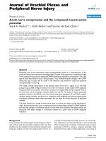
Figure 1
Potential mechanisms of increased tissue lactate production in sepsis. GLUT1, glucose transporter 1; TCA, tricarboxylive acid cycle; acetyl-CoA,
acetyle-coenzyme A.
Potential mechanisms of lung lactate production by the
injured lung may include not only the onset of anaerobic
metabolism in hypoxic zones, but also direct cytokine effects
on pulmonary cells and an accelerated glucose metabolism in
both the parenchymal and the inflammatory cells infiltrating
the lung tissue. Experimental evidence in vitro [11] and in
vivo [12,13] indicates that lung metabolism tolerates severe
reductions of intracellular oxygen availability, suggesting that
lung hypoxia is not the main factor responsible for increasing
lactate release from the injured lung. In severe cardiac failure
[14] and during acute hepatic failure [15], an increased lung
lactate production appeared to be directly related to systemic
lactate levels. In addition, preliminary data from our laboratory
indicate that septic ARDS patients with no direct lung injuries
and with normal oxygen tissue delivery release lactate from
lung tissue at rates three to four times greater than that from
skeletal muscle [16]. These patients also exhibited a negative
lung protein balance and a large lung release of neogluco-
genic amino acids [17].
These observations suggest that, as skeletal muscle, lung
tissue may show metabolic adaptations in response to sys-
temic mediators (e.g. cytokines) and may contribute to the
systemic metabolic response to severe illness even in the
absence of direct tissue abnormalities.
Competing interests
None declared.
References
1. Evans CL, Hsu FY, Kosaka T: Utilization of blood sugar and for-
mation of lactic acid by the lungs. J Physiol 1934, 82:41-60.
2. Rochester DF, Wichern A, Fritts W, Caldwell PR, Lewis ML, Glun-
tial C, Garfield JW: Arteriovenous differences of lactate and
pyruvate across healthy and diseased human lungs. Am Rev
Respir Dis 1973, 107:442-448.
3. Mitchell AM, Cournand A: The fate of circulating lactic acid in
the human lung. J Clin Invest 1955, 34:471-476.
4. Harris P, Bailey T, Bateman M: Lactate, pyruvate, glucose and
free fatty acid in mixed venous and arterial blood. J Appl
Physiol 1963, 18;933-936.
5. Bellomo R, Kellum JA, Pinsky MR: Visceral lactate fluxes during
early endotoxinemia in the dog. Chest 1996, 110:195-204.
6. De Backer D, Creteur J, Zhang H, Norrenberg M, Vincent JL:
Lactate production by the lungs in acute lung injury. Am J
Respir Crit Care Med 1997, 156:1099-1104.
7. Brown SD, Clark C,Guttierez G: Pulmonary lactate release in
patients with sepsis and ARDS. J Crit Care 1996, 11:2-8.
8. Douzinas EE, Tsidemiadou PD, Pitaridis MT, Andrianskis I,
Bobota-Chloraki A, Katsouyanno K, Sfyras D, Malagari K, Roussos
C: The regional production of cytokines and lactate in sepsis-
related multiple organ failure. Am J Respir Crit Care Med
1997, 155:53-59.
9. Kellum JA, Kramer DJ, Lee K, Mankad S, Beliomo R, Pinsky MR:
Release of lactate by the lung in acute lung injury. Chest
1997, 111:1301-1305.
10. Iscra F, Biolo G, Randino A, Gullo A: Transpulmonary lactate
flux in ALI/ARDS patients [abstract]. Int Care Med 1999, 25
(suppl 1):133.
11. Fischer AB, Dodia C: Lung as a model for evaluation of critical
intracellular PO
2
and PcO
2
. Am J Physiol 1981, 241:E47-E50.
12. Routsi C, Bardouniotou H, Ioannidou VD, Kazi D, Roussos C,
Zakynthinos S: Pulmonary lactate release in patients with
acute lung injury is not attributable to lung hypoxia. Crit Care
Med 1999, 27:2469-2473.
13. Longmore WJ, Cournand A: Lactate production in isolated per-
fused rat lung. Am J Physiol 1976, 231:351-354.
14. Tagan D, Fehil F, Perret C: Massive pulmonary lactate produc-
tion in states of severe tissue hypoxia [abstract]. Am Rev
Resp Dis 1992, 145:A319.
15. Walsh TS, McLellan S, Mackenzie SJ, et al.: Hyperlactacidemia
and pulmonary lactate production in patients with fulminant
hepatic failure. Chest 1999, 116:471-476.
16. Iscra F, Biolo G, Randino A, Piller F, Pagnin A, Balbi M, Situlin R,
Gullo A: Lung vs. skeletal muscle metabolism in ARDS
patients: lactate production and glucose uptake. Int Care Med
2000, 26:S341.
17. Iscra F, Biolo G, Randino A, Piller F, Pagnin A, Balbi M, Situlin R,
Gullo A: Lung vs. skeletal muscle amino acid flow in septic
ARDS patients [abstract]. Int Care Med 2001, 27:S243.
Available online />