Báo cáo y học: " Human lung cancer cells express functionally active Toll-like receptor 9" potx
Bạn đang xem bản rút gọn của tài liệu. Xem và tải ngay bản đầy đủ của tài liệu tại đây (973.6 KB, 10 trang )
BioMed Central
Page 1 of 10
(page number not for citation purposes)
Respiratory Research
Open Access
Research
Human lung cancer cells express functionally active Toll-like
receptor 9
Daniel Droemann*
1
, Dirk Albrecht
1,2
, Johannes Gerdes
2
, Artur J Ulmer
2
,
Detlev Branscheid
3
, Ekkehard Vollmer
4
, Klaus Dalhoff
5
, Peter Zabel
1,5
and
Torsten Goldmann
4
Address:
1
Medical Clinic, Research Center Borstel, D-23845 Borstel, Germany,
2
Department of Immunology and Cell Biology, Research Center
Borstel, D-23845 Borstel, Germany,
3
Department for Thoracic Surgery, Krankenhaus Großhansdorf, D-22927 Großhansdorf, Germany,
4
Clinical
and Experimental Pathology, Research Center Borstel, D-23845 Borstel, Germany and
5
Medical Clinic III, University of Lübeck, D-23538 Lübeck,
Germany
Email: Daniel Droemann* - ; Dirk Albrecht - ; Johannes Gerdes - ;
Artur J Ulmer - ; Detlev Branscheid - ; Ekkehard Vollmer - ;
Klaus Dalhoff - ; Peter Zabel - ; Torsten Goldmann -
* Corresponding author
Abstract
Background: CpG-oligonucleotides (CpG-ODN), which induce signaling through Toll-like receptor 9
(TLR9), are currently under investigation as adjuvants in therapy against infections and cancer. CpG-ODN
function as Th-1 adjuvants and are able to activate dendritic cells. In humans TLR9 has been described to
be strongly expressed in B-lymphocytes, monocytes, plasmacytoid dendritic cells and at low levels in
human respiratory cells. We determined whether a direct interaction of bacterial DNA with the tumor
cells themselves is possible and investigated the expression and function of TLR9 in human malignant solid
tumors and cell lines. TLR9 expression by malignant tumor cells, would affect treatment approaches using
CpG-ODN on the one hand, and, on the other hand, provide additional novel information about the role
of tumor cells in tumor-immunology.
Methods: The expression of TLR9 in HOPE-fixed non-small lung cancer, non-malignant tissue and tumor
cell lines was assessed using immunohistochemistry, confocal microscopy, in situ hybridization, RT-PCR
and DNA-sequencing. Apoptosis and chemokine expression was detected by FACS analysis and the Bio-
Plex system.
Results: We found high TLR9 signal intensities in the cytoplasm of tumor cells in the majority of lung
cancer specimens as well as in all tested tumor cell lines. In contrast to this non-malignant lung tissues
showed only sporadically weak expression. Stimulation of HeLa and A549 cells with CpG-ODN induced
secretion of monocyte chemoattractant protein-1 and reduction of spontaneous and tumor necrosis
factor-alpha induced apoptosis.
Conclusions: Here we show that TLR9 is expressed in a selection of human lung cancer tissues and
various tumor cell lines. The expression of functionally active TLR9 in human malignant tumors might affect
treatment approaches using CpG-ODN and shows that malignant cells can be regarded as active players
in tumor-immunology.
Published: 04 January 2005
Respiratory Research 2005, 6:1 doi:10.1186/1465-9921-6-1
Received: 16 August 2004
Accepted: 04 January 2005
This article is available from: />© 2005 Droemann et al; licensee BioMed Central Ltd.
This is an Open Access article distributed under the terms of the Creative Commons Attribution License ( />),
which permits unrestricted use, distribution, and reproduction in any medium, provided the original work is properly cited.
Respiratory Research 2005, 6:1 />Page 2 of 10
(page number not for citation purposes)
Background
The Toll gene, the expression of one of it's relatives we are
reporting here concerning human malignant tumors,
originally was characterized for its role in specifying dors-
oventral polarity of the Drosophila embryo[1]. Since
homologues of Toll are also present in plants, mammalian
toll-like genes are products of an ancient evolutionary
process beginning before the separation of animals and
plants [2]. Within the genome of Drosophila thus far nine
toll-like genes were identified, ten different human toll-
like genes are currently described. In contrast to Dro-
sophila, the mechanisms taking place in mammalian
embryogenesis concerning TLR are widely unknown. The
discovery of immune function for Toll in Drosophila led to
a new understanding of innate immunity mechanisms.
Human TLR recognize pathogen-derived products, also
termed pathogen-associated molecular patterns (PAMP)
[3]. These are bacterial lipoproteins (sBLP) [4], viral dou-
ble stranded RNA/poly (I:C) [5], lipopolysaccharides
(LPS) [6], flagellin [7] and bacterial DNA [8], which
engage TLR2, TLR3, TLR4, TLR5 and TLR9, respectively.
All functionally characterized TLR signal via the cytoplas-
mic Toll/interleukin-1 receptor domain (TIR) leading to
activation of transcription factors like activator protein-1
(AP-1) and nuclear factor-κB (NF-κB) [9]. TLR9, in con-
trast to the other TLR, is not located at the cell surface, but
intracellularily and, therefore, inhibition of endocytosis
or endosome formation completely ablates the effects of
CpG-ODN [10].
Different studies show an immunostimulatory capacity of
bacterial components which can mediate anti-tumor
activity. The first reported use of such a therapy for a non-
bacterial disease took place 1890, evaluating the anti-
tumor activity of living streptococci directly injected into
tumor masses [11]. Shimada demonstrated that bacterial
DNA itself can stimulate the immune system [12]. Over
the past years there has been an enormous increase in the
understanding of the molecular and cellular effects of
CpG-ODN [13], which have been found to function as
Th-1 adjuvants [14], and are able to activate dendritic cells
[15]. This led to the idea to utilize CpG-ODN for induc-
tion of anti-tumor immune response as an adjuvant ther-
apeutic strategy [16-18].
In order to characterize possible interactions between
malignant cells and CpG-ODN, we investigated whether
TLR9 is present in malignant tumors. A variety of malig-
nant solid tumors and cell lines were tested for TLR9
expression; in addition, we examined direct effects of
CpG-ODN upon apoptosis and chemokine production of
tumor cells.
Methods
Tissues
Samples of human tumors and tumor-free tissues were
obtained from lobectomies because of lung cancer.
Tumor-free tissues were taken at least 5 cm away from the
tumor-border. The specimens were fixed and paraffin-
embedded using the HOPE-technique [19]. Sections were
cut, mounted, and deparaffinized as described elsewhere
[20].
For increased comparability of the staining intensities in
malignant and non malignant cells we additionally per-
formed IHC on tumor-bearing and tumor free lung tissues
which have been assembled on one slide by use of a
mechanical tissue arrayer device (MTA1, Alphametrix,
Germany).
Cell culture
A549 cells and HeLa cells were grown in 25 cm
2
polysty-
rene flasks with Dulbecco's modified Eagle's medium
DMEM (Sigma) with 10 % heat-inactivated fetal calf
serum (PAA Laboratories), 100 µg/ml penicillin G, 100
µg/ml streptomycin and 2 mM L-glutamine (Sigma),
maintained under 5 % CO
2
by routine passage every 3
days. Cells were seeded in 35-mm dishes (Nunc).
For IHC cells were cytocentrifuged and treated by the
HOPE-technique [21], the cell lines used were: A549,
HeLa, NCI-H727, Jurkat, L428, CPC-N, Raji, H23, U937,
H157, H125, L428, and DV90.
Preparation of the probes
Total RNA was extracted from lung tissues according to
the manufacturer's recommendations (RNeasy, Qiagen).
After destroying residual DNA with DNase (Invitrogen),
cDNA was synthesized by reverse transcription [22]. PCR
was performed targeting a 393 bp fragment of human
TLR9-mRNA (TLR9 forward: AAC TGG CTG TTC CTG
AAG TC; TLR9 reverse: TGC CGT CCA TGA ATA GGA AG).
PCR-products were separated on 2 % agarose gels stained
by ethidiumbromide. Cycle sequencing confirmed 100 %
identity with the human TLR9 wild-type-sequence. Probes
were labeled with digoxigenin using High-Prime (Roche)
according to the manufacturer's recommendations [23].
ISH
Hybridization, detection of signals and controls were car-
ried out as previously described (concentration of probe 2
ng/µl, hybridization temperature 46°C) [20,22].
IHC
Primary antibody (mouse anti-human TLR9, clone
26C593, Imgenex) was applied in a dilution of 1/100 in
PBS for 16 h at 4°C. Negative controls comprised omis-
sion of the primary antibody. Detection was performed by
Respiratory Research 2005, 6:1 />Page 3 of 10
(page number not for citation purposes)
horseradish-peroxidase labeled streptavidine-biotin tech-
nique (LSAB2, Dako) [24].
RT-PCR/Cell lines
A549, HeLa, BEAS 2b, U937, and NCI-H727 cell lines
were used. RT-PCR was performed like described above
using TLR9 specific primers (forward: 5'CATGCCCT-
GCGCTTCCTATTCA; reverse: 5'TGGGCCAG-
CACAAACAGCGTCTT) spanning an amplicon of 260 bp.
Mononuclear cells were included as positive control as
well as RT-PCR targeting glyceraldehyde-3-phosphate
dehydrogenase (GAPDH) (forward: GTCATCATCTC-
CGCCCCTTCTGC; reverse: GATGCCTGCTTCACCACCT-
TCTTG) (not shown). PCR-products were separated along
with a molecular weight marker (MW8, Roche) using 2 %
agarose gels (Fig. 1).
Transfection
A549-cells were seeded in 35-mm glass bottom dishes
(MatTek Corp.) overnight. Cells were transfected with
GFP-huTLR9 using Polyfect (Qiagen) according to the
manufacturer's instructions or incubated in medium.
Confocal Microscopy
Cells were washed in tris-buffered-saline, containing 0.2
% Tween 20 (TTBS), fixed with 4 % paraformaldehyde in
phosphate-buffered-saline (PBS) for 10 min on ice, and
permeabilized with 0.25 % Triton-X100 (Roche) in PBS
for 10 min. Cells were washed with TTBS, blocked with 10
% bovine-serum-albumine (BSA) in TBS for 20 min, and
incubated with primary antibody (clone 26C593,
Imgenex) or isotype (Mouse IgG1, Jackson ImmunoRe-
search Laboratories) 1:150 in TBS 10 % BSA for 30 min.
Cells were washed with TTBS, incubated for 30 min with
Alexa-568/goat-anti-MouseIgG1 (Molecular Probes Inc.)
1:500 in TBS containing 10 % BSA, and washed with
TTBS. Counterstaining was achieved using TOTO-3 1:500
in TBS containing 10 % BSA. Cells were washed with
TTBS, fixed again as above, mounted and analyzed using
a confocal laser microscope. The GFP-TLR9 plasmid was
kindly provided by Terje Espevik, Trondheim, Norway.
Treatment Protocols
For CpG-ODN stimulation the M362 sequence was used
in a concentration of 1 µM; as control M383 was used as
described by Hartmann et al. [25] (MWG-Biotech).
Human tumor necrosis factor-alpha (TNF-α, Roche) in
PBS containing 0.5 % bovine serum albumin was added
to the cultures in a concentration of 10 ng/ml. CHX
(Sigma) was dissolved in PBS and added in a concentra-
tion of 10 µM.
Flow cytometry
Annexin-V FITC apoptosis kit I and PE-conjugated active
caspase-3 apoptosis kit I were used according to the man-
ufacturer's instructions (BD Pharmingen). TLR9 antibody
and isotype control (eBioscience, clone: eB72-1665) were
stained after fixation and permeabilization using Intrap-
rep (Beckmann Coulter) according to the manufacturer's
instructions. Flowcytometric data (FACS Calibur) col-
lected from 10,000 cells are reported as percentages of
positive cells (Becton Dickinson).
Cytokine assays
Cell culture supernatant (50 µl per sample) was analyzed
using the Bio-Plex system and a Luminex 100TM analyzer
(BioRad) according to manufacturer's instructions.
Stimulation of tumor-tissues and RT-PCR
Tissue blocks from lung cancer specimens (edge length
approximately 0.5 cm) were cultivated in RPMI 1640 at
37°C and 5 % CO
2
for 24 h, and either stimulated or not
stimulated with 1 µM of CpG-ODN (M362 sequence).
These blocks from adjacent locations of the same lung-
tumors were fixed using the HOPE-technique and paraffin
embedded. RT-PCR was carried out like described above
using primers targeting human MCP-1 (forward: AAAG-
CACCAGTCAACTGGAC; reverse: AGCGCTTGGTGATGT-
GCTTT) resulting in a 149 bp PCR-product and GAPDH
(forward: AGAACGGGAAGCTTGTCATC; reverse: TGCT-
GATGATCTTGAGGCTG) resulting in a 257 bp PCR-prod-
uct. PCR products were separated on 2 % agarose gels
along with a molecular weight marker (pBR322-Msp1)
and the results displayed in figure 4b.
Results
Expression of TLR9 in malignant tumors
To investigate the expression of TLR9 in human lung
tumors and lung tumor cell lines we used the recently
described HOPE-fixation method. HOPE-fixed [19] spec-
imens showed superior preservation of morphology after
in situ hybridization (ISH). The generation of TLR9-sig-
nals was achieved within 10 minutes, whereas unspecific
signals were not detected in the control preparations. We
found high signal intensities for TLR9 transcripts in the
cytoplasm of tumor cells in the majority of lung cancer
specimens. Immunohistochemistry (IHC) revealed strong
TLR9 protein expression within tumor cells of tissues and
cell lines. In contrast normal lung tissues sporadically
showed weak expression of TLR9 mainly in cells revealing
morphological characteristics of alveolar macrophages
and alveolar epithelial cells as displayed in figure 1. Neg-
ative control specimens did not display signals. The
results are summarized in table 1; some representative
results of ISH and IHC are displayed in figure 1. To con-
firm the results obtained by ISH we analyzed TLR9-tran-
scripts in tumor cell lines by RT-PCR. As shown in figure
1, we found that all tumor cell lines indeed express TLR9.
Respiratory Research 2005, 6:1 />Page 4 of 10
(page number not for citation purposes)
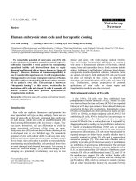
Immunohistochemistry (IHC) (A-C) for TLR9 detected by a mouse monoclonal antibodyFigure 1
Immunohistochemistry (IHC) (A-C) for TLR9 detected by a mouse monoclonal antibody. Adenocarcinoma of the lung (A).
Squamous cell carcinoma of the lung (B). A549 cells (all 600 ×) (C). In situ hybridization (ISH) targeting mRNA of human TLR9
with a digoxigenin-labeled DNA-probe in a squamous cell carcinoma of the lung (600 ×) (D). Immunohistochemical staining of
TLR9-expression-levels in nonmalignant (E) and malignant tissues (F) derived from the same lungs an stained by the use of tis-
sue arrays. Results of RT-PCR targeting TLR9 in cell lines (G). M: molecular-weight marker (MW8, Roche). 1: negative control;
2: A549; 3: NCI-H727; 4: BEAS 2b; 5: Mononuclear cells from a healthy human donor. Confocal laser microscopy of A549 cells
transiently transfected with a GFP-TLR9 plasmid: Cytoplasmic expression of TLR9 is observable in all cells, while successful
transfection led to overexpression of TLR9 resulting in bright GFP signals completely superimposed by the TLR9 antibody sig-
nal (H). Nuclear counterstain was performed with TOTO3.
Respiratory Research 2005, 6:1 />Page 5 of 10
(page number not for citation purposes)
A cytoplasmic localization of TLR9 was confirmed by con-
focal microscopy (fig. 1). This finding is in agreement
with previous studies on the distribution of TLR9 in
RAW264.7 cells [10]. Furthermore, immunostaining of
GFP-TLR9 transfected A549 cells verified the specificity of
the TLR9 antibody: Only those cells which were success-
fully transfected as demonstrated by the GFP-dependent
fluorescence also stained brightly with the TLR9 antibody.
CpG-ODN stimulation reduces spontaneous and tumor
necrosis factor-alpha (TNF-
α
)/Cycloheximide (CHX)-
induced apoptosis
The expression of TLR9 in tumor cells and cell lines rises
up the question, whether this receptor is functional active
in these cells. As shown in figure 2a, CpG-ODN decrease
the rate of spontaneous and induced apoptosis in HeLa
and A549 cells after treatment with TNF-α and CHX.
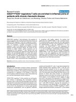
MCP-1 secretion in response to CpG-ODN-stimulation in the presence or absence of TNF-α by HeLa and A549 cells (A)Figure 4
MCP-1 secretion in response to CpG-ODN-stimulation in the presence or absence of TNF-α by HeLa and A549 cells (A).
Data are expressed as the mean ± SD (n = 6). Student's t test was used for statistical analysis. RT-PCR targeting mRNA of
MCP-1 in human non-small cell lung cancer tissue stimulated with CpG-ODN for 24 h (B) (M = pBR322-Msp1). Lanes 2 and 3,
as well as lanes 4 and 5 respectively show results of tissue samples from the same tumors either in the absence or presence of
CpG-ODN.
Respiratory Research 2005, 6:1 />Page 6 of 10
(page number not for citation purposes)
Table 1: Summarized results of immunohistochemistry (IHC) targeting TLR9 in tumor tissues and cell lines.
Entity N* No expression Weak expression Strong expression
Adenocarcinoma of the lung 21 1 7 13
Squamous cell carcinoma of the lung 23 1 14 8
Large cell carcinoma of the lung 3021
Cell lines** 13 0 1 12
Total 60 2 24 34
* Number of analyzed specimens
** See methods
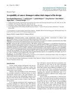
CpG-ODN-stimulation decreases apoptosis in HeLa and A549 cellsFigure 2
CpG-ODN-stimulation decreases apoptosis in HeLa and A549 cells. Cells were stained with Annexin-V after CpG-ODN-stim-
ulation in the presence or absence of TNF-α and CHX after 24 h (A). Data are expressed as the mean ± SD (n = 6). Student's
t test was used for statistical analysis. Representative histograms are shown from experiments with HeLa cells after CpG-
ODN-stimulation in the absence (B) or presence (C) of TNF-α and CHX. Caspase 3 expression in HeLa cells is shown after
incubation with TNF-α and CHX (D). In the presence of CpG-ODN the expression is decreased (E). The percentage of pos-
itive cells in each sample is indicated.
Respiratory Research 2005, 6:1 />Page 7 of 10
(page number not for citation purposes)
Representative histograms demonstrate the detection of
annexin in the presence or absence of CpG-ODN and
TNF-α/CHX (Fig. 2b and 2c). The induction of apoptosis
after stimulation with TNF-α/CHX was further verified by
the expression of active caspase 3 as shown in figure 2d. In
the presence of CpG-ODN the expression was reduced
analogous to the reduction of annexin-staining (Fig. 2e).
Influence of induced apoptosis on TLR9 expression
Here we investigated, whether CpG-ODN can modulate
their own receptor. We found no differences in TLR9
expression with and without CpG-ODN stimulation.
However, in the presence of TNF-α/CHX the expression of
TLR9 was strongly reduced, whereas CpG-ODN stimula-
tion counteracted this downregulation (Fig. 3a and 3b).
Secretion of MCP-1 in response to CpG-ODN and TNF-
α
In order to obtain further information about the func-
tional activity of TLR9 in tumors we studied cytokine
release upon CpG-ODN stimulation. The measurement of
cytokines from stimulated HeLa and A549 cells revealed a
significantly enhanced release of monocyte chemoattract-
ant protein-1 (MCP-1) after 24 h of stimulation in
response to CpG-ODN or TNF-α (Fig. 4a). The production
was further enhanced when stimulated with a combina-
tion of CpG-ODN and TNF-α (Fig. 4a). There was no
effect of CpG-ODN on TNF-α production (data not
shown). To verify the induction of MCP-1 by CpG-ODN
in cell lines we additionally analyzed human tumor tis-
sues by RT-PCR; the results are shown in figure 4b. The rel-
ative amounts of RT-PCR-signals for MCP-1 in relation to
GAPDH were higher in the specimens treated with CpG-
ODN if compared with the controls confirming the results
obtained in cell culture experiments on the tissue level.
Discussion
By application of a novel fixation technique we specify for
the first time the expression of TLR9 protein and mRNA in
a selection of human non small cell lung cancer tissues as
well as cell lines. Stimulation of the TLR-9 expressing cell
lines A549 and HeLa with CpG-ODN showed a marked
antiapoptotic effect. In addition, there was substantially
enhanced release of MCP-1 from the cell lines upon CpG-
ODN stimulation which was also shown in ex vivo experi-
ments. We conclude the expression of a functionally
active TLR9 in human malignant tumors.
The presence of molecules involved in ontogenesis e.g. the
carcinoembryonic antigen (CEA) is frequently observed in
malignant tumors suggesting a kind of "shift-back"
towards earlier developmental stages [26]. The signifi-
cance and underlying mechanisms of this phenomenon
are poorly understood; nevertheless, the detection of such
molecules is used for diagnostic purposes in cancer [27].
The role of TLR in mammalian embryogenesis is
unknown, and thus far there is no evidence for an endog-
enous TLR9 ligand homologous to Spaetzle. Such a ligand
could play a role for the activation of human TLR9.
Whether the expression of TLR9 in human malignant cells
takes advantage of TLR9-function in embryogenesis there-
fore remains unclear.
On the other hand TLR9 in malignant cells could have
similar functions as in cells of the innate and adaptive
TLR9 expression after CpG-ODN-stimulation in HeLa cells: There is no difference in TLR9 expression with and without CpG-ODN-stimulation after 24 h (A)Figure 3
TLR9 expression after CpG-ODN-stimulation in HeLa cells: There is no difference in TLR9 expression with and without CpG-
ODN-stimulation after 24 h (A). CpG-ODN partially inhibit downregulation of TLR9 which is induced by TNF-α and CHX
(B). FI = fluorescence intensity.
Respiratory Research 2005, 6:1 />Page 8 of 10
(page number not for citation purposes)
immune system. In humans TLR9 has been described to
be mainly expressed in B-lymphocytes, monocytes and
plasmacytoid dendritic cells [28]. In addition Platz et al.
reported a weak expression in respiratory epithelial cell
lines and primary epithelial cells [29].
The CpG-ODN sequence M362 used in our study is
known to potently activate TLR9-expressing immune cells
in humans including plasmacytoid dendritic cells and B
cells as shown by Hartmann et al. [25] B cells are induced
to proliferate and secrete immunoglobulin in response to
CpG-ODN, dendritic cells produce a wide array of
cytokines and apoptosis is inhibited [30,31].
These mechanisms are both reflected in the results we
obtained in our study after CpG-ODN stimulation of
malignant cells:
Firstly, stimulation of the A549 and HeLa cells with CpG-
ODN showed an antiapoptotic effect. This was demon-
strated for spontaneous as well as induced apoptosis with
TNF-α and CHX after 24 h. Our observation is consistent
with previous evidence in other cell lines. Yi et al.
demonstrated antiapoptotic effects of CpG-ODN in a
mouse B lymphoma cell line [32], and similar changes
were described in chronic lymphocytic leukemia cells
[33,34]. Previous data of systemic administration of bac-
terial DNA as a single agent in vivo showed anti-tumor
effects. However, this anti-tumor effect appears to be
effective indirectly and is related to enhanced NK cell
activity. In a murine model of lymphoma the immunos-
timulatory effect of CpG-ODN was demonstrated to be
responsible for the observed anti-tumor effects [35]. Car-
pentier et al. have shown that CpG-ODN in vivo induced
rejection of neuroblastoma xenografts [36]. In contrast
CpG-ODN had no effect on survival in mice inoculated
with the 38C13 murine B cell lymphoma. However, a sin-
gle injection of CpG-ODN enhanced the response to anti-
tumor antibody therapy [37]. To what extent the antiap-
optotic effects of CpG-ODN on tumor cells demonstrated
in our study affect the tumorbiology in vivo requires fur-
ther investigation.
Secondly, tumor cell lines (A549 and HeLa) stimulated
with CpG-ODN showed strong secretion of the CC chem-
okine MCP-1. Furthermore a similar effect was observed
in the investigated tumor tissues. Immunostimulatory
properties together with anti-tumor activity of bacterial
DNA were initially reported for a DNA fraction derived
from mycobacteria by Tokunaga and coworkers [38]. It is
known that such DNA induces enhanced production of
various cytokines with anti-tumoral activity in NK cells, B
cells, monocytes, macrophages and dendritic cells, such as
TNF-α, IL-12, and IFN-γ [39]. In our study a substantial
costimulatory effect in addition to CpG-ODN was
achieved using TNF-α. MCP-1 has various biological activ-
ities including the induction of increased cytotoxic activity
of monocytes and NK cells. Transfection of MCP-1 into a
human malignant glioma cell line tested on nude mice
did not reduce the tumor mass but was associated with the
infiltration of large numbers of NK cells and monocytes at
the tumor site [40]. A further study by Nokihara et al. per-
formed with transfection of the MCP-1 gene into human
lung adenocarcinoma cells showed reduced systemic
spread of transfected cells inoculated i.v. in NK cell-intact
severe combined immunodeficient (SCID) mice. These
findings suggest that locally produced MCP-1 suppresses
tumor progression by a NK cell-mediated mechanism
[41]. Thus, apart from the direct activation of immune
cells, the effect of CpG-ODN stimulation on the secretion
of MCP1 by TLR9 expressing tumor cells could possibly
lead to anti-tumoral effects due to an increase of local
MCP1 production which then might lead to attraction of
immune cells. The costimulatory effect of TNF-α as dem-
onstrated in vitro in this study could further enhance this
scenario.
Regarding TLR9 expression in nonmalignant lung tissue
our data confirm the findings of low TLR9 expression in
respiratory cells of Platz et al. [29], who have been work-
ing on single cell preparations. However TLR9 expression
was only seen sporadically weak in nonmalignant lung
tissue.
Biological explanations for the TLR9 expression in malig-
nant cells require further investigations. Three possibili-
ties are conceivable: Either this could represent a
bystander phenomenon, a side effect of a pathway func-
tional to a different purpose. Secondly the upregulation of
TLR9 could be beneficial to the tumor, promoting tumor
cell survival. Thirdly, it even might help immune control
strategies of the organisms an element of a pathway direct-
ing defense mechanisms against malignantly transform-
ing cells. While the first possibility seems unlikely in the
light of our findings of a functionality of the receptor in
various in vitro and ex vivo experiments, our data provide
evidence for the second as well as the third possibility; the
sum effect of these two counteracting mechanisms in an in
vivo setting can not be estimated from these experiments
and could even differ from tumor entity to tumor entity.
Conclusions
In conclusion, we showed in a selection of samples that
human malignant tumors express functionally active
TLR9 and respond to CpG treatment with prolonged sur-
vival and chemokine release. This might influence the
effects of CpG-ODN based anti-tumor therapies. Broad
screening approaches will be worthwhile to further sub-
stantiate these initial results.
Respiratory Research 2005, 6:1 />Page 9 of 10
(page number not for citation purposes)
While recent strategies in tumor-immunology mainly tar-
get a strengthening of the host-defense, we provide
evidence that the malignant cells themselves can be
regarded active players in the complex struggle between
tumor and host. In any case CpG-ODN based anti-tumor
therapies should be reconsidered in the light of our find-
ings since CpG-ODN products are currently in Phase I/II
clinical trials both as a monotherapy and as part of multi-
drug regimens.
Author's contributions
DD carried out the flow cytometry and cytokine assays
and was involved in the design and coordination of the
study and drafting the manuscript. DA and AJU carried
out the confocal microscopy, RT-PCR with cell lines and
were involved in drafting the manuscript. JG was involved
in immunohistochemistry of cell lines and the design of
the study. DB conducted the surgical part of the study. EV
conducted the pathological part of the study and was
involved in the design of the study. KD and PZ conducted
the clinical part of the study and were involved in the
design and coordination of the study. TG performed the
immunohistochemistry, in situ hybridization and RT-PCR
with tissues and conceived of the study. All authors read
and approved the final manuscript.
Acknowledgements
The authors thank H. Kühl, D. Bubritzki, S. Adrian, J. Hofmeister and S.
Ross for excellent technical assistance, Elvira Richter for sequencing the
PCR-products and Maria Manoukian for help with the confocal microscopy.
References
1. Belvin MP, Anderson KV: A conserved signaling pathway: the
Drosophila toll-dorsal pathway. Annu Rev Cell Dev Biol 1996,
12:393-416.
2. Beutler B, Rehli M: Evolution of the TIR, tolls and TLRs: func-
tional inferences from computational biology. Curr Top Micro-
biol Immunol 2002, 270:1-21.
3. Medzhitov R, Janeway C Jr: Innate immunity. N Engl J Med 2000,
343(5):338-344.
4. Aliprantis AO, Yang RB, Mark MR, Suggett S, Devaux B, Radolf JD,
Klimpel GR, Godowski P, Zychlinsky A: Cell activation and apop-
tosis by bacterial lipoproteins through toll-like receptor-2.
Science 1999, 285(5428):736-739.
5. Alexopoulou L, Holt AC, Medzhitov R, Flavell RA: Recognition of
double-stranded RNA and activation of NF-kappaB by Toll-
like receptor 3. Nature 2001, 413(6857):732-738.
6. Poltorak A, He X, Smirnova I, Liu MY, Van Huffel C, Du X, Birdwell
D, Alejos E, Silva M, Galanos C, Freudenberg M, Ricciardi-Castagnoli
P, Layton B, Beutler B: Defective LPS signaling in C3H/HeJ and
C57BL/10ScCr mice: mutations in Tlr4 gene. Science 1998,
282(5396):2085-2088.
7. Hayashi F, Smith KD, Ozinsky A, Hawn TR, Yi EC, Goodlett DR, Eng
JK, Akira S, Underhill DM, Aderem A: The innate immune
response to bacterial flagellin is mediated by Toll-like recep-
tor 5. Nature 2001, 410(6832):1099-1103.
8. Hemmi H, Takeuchi O, Kawai T, Kaisho T, Sato S, Sanjo , Matsumoto
M, Hoshino K, Wagner H, Takeda K, Akira S: A Toll-like receptor
recognizes bacterial DNA. Nature 2000, 408(6813):740-745.
9. Muzio M, Natoli G, Saccani S, Levrero M, Mantovani A: The human
toll signaling pathway: divergence of nuclear factor kappaB
and JNK/SAPK activation upstream of tumor necrosis factor
receptor-associated factor 6 (TRAF6). J Exp Med 1998,
187(12):2097-2101.
10. Ahmad-Nejad P, Hacker H, Rutz M, Bauer S, Vabulas RM, Wagner H:
Bacterial CpG-DNA and lipopolysaccharides activate Toll-
like receptors at distinct cellular compartments. Eur J Immunol
2002, 32(7):1958-1968.
11. Coley WB: The treatment of malignant tumors by repeated
inoculations of erysipelas. With a report of ten original cases.
1893. Clin Orthop 1991:3-11.
12. Shimada S, Yano O, Tokunaga T: In vivo augmentation of natural
killer cell activity with a deoxyribonucleic acid fraction of
BCG. Jpn J Cancer Res 1986, 77(8):808-816.
13. Steinman RM, Dhodapkar M: Active immunization against can-
cer with dendritic cells: the near future. Int J Cancer 2001,
94(4):459-473.
14. Roman M, Martin-Orozco E, Goodman JS, Nguyen MD, Sato Y, Ron-
aghy A, Kornbluth RS, Richman DD, Carson DA, Raz E: Immunos-
timulatory DNA sequences function as T helper-1-
promoting adjuvants. Nat Med 1997, 3(8):849-854.
15. Sparwasser T, Vabulas RM, Villmow B, Lipford GB, Wagner H: Bac-
terial CpG-DNA activates dendritic cells in vivo: T helper
cell-independent cytotoxic T cell responses to soluble
proteins. Eur J Immunol 2000, 30(12):3591-3597.
16. Heckelsmiller K, Beck S, Rall K, Sipos B, Schlamp A, Tuma E, Rothen-
fusser S, Endres S, Hartmann G: Combined dendritic cell- and
CpG oligonucleotide-based immune therapy cures large
murine tumors that resist chemotherapy. Eur J Immunol 2002,
32(11):3235-3245.
17. Okamoto M, Sato M: Toll-like receptor signaling in anti-cancer
immunity. J Med Invest 2003, 50(1–2):9-24.
18. Wooldridge JE, Weiner GJ: CpG DNA and cancer immuno-
therapy: orchestrating the antitumor immune response. Curr
Opin Oncol 2003, 15(6):440-445.
19. Olert J, Wiedorn KH, Goldmann T, Kuhl H, Mehraein Y, Scherthan
H, Niketeghad F, Vollmer E, Muller AM, Muller-Navia J: HOPE fixa-
tion: a novel fixing method and paraffin-embedding tech-
nique for human soft tissues. Pathol Res Pract 2001,
197(12):823-826.
20. Goldmann T, Wiedorn KH, Kuhl H, Olert J, Branscheid D, Pechko-
vsky D, Zissel G, Galle J, Muller-Quernheim J, Vollmer E: Assess-
ment of transcriptional gene activity in situ by application of
HOPE-fixed, paraffin-embedded tissues. Pathol Res Pract 2002,
198(2):91-95.
21. Umland O, Ulmer AJ, Vollmer E, Goldmann T: HOPE fixation of
cytospin preparations of human cells for in situ hybridization
and immunocytochemistry. J Histochem Cytochem 2003,
51(7):977-980.
22. Droemann D, Goldmann T, Branscheid D, Clark R, Dalhoff K, Zabel
P, Vollmer E: Toll-like receptor 2 is expressed by alveolar epi-
thelial cells type II and macrophages in the human lung. His-
tochem Cell Biol 2003, 119(2):103-108.
23. Roche Molecular Biochemicals: Nonradioactive in situ hybridiza-
tion application manual. Roche Diagnostics, Boehringer Mannheim,
Mannheim, Germany. Ref Type: Generic 21996.
24. Goldmann T, Vollmer E, Gerdes J: What's Cooking? Detection of
Important Biomarkers in HOPE-Fixed, Paraffin-Embedded
Tissues Eliminates the Need for Antigen Retrieval. Am J Pathol
2003, 163(6):2638-2640.
25. Hartmann G, Battiany J, Poeck H, Wagner M, Kerkmann M, Lubenow
N, Rothenfusser S, Endres S: Rational design of new CpG oligo-
nucleotides that combine B cell activation with high IFN-
alpha induction in plasmacytoid dendritic cells. Eur J Immunol
2003, 33(6):1633-1641.
26. Gold P, Freedman SO: Demonstration of tumor-specific anti-
gens in human colonic carcinomata by immunological toler-
ance and absorbtion techniques:. J Exp Med 1965, 121:439-462.
27. Grunert F, Luckenbach GA, Haderlie B, Schwarz K, von Kleist S:
Comparison of colon-, lung-, and breast-derived carcinoem-
bryonic antigen and cross-reacting antigens by monoclonal
antibodies and fingerprint analysis. Ann N Y Acad Sci 1983,
417:75-85.
28. Takeshita F, Leifer CA, Gursel I, Ishii KJ, Takeshita S, Gursel M, Klin-
man DM: Cutting edge: Role of Toll-like receptor 9 in CpG
DNA-induced activation of human cells. J Immunol 2001,
167(7):3555-3558.
29. Platz J, Beisswenger C, Dalpke A, Koczulla R, Pinkenburg O, Vogel-
meier C, Bals R: Microbial DNA induces a host defense reac-
Publish with BioMed Central and every
scientist can read your work free of charge
"BioMed Central will be the most significant development for
disseminating the results of biomedical research in our lifetime."
Sir Paul Nurse, Cancer Research UK
Your research papers will be:
available free of charge to the entire biomedical community
peer reviewed and published immediately upon acceptance
cited in PubMed and archived on PubMed Central
yours — you keep the copyright
Submit your manuscript here:
/>BioMedcentral
Respiratory Research 2005, 6:1 />Page 10 of 10
(page number not for citation purposes)
tion of human respiratory epithelial cells. J Immunol 2004,
173(2):1219-1223.
30. Medzhitov R: CpG DNA: security code for host defense. Nat
Immunol 2001, 2(1):15-16.
31. Park Y, Lee SW, Sung YC: Cutting Edge: CpG DNA inhibits den-
dritic cell apoptosis by up-regulating cellular inhibitor of
apoptosis proteins through the phosphatidylinositide-3'-OH
kinase pathway. J Immunol 2002, 168(1):5-8.
32. Yi AK, Hornbeck P, Lafrenz DE, Krieg AM: CpG DNA rescue of
murine B lymphoma cells from anti-IgM-induced growth
arrest and programmed cell death is associated with
increased expression of c-myc and bcl-xL. J Immunol 1996,
157(11):4918-4925.
33. Decker T, Schneller F, Kronschnabl M, Dechow T, Lipford GB, Wag-
ner H, Peschel C: Immunostimulatory CpG-oligonucleotides
induce functional high affinity IL-2 receptors on B-CLL cells:
costimulation with IL-2 results in a highly immunogenic
phenotype. Exp Hematol 2000, 28(5):558-568.
34. Decker T, Schneller F, Sparwasser T, Tretter T, Lipford GB, Wagner
H, Peschel C: Immunostimulatory CpG-oligonucleotides
cause proliferation, cytokine production, and an immuno-
genic phenotype in chronic lymphocytic leukemia B cells.
Blood 2000, 95(3):999-1006.
35. Smith JB, Wickstrom E: Antisense c-myc and immunostimula-
tory oligonucleotide inhibition of tumorigenesis in a murine
B-cell lymphoma transplant model. J Natl Cancer Inst 1998,
90(15):1146-1154.
36. Carpentier AF, Chen L, Maltonti F, Delattre JY: Oligodeoxynucle-
otides containing CpG motifs can induce rejection of a neu-
roblastoma in mice. Cancer Res 1999, 59(21):5429-5432.
37. Wooldridge JE, Ballas Z, Krieg AM, Weiner GJ: Immunostimula-
tory oligodeoxynucleotides containing CpG motifs enhance
the efficacy of monoclonal antibody therapy of lymphoma.
Blood 1997, 89(8):2994-2998.
38. Tokunaga T, Yamamoto H, Shimada S, Abe H, Fukuda T, Fujisawa Y,
Furutani Y, Yano O, Kataoka T, Sudo T: Antitumor activity of
deoxyribonucleic acid fraction from Mycobacterium bovis
BCG. I. Isolation, physicochemical characterization, and
antitumor activity. J Natl Cancer Inst 1984, 72(4):955-962.
39. Ballas ZK, Rasmussen WL, Krieg AM: Induction of NK activity in
murine and human cells by CpG motifs in oligodeoxynucle-
otides and bacterial DNA. J Immunol 1996, 157(5):1840-1845.
40. Nagai M, Masuzawa T: Vaccination with MCP-1 cDNA trans-
fectant on human malignant glioma in nude mice induces
migration of monocytes and NK cells to the tumor. Int
Immunopharmacol 2001, 1(4):657-664.
41. Nokihara H, Yanagawa H, Nishioka Y, Yano S, Mukaida N, Matsu-
shima K, Sone S: Natural killer cell-dependent suppression of
systemic spread of human lung adenocarcinoma cells by
monocyte chemoattractant protein-1 gene transfection in
severe combined immunodeficient mice. Cancer Res 2000,
60(24):7002-7007.