Báo cáo y học: "Dynamic changes of serum SARS-Coronavirus IgG, pulmonary function and radiography in patients recovering from SARS after hospital discharge" ppt
Bạn đang xem bản rút gọn của tài liệu. Xem và tải ngay bản đầy đủ của tài liệu tại đây (312.39 KB, 7 trang )
BioMed Central
Page 1 of 7
(page number not for citation purposes)
Respiratory Research
Open Access
Research
Dynamic changes of serum SARS-Coronavirus IgG, pulmonary
function and radiography in patients recovering from SARS after
hospital discharge
Lixin Xie
1
, Youning Liu*
1
, Baoxing Fan
1
, Yueyong Xiao
2
, Qing Tian
1
,
Liangan Chen
1
, Hong Zhao
2
and Weijun Chen
3
Address:
1
Department of Respiratory Medicine, Chinese PLA General Hospital, 28 Fuxing Road, Beijing, 100853, P.R. China,
2
Department of
Radiology, Chinese PLA General Hospital, 28 Fuxing Road, Beijing, 100853, P.R. China and
3
BGI-GBI Biotech Company, Beijing, P.R. China
Email: Lixin Xie - ; Youning Liu* - ; Baoxing Fan - ;
Yueyong Xiao - ; Qing Tian - ; Liangan Chen - ;
Hong Zhao - ; Weijun Chen -
* Corresponding author
Severe acute respiratory syndrome (SARS)SARS-CoV IgG antibodyPulmonary functionPulmonary fibrosisAvascular necrosis of femoral head
Abstract
Objective: The intent of this study was to examine the recovery of individuals who had been hospitalized for severe acute
respiratory syndrome (SARS) in the year following their discharge from the hospital. Parameters studied included serum levels
of SARS coronavirus (SARS-CoV) IgG antibody, tests of lung function, and imaging data to evaluate changes in lung fibrosis. In
addition, we explored the incidence of femoral head necrosis in some of the individuals recovering from SARS.
Methods: The subjects of this study were 383 clinically diagnosed SARS patients in Beijing, China. They were tested regularly
for serum levels of SARS-CoV IgG antibody and lung function and were given chest X-rays and/or high resolution computerized
tomography (HRCT) examinations at the Chinese PLA General Hospital during the 12 months that followed their release from
the hospital. Those individuals who were found to have lung diffusion abnormities (transfer coefficient for carbon monoxide
[D
L
CO] < 80% of predicted value [pred]) received regular lung function tests and HRCT examinations in the follow-up phase
in order to document the changes in their lung condition. Some patients who complained of joint pain were given magnetic
resonance imaging (MRI) examinations of their femoral heads.
Findings: Of all the subjects, 81.2% (311 of 383 patients) tested positive for serum SARS-CoV IgG. Of those testing positive,
27.3% (85 of 311 patients) were suffering from lung diffusion abnormities (D
L
CO < 80% pred) and 21.5% (67 of 311 patients)
exhibited lung fibrotic changes. In the 12 month duration of this study, all of the 40 patients with lung diffusion abnormities who
were examined exhibited some improvement of lung function and fibrosis detected by radiography. Of the individuals receiving
MRI examinations, 23.1% (18 of 78 patients) showed signs of femoral head necrosis.
Interpretation: The lack of sero-positive SARS-CoV in some individuals suggests that there may have been some misdiagnosed
cases among the subjects included in this study. Of those testing positive, the serum levels of SARS-CoV IgG antibody decreased
significantly during the 12 months after hospital discharge. Additionally, we found that the individuals who had lung fibrosis
showed some spontaneous recovery. Finally, some of the subjects developed femoral head necrosis.
Published: 08 January 2005
Respiratory Research 2005, 6:5 doi:10.1186/1465-9921-6-5
Received: 18 November 2004
Accepted: 08 January 2005
This article is available from: />© 2005 Xie et al; licensee BioMed Central Ltd.
This is an Open Access article distributed under the terms of the Creative Commons Attribution License ( />),
which permits unrestricted use, distribution, and reproduction in any medium, provided the original work is properly cited.
Respiratory Research 2005, 6:5 />Page 2 of 7
(page number not for citation purposes)
Introduction
Severe acute respiratory syndrome (SARS) is a new infec-
tious disease in humans. The first victim of SARS to be
diagnosed was a businessman from the city of Foshan in
Guangdong Province, China. SARS patients may present
with a spectrum of symptoms and signs, ranging from rel-
atively asymptomatic to fulminant pneumonitis and
death [1]. Lung injury caused by the SARS coronavirus
(SARS-CoV) is one of the main clinical manifestations in
SARS patients, significantly affecting their prognosis. A
regular follow-up survey of SARS patients in the convales-
cent phase would be helpful to evaluate any changes in
acquired immune function, pulmonary function, bones
and joints over the course of time. At present, there have
been few reports about the relationship between the prog-
nosis for recovery and the degree of lung injury caused by
the SARS-CoV. In addition, a study of the serum levels of
the specific IgG antibody against SARS-CoV is needed
because it is the major immunologic protection to aid in
recovery and is essential to avoid repeated infection with
SARS-CoV. It has been 14 months since the World Health
Organization officially declared the global outbreak of
SARS to be under control [2]. The present study focused
on the dynamic changes in the IgG antibody levels against
SARS-CoV and in lung lesions in the discharged but recov-
ering SARS patients as measured by lung function and
imaging tests. The phenomenon of femoral head necrosis
was also investigated in those SARS patients who com-
plained of chronic bone and joint pain during the one
year follow-up after discharge from the hospital.
Methods
All of the subjects of this study were discharged from Bei-
jing Xiaotangshan Hospital, Beijing Armed Police Hospi-
tal, and Chinese 309 PLA Hospital, and all gave their
informed consent.
Study Protocol
The subjects of our investigation were 383 clinically diag-
nosed SARS patients in the convalescent phase (160 male
and 223 female, average age 38.2 ± 13.6 years) undergo-
ing testing from May, 2003 to June, 2004. Each clinical
diagnosis was based on the Clinical Diagnosis Standard
for SARS Patients issued by the Ministry of Chinese Public
Health [3]. All participants in the study had met the spec-
ified criteria for discharge from the hospital [4]. On the
first visit, each patient was given a routine pulmonary
function test (ventilation and diffusion function: Sensor-
Medics 2200 pulmonary function test apparatus, U.S.A.),
a chest X-ray examination and serum SARS-CoV specific
antibody (SARS-CoV IgG) test at the Chinese PLA General
Hospital, Beijing, P.R. China. Those individuals suspected
of having pulmonary fibrotic changes received high reso-
lution computerized tomography (HRCT) examination of
their lungs. Individuals who complained of chronic pain
in their bones and joints or who had difficulty walking
received femoral head magnetic resonance imaging (MRI)
examinations.
Each patient returned a month after the first visit followed
by one visit every 3 months. Serum SARS-CoV IgG was
tested at each return visit. If negative results were obtained
twice consecutively, the case was regarded as a misdiagno-
sis and the patient did not undergo a follow-up survey.
Patients with positive SARS-CoV IgG and abnormal pul-
monary diffusion received regular pulmonary function
tests and those showing pulmonary fibrosis in imaging
examinations received further regular HRCT examina-
tions. Some individuals observed to have avascular necro-
sis of the femoral head received MRI examinations 3–6
months later.
Clinical Diagnostic Criteria for the Patients with SARS
Disease in Mainland China [3]
(1) Epidemiological history
(1.1) The individual has a history of close contact with
SARS patients or is part of a cluster of cases of SARS or
there is clinical evidence of having infected other patients.
(1.2) The individual has a history of recent travel to an
area where SARS cases have been reported within 2 weeks
and secondary infected SARS cases have been found.
(2) Symptoms and signs
Acute onset of SARS generally begins with a prodrome of
fever with a temperature >38°C, sometimes accompanied
by chills, myalgia and anthralgia, headaches, and fatigue.
Upper respiratory tract symptoms of catarrh are not prom-
inent, although cough may be present. If present, it is
mainly a dry cough, occasionally with blood streak spu-
tum. Some individuals have chest discomfort, and severe
cases may present with tachypnea, panting, and even res-
piratory distress.
Generally, there are no obvious pulmonary signs among
SARS patients. Wet rales and signs of lung consolidation,
as well as decreased respiration and other signs of pleural
effusion can occasionally be found.
Note: Some patients do not show initial symptoms of
fever, especially those who have had recent surgery or
those having chronic diseases.
(3) Routine laboratory examinations
White blood cell counts are generally normal or below
normal, with decreased absolute lymphocyte counts.
(4) Chest radiological examinations
The typical imaging profile of SARS is of multiple patchy
opacities with bilateral distribution. The opacities are
Respiratory Research 2005, 6:5 />Page 3 of 7
(page number not for citation purposes)
usually ground-glass in appearance, sometimes with air
space consolidation, evolving progressively over the
course of the disease. The evolution is very rapid in some
cases, resulting in the confluence of lesions and large areas
of opacification in a short time. If the chest radiological
examination is negative, reexamination after 1 to 2 days
should be done.
(5) Antibiotic therapy is ineffective
Suspected cases: In accordance with 1.1+2+3, 1.2+2+4 or
2+3+4.
Clinically diagnosed cases: In accordance with 1.1+2+4,
1.2+2+4+5, or 1.2+2+3+4.
SARS-CoV IgG Antibody Test
The SARS-CoV IgG antibody in serum specimens from
recovering SARS patients was assayed by the BGI-GBI Bio-
tech Company with an enzyme-linked immunosorbent
assay (ELISA) kit (No. S20030003, BGI-GBI Biotech Com-
pany, Beijing, P.R. China). The wells containing polysty-
rene microplate strips were coated with two recombinant
SARS-CoV antigens that are well-characterized. Recover-
ing SARS patients' serum samples in the diluent buffer
(1:10) were incubated in the coated wells for 30 min at
37°C and then the wells were washed 5 times with the
washing buffer. The dilutedenzyme-labeled anti-human
IgG (100 µl) was added to the wells and incubated for 20
min at 37°C. The wells were washed 5 times with the
washing buffer. A tetramethyl-benzidine substrate was
then added to each well. The presence of specific antibod-
ies was indicated by a yellow color developing after sub-
strate addition. The reaction was terminated by addition
of hydrochloric acid. The intensity of the color was meas-
ured spectrophotometrically at 450 nm to quantify the
amount of antibody in the specimen. The optical density
(OD) measured was compared with a standard calibra-
tion curve constructed for each lot, yielding concentration
values for the samples. The OD values of both the positive
and negative controls were determined. The threshold
value for IgG was 0.18 OD units, calculated as the mean +
2 standard deviation (SD) levels of the readings given by
1000 control blood donor sera samples. If the OD was
above the threshold value, the sample was considered to
be positive for SARS-CoV IgG [5].
Pulmonary Function Test
Each recovering SARS patient underwent a standard pul-
monary function test (SensorMedics 2200, Yorba Linda,
U.S.A) for forced expiratory volume in 1 second (FEV
1
),
vital capacity (VC), forced vital capacity (FVC), total lung
capacity (TLC), transfer coefficient for carbon monoxide
(D
L
CO), and carbon monoxide diffusion constant
(D
L
CO/V
A
) measured by means of the single-breath test.
The hemoglobin level was also measured to adjust the
D
L
CO value. The results were compared with those of age-
and sex-matched controls and expressed as a percentage of
predicted values. Pulmonary function was regarded as
abnormal when the D
L
CO was less than 80% of predicted
values (pred). This was considered a diffusion deficit [6].
Chest Radiography and Evaluation
Frontal chest X-ray radiographs (CXR) were obtained at
the first follow-up visit for each recovering SARS patient.
If abnormities were found in the CXR or if the D
L
CO was
<80% pred despite a normal CXR, the patient was sent for
HRCT scanning (GE Light Speed, GE, U.S.A. 1-mm section
in thickness with a 10-mm gap, supine position, scanning
during inspiration, 1 second per scan, 140 kv, 200 mA).
All CXR and HRCT images were assessed by three radiolo-
gists via a viewing console. The three radiologists were
aware of the patients' clinical diagnosis at the time of their
review of the radiographs. The final conclusions were
established by consensus. Each segment of the lung was
reviewed for ground-glass opacification, interstitial thick-
ening, bronchiectasis, and architectural distortion. Abnor-
malities were magnified by means of a zoom function and
were examined for intralobular interstitial, interlobular
septal, or peribronchovascular interstitial thickening.
Attention was also paid to the presence or absence of nod-
ules or masses, cavitation or calcification, and emphy-
sema. The presence of parenchymal bands, irregular
interfaces (bronchovascular, pleural, or mediastinal),
thickened interstitium, and traction bronchiectasis were
considered as evidence of fibrotic changes [7].
Magnetic Resonance Imaging (MRI) Examination
All MRI examinations were done using a 1.5 T Signa CVi
imager (GE Medical Systems, Milwaukee, WI, U.S.A.). For
the patients who complained of chronic bone and joint
pain, coronal T
1
-weighted (spin echo; time to repetition
[TR], 440–500; time to echo [TE) 11–14] scans of the hips
were done. If there were any abnormalities noted in the
T
1
-weighted images, further T
1
-weighted sagittal images
and coronal short tau inversion recovery (inversion time
145, TR 3500–5000, TE 80–120) or turbo-spin-echo T
2
-
weighted images with fat suppression (TR 2500–3000, TE
80–120) were obtained. Images 3 mm thick were
obtained for the coronal studies, and 4 mm thick images
were obtained for the sagittal studies. Osteonecrosis was
diagnosed by the presence of a band of low signal inten-
sity in T
1
-weighted images [8].
Statistical Analysis
All data were expressed as the ± SD unless otherwise
indicated. Statistical analyses were done by one-way anal-
ysis of variance (ANOVA), Student-Newman-Keuls, and
Chi-square test for multiple comparisons. We used the
STATA™ 7.0 statistical analysis software for Windows
(STATA Statistical Software, Inc., U.S.A.) for evaluating the
X
Respiratory Research 2005, 6:5 />Page 4 of 7
(page number not for citation purposes)
results of our study. With each statistical test, the criterion
for significance was a p value of less than 0.05.
Results
The interval from hospital discharge to the first follow-up
visit was 45.0 ± 20.7 days (Range: 11–104 days). Of the
383 individuals participating in our study, 311 patients
(81.2%) tested positive for SARS-CoV IgG and 72 (18.8%)
tested negative. (All patientswere tested twice for SARS-
CoV IgG.) Of these, 33 patients (13 male and 20 female,
average age 35.7 ± 12.1) with positive SARS-CoV IgG and
abnormal pulmonary diffusion received regular follow-up
examinations each month, from June to December in
2003, and every two months, from January to June in
2004. Tables 1 and 2 show the dynamic changes of SARS-
CoV IgG in patients with positive tests for SARS-CoV IgG
within the year after discharge, indicating that the serum
SARS-CoV IgG remained at high levels, although it
decreased significantly over the course of time.
There were 88 individuals (23.0%) with abnormal D
L
CO
among the 383 patients participating in our study. Of the
311 individuals testing positive for SARS-CoV IgG, there
were 85 with abnormal D
L
CO (27.3%, 85/311), in con-
trast to just 3 cases with abnormal D
L
CO among the 72
subjects testing negative for SARS-CoV IgG (4.2%, 3/72).
There was a statistically significant difference between
positive and negative SARS-CoV IgG groups in D
L
CO val-
ues (table 3).
Among the 85 patients (29 male and 56 female, average
age 42.2 ± 11.9 years) with abnormal D
L
CO and positive
SARS-CoV IgG, 40 individuals received pulmonary func-
tion tests 4 times within the year at 42.0 ± 10.4, 70.0 ±
11.8 and 155.1 ± 42.9 day intervals. Among these 40
patients, there were 23 who exhibited abnormal D
L
CO at
their second pulmonary function examination, 23 at the
third examination, and 20 at the fourth examination
(table 4).
.
Pulmonary fibrosis was detected by CXR and confirmed
by HRCT examination in 72 SARS patients in the conva-
lescent phase. Among these, there were 4 individuals with
negative and 67 with positive SARS-CoV IgG. Of the 40
patients who received HRCT examinations at least 4
times, all showed improvement in the fibrotic condition
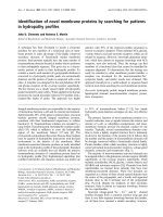
(Figure 1).
Of the 311 convalescent SARS patients with sero-positive
SARS-CoV IgG, 78 received femoral head MRI examina-
tions. The Imaging showed that 18 of these patients
(23.1%, 18/78) had avascular necrosis of the femoral
head. Of these 18 individuals, 8 had avascular necrosis of
Table 1: Dynamic changes of serum SARS-CoV IgG antibody
levels in patients recovering from SARS
Samples (n) ± SD (OD units)
May, 2003 35 1.240 ± 0.350
June, 2003 74 1.087 ± 0.284
July, 2003 172 1.203 ± 0.306
Aug., 2003 152 1.061 ± 0.376
Sept., 2003 123 1.105 ± 0.378
Oct., 2003 35 1.097 ± 0.282
Nov., 2003 77 0.835 ± 0.327†‡§¶*
Dec., 2003 35 0.829 ± 0.232†§*
Jan.–Feb., 2004 67 0.737 ± 0.169†‡§¶*#
Mar–Apr, 2004 34 0.678 ± 0.179†‡§¶*#
May–June, 2004 46 0.621 ± 0.181†‡§¶*#
F value 30.62
p value 0.0000
Note: Statistical analyses were done by one-way analysis of variance
(ANOVA) and Student-Newman-Keuls for multiple comparisons, and
values are given as mean ± SD;
† p < 0.05 vs. SARS-CoV IgG antibody results in May, 2003.
‡ p < 0.05 vs. SARS-CoV IgG antibody results in June, 2003.
§ p < 0.05 vs. SARS-CoV IgG antibody results in July, 2003.
¶ p < 0.05 vs. SARS-CoV IgG antibody results in August, 2003.
* p < 0.05 vs. SARS-CoV IgG antibody results in Sepember, 2003.
# p < 0.05 vs. SARS-CoV IgG antibody results in October, 2003.
Table 2: Dynamic changes of serum SARS-CoV IgG antibody
levels in 33 regular follow-up examinations of patients
recovering from SARS
Samples (n) ± SD (OD units)
June, 2003 33 1.104 ± 0.267
July, 2003 33 1.325 ± 0.357
Aug., 2003 33 1.092 ± 0.249
Sept., 2003 33 1.121 ± 0.432
Oct., 2003 33 1.056 ± 0.309
Nov., 2003 33 0.895 ± 0.203‡¶
Dec., 2003 33 0.800 ± 0.170†‡§¶
Jan.–Feb., 2004 33 0.726 ± 0.163†‡§¶*
Mar–Apr, 2004 33 0.675 ± 0.181†‡§¶*
May–June, 2004 33 0.610 ± 0.167†‡§¶*#
F value 25.69
p value 0.0000
Note: Statistical analyses were done by one-way analysis of variance
(ANOVA) and Student-Newman-Keuls for multiple comparisons, and
values are given as mean ± SD;
† p < 0.05 vs. SARS-CoV IgG antibody results in June, 2003.
‡ p < 0.05 vs. SARS-CoV IgG antibody results in July, 2003.
§ p < 0.05 vs. SARS-CoV IgG antibody results in August, 2003.
¶ p < 0.05 vs. SARS-CoV IgG antibody results in September, 2003.
* p < 0.05 vs. SARS-CoV IgG antibody results in October, 2003.
# p < 0.05 vs. SARS-CoV IgG antibody results in November, 2003.
X
X
Respiratory Research 2005, 6:5 />Page 5 of 7
(page number not for citation purposes)
both femoral heads and 10 had avascular necrosis of one
femoral head. Ten of the 18 patients showed first stage
changes and 8 had secondary changes. During the 3–6
month follow-up visits for these individuals, there were
no obvious changes in the avascular necrosis for these
patient.
Discussion
Since the outbreak of SARS at the end of 2002, despite the
great efforts that have been extended, the mechanisms,
clinical characteristics, prognosis and effective therapeu-
tics for this disease have not been adequately clarified.
Both the SARS virus itself and the anti-viral therapy (such
as high-dose glucocorticoids) used in treatment can cause
various degrees of toxicity and side effects, including pul-
monary fibrosis and avascular necrosis of the femoral
head, even in the convalescent phase. Follow-up surveys
of SARS patients in the convalescent phase are needed for
recognizing the clinical characteristics of this disease and
reevaluation of the therapeutic treatments [2,7].
In our study, 72 individuals (18.8%) showed negative
results in the SARS-CoV IgG antibody test for at least two
tests, suggesting that there may have been misdiagnosis of
some clinically diagnosed SARS patients. Comparison of
the Chinese clinical diagnosis standard (published April,
2003) [3] to the Center for Disease Control (CDC) SARS
case definition (published April 30, 2003) [9], indicates
that both of them emphasize the importance of epidemi-
ological history, clinical manifestations and chest radio-
logical changes for the clinical diagnosis of SARS disease.
The CDC SARS case definition especially emphasizes the
importance of laboratory criteria for the confirmation of a
SARS diagnosis. This is accomplished by detecting the
dynamic changes in the titration of specific antibodies
against SARS CoV and positive detection of SARS-CoV
RNA by PCR. In contrast, the Chinese clinical diagnosis
standard did not mention the importance of a laboratory
SARS-CoV test for the confirmation of a SARS diagnosis.
This might have resulted in the misdiagnosis of SARS in
some cases. During follow-up examinations, we found
that those individuals with positive SARS-CoV IgG
remained positive for a year, although the level of the anti-
body decreased gradually. Therefore, those inoculated
with a SARS vaccine or infected by the SARS virus might
not receive lifetime immunity, but only immunity for a
The results of chest HRCT examination in a SARS patient in the convalescent phase, showing marked reversal of pulmonary fibrosisFigure 1
The results of chest HRCT examination in a SARS patient in the convalescent phase, showing marked reversal of pulmonary
fibrosis.
Respiratory Research 2005, 6:5 />Page 6 of 7
(page number not for citation purposes)
limited duration. Certainly, our findings must be con-
firmed by further studies [7,8].
By regular examination of pulmonary function and CXR,
we found that those with pulmonary fibrotic changes
were able to heal on their own. The fibrotic tissue was
absorbed and pulmonary diffusion and VC improved
with time, suggesting that the mechanism of lung injury
and lung fibrosis caused by the SARS-CoV may have a dif-
ferent pathophysiological process compared to other lung
diseases, such as idiopathic pulmonary fibrosis or pulmo-
nary fibrosis secondary to adult respiratory stress syn-
drome. The reason is not clear. However, in our follow-up
study, we found some ground-glass-like changes in the
HRCT images from SARS patients one year after discharge.
This result shows that changes in the lung can still be
observed in convalescents [7,9].
Recent concern has focused on a complication of SARS in
the convalescent phase, when avascular necrosis develops
on the femoral head. The morbidity of this condition is
reported to be 15% to 30% in some SARS patients in
Mainland China [8,10]. Among the 78 patients receiving
an MRI examination, there were 18 cases of complicated
necrosis of the femoral head to different degrees. The
causes of this complication include SARS itself and the
drugs (such as glucocorticoids) used in treatment, with
the latter being more important than the former [11-13].
We didn't find any worsening or improvement of the avas-
cular necrosis of the femoral head in these patients during
our follow-up examinations. Although most patients
received magnetotherapy, hyperbaric oxygen chamber
therapy, local kerotherapy and Chinese traditional medi-
cine to promote local blood circulation, there was no
apparent short-term therapeutic effectiveness of these
methods for recovery of the femoral head.
In conclusion, SARS, as a new disease, remains unfamiliar
to mankind. It has high rates of morbidity and mortality
in the acute phase. A significant proportion of patients
surviving the acute illness have impairment in their over-
all functional capacity and health status in the convales-
cent phase after discharge from the hospital. Follow-up
surveys of SARS patients in the convalescent phase are
needed to understand the clinical characteristics of this
disease. Our findings suggest that follow up studies of
these patients are required for a longer duration, includ-
ing comprehensive assessments for detection and appro-
priate management of any persistent or emerging
sequelae. These types of investigation may facilitate the
search for effective therapeutics and aid in ultimately con-
quering this disease.
Acknowledgments
We are thankful for the research funding from the National High Technol-
ogy Research and Development Program of China (863 Program)
Table 3: D
L
CO results for the convalescent SARS patients with sero-positive or sero-negative SARS-CoV IgG
Positive Negative Total X
2
P value
D
L
CO normal cases 226 69 295
D
L
CO abnormal cases 85 3 88
Total 311 72 383 17.7269 0.000
Note: Analyzed with Chi-square test.
Table 4: Pulmonary function test results from the 4 follow-up examinations of 40 convalescent SARS patients ( ± SD)
Follow-up* VC(% pred) FEV
1
(% pred) D
L
CO(% pred) D
L
CO/V
A
(% pred)
Two months 87 ± 15 (51~114) 83 ± 13 (60~108) 69 ± 9 (47~79) 95 ± 14 (58~123)
Four months 94 ± 14 (61~123) 90 ± 13 (65~121) 76 ± 11 (48~94) 99 ± 14 (67~126)
Six months 100 ± 15† (66~136) 93 ± 12† (66~114) 76 ± 11† (52~98) 97 ± 14 (62~129)
Eleven months 103 ± 15† (66~142) 96 ± 11† (67~115) 79 ± 12† (56~98) 97 ± 14 (59~128)
F value 9.23 7.84 6.15 0.63
P value 0.0000 0.0001 0.0006 0.5936
Note: *: Indicating as time after discharge from acute illness.
Statistical analyses were done by one-way analysis of variance (ANOVA) and Student-Newman-Keuls for multiple comparisons, and values are given
as mean ± SD;
†: Compared to those in the first follow-up exam, p < 0.05.
X
Publish with BioMed Central and every
scientist can read your work free of charge
"BioMed Central will be the most significant development for
disseminating the results of biomedical research in our lifetime."
Sir Paul Nurse, Cancer Research UK
Your research papers will be:
available free of charge to the entire biomedical community
peer reviewed and published immediately upon acceptance
cited in PubMed and archived on PubMed Central
yours — you keep the copyright
Submit your manuscript here:
/>BioMedcentral
Respiratory Research 2005, 6:5 />Page 7 of 7
(page number not for citation purposes)
2003AA208107 from Ministry of Science and Technology, the People's
Republic of China.
References
1. Chinese Respiratory Association: Consensus for the diagnosis
standard and management of severe acute respiratory
syndrome. Zhonghua Jie He He Hu Xi Za Zhi 2003, 26:323-325.
2. Chinese Medical Association: Consensus for the management of
severe acute respiratory syndrome. Chin Med J 2003,
116:1603-1635.
3. Ministry of Chinese Public Health: Clinical diagnosis consulting
criteria for the patients with severe acute respiratory syn-
drome disease (Apr 4, 2003). [ />open.aspx?n_id=7310&seq=0].
4. Ministry of Chinese Public Health: Diagnosis standard, treatment
proposal and discharge consulting criteria for severe acute
respiratory syndrome. [ />open.aspx?n_id=7286&seq=0].
5. Chen W, Xu Z, Mu J, Yang L, Gan H, Mu F, Fan B, He B, Huang S, You
B, Yang Y, Tang X, Qiu L, Qiu Y, Wen J, Fang J, Wang J: Antibody
response and viraemia during the course of severe acute res-
piratory syndrome (SARS)-associated coronavirus infection.
J Med Microbiol 2004, 53:435-438.
6. Xie LX, Liu YN, Fan BX, Chen LA, Hao FY, Cao L, Tian Q, Ma L:
Prognostic Analysis of Serum SARS-Co V IgG antibody,
Lung function and Lung Radiographic Changes of SARS
Patients in Six Months after Discharge. Chin PLA Med J 2004,
29:762-764.
7. Antonio GE, Wong KT, Hui DSC, Wu A, Lee N, Yuen EH, Leung CB,
Rainer TH, Cameron P, Chung SS, Sung JJ, Ahuja AT: Thin-section
CT in patients with severe acute respiratory syndrome fol-
lowing hospital discharge: preliminary experience. Radiology
2003, 228:810-815.
8. Li YM, Wang SX, Gao HS, Wang JG, Wei CS, Chen LM, Hui WL, Yuan
SL, Jiao ZS, Yang Z, Su B: Factors of avascular necrosis of femo-
ral head and osteoporosis in SARS patients' convalescence.
Zhonghua Yi Xue Za Zhi 2004, 84:1348-1353.
9. Shichijo S, Keicho N, Long HT, Quy T, Phi NC, Ha LD, Ban VV,
Itoyama S, Hu CJ, Komatsu N, Kirikae T, Kirikae F, Shirasawa S, Kaji
M, Fukuda T, Sata M, Kuratsuji T, Itoh K, Sasazuki T: Assessment of
synthetic peptides of severe acute respiratory syndrome
coronavirus recognized by long-lasting immunity. Tissue
Antigens 2004, 64:600-607.
10. Chan CW, Chiu WK, Chan CC, Chow EY, Cheung HM, Ip PL:
Osteonecrosis in children with severe acute respiratory
syndrome. Pediatr Infect Dis J 2004, 23:888-890.
11. Hong N, Du XK: Avascular necrosis of bone in severe acute
respiratory syndrome. Clin Radiol 2004, 59:602-608.
12. Chan KS, Zheng JP, Mok YW, Li YM, Liu YN, Chu CM, Ip MS: SARS:
prognosis, outcome and sequelae. Respirology 2003,
8(Suppl):S36-S40.
13. Koo KH, Kim R, Kim YS, Ahn IO, Cho SH, Song HR, Park YS, Kim H,
Wang GJ: Risk period for developing osteonecrosis of the fem-
oral head in patients on steroid treatment. Clin Rheumatol 2002,
21:299-303.