Báo cáo y học: " Local therapy with CpG motifs in a murine model of allergic airway inflammation in IFN-β knock-out mice" doc
Bạn đang xem bản rút gọn của tài liệu. Xem và tải ngay bản đầy đủ của tài liệu tại đây (518.36 KB, 12 trang )
BioMed Central
Page 1 of 12
(page number not for citation purposes)
Respiratory Research
Open Access
Research
Local therapy with CpG motifs in a murine model of allergic airway
inflammation in IFN-β knock-out mice
Victor Matheu*
1,2
, Alexandra Treschow
1
, Ingrid Teige
1
, Vaidrius Navikas
1
and
Shohreh Issazadeh-Navikas
1
Address:
1
Section of Medical Inflammation Research, Department of Cell & Molecular Biology; Lund University; Sweden and
2
Fundación Rafael
Clavijo de Investigación Biomédica, Tenerife, Spain
Email: Victor Matheu* - ; Alexandra Treschow - ;
Ingrid Teige - ; Vaidrius Navikas - ; Shohreh Issazadeh-
Navikas -
* Corresponding author
IFN-βCpG motifsallergyasthmainflammationsynovitisarthritiseosinophilIFN-γTh1-responseknockoutlung
Abstract
Background: CpG oligodeoxynucleotides (CpG-ODN) are capable of inducing high amounts of
type I IFNs with many immunomodulatory properties. Furthermore, type-I IFNs have been
proposed to play a key role in mediating effects of CpG-ODN. The precise role of IFN-β in the
immunomodulatory effects of CpG-ODN is not known.
Objective: Here, we aimed to elucidate the role of IFN-β in the anti-allergic effect of CpG motifs.
Methods: We assessed the immune response in OVA-primed/OVA-challenged IFN-β knockout (-
/-) mice compared to wild type (WT) control, after intranasal and systemic treatment with
synthetic CpG motifs.
Results: Vaccination with CpG-ODN reduced the number of cells in airways of OVA-sensitized
WT but not IFN-β-/- mice. Although airway eosinophilia was reduced in both treated groups, they
were significantly higher in IFN-β
-
/- mice. Other inflammatory cells, such as lymphocytes and
macrophages were enhanced in airways by CpG treatment in IFN-β-/- mice. The ratio of IFN-γ/IL-
4 cytokines in airways was significantly skewed to a Th1 response in WT compared to IFN-β
-
/-
group. In contrast, IL-4 and IgE were reduced with no differences between groups. Ag-specific T-
cell proliferation, Th1-cytokines such as IFN-γ, IL-2 and also IL-12 were significantly lower in IFN-
β-/- mice. Surprisingly, we discovered that intranasal treatment of mice with CpG-ODN results in
mild synovitis particularly in IFN-β-/- mice.
Conclusion: Our results indicate that induction of Th1 response by therapy with CpG-ODN is
only slightly and partially dependent on IFN-β, while IFN-β is not an absolute requirement for
suppression of airway eosinophilia and IgE. Furthermore, our finding of mild synovitis is a warning
for possible negative effects of CpG-ODN vaccination.
Published: 05 March 2005
Respiratory Research 2005, 6:25 doi:10.1186/1465-9921-6-25
Received: 28 June 2004
Accepted: 05 March 2005
This article is available from: />© 2005 Matheu et al; licensee BioMed Central Ltd.
This is an Open Access article distributed under the terms of the Creative Commons Attribution License ( />),
which permits unrestricted use, distribution, and reproduction in any medium, provided the original work is properly cited.
Respiratory Research 2005, 6:25 />Page 2 of 12
(page number not for citation purposes)
Introduction
Allergic diseases are characterized by elevated serum IgE,
an inflammatory reaction with increased number of eosi-
nophils, mast cells and an adaptative immune responses
orchestrated by Th2-like CD4+ memory T cells secreting
an array of cytokines such as IL-4, IL-5 and IL-13. Thus,
there are major efforts focused on a therapeutic treatment
which will decrease the Th2 profile and/or re-direct the
immune response from a Th2, IgE-mediated allergic
hypersensitivity reaction towards the more favorable Th1
response. IL-12 and IFN-γ are of primary importance in
modulating the Th1/Th2 balance. IFN-γ has been shown
to attenuate eosinophil recruitment[1], and also inhibit
the development of secondary allergic response [2-4].
There has also been extensive research into therapeutic
use of IL-12[5]. However, difficulties with precise dosing
and toxicity associated with the direct administration of
these cytokines may preclude their therapeutic
application.
Another approach is to use natural up-regulators to ele-
vate endogenous levels of IL-12 or IFN-γ. Many microbial
products, including heat-killed bacteria and CpG motifs
can up-regulate Th1 cytokines. Oligodeoxynucleotides
(ODN) containing unmethylated cytosine-guanine
motifs (CpG) have powerful immunomodulatory activity
in human and murine lymphocytes in both Th1 and Th2
associated diseases [6-12]. It is believed that CpG exert
their effect through antigen presenting cells by inducing
cytokines such as TNF-alpha, IL-12, IL-18, and IFNs
[9,13,14].
Type I IFNs have been proposed as mediators of immu-
nomodulatory effects of CpG oligonucleotides [15].
Importantly, some studies have suggested that endog-
enous type I IFN might contribute to the downregulation
of eosinophil infiltration in murine asthma model [16].
Furthermore, reduced inflammatory infiltration and IgE
production have been shown after administration of
recombinant IFN-β[17,18]. We have recently demon-
strated that lung eosinophilic inflammatory response was
exacerbated by the lack of IFN-β gene[19]. Even though it
is believed that immunomodulatory effects of CpG-ODN
are mediated by type I IFNs, the relative role of IFN-β has
not been defined.
In this report, we examined the role of IFN-β in the
immune response after CpG treatment in a murine model
of allergic inflammation. Our results indicate that induc-
tion of Th1 response by therapy with CpG-ODN is par-
tially dependent on IFN-β, while IFN-β is not an absolute
requirement for suppression of eosinophilia and IgE.
Materials and methods
Animals
Groups of pathogen-free female[20,21] 8-10-week-old,
17-20 g, B10.RIII mice (n = 5 mice per group) were used
in the experiments. IFN-β deficient mice (IFN-β-/-) were
kindly provided by Dr Leanderson[22]. Genotyping of the
offspring has been described before[23]. All animal care
and experimentation were conducted at the animal unit of
Medical Inflammation Research in Lund in accordance
with the current protocols in Lund University.
Induction of disease and treatment protocol
Immunization and allergen challenge of the mice were
carried out according to a short term allergy model proto-
col by Sur and colleagues [24] with slight modification.
Mice were sensitized by i.p. injection on days 0 and 4 with
OVA 50 µg (Sigma Chemical Co., St Louis, Mo), with 5 mg
alum (Sigma Chemical Co.). At day 14 and 16 after
immunization, mice were challenged with 50 µg of OVA
plus 5 µg of CpG-ODN (Scandinavian Gene Synthesis AB,
Köping, Sweden) delivered through the airways as intra-
nasal drops after light anesthesia. Control mice were
immunized with 5 mg alum with PBS, and challenge with
PBS using the same schedule as OVA immunized mice.
Our previous studies have confirmed that control mice
did not show any remarkable allergy changes[19]. The
ODNs were designed using published sequences[8,25]
consisting of a single-stranded phosphorothioate-modi-
fied ODNs with 22 bases containing two CpG motifs (5'-
TGACTGTGAACGTTCGAGATGA-3'), highly purified with
undetectable levels of LPS (detection limit: 1 ng/mg
DNA): and were dissolved in PBS with a final concentra-
tion of 1 µg/µl [11]. Mice received either 5 µg of CpG-
ODN in PBS or PBS alone intranasally in conjunction
with OVA challenges. On day 17 (i.e. 24 h after the last
challenge) mice were assessed for lung allergic inflamma-
tory response.
In the prevention study (vaccination), mice were pre-
treated i.p. with 5 µg of CpG-ODN in PBS on day 0. On
the same day, mice were sensitized by i.p. injection with
OVA complexed with 5 mg alum (Sigma). On day 4 mice
were injected i.p. OVA (50 µg) in Alum (5 mg). On days
14 and 16 after immunization mice were challenged with
50 µg of OVA delivered through the airways as intranasal
drops after light anesthesia. On day 17 mice were assessed
for lung allergic inflammatory response, 24 hours (h)
after the last challenge.
Bronchoalveolar lavage Fluid (BALF)
Mice were deeply anesthetized with an ip injection of 0.2
ml avertin (20 mg/ml; 2,2,2 tribromoethanol, Sigma-
Aldrich) and sacrificed 24 hours after the last OVA expo-
sure. After thoracotomy, the trachea was cannulated and
BAL was collected twice with 0.5 mL of PBS and the
Respiratory Research 2005, 6:25 />Page 3 of 12
(page number not for citation purposes)
collected fluid was pooled. Total cell counts were deter-
mined using an automated hemocytometer (Sysmex
CDA-500, Toa Medical Electronics CO., Ltd., Kobey,
Japan), and the fluid was centrifuged (1.000 rpm, 4°C, 10
min). The supernatant was used to determine the airway
cytokine and IgE levels contents. The cells were applied to
slides using a cytospin apparatus (Auto-smear CF-12DE,
Sakura Finetek Europe BV, Zoeterwoude, The Nether-
lands) and were stained with May-Grunwald-Giemsa
staining. Eosinophils were specifically detected by histo-
chemical staining of cyanide-resistant eosinophil peroxi-
dase activity (CREPA) using as substrate 3,3
diaminobenzidine tetrahydrochlorhid (DAB), as
described before[26]. Briefly, samples were dried over-
night at room temperature and fixed with 4% paraformal-
dehide for 5 min and PBS for 2 min. Then, samples were
incubated in PBS buffer with DAB 60%, H
2
O
2
30% and
NaCN 120% for 7 min. After washing with PBS, samples
were counterstained with hemtoxiline 30" and mounted
with Kaiser medium (Merck, Darmstadt, Germany). Eosi-
nophils were easily detected by its dark brown color. The
slides were examined by light microscopy (×40 magnifica-
tion) in a blinded fashion counting at least 400 cells per
slide
Allergen specific T cell proliferation
At the time of sacrifice spleens were dissected and a single
cell suspensions from each mouse was prepared in DMEM
with glutamax I (Gibco BRL, Life Technologies), supple-
mented with 10% heat-inactivated fetal calf serum, 10
mmol/l HEPES, 50 mmol/l β-mercaptoethanol, 100 U/ml
penicillin G, and 100 µg/ml streptomycin. Cells were cul-
tured (5 × 10
6
/ml) in triplicates in 96-well flat-bottomed
plates at 37°C, 5% CO2 in a humidified incubator. Cells
were cultured in absence or presence of OVA (111 µM),
CpG-ODN (1 µg/ml) or concavalin A (4 µg/ml).
3
H-thy-
midine (100 µCi/ml) was added 54 h later, and after a fur-
ther 18 hr later incubation, a beta-scintillation counter
measured incorporation.
Cytokine Assays
Splenocytes were isolated as described and incubated for
48 h with or without OVA (Sigma-Aldrich) (111 µM) in
48-well plates. Enzyme immunoassays were performed as
described before[23,27] using monoclonal Ab (anti-IL-2,
anti-IL-4, anti-IL-5, anti-IL-12, anti-IFN-γ (BD Pharmin-
gen, San Diego, CA, USA) and reading by chemilumines-
cence (Victor
®
; 1420 Multilabel Counter
©
, Wallac Oy; EG
& G Turku, Finland).
Determination of total and OVA-specific IgE levels
Mice were bled at the time of sacrifice. A sandwich ELISA
(BD Pharmingen) was used to measure levels of IgG and
IgE as described previously[28]. To determine OVA-spe-
cific IgE plates were incubated with OVA 10 µg/ml in PBS
buffer (pH 7.'5). Procedure was the same as total IgE.
Standard curve was performed with sera with known lev-
els of specific IgE as it has been published before [29].
Briefly, real concentration of specific IgE in ng/ml of a
pooled serum was determined indirectly by absorption of
50 µl of serum with either conjugated BSA in Sepharose
(Pharmacia, Uppsala, Suecia) or conjugated OVA in
Sepharose. Total IgE ELISA, as mentioned before, deter-
mined the level of not absorbed specific IgE. The percent-
age of OVA-specific IgE was calculated by reciprocal value
of: (IgE not absorbed by OVA-Sepharose/IgE not
absorbed by BSA-Sepharose) × 100. The result of a pool of
sera from several immunized mice by this method was
402 ng/ml of OVA-specífica IgE. In next experiments this
serum was used as standard pattern. For that, plates were
coated with OVA (10 µg/ml) overnight 4°C and blocked
with 1% BSA in PBS 1 h room temperature. The remainder
steps were performed as total IgE ELISA, as described
before.
Flow cytometry
At time of sacrifice spleens were removed and a single cell
suspension was made, cells were then lysed with 0.84%
NH
3
Cl
2
and washed in PBS with 1% BSA and 0.01%
sodium azide. After blocking Fc receptors, using 24.G2
(from our hybridoma collection), cells were stained with
the following antibodies (BD PharMingen); PE conju-
gated anti-B7.1 (clone 16-10A1), FITC conjugated anti-
B7.2 (GL1), cytochrome conjugated anti-B220 (RA3-
6B2), APC conjugated anti-Thy1.2 (53-2.1), PE conju-
gated anti-CD4 (H129.19), cytochrome conjugated anti-
CD8 (53-6.7). The cells were then analyzed by flow
cytometry FACSort (Becton Dickinson, Franklin Lakes, NJ,
USA), using the BD Cell-Quest™ Pro, Version 4.0.1 soft-
ware (Becton Dickinson). Three individuals per time
point and group were analyzed. The program then dis-
plays the percentage of events, which express the CD86
molecule and this percentage is the compared between
the groups.
Clinical and Histological analysis of joints for arthritis
Seventeen days post CpG-ODN or control vaccination,
paws were visually assessed looking for swelling or defor-
mation with redness in one joint, several joints or severe
swelling of the entire paw and/or ankylosis[30]. Then,
mice were sacrificed and paws were dissected and were
fixed in 4% formaldehyde, decalcified with EDTA (for 2–
3 weeks), embedded in paraffin, sectioned at 5µm and
stained with hematoxylin and erythrosine. Approxi-
mately, 20–30 sections were made from each paw (2 paws
per mouse, i.e. front and back paws). The sections were
then evaluated blindly for pathological changes in joints
(synovitis, erosion or destruction)[31].
Respiratory Research 2005, 6:25 />Page 4 of 12
(page number not for citation purposes)
Statistic analysis
The significance of changes was evaluated using Mann-
Whitney U test. Significance was assumed at p values ≤
0.05.
Results
Treatment with different dose of intranasal CpG-ODN
showed similar results
The percentage of local eosinophils in airways was
increased after immunization and challenge with OVA in
BALF of WT and IFN-β-/- compared to non immunized
mice. Preliminary data with different dose of CpG admin-
istered intranasally with OVA (5 µg, 10 µg or 20 µg) to
both strain of mice resulted in similar reduction of per-
centage of infiltrating eosinophils in BALF (Table 1).
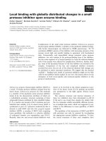
Treatment with CpG-ODN inhibits total number of
infiltrating cells in airways in WT but not in IFN-
β
-/- mice
The treatment with 5 µg of CpG administered intranasally
with OVA resulted in significant reduction of total
number of infiltrating cells in BALF in WT group while it
had no effect in IFN-β-/- group (Figure 1A). We examined
the number of recruited cells in lung airways after admin-
istration of PBS, OVA or CpG-ODN plus OVA and chal-
lenge with OVA. We found that OVA nasal challenge
increased significantly the number of cells recruited in air-
ways of OVA-primed mice compared to PBS group. CpG-
ODN vaccinated mice had reduced the number of cells in
OVA-sensitized B10.RIII mice but not in IFN-β-/
Suppression of eosinophilia by CpG-ODN in airways is
only partially dependent on IFN-
β
gene
Next, we were interested in the effect of CpG-ODN treat-
ment on eosinophilia. As expected, we found that OVA-
sensitized/OVA-challenge WT mice had a dramatic
increase in numbers of eosinophils compared with non-
treated WT. Vaccination with CpG-ODN diminished dra-
matically the number of eosinophils in WT mice while it
was only partially effective in prevention of eosinophilia
in IFN-β
-
/- mice, and the difference between the CpG-
ODN vaccinated and PBS vaccinated mice was statistically
significant for both WT and IFN-β
-
/- (figure 1B).
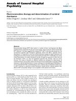
IFN-
γ
induction in the airways by CpG-ODNs vaccination
is impaired in IFN-
β
-/- mice
We were interested in investigating if disease mediated
Th2 cytokines or disease counter-acting cytokine, IFN-γ,
was effected by the CpG-ODN vaccination. We observed
that the level of IL-4 in BALF was reduced from 65 ± 7 pg/
ml to 43 ± 6 pg/ml (33% of reduction) in WT mice and
from 62 ± 8 pg/ml to 46 ± 87 pg/ml (26%) in IFN-β-/-
mice respectively after CpG-ODN vaccination. The levels
of IL-5 were significantly reduced in both groups with no
difference between groups (figure 2A). IFN-γ production
in airways of WT mice was enhanced upon CpG-ODN
vaccination and it was dependent on IFN-β gene since its
induction was impaired in IFN-β-/- mice (figure 2B).
Hence, the ratio IFN-γ/IL-4 determining the Th1/Th2
ratio, was skewed to a Th1 response in both groups
although much stronger in WT mice (figure 2C).
Table 1: Eosinophils in airways with different dose of intranasal
CpG-ODN
Treatment Genotype Eosinophils
PBS B10.RIII 0.5 %
IFN-β
-
/- 0.7 %
OVA B10.RIII 55 %
IFN-β
-
/- 62 %
OVA+CPG 5 µ B10.RIII 2.1 %
IFN-β
-
/- 9.2 %
OVA+CPG 10 µ B10.RIII 1. 9 %
IFN-β
-
/- 9.4 %
OVA+CPG 20 µ B10.RIII 1.9 %
IFN-β
-
/- 9.0 %
B10.RIII/WT (ᮀ) and IFN-β
-
/- (■) mice were sensitized to OVA by
intraperitoneal injection and subsequently challenged with OVA either
alone or with different dose of CpG-ODN by intranasal drops on days
14 and 16. Eosinophil percentage in bronchoalveolar lavage with
different dose of intranasal CpG-ODN were similar in all IFN-β
-
/-
treated mice.
Effects of treatment with CpG-ODN on total BALF cell recruitment (A), eosinophils (B)Figure 1
Effects of treatment with CpG-ODN on total BALF cell
recruitment (A), eosinophils (B). B10.RIII/WT (ᮀ) and IFN-β
-
/- (■) mice were sensitized to OVA by intraperitoneal injec-
tion and subsequently challenged with OVA either alone or
with CpG-ODN by intranasal drops on days 14 and 16. Cells
were harvested on day 17
th
. n = 5/group, *P < 0.05 vs. OVA
groups. † P < 0.05 vs OVA-treated WT mice.
Respiratory Research 2005, 6:25 />Page 5 of 12
(page number not for citation purposes)
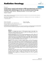
Vaccinated with CpG-ODN induces CD86 expression on B
cells in IFN-
β
-/- mice
In order to observe any differences between cell surface
markers between IFN-β
-
/- and wild type mice treated with
CPG-ODN or with PBS, splenocytes were analyzed by
flow cytometry. We could not see any difference in T cell
population, in regards to both CD4:CD8 ratio and expres-
sion of CD86 (B7.2) on T cells. However, there was a
significant difference in CD86 (B7.2) expression on B
cells. This difference was observed between CpG-ODN
vaccinated IFN-β
-
/- mice and PBS control IFN-β
-
/- mice as
well as between CpG-ODN vaccinated IFN-β
-
/- and CpG-
ODN vaccinated wild type mice (Figure 3).
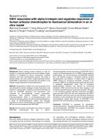
CpG-ODN vaccination induces mild synovitis particularly
in IFN-
β
-/- mice
Mice did not show any clinical visually deformation.
While surveying the capacity of CpG-ODN vaccination to
induce IFN-β in different tissues, it was noticeable that
there were pathological changes in joints of some mice.
Thus, we stained the paws of mice (n = 3) with hematox-
ylin and erythrosine and evaluated the pathologic changes
in joints. Data revealed mild synovitis and pannus forma-
tion in multiple joints of CpG-ODN vaccinated mice
while no control mice had any pathologic changes. Fur-
thermore, we discovered that mice lacking IFN-β were
more affected than their wild type littermates (table 2 and
figure 4).
BALF cytokine (protein) concentrations after intranasal CpG-ODNFigure 2
BALF cytokine (protein) concentrations after intranasal CpG-ODN. BALF were collected 24 h after the last challenge from
each group (n = 5/group) and cytokine levels determined by ELISA in non-immunized, OVA-challenged, and OVA-challenged/
CpG-treated B10.RIII (ᮀ) and IFN-β
-
/- (■) mice at days 14 and 16. IL-5 (A) levels were significantly augmented after OVA chal-
lenge and diminished after CpG vaccination in both strains similarly. IFN-γ (B) was not induced in OVA/primed-OVA/challenge,
but was induced after CpG vaccination. IFN-γ was stronger induced in B10.RIII than in IFN-β
-
/- mice. Th1/T2 ratio was
stronger skewed to Th1-profile in B10.RIII than in IFN-β
-
/- mice. Data are given as mean ± SEM, *P < 0.05 vs. OVA groups. † P
< 0.05 vs B10.RIII mice treated with CpG
Respiratory Research 2005, 6:25 />Page 6 of 12
(page number not for citation purposes)
Cell profile in airways after vaccination withCpG-ODN
The CpG-ODN vaccination reduced the number of cells in
OVA-sensitized B10.RIII mice. However, the number of
cells recovered in IFN-β
-
/- mice did not significantly
change (table 3). ODN vaccinated mice had a slight
increase in numbers of eosinophils compared with non-
treated WT. CpG-ODN therapy diminished the number of
eosinophils in WT mice, while it was only partially effec-
tive in prevention of eosinophilia in IFN-β
-
/- mice with
significant differences between the CpG-ODN treated and
non-treated mice in WT and IFN-β
-
/- (table 3). Similarly,
vaccination with CpG-ODN showed an enhanced
response of macrophages in IFN-β
-
/- mice compared to
WT mice, but this macrophage response was similar in
treated and non-treated WT mice. Lymphocyte and
neutrophil response in airways of treated-IFN-β
-
/- mice
was also significantly enhanced compared to WT mice.
Inhibition of OVA-specific IgE in the prevention study
(vaccination) by CpG-ODNs is independent of IFN-
β
It has been shown that systemic administration of CpG-
ODN do not inhibit established IgE response while vacci-
nation inhibits IgE production[32], however the role of
INF-β was not investigated. Here, we examined what the
function of IFN-β was in prevention of OVA-specific IgE in
CpG-ODN vaccine. We found that CpG-ODN vaccine
resulted in inhibition of OVA-Specific IgE in both WT and
IFN-β-/- mice (figure 5). IgG2a levels were similar in both
WT (118 ± 15 µg/ml) and IFN-β-/- (135 ± 25 µg/ml) mice.
Allergen specific Th1 response as a result of CpG-ODN
vaccination is partly impaired in the absence of IFN-
β
To address if splenocytes from WT and IFN-β
-
/- respond
differently in vitro, cells from naïve mice were stimulated
and cell proliferation was measured. Splenocytes from
both groups, WT and IFN-β
-
/-, had the same proliferation
levels after stimulation with concavalin A, CpG or culture
media (figure 6A). However, cells from WT immunized
mice vaccinated with CpG in vivo had more cell prolifera-
tion after restimulation with OVA than IFN-β
-
/- immu-
nized and CpG vaccinated mice (figure 6B). Next we
assessed whether OVA specific Th1 response, i.e. IFN-γ, IL-
2 and IL-12, were affected by CpG-ODN vaccination plus
OVA treatment in vivo. We found that IFN-γ, IL-12 and IL-
2 were significantly lower in OVA-primed/OVA-challenge
IFN-β-/- mice compared to WT mice (figure 6C).
Discussion
Synthetic unmethylated CG dinucleotides within particu-
lar sequence context (CpG motifs) mimic bacterial DNA,
and are responsible for the immunostimulatory activity of
that [6]. CpG oligonucleotides have shown to produce a
strong activation of B cells[33], NK cells [34], macro-
phages[35] and dendritic cells[36] by a direct mechanism.
However CpG have also the ability to exert activation of T
cells by an indirect mechanism through via IFN-α/β
[37,38]. Furthermore, CpG in mice results in production
of inflammatory and antiinflammatory cytokines includ-
ing IL-1, IL-2, IL-6, IL-18, TNF-α, type I IFN (IFN-α/β) and
type II IFN (IFN-γ) [39-41]. Type I IFNs (IFN-α/β) have
pleiomorphic effect on the immune system with activa-
Percent of expression of CD86/B7.2 on B cells in splenocytes of mice at day 17 after immunization and vaccination of ODN-CpG in IFN-β
-
/- mice (KO) and wild type litter-mates (WT)Figure 3
Percent of expression of CD86/B7.2 on B cells in splenocytes
of mice at day 17 after immunization and vaccination of
ODN-CpG in IFN-β
-
/- mice (KO) and wild type litter-mates
(WT).
Table 2: Histopathologic evaluation of joints for arthritis
changes.
Groups Vaccination
CpG-ODN Control
IFN-β
-
/- (n.1) ++ -
IFN-β
-
/- (n.2) ++ -
IFN-β
-
/- (n.3) + -
WT (n.1) ++ -
WT (n.2) - -
WT (n.3) - -
Hematoxylin-eosin staining of joints from four different groups of
mice (IFN-β
-
/- and their WT littermates with CPG-ODN treatment
or control) were analyzed. This revealed mild synovitis and pannus
formation in 3/3 IFN-β-/- mice treated with CPG-ODN and 1/3 WT
treated with CPG-ODN while no pathological changes were observed
in these two non-treated groups.
Respiratory Research 2005, 6:25 />Page 7 of 12
(page number not for citation purposes)
tion of macrophages and stimulation of NK cells to
produce IL-12, which in turn induces Th1 cell
development[42].
Some of these immunostimulatory effects have been
applied in animal models of several diseases including
allergic disorders[8,43-50]. It have been shown that ther-
apies using oligonucleotides containing CpG have the
ability of immunomodulation with a downregulation of
elevated IgE and eosinophilic inflammation in the air-
ways, both of which are orchestrated by cytokines elabo-
rated by Th2 cells. However, systemic administration of
CpG has been reported to increase side effects, owing in
part to high dose of these oligonucleotides. Systemic
immunization, even with adjuvants, induces robust adap-
tive immune responses at systemic sites but weak in the
airways, while local immunization can elicit both sys-
temic and mucosal responses [51-53]. In this report, we
have demonstrated that concomitant intranasal adminis-
tration of low doses of CpG and the offending antigen
Illustration of joint synovitis after hematoxylin-eosin stainingFigure 4
Illustration of joint synovitis after hematoxylin-eosin staining. A. It shows synovitis and pannus formation in IFN-β
-
/- mice
treated with CPG-ODN. B. It shows no pathologic changes in a control treated IFN-β
-
/- mice.
Table 3: Effects of vaccination with CpG-ODN (prevention study) on eosinophil and total BAL cell recruitment.
Treatment Genotype Total cells Eosinophils Monocytes Lymphocytes Neutrophils
PBS B10.RIII 245 ± 43 3 ± 1 232 ± 20 5 ± 1 5 ± 1
IFN-β
-
/- 259 ± 14 3 ± 1 242 ± 33 6 ± 1 8 ± 2
OVA B10.RIII 622 ± 37* 381 ± 43* 144 ± 17 62 ± 3* 35 ± 2*
IFN-β
-
/- 683 ± 66* 427 ± 83* 178 ± 22 55 ± 8* 22 ± 4*
OVA+CpG B10.RIII 227 ± 18† 2.7 ± 2† 142 ± 19 67 ± 4 14 ± 1
IFN-β
-
/- 574 ± 32 52 ± 7 † ‡ 321 ± 39† ‡ 130 ± 38† ‡ 70 ± 22† ‡
Cell types quantified in BALF were eosinophils, macrophages, lymphocytes and neutrophils and are expressed as no. of cells × 10
3
/ml. n = 5/group,
*P < 0.05 vs. untreated groups. † P < 0.05 vs OVA-treated mice. ‡ P < 0.05 vs WT mice treated with CpG-ODN. OVA-treated mice and control
groups.
Respiratory Research 2005, 6:25 />Page 8 of 12
(page number not for citation purposes)
exerted significant reduction of total number of infiltrat-
ing cells, including eosinophils in BALF (table 1).
As mentioned before, CpG in mice results in production
of several cytokines including type I IFN (IFN-α/
β)[37,38,54-56] which have the ability to exert indirect
activation of T cells [37,38]. IFN-β treatment, used by
either oral[18] or parenteral[17] via in mice, have shown
to produce an inhibition of antigen-induced bronchial
inflammation and airway hyperresponsiveness [17,18]
probably influenced by the inhibition of Th-2 airway
eosinophilia by the suppressive effect on eosinopoiesis
[57]. We have recently demonstrated that lung eosi-
nophilic inflammatory response was exacerbated by the
lack of IFN-β gene[19]. Even though it is believed that
immunomodulatory effects of CpG-ODN may be medi-
ated by type I IFNs [15], the relative role of IFN-β, a type I
IFN, has not been defined. Here, we aimed to elucidate
whether IFN-β have a key role in the anti-allergic effect of
CpG motifs. Our results demonstrate that therapy with
CpG-ODN prior to and after the allergen challenge
resulted in significant reduction of total number of infil-
trating cells, including eosinophils, in BALF in WT mice
while CpG-ODN did show an enhanced response of mac-
rophages, lymphocytes and neutrophils in airways of IFN-
β-/- mice. These findings might be explained since CpG
motifs in bacterial DNA can delay apoptosis of neutrophil
granulocytes [58] and macrophages [59], indicating a
possibility of inhibition of macrophage apoptosis by CpG
and a difference of cellular responses downstream of dif-
ferent Toll-like receptors [59]. Another possibility might
be that phosphorothioated ODNs used in our experi-
ments might have been chemoattractants for primary
macrophages[60] in the absence of IFN-β. This chemoat-
tractant activity have been exposed as independent of
CpG activity[60], since it has not been seen with phos-
phodiester CpG-ODNs. However, up to our knowledge
this is the first reference about the influence of CpG on
neutrophils.
It has been shown that systemic administration of CpG-
ODN do not inhibit established IgE response while vacci-
nation inhibits IgE production[32]. We found that CpG-
ODN vaccine resulted in inhibition of OVA-Specific IgE in
both WT and IFN-β
-
/- mice (figure 5). These data under-
line that IFN-β is not required for the beneficial effect of
CpG-ODN vaccine in a model of allergic inflammation.
Vaccination with a single low dose of CpG-dinucleotide
inhibited OVA-specific IgE production with subsequent
upregulation of IgG2a in both groups. The success in
inhibiting established IgE response is most likely due to
the timing of the protocol where mice received CpG-ODN
at the time of priming. This early intervention presumably
prevents presence of IgE-plasma cells in the bone marrow
as suggested earlier by Peng et al [32].
Production of the Th1 cytokine, IFN-γ, has been reported
to be dependent on CpG-ODN-induced IFN-α/β as
demonstrated by antibodies that block IFN-α/β[54].
Since, earlier reports target both IFN-α and β, it was
unclear if one or both of these cytokines mediate the bio-
logical effects of CpG-ODN. In addition, we have recently
reported that IFN-β knock out mice do not have any fail-
ing mounting a T
H
1 response, measured by IFN-γ
production. In contrary, IFN-γ production was signifi-
cantly elevated as a result of experimental autoimmune
encephalomyelitis (EAE), a T
H
1-mediated disease model
for multiple sclerosis. Consequently IFN-β knock out
mice had more severe and chronic symptoms than their
WT littermates with more extensive CNS inflammation
and higher demyelination [23]. Thus, here we aimed to
investigate the profile of OVA-specific Th1 cytokines after
CpG-ODN vaccination in the absence of IFN-β. We found
a clear reduction in Th1 response (IL-2 and IFN-γ) in IFN-
OVA-specific IgE levels in the prevention study (vaccination)Figure 5
OVA-specific IgE levels in the prevention study (vaccination).
B10.RIII/WT (ᮀ) and IFN-β
-
/- (■) were sensitized to OVA
by intraperitoneal injection either OVA alone or with CpG-
ODN and subsequently challenged with OVA by intranasal
drops on days 14 and 16; control mice received PBS alone.
Cells were harvested on day 17. n = 5/group, *P < 0.05 vs.
OVA groups. † P < 0.05 vs OVA-treated WT mice.
Respiratory Research 2005, 6:25 />Page 9 of 12
(page number not for citation purposes)
Ex-vivo immune response in the prevention study (vaccination)Figure 6
Ex-vivo immune response in the prevention study (vaccination). A. In vitro stimulation of splenocytes from naïve mice with con
A and CpG does not show any difference between B10.RIII (ᮀ) and IFN-β-/- mice (■). B. In vitro proliferation of OVA restim-
ulated T cells from in vivo CpG-vaccinated OVA-primed B10.RIII (ᮀ) and IFN-β-/- mice (■). Mice were primed and challenged
as in Figure 2. In vitro proliferation after recall with OVA was weaker in IFN-β-/- mice (■) than B10.RIII mice (ᮀ). C. Th-1
cytokines from supernatants after in vitro proliferation of OVA restimulated T cells in OVA-primed/CpG-vaccinated mice. IFN-
γ, IL-12 and IL-2 production in supernatants from cell cultures was higher in B10.RIII than in IFN-β-/- mice. n = 5/group *P <
0.05 vs. OVA-treated B10.RIII mice.
Respiratory Research 2005, 6:25 />Page 10 of 12
(page number not for citation purposes)
β knock out mice vaccinated with CpG-ODN which was
in agreement with earlier reports[55]. As Th1-promoting
activity of CpG-ODN is controlled by IL-12[12], we meas-
ured the levels of IL-12 and found that production was
elevated in the CpG-ODN WT group. We also found that
its induction is partially under the influence of IFN-β trig-
gered by synthetic CpG sequences. Since IFN-γ is almost
undetectable in non-treated mice, at least under the con-
ditions used in this study, the results also suggest that CpG
is capable of inducing IFN-β in substantial amounts to
trigger IFN-γ production. Our findings of Th1 mediated
response in systemic immune response were moreover
supported by the fact that IFN-γ production was also
defective in the inflammatory organ measured in BALF.
Moreover, our results also provide evidence that IFN-β is
an important cofactor for IFN-γ production through
induction of IL-12 pathway as it has been suggested by
Sun et al[37] While, it is crucial to underline that IFN-β-/-
mice do not have a general defect on mounting a Th1
immune response[23] therefore it is more likely that the
defect in inducing a proper Th1 response in IFN-β-/- mice
is due to malfunctioning IL-12 and IFN-γ induction
through TLR9 pathway as a result of CPG-ODN vaccina-
tion. This might also explain the lower proliferative
response of OVA-specific Th1 cells in IFN-β-/- mice
reported here. Once more, it should be mentioned that
IFN-β-/- mice are capable of inducing significantly higher
OVA-specific T cell proliferation of Th2 character [19]
which might also partly contribute to suppression of a
more profound Th1 response. It has been reported that
CpG-ODNs do not directly stimulate T cells, but by induc-
ing production of IFN-γ from APCs, thus activating T cells
to express CD69 and B7.2[9,37], while their proliferative
responses are reduced[37]. It was also shown that CpG
stimulate T cells by inducing APCs to synthesize IFN-I,
which then act directly on T cells via IFNAR[37]. In addi-
tion, it has been suggested that production of type I IFNs
by APCs is through increased availability of costimulatory
signals on activated DC[37,36]. It has also been reported
that stimulation with CpG motifs induces the changes in
surface molecules of APCs[25,55,37]. However, the
reduced OVA-specific Th1 response in IFN-β-/- mice is less
likely to be mediated by lack of upregulation of costimu-
latory molecules on APCs as we have previously reported
that these mice have upregulated B7.1/2 on APCs[19].
After treatment with CPG-ODN we made an interesting
observation that the mice developed a mild synovitis,
which to our knowledge is the first report of mucosal
administration of CPG-ODN causing joint modification.
Synovitis is one of the phenotype features of the
experimental murine animal models of autoimmune
arthritis, such as collagen-induced arthritis (CIA), which is
an extensive investigated model of human rheumatoid
arthritis. This model can be elicited in susceptible strains
by immunization with type II collagen (CII), the major
protein of articular cartilage. Assessment of disease
includes visual/clinical evaluation of arthritis severity,
measurement of humoral and cellular immune responses,
including CII-specific antibody titers and T cell responses
to CII. In these models, joints are histologically scored for
the changes of inflammation including synovitis and peri-
articular, pannus formation, cartilage damage with mar-
ginal erosions or diffuse changes, and bone damage
including resorption and periosteal proliferation[31]. It is
known that unmethylated CpG-ODN are responsible for
induction of arthritis triggered by bacterial DNA[11,61-
63] that supports our data. Our finding that mucosal
administration of CpG-ODN causes mild synovitis points
out a potential hazardous side effect when using CpG-
ODN as a treatment.
In summary, we have demonstrated that the CpG-ODNs
can partly prevent the development of eosinophilic airway
inflammation and allergen specific IgE response in the
absence of IFN-β, while Th1 response is defective. In addi-
tion, these results demonstrate that mucosal administra-
tion of CpG-ODN before allergen exposure could be a less
harmful form of active immunotherapy in allergic dis-
eases without impeding systemic immune responses as
earlier suggested [8,51]. However, due to the potential of
hazardous side effects, meticulous caution must be under-
taken prior to considering it as a therapy in allergic
asthma.
Abbreviations
APC: Antigen presenting cells; CpG, cytosine-guanine
motifs; ODNs, oligodeoxynucleotides; DAB, 3.3 diamino
benzidine tetrahydrochlorhide; BALF, bronchoalveolar
lavage fluid; CREPA, (cyanide-resistant eosinophil perox-
idase activity); IFNAR, type I IFN receptor; APC, antigen-
presenting cells; DC, dendritic cells.
Authors' contributions
VM conceived of the study, participated in its design and
coordination, performed the experiments and drafted the
manuscript. AT carried out the analysis of flow cytometry,
prepared histological samples of joints and performed the
clinical and histological analysis of joints for arthritis. AT
and IT generated crossing of IFN-β ko mice to B10.RIII
strain of mice, genotyped, backcrossed and maintained
the IFN- β-/- mouse line. VN participated in the design
and coordination of the study. SI-N participated in the
direction of the study, performed histological analysis of
joints, as well as writing and preparing the manuscript. All
authors read and approved the final manuscript.
Acknowledgements
We thank Sandy Liedholm, Isabelle Bohlin, Rebecka Ljungqvist and Carlos
Palestro for taking excellent care of the animals and Emma Mondoc and
Margareta Svejme for help with histological analysis. This work has been
Respiratory Research 2005, 6:25 />Page 11 of 12
(page number not for citation purposes)
supported by grants from The Swedish Foundation for Health Care Sci-
ences and Allergy Research, The Crafoord Foundations, The Edvard
Welander Foundation, King Gustaf V's 80-year Foundation, Fundación
SEAIC and Tore Nilsson's Foundation for Medical Research.
References
1. Lack G, Bradley KL, Hamelmann E, Renz H, Loader J, Leung DY,
Larsen G, Gelfand EW: Nebulized IFN-gamma inhibits the
development of secondary allergic responses in mice. J
Immunol 1996, 157:1432-1439.
2. Boguniewicz M, Martin RJ, Martin D, Gibson U, Celniker A, Williams
M, Leung DY: The effects of nebulized recombinant inter-
feron-gamma in asthmatic airways. J Allergy Clin Immunol 1995,
95:133-135.
3. Hofstra CL, Van Ark I, Hofman G, Nijkamp FP, Jardieu PM, Van Oost-
erhout AJ: Differential effects of endogenous and exogenous
interferon-gamma on immunoglobulin E, cellular infiltra-
tion, and airway responsiveness in a murine model of allergic
asthma. Am J Respir Cell Mol Biol 1998, 19:826-835.
4. Dow SW, Schwarze J, Heath TD, Potter TA, Gelfand EW: Systemic
and local interferon gamma gene delivery to the lungs for
treatment of allergen-induced airway hyperresponsiveness
in mice. Hum Gene Ther 1999, 10:1905-1914.
5. Gavett SH, O'Hearn DJ, Li X, Huang SK, Finkelman FD, Wills-Karp M:
Interleukin 12 inhibits antigen-induced airway hyperrespon-
siveness, inflammation, and Th2 cytokine expression in
mice. J Exp Med 1995, 182:1527-1536.
6. Krieg AM, Yi AK, Matson S, Waldschmidt TJ, Bishop GA, Teasdale R,
Koretzky GA, Klinman DM: CpG motifs in bacterial DNA trig-
ger direct B-cell activation. Nature 1995, 374:546-549.
7. Kline JN, Waldschmidt TJ, Businga TR, Lemish JE, Weinstock JV,
Thorne PS, Krieg AM: Modulation of airway inflammation by
CpG oligodeoxynucleotides in a murine model of asthma. J
Immunol 1998, 160:2555-2559.
8. Broide D, Schwarze J, Tighe H, Gifford T, Nguyen MD, Malek S, Van
Uden J, Martin-Orozco E, Gelfand EW, Raz E: Immunostimulatory
DNA sequences inhibit IL-5, eosinophilic inflammation, and
airway hyperresponsiveness in mice. J Immunol 1998,
161:7054-7062.
9. Lipford GB, Bauer M, Blank C, Reiter R, Wagner H, Heeg K: CpG-
containing synthetic oligonucleotides promote B and cyto-
toxic T cell responses to protein antigen: a new class of vac-
cine adjuvants. Eur J Immunol 1997, 27:2340-2344.
10. Shirota H, Sano K, Kikuchi T, Tamura G, Shirato K: Regulation of
murine airway eosinophilia and Th2 cells by antigen- conju-
gated CpG oligodeoxynucleotides as a novel antigen-specific
immunomodulator. J Immunol 2000, 164:5575-5582.
11. Deng GM, Nilsson IM, Verdrengh M, Collins LV, Tarkowski A: Intra-
articularly localized bacterial DNA containing CpG motifs
induces arthritis. Nat Med 1999, 5:702-705.
12. Chiaramonte MG, Hesse M, Cheever AW, Wynn TA: CpG oligonu-
cleotides can prophylactically immunize against Th2-medi-
ated schistosome egg-induced pathology by an IL-12-
independent mechanism. J Immunol 2000, 164:973-985.
13. Klinman DM, Yi AK, Beaucage SL, Conover J, Krieg AM: CpG motifs
present in bacteria DNA rapidly induce lymphocytes to
secrete interleukin 6, interleukin 12, and interferon gamma.
Proc Natl Acad Sci U S A 1996, 93:2879-2883.
14. Sato Y, Roman M, Tighe H, Lee D, Corr M, Nguyen MD, Silverman
GJ, Lotz M, Carson DA, Raz E: Immunostimulatory DNA
sequences necessary for effective intradermal gene
immunization. Science 1996, 273:352-354.
15. Hafner M, Zawatzky R, Hirtreiter C, Buurman WA, Echtenacher B,
Hehlgans T, Mannel DN: Antimetastatic effect of CpG DNA
mediated by type I IFN. Cancer Res 2001, 61:5523-5528.
16. Nakajima H, Nakao A, Watanabe Y, Yoshida S, Iwamoto I: IFN-alpha
inhibits antigen-induced eosinophil and CD4+ T cell recruit-
ment into tissue. J Immunol 1994, 153:1264-1270.
17. Maeda Y, Musoh K, Shichijo M, Tanaka H, Nagai H: Interferon-beta
prevents antigen-induced bronchial inflammation and air-
way hyperreactivity in mice. Pharmacology 1997, 55:32-43.
18. Satoh Y, Kasama K, Kuwabara M, Yimin, Diao HY, Nakajima H,
Kohanawa M, Minagawa T: Suppression of late asthmatic
response by low-dose oral administration of interferon-beta
in the guinea pig model of asthma. J Interferon Cytokine Res 1999,
19:887-894.
19. Matheu V, Treschow A, Navikas V, Issazadeh-Navikas S: Upregula-
tion of B7 molecules (CD80 and CD86) and exacerbated
eosinophilic pulmonary inflammatory response in mice lack-
ing the IFN-beta gene. J Allergy Clin Immunol 2003, 111:550-557.
20. Hayashi T, Adachi Y, Hasegawa K, Morimoto M: Less sensitivity for
late airway inflammation in males than females in BALB/c
mice. Scand J Immunol 2003, 57:562-567.
21. Yamatomo T, Okano M, Ono T, Nakayama E, Yoshino T, Satoskar
AR, Harn DAJ, Nishizaki K: Sex-related differences in the initia-
tion of allergic rhinitis in mice. Allergy 2001, 56:525-531.
22. Erlandsson L, Blumenthal R, Eloranta ML, Engel H, Alm G, Weiss S,
Leanderson T: Interferon-beta is required for interferon-alpha
production in mouse fibroblasts. Curr Biol 1998, 8:223-226.
23. Teige I, Treschow A, Teige A, Mattsson R, Navikas V, Leanderson T,
Holmdahl R, Issazadeh-Navikas S: IFN-beta gene deletion leads
to augmented and chronic demyelinating experimental
autoimmune encephalomyelitis. J Immunol 2003,
170:4776-4784.
24. Sur S, Wild JS, Choudhury BK, Sur N, Alam R, Klinman DM: Long
term prevention of allergic lung inflammation in a mouse
model of asthma by CpG oligodeoxynucleotides. J Immunol
1999, 162:6284-6293.
25. Martin-Orozco E, Kobayashi H, Van Uden J, Nguyen MD, Kornbluth
RS, Raz E: Enhancement of antigen-presenting cell surface
molecules involved in cognate interactions by immunostim-
ulatory DNA sequences. Int Immunol 1999, 11:1111-1118.
26. Ten RM, Pease LR, McKean DJ, Bell MP, Gleich GJ: Molecular clon-
ing of the human eosinophil peroxidase. Evidence for the
existence of a peroxidase multigene family. J Exp Med 1989,
169:1757-1769.
27. Teige A, Teige I, Lavasani S, Bockermann R, Mondoc E, Holmdahl R,
Issazadeh-Navikas S: CD1-dependent regulation of chronic cen-
tral nervous system inflammation in experimental autoim-
mune encephalomyelitis. J Immunol 2004, 172:186-194.
28. Matheu V, Navikas V, Issazadeh S: Susceptibility of B10.RIII
mouse strain to develop inflammatory allergic pulmonary
disease. Alergol Inmunol Clin 2001, 16:282-290.
29. Zuberi RI, Apgar JR, Chen SS, Liu FT: Role for IgE in airway secre-
tions: IgE immune complexes are more potent inducers than
antigen alone of airway inflammation in a murine model. J
Immunol 2000, 164:2667-2673.
30. Svensson L, Jirholt J, Holmdahl R, Jansson L: B cell-deficient mice
do not develop type II collagen-induced arthritis (CIA). Clin
Exp Immunol 1998, 111:521-526.
31. Johansson AC, Nakken B, Sundler M, Lindqvist AK, Johannesson M,
Alarcon-Riquelme M, Bolstad AI, Humphreys-Beher MG, Jonsson R,
Skarstein K, Holmdahl R: The genetic control of sialadenitis ver-
sus arthritis in a NOD.QxB10.Q F2 cross. Eur J Immunol 2002,
32:243-250.
32. Peng Z, Wang H, Mao X, HayGlass KT, Simons FE: CpG oligodeox-
ynucleotide vaccination suppresses IgE induction but may
fail to down-regulate ongoing IgE responses in mice. Int
Immunol 2001, 13:3-11.
33. Liang H, Nishioka Y, Reich CF, Pisetsky DS, Lipsky PE: Activation of
human B cells by phosphorothioate oligodeoxynucleotides. J
Clin Invest 1996, 98:1119-1129.
34. Ballas ZK, Rasmussen WL, Krieg AM: Induction of NK activity in
murine and human cells by CpG motifs in oligodeoxynucle-
otides and bacterial DNA. J Immunol 1996, 157:1840-1845.
35. Takeshita S, Takeshita F, Haddad DE, Ishii KJ, Klinman DM: CpG oli-
godeoxynucleotides induce murine macrophages to up-reg-
ulate chemokine mRNA expression. Cell Immunol 2000,
206:101-106.
36. Jakob T, Walker PS, Krieg AM, Udey MC, Vogel JC: Activation of
cutaneous dendritic cells by CpG-containing oligodeoxynu-
cleotides: a role for dendritic cells in the augmentation of
Th1 responses by immunostimulatory DNA. J Immunol 1998,
161:3042-3049.
37. Sun S, Zhang X, Tough DF, Sprent J: Type I interferon-mediated
stimulation of T cells by CpG DNA. J Exp Med 1998,
188:2335-2342.
38. Rothenfusser S, Hornung V, Krug A, Towarowski A, Krieg AM,
Endres S, Hartmann G: Distinct CpG oligonucleotide sequences
Publish with Bio Med Central and every
scientist can read your work free of charge
"BioMed Central will be the most significant development for
disseminating the results of biomedical research in our lifetime."
Sir Paul Nurse, Cancer Research UK
Your research papers will be:
available free of charge to the entire biomedical community
peer reviewed and published immediately upon acceptance
cited in PubMed and archived on PubMed Central
yours — you keep the copyright
Submit your manuscript here:
/>BioMedcentral
Respiratory Research 2005, 6:25 />Page 12 of 12
(page number not for citation purposes)
activate human gamma delta T cells via interferon-alpha/-
beta. Eur J Immunol 2001, 31:3525-3534.
39. Sun S, Zhang X, Tough D, Sprent J: Multiple effects of immunos-
timulatory DNA on T cells and the role of type I interferons.
Springer Semin Immunopathol 2000, 22:77-84.
40. Sun S, Sprent J: Role of type I interferons in T cell activation
induced by CpG DNA. Curr Top Microbiol Immunol 2000,
247:107-117.
41. Sprent J, Zhang X, Sun S, Tough D: T-cell proliferation in vivo and
the role of cytokines. Philos Trans R Soc Lond B Biol Sci 2000,
355:317-322.
42. Roman M, Martin-Orozco E, Goodman JS, Nguyen MD, Sato Y, Ron-
aghy A, Kornbluth RS, Richman DD, Carson DA, Raz E: Immunos-
timulatory DNA sequences function as T helper-1-
promoting adjuvants. Nat Med 1997, 3:849-854.
43. Spiegelberg HL, Broide D, Tighe H, Roman M, Raz E: Inhibition of
allergic inflammation in the lung by plasmid DNA allergen
immunization. Pediatr Pulmonol Suppl 1999, 18:118-121.
44. Spiegelberg HL, Orozco EM, Roman M, Raz E: DNA immunization:
a novel approach to allergen-specific immunotherapy. Allergy
1997, 52:964-970.
45. Spiegelberg HL, Tighe H, Roman M, Broide D, Raz E: Inhibition of
IgE formation and allergic inflammation by allergen gene
immunization and by CpG motif immunostimulatory
oligodeoxynucleotides. Allergy 1998, 53:93-97.
46. Broide D, Raz E: DNA-Based immunization for asthma. Int Arch
Allergy Immunol 1999, 118:453-6.
AA&action=render&rendertype=fulltext&uid=IAA.iaa18453.
47. Broide DH, Paine MM, Firestein GS: Eosinophils express inter-
leukin 5 and granulocyte macrophage-colony- stimulating
factor mRNA at sites of allergic inflammation in asthmatics.
J Clin Invest 1992, 90:1414-1424.
48. Broide DH, Stachnick G, Castaneda D, Nayar J, Miller M, Cho J,
Rodriquez M, Roman M, Raz E: Immunostimulatory DNA medi-
ates inhibition of eosinophilic inflammation and airway
hyperreactivity independent of natural killer cells in vivo. J
Allergy Clin Immunol 2001, 108:759-763.
49. Broide DH, Stachnick G, Castaneda D, Nayar J, Miller M, Cho JY,
Roman M, Zubeldia J, Hayashi T, Raz E: Systemic administration
of immunostimulatory DNA sequences mediates reversible
inhibition of Th2 responses in a mouse model of asthma. J Clin
Immunol 2001, 21:175-182.
50. Cho JY, Miller M, Baek KJ, Castaneda D, Nayar J, Roman M, Raz E,
Broide DH: Immunostimulatory DNA sequences inhibit respi-
ratory syncytial viral load, airway inflammation, and mucus
secretion. J Allergy Clin Immunol 2001, 108:697-702.
51. Shirota H, Sano K, Kikuchi T, Tamura G, Shirato K: Regulation of
T-helper type 2 cell and airway eosinophilia by transmucosal
coadministration of antigen and oligodeoxynucleotides con-
taining CpG motifs. Am J Respir Cell Mol Biol 2000, 22:176-182.
52. Magone MT, Chan CC, Beck L, Whitcup SM, Raz E: Systemic or
mucosal administration of immunostimulatory DNA inhibits
early and late phases of murine allergic conjunctivitis. Eur J
Immunol 2000, 30:1841-1850.
53. Takabayashi K, Libet L, Chisholm D, Zubeldia J, Horner AA: Intrana-
sal immunotherapy is more effective than intradermal
immunotherapy for the induction of airway allergen toler-
ance in Th2-sensitized mice. J Immunol 2003, 170:3898-3905.
54. Krug A, Rothenfusser S, Hornung V, Jahrsdorfer B, Blackwell S, Ballas
ZK, Endres S, Krieg AM, Hartmann G: Identification of CpG oli-
gonucleotide sequences with high induction of IFN-alpha/
beta in plasmacytoid dendritic cells. Eur J Immunol 2001,
31:2154-2163.
55. Cho HJ, Hayashi T, Datta SK, Takabayashi K, Van Uden JH, Horner A,
Corr M, Raz E: IFN-alphabeta Promote Priming of Antigen-
Specific CD8(+) and CD4(+) T Lymphocytes by Immunos-
timulatory DNA-Based Vaccines. J Immunol 2002,
168:4907-4913.
56. Van Uden JH, Tran CH, Carson DA, Raz E: Type I interferon is
required to mount an adaptive response to immunostimula-
tory DNA. Eur J Immunol 2001, 31:3281-3290.
57. Klimpel GR, Fleischmann WRJ, Klimpel KD: Gamma interferon
(IFN gamma) and IFN alpha/beta suppress murine myeloid
colony formation (CFU-C)N: magnitude of suppression is
dependent upon level of colony-stimulating factor (CSF). J
Immunol 1982, 129:76-80.
58. Jozsef L, Khreiss T, Filep JG: CpG motifs in bacterial DNA delay
apoptosis of neutrophil granulocytes. Faseb J 2004,
18:1776-1778.
59. Kim SO, Ono K, Han J: Apoptosis by pan-caspase inhibitors in
lipopolysaccharide-activated macrophages. Am J Physiol Lung
Cell Mol Physiol 2001, 281:L1095-105.
60. Baek KH, Ha SJ, Sung YC: A novel function of phosphorothioate
oligodeoxynucleotides as chemoattractants for primary
macrophages. J Immunol 2001, 167:2847-2854.
61. Deng GM, Tarkowski A: Synovial cytokine mRNA expression
during arthritis triggered by CpG motifs of bacterial DNA.
Arthritis Res 2001, 3:48-53.
62. Miyata M, Kobayashi H, Sasajima T, Sato Y, Kasukawa R: Unmethyl-
ated oligo-DNA containing CpG motifs aggravates collagen-
induced arthritis in mice. Arthritis Rheum 2000, 43:2578-2582.
63. Svelander L, Erlandsson Harris H, Lorentzen JC, Trollmo C,
Klareskog L, Bucht A: Oligodeoxynucleotides containing CpG
motifs can induce T cell-dependent arthritis in rats. Arthritis
Rheum 2004, 50:297-304.